Embed Size (px)
Citation preview

....
Conlracl AIR3 CT94.2487
Final Consolidaled Report.
Development of Mussel-Specific Expression Vectors Suitable for Transgenic Organisms: Implication on the Establishment of
MoUusc Continuous Cell lines
conlracl W AIR3-CT94-2487
Coordinator
, Claude Delserl
Lat:lOratoire Génome et Populations Station Méditerranéenne de l'Environnement Littoral. 1 quai de la Dorade. 34200 Sète Tel: 33/4/67 46 33 90; Fax: 33/4/67 46 02 56; E·mail: cdelsert@u niv-montp2.fr
Participant
Leonor Cancela da Fonseca Universidade do Algarve Unidade de Ciencas e Tecnologias dos Recursos Aquaticos Campus de Gambelas 8000 Faro - Portugal Tel: 351 /89 800971; Fax: 351 /89.818353; E-mail : Icancela @mozart.si.ualg .pt
-
'Present address: Centre de Recherche en Biologie Moléculaire - Centre National de la Recherche Scientifique - Route de Mende - 34293 Montpellier cedex5 Tel: 33/4/6761 33 30; Fax: 33/4/67 52 15 59; E-mail : cdelsert @xerxes.crbm .cnrs-mop.fr
Issuing date: July 1998.
1

1 1
1
1
1
1
Contract AIR3 CT94.2487
CONTENTS
Summary
Executive Summary
Chapter 1. Introduction
Chapter 2. Materials and Methods.
Chapter 3. Results
Chapter 4. Discussion.
Chapter 5. Finalization of tasks
2

k:
Contract AIR3 CT94.2487
SUMMARY
The main objective of this project was to develop the tools for the in-depth understanding of
mollusc biology and pathology. Such a goal has been reached in other biological systems through
the development of molecular biology and cell culture. Indeed, development of a mollusc cell
culture system is an essential step to approach many of the problems related to mollusc
pathology, in particular to use molecular biology tools to study viral infection of these
organisms. No continous cell line is available from bivalves and while cell cultures have been
obtained from these organisms they are difficult to develop and labor intensive, survival
cultures being maintained only for a few weeks. Our goal has been to study in detail many of the
parameters involved in maintaining primary cell cultures from bivalve molluscs in an effort
to ease the burden associated with maintenance of these ce Ils in culture. We then studied
different ways to transform primary cell culture, in an effort to obtain continuous cell lines.
The initial part of this project was devoted to the development of mussel-specific expression
vectors in order to help the transformation of mollusc primary cell culture. This approach
required (i) the development of mollusc-specific expression vectors and (ii) the
establishment of optimal conditions for primary cell cultures and (3i) transfection of
expression vectors and detection of the expression of reporter genes and (4i) expression of
oncogenes in primary cell cultures. A mussel genomic DNA library was built and was screened
using the actin A3 gene of Bombyx mari as a probe. Clones corresponding to the mussel actin
gene family were isolatep and characterized. The promoter regions of five of these genes were
subcloned and used ta build mussel-specific expression vectors carrying the B-galactosidase
or Luciferase reporter sequence. In addition, the large T antigen of the simian SV40 virus, a
potent oncogene in mammalian systems, was inserted in one of the expression vectors.
Expression vectors were then transfected in primary cell cultures which were maintained in
optimal culture conditions, as previously defined. Some success has been recorded as shown by
detection of the reporter gene activity although the rate of transfection remained somehow low.
Meanwhile, further experiments were performed (i) to determine transcription parameters
such as the transcription start site and the tissue-specificity for each gene (ii) to analyze the
level of expression of these genes at various developmental stages (iii) to look for cellular
transformation by using carcinogenic drugs to treat different primary cell cultures or mollusc
larvaes maintained in cell culture media (4i) to assess transient expression of a reporter gene
by electroporation ofaxenic oyster larvae (5i) to uncover through a cDNA subtraction
approach, a number of genes differentially expressed du ring in vitro cell culture as compared
to gene expression in non-dispersed tissues.
Data concerning the molecular biology of molluscs and our observations regarding the obtention
and transfection of mollusc primary cell cultures are discussed.
3

Contraet AIR3 CT94.2487
EXECUTIVE SUMMARY
The main objective of this project was to develop the tools for the in-depth understanding of
mollusc biology and pathology. Such a goal has been reached in other biological systems through
the development of molecular biology and cell culture. Indeed, development of a mollusc cell
culture. system is an essential step to approach many of the problems related to mollusc
pathology, in particular to use molecular biology tools to study viral infection of these
organisms. No conti nous cell line is available from bivalves and while cell cultures have been
obtained from these organisms they are difficult to develop and labor intensive, survival
cultures being maintained only for a few weeks.
The initial part of this project was devoted to the development of mussel-specific expression
vectors in order to help the translormation of mollusc 'primary cell culture. The research
programme contained the following main steps: (i) the development 01 mollusc-specilic
expression vectors, (ii) the establishment of optimal conditions for primary cell cultures,
(3i) the transfection 01 expression vectors and subsequent detection of the reporter genes
activity and (4i) the expression of oncogenes in primary cell cultures to help cell
transformation.
Although a number 01 mollusc cDNA sequences have been reported, few reports concerned
bivalves and yet none provided information about gene structure, especially about their
promoter sequence. Therelore, the programme initial step was to build a genomic library with
the DNA of blue mussel, Mytilus galloprovincialis and to isolate mussel genes and to analyze
their structure.
Cloning of mussel aetin genes.
A genomic DNA library was made by obtention 01 9 to 23 kb-Iong DNA Iragments made by the
partial enzymatic digestion of high-Iength mussel-DNA Iragments, then ligated in the phage
lambda Fix2. The resulting genomic DNA library contained 1.2 million independent clones.
This number is equivalent to about 3 lold the number of clones required lor a 99% chance 01
isolating an individual sequence Irom the lull-Iength mussel genome (estimated to 1 billion
base pairs), provided the DNA inserts have an average size 01 15 kb. To identily and isolate
mussel genes, a probe was initially made with a DNA Iragment corresponding to the 5'end 01 the
actin gene 01 the lepidoptera Bombyx mari.
Screening was perlormed with the insect actin probe on 300,000 clones, a number 01 clones
sufficient to provide a 99% chance 01 isolating any sequence of the genome and which i s
equivalent to 4.5 fold the length 01 DNA contained in the whole genome. Only 160 bp 01 the
4

Contract AIR3 CT94.2487
400-bp-long actin probe overlapped the sequence coresponding to the N-terminal part of the
actin coding sequence, the remainder corresponding to the 5'·untranslated region. Successsive
rounds of screening were performed to isolate 32 distinct clones of which 19 were further
characterized.
The average size of the Iibrary inserts was found to be about 14 kb, a size too large to be
conveniently manipulated. Shorter DNA fragments corresponding to the targeted regions were
then subcloned into plasmid vectors alter identification according to the positive restriction
subfragments identified alter performing Southern blot analysis with the same insect probe.
Sequencing of the positive subclones from three clones isolated from the library allowed the
identification of an open reading frame encoding an amino acid sequence related to the we 11-
conserved N-terminal part of the actin protein. These data allowed to identify two distinct actin
genes, termed mac-1 and mac-2 and to design specific primers in the 5'·untranslated region.
These gene-specific primers were used in a rapid PCR-te?t to sort the remaining ciones. This , analysis revealed that the pool of 19 initial clones contained 8 mac-1, 3 mac-2, and 8 actin
genes of a different type.
Analysis of the 8 distinct clones through Southern hybridization with the same probe as above,
subcloning and subsequent sequencing revealed 3 new actin genes: 2 mac-3, 1 mac-4 and 1
mac-5. The four remaining clones did not provide a clear hybridization signal and were not
further characterized. Later on, while carrying out a population genetic study of the species
complex Mytilus, we identified an additional actin gene by PCR amplification using degenerated
primers on both genomic and complementary DNA (RT-PCR).
We th us have identified a family of 6 actin genes in Mytilus, a number which is comparable to
what was observed in Drosophila. The different frequency of isolation of the distinct actin genes
is likely to be Inherent to the cloning procedure. It is likely that the incubation of genomic DNA
with a unique restriction enzyme (Sau-3A) provided a slightly heterogenous pattern of
digestion. The GC content is indeed known to be variable between distinct regions of the genome,
thus leading to over- or under-digestion of these regions depending upon the sequence motif
constituting the restriction site. For instance, we isolated either GC- or AT -rich
microsatelitte sequences depending upon the restriction enzymes used when fully digesting the
genomic DNA of another mollusc, Ostrea edufis (our unpublished observation).
Nevertheless, we believe that this genomic DNA library is representative of the mussel genome
since we have been able to isolate clones each time we screened for various genes (0 u r
unpublished observation).
Sequence analysis of the isolated clones revealed they contained an open reading frame encoding
for the first 42 amino acids and thus corresponding to the first exon of the actin gene. Such a
structure is organized as in vertebrate actins, carrying a putative splicing donor site located at
5

Contract AIR3 CT94.2487
od the second nucleotide of the 42 codon. In addition, a codon stop is found immediately alter the
last codon in ail the mussel actin genes but mac-1 where it is located 23 codons downstream.
The N-terminal sequence was found identical for mac-1 and mac-2 but distinct for the other
genes. Alignment revealed that, as it is usually the case among invertebrates, mussel actin
sequences are more related to the human cytoplasmic actin th an to the skeletic actin. The N -
terminus of ail actins contains a series of 3 or 4 contiguous acidic residues between position 1
and 7 that constitutes the myosin binding site. Interestingly, mac-4 appears to be unique in
that it contains an alkaline residue (lysine in position 4) in the series of acidic residues.
Whether this has any significant biological effect is unknown. In addition, mac-5 sequence
car ries a serine (hydrophilic) in position 7 instead of the usual alanine (hydrophobie). mac-6
sequence comprises between 0 and 3 amino acid substitutions on the first 15 residues and it i s
noteworthy that the substitution in position 12 is unique since an aspartic acid (pKa: 3.8) i s
replaced with a histidine (pKa: 6).
Comparison of the 5'-untranslated region from genomic and cDNA sequences provided the
evidence that the exon encoding the N-terminus of mac-6 is not the tirst exon but instead that
it is preceded by a first short and distant non-coding exon. Interestingly, this gene was found to
be present as at least 3 copies per haploid genome and as many as 5 distinct protein sequences
were identified for this gene in Mytilus populations. More data will be required to determine
whether the observed protein sequence polymorphism is a mere result of the presence of this
gene as a multi-copy gene or if instead the polymorphism is observed because it provides
adaptative advantages.
Transcription and 5'-end structure of actin genes
Determination of the transcription start site was performed using a combination of techniques
such as primer extension, RNase protection assay and sequencing of 5' RACE PCR products.
Results of these experiments show that the leader sequences are 56-, 66-, 60-, 42-, 34-
and 55-nt-long for mac-1 to -6, respectively. These data provide the approximate cap-site
location for mac-1 to -4, which is about 25 nt downstream a putative TATAA box. By contrast
no such informations were provided for mac-5 and mac-6 since the genomic region in front of
their exon-1 are not cloned. The size of the intron-1 of mac-6 is not determined but partial
sequencing revealed that the intron length exceeds 1.4 kb.
ln an attempt to localize the exon-2 of mussel actin genes, the entire open reading frame of the
human actin was used as a probe but it faHed to hybridize to the genomic clones, thus indicating
that the intron-1 for mac-1 and mac-2 is larger than 7 kb and 1 kb, respectively.
Sequence analysis of the region upstream to the exon-1 revealed a promoter-like region with
putative TATAA box motifs and Ge and CArG boxes which are well-known enhancer motifs in
other eukaryotic organisms. Ge box is the binding site for the sp1 transcription factor while
6

Contract AIR3 CT94.2487
CArG box is the binding motif for the protein belonging to the Serum Response Transcription
Factor (SRF) famity. These enhancers are commonly found in the promoter region of actin
genes and have been shown to participate to the early regulation of the alpha cytoplasmic actin
gene in mammals. Interaction between SRF and other factors suggests a mechanism through
which cells can coordinate intrinsec information on cell specificity with the response to
external regulatory signais and a mechanism which can control cell-type-specific expression
and differentiation in both a positive and negative manner. Together with the informations
provided by the cap-site determination, these data demonstrated that the 5' region of mac-1 to
-4 is the promoter region that had to be cloned in order to build mussel-specific expression
vectors.
We further characterized the actin messengers and their level of expression in various tissues.
Organs were dissected (including some of the surrounding· tissues) and RNA was extracted as
previously described by Chomczynski and Sacchi (1989) a~d then polyA selected.
Northern blot analysis revealed a 1.9-kb-long signal for ail the actin genes. Quantitative PCR
was performed on RNA extracted fram tissues obtained after dissection and it revealed some
degree of tissue-specificity in the expression of the distinct actin genes. Comparison of the
level of expression of a single gene in the different tissues revealed that mac-1 and mac-2
patterns are simitar although mac-1 is rather expressed in the mantle border while mac-2 i s
more expressed in the internai part of the mantle. mac-3, mac-4 and mac-6 are more
expressed in the gill white mac-4 is the unique gene to be substantially expressed in the foot.
mac-5 is more expressed in the adductor muscle and in the mantle border. More experiments
are underway to better define the tissue-specificity of these genes. In addition, RNA was
prepared from larvaes at different stages and determination of the stage-specificity of each
actin gene is currently underway.
Intron-Iength polymorphism as a genetic marker.
White sequencing various clones of the mac-1 gene isolated from different individuals, we
noticed nucleotide polymorphism in the intron-1 region. Further study brought evidence for
an insertion/deletion event in the intron-1 region thus providing a valuable genetic marker
usable through PCR analysis and potentially useful for the analysis of population structure of
the genus Mytilus, M. galloprovincialis and M. edulis.
The length polymorphism reported here provides a new nuclear marker ta discriminate
between Mytilus galloprovincialis and M. edulis populations. This is an interesting feature in
view of the thorny systernatics of the genus Mytilus, of the process of introgression in areas
where the two species hybridize and of the selective pracesses inferred to affect e.g. edulis
like genomes in some areas of overlap. We believe that our understanding of the geographic
7

Contract AIR3 CT94.2487
structure and history of Mytilus natural populations will benefit from additional studies using
the length-polymorphism at Locus mac-1 as a genetic marker.
The determination of the intron-1 sequence of mac-1 in M. galloprovincialis revealed the
presence of a large (65 bp) insert in allelomorphs mac-1b and mac-1c. An octonucleotide that
was present as a single element in the mac-1 a sequences was as a direct repeat flan king the
large insert in mac-1b sequences, and it was absent at the 5' end of the insert in mac-1c. It is
thus tempting to propose that the octonucleotide was duplicated through the insertion process,
as thishas been reported for genetic mobile elements, that gave the allele ancestral to both
mac-1 band mac-1 c alleles. Subsequently, a short deletion immediately upstream the inse rt
presumably deleted the 5' octonucleotide, which led to mac-1 c. PCR-amplification using
primers located in more conserved regions of mac-1 Intron-1 allowed the characterization of
the three allelomorphs mac-1 a, band c. Some preliminary results on other non-coding
regions of the M. galloprovincialis genome showed tha!' length polymorphism is probably
common in the non-coding DNA of this species, and can I~ad to the development of population
genetic markers with simiJar properties (Our unpublished observation). The sequences of
mac-1 haplotypes reported here, although limited in number, demonstrate at once that
allelomorphs may consist of alleles whose divergence is probably very old (e.g. mac-1 a), and
also that identical alleles (mac-1c) occur in different species (M. galloprovincialis and M.
edulis) probably as a result of recent introgression. Future in-depth phylogeographic studies
involving the sequencing of large numbers of mac-1 alleles in samples of individuals from
different M. galloprovincialis and M. edulis populations are expected to reveal the apportion of
ancestral polymorphism vs recent introgression in the genus Mytilus. A more detailed report
and discussion of this work has been published elsewhere (Ohresser et al, 1997).
Cloning of a series of mussel genes and cONAs
MeanwhiJe, other DNA fragments were PCR-amplified from mussel cDNA using degenerated
primers designed among phylogenetically conserved sequences. This alternative approach led to
the identification of partial sequences corresponding to several mussel genes. Among the
amplified sequences we identified cyclin B1, protein phi 1 and phi 2, rho oncogene, and more
interestingly two types of proteins which are tissue- and/or development-specific
transcription factors, such as a family of nuclear receptors and a POU protein.
The short DNA fragment encoding a phylogenetically conserved domain of the POU protein was
used to screen the mussel genomic DNA library. It led to the isolation of several clones, one of
which was subcloned and fully sequenced. It revealed an intronless structure as in higher
vertebrates. By contrast, the coding sequence showed no homology with mammalian sequence
outside of the conserved domain. In mammals, this POU protein was shown to be a transcription
factor specifie of neuronal tissues and its response element (DNA binding site) has been
identified. This mammalian POU protein was found to activate a promoter carrying its response
8

Contract AIR3 CT94.2487
element when transiently expressed. However, preliminary data show that expression of the
mussel POU protein in the same context as above does not activate mammalian promoter. More
experiments are needed to determine whether this absence of function is due to a different
response element or to a diffferent cellular environment (Collaboration with Dr. M. Wegner,
University of Hamburg).
More recently, RT -PCR was performed on mussel cDNA using degenerated primers designed on
phylogenetically conserved domains of snail and twist, two homeo-domain proteins involved in
the development of mesoderm. Sequence analysis revealed that their predicted amino acid
sequences are homologous to vertebrate homeodomain. These DNA fragments were used to
screen the mussel genomic library. Partial sequencing of the isolated clones confirmed they
encode homeodomain proteins homologous to vertebrate snail and twist. More work is underway
to characterize these mussel genes in order to determine the phylogeny of mesoderm
(collaboration with Dr. M. Cassan, CNRS, Gif-sur-Yvettej.
Construction of bivalve-specifie expression vectors.
The final goal of this work was to built mussel-specific expression vectors in order to express
oncogenes into transfected primary mollusc cell cultures. Gene-specific primers were used to
PCR-amplify the 5'-untranslated region (over 1 kb) of the four actin genes (mac-1 to - 4 )
for which the promoter region was characterized. PCR fragments were size-selected, purified
and blunt-ended with Klenow polymerase. Fragments were ligated in the linearized
pB.Gal.basic vector, a commercially available plasmid (Clontech, USA) that carries the B
galactosidase reporter gene and the SV40 termination signal. This cloning provided the p.mac-
1 to -4 B Gal vector series. To potentially increase the actin promoter activity, the SV40
enhancer element was inserted infront of the actin promoter of each constructs thus providing
the p mac-1 to -4 e-B Gal vector series. These two series of mussel-specific expression
vectors carrying the B-galactosidase reporter gene were built in order to test transfection
efficiency and the sensitivity of different detection assays. In addition, the B-galactosidase
activity was to be tested after transfection with various mammalian (early promoters of
cytomegalovirus and SV40, late promoter of human adenovirus) and insect (cytoplasmic actin
A3 of Bombyx mori) expression vectors. Later on the SV40 early coding region was PCR
amplified and ligated in p mac-4 after deletion of the B-galactosidase reporter gene. This was
made in order to transiently express the SV 40 large T antigen, a potent oncogene commonly
used to transform mammalian cells. Last, the luciferase gene, a most sensitive reporter gene,
is currently being inserted into the expression vectors in order to improve the signal to noise
ratio in detecting the reesults of transfection events.
9

Contra ct AIR3 CT94.2487
Bivalve cell culture
Development of bivalve continuous cell line through the use of molecular biology tools is the
ultimate goal of this programme. the initial step of this second part of the programme was to
study in detail many of the parameters involved in maintaining primary cell cultures from
bivalve molluscs in an effort to ease the burden associated with maintenance of these cells in
culture. We th en studied different ways to transform primary cell culture in an effort to
obtain continuous cell lines.
Parameters for mollusc primary cell culture have been studied such as conditions of culture
sterility, determination of the best type of cell tissue donor, composition of culture media and
cell atlachment conditions. Cells originating from mussel tissues and in particular from mantle
(a tissue found to be a good cell donor) were consistently more difficult to disaggregate and to
obtain as individualized cells; in addition, many cells are flagellated and therefore do not attach
to the plate surface. Another aspect has been the difficultY,to obtain immature organisms since
in the Algarve coasts, the reproductive season is extensive and most of the mussels obtained
have their mantle invaded by gonads, which induces a great heterogeneity in the cultures. We
therefore concentrated most of our effort on oyster where we found that heart tissue is the best
cell donor. In order to improve cell attachment, different types of cell culture dishes were
assayed as weil as different ways of coating the dishes prior to plating the cells but this did not
lead to a significant improvement of cell atlachment. Because of the lack of commercially
available culture media, supplementation of 3x L 15 media with various extracts or drugs was
assayed and in some cases it increased cell survival but not cell spreading. Interestingly, it was
found that mollusc hemolymph, algae extract and gonad extract improved the constitution of a
cell layer. Indeed, results obtained with calf serum supplementation were consistently less
goOO th an with hemolymph, which remains the best supplement for these cells. These data
conlirm previous report by Odintsova and coauthors (1994) showing that cali serum cannot be
higher than 2% without altering cell morphology and survival. We also tried other serum
substitutes such as ultraser (Gibco) but this did not improve cell division or survival.
Transient expression in oyster cells
While studying transfection conditions, we used two different . procedures in order to prepare
cells 01 two distinct types: epithelial-like cells and cardiomyocyte-like cells which were
prepared according to the procedure we have developed (this programme, reports 1 and 2).
Both cell types were transiently translected the different vectors prepared by participant 1
after introducing DNA into oyster ce Ils and embryos either by lipofection or electroporation.
We also assayed another invertebrate system as positive control (i) the A31acz vector under
the control of the actin A3 promoter Irom Bombyx mori in insect (SI9) cells from Spodoptera
10

Contract AIR3 CT94.2487
frugiperda and (ii) in vertebrale epithelial A6 cells from Xenopus laevis. interestingly,
following Iipofection, the insect expression vector A31acz was found to be active in both
vertebrate and invertebrate cells (insect and mollusc primary cells). In addition, it i s
noteworthy to observe that the A31acz expression vector is functional when introduced into
oyster embryos thus confirming the usefulness of this heterologous expression vector.
Ali vectors were transfected into Sf9 cells and as expected the best efficiency was found for the
A31acz vector for which the BGalactosidase activity reached two orders of magnitude higher
than the control. The next best performance was obtained with the CMV promoter (10% of the
level of expression induced by the A3 promoter) while the mollusc-specific vectors did not
show a significant level of expression in insect cells.
Because the major problem was the high signal to noise ratio obtained during detection of the
reporter gene expression, we tried to increase the sensilivity of our detection assay by i)
using a luminiscent enzymatic detection assay and ii) substituting the lacz with the most
sensitive luciferase reporter gene. We also tested a number of constructs containing different
promoter regions and/or derived from different mussel aclin genes which were constructed by
participant 1. Despite this effort, we were unable to improve the reproducibility of our
detection assay; this is most Iikely due 10 a combination of a too low transfection efficiency and
a degree of variabilily Inherent to primary cell work as it has been described in previous
reports.
Genes differentially expressed in cell culture conditions.
The lack of cell division in marine mollusc system may not be solely due to the lack of adequate
cell culture medium adapted to the marine environment, but il may instead reflect the lack of
basic knowledge of the cell biology of these organisms. In fact, !ittle is known about mollusc
cell biology including their interactions and their response to various stimuli and the factors
controling their growth and development. To address this issue we tried to determine at the
molecular level, some of the modifications sustained by the cells wh en placed in culture
conditions. We used a cDNA subtraction approach (Vieto and Huber,1997; Kuang et al., 1 998).
to clone some of the genes that are differentially expressed in in vitro cell culture as compared
to whole tissue and to determine their RNA steady state level in response to different cell
culture conditions. We identified a set of genes differentially expressed between the in vivo and
in vitro conditions and we characterized the genes overexpressed in cell culture conditions.
This study should provide molecular tools for a beller understanding of the biology of these
cells once placed in culture. In the course of this programme, while working on a p ri ma ry
culture of oyster larvae we isolated a eukaryotic cell growing in culture conditions. Sequence
analysis of the 18s ribosomal RNA gene showed that it is phylogenetically related to a
unicellular parasite of oysters. We are currently working on the identification of this potential
11

1
1
Contract AIR3 CT94.2487
parasite and il indeed this is conlirmed, we then already have molecular tools to study this
parasite.
Thus, the initial part 01 the AIR programme was successlul in providing the expression vectors
necessary for the second part of the programme. On a longer term prospect, the programme
allowed the uncovering of informations necessary to approach new aspects 01 the biology 01
molluscs. Among the outcomes are the cloning of genes involved in the development 01 the
organism and/or of specifie tissues such as neuronal or mesoderm tissue. These approaches are
being pursued through collaborations with more specialized laboratories. More interestingly,
sequences homologous to mammalian nuclear factors were identified that indicate that the
neuroendocrine system is weil developed in mussels. This particular research domain is vast
and it will require a lot more research to figure out its degr.ee of complexity when compared to
vertebrates. Because the neuroendocrine system is involved in the control 01 development,
growth and reproduction, the availability of molecular informations should induce a rapid
development of this research area.
12
Contract AIR3 CT94.2487
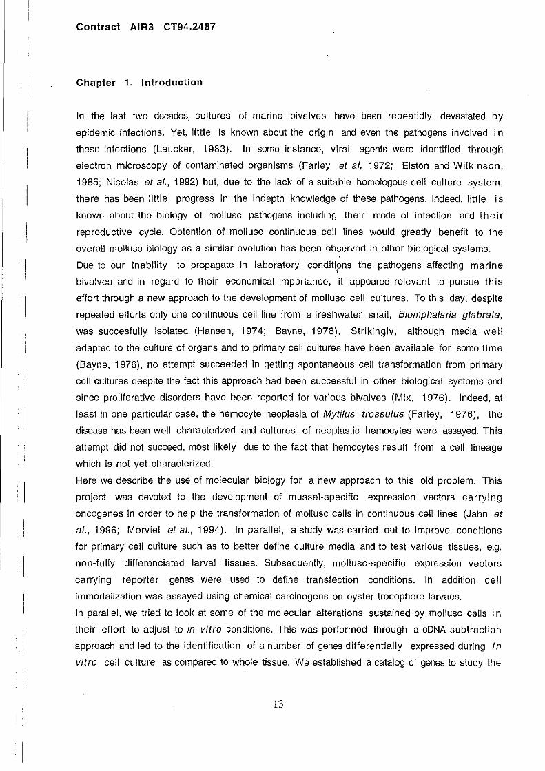
Chapter 1. Introduction
ln the last two decades, cultures of marine bivalves have been repeatidly devastated by
epidemic infections. Yet, little is known about the origin and even the pathogens involved in
these infections (Laucker, 1983). In some instance, viral agents were identified through
electron microscopy of contaminated organisms (Farley et al, 1972; Eiston and Wilkinson,
1985; Nicolas et al., 1992) but, due to the lack of a suitable homologous cell culture system,
there has been little progress in the indepth knowledge of these pathogens. Indeed, little i s
known about the biology of mollusc pathogens including their mode of infection and the i r
reproductive cycle. Obtention of mollusc continuous cell lines would greatly benefit to the
ove rail mollusc biology as a similar evolution has been observed in other biological systems.
Due to our inability to propagate in laboratory conditipns the pathogens affecting marine , bivalves and in regard to their economical importance, it appeared relevant to pursue this
effort through a new approach to the development of mollusc cell cultures. To this day, despite
repeated efforts only one continuous cell line from a freshwater snail, Biomphalaria glabrata,
was succesfully isolated (Hansen, 1974; Bayne, 1978). Strikingly, although media weil
adapted to the culture of organs and to primary cell cultures have been available for some time
(Bayne, 1976), no attempt succeeded in getting spontaneous cell transformation from primary
cell cultures des pite the fact this approach had been successful in other biological systems and
since proliferative disorders have been reported for various bivalves (Mix, 1976). Indeed, at
least in one particular case, the hemocyte neoplasia of Mytilus trossulus (Farley, 1976), the
disease has been weil characterized and cultures of neoplastic hemocytes were assayed. This
attempt did not succeed, most likely due to the fact that hemocytes result from a cell lineage
which is not yet characterized.
Here we describe the use of molecular biology for a new approach to this old problem. This
project was devoted to the development of mussel-specific expression vectors carrying
oncogenes in order to help the transformation of mollusc cells in continuous cell lines (Jahn et
al., 1996; Merviel et al., 1994). In parallel, a study was carried out to improve conditions
for primary cell culture such as to better define culture media and to test various tissues, e.g.
non-Iully differenciated larval tissues. Subsequently, mollusc-specific expression vectors
carrying reporter genes were used to define transfection conditions. In addition cell
immortalization was assayed using chemical carcinogens on oyster trocophore larvaes.
ln parallel, we tried to look at some 01 the molecular alterations sustained by mollusc cells in
their effort to adjust to in vitro conditions. This was performed through a cDNA subtraction
approach and led to the identification of a number of genes differentially expressed during in
vitro cell culture as compared to whole tissue. We established a catalog 01 genes to study the
13

Contract AIR3 CT94.2487
biology of mollusc cells in culture. This should provide useful information to apprehend the
complexity of marine mollusc cells.
This report provides the details and analysis of the most significant results obtained during
this programme.
14

Contract AIR3 CT94.2487
Chapter 2. Materials and Methods
Cloning and characterization of the actin gene family
High-Iength genomic ONA fragments 01 the mussel Mytilus galloprivincialis were obtained
essentiallyas described (Ausubel et al., 1996). Brielly, a whole animal was lrozen in liquid
nitrogen and was ground in a tissuemizer and incubated in the digestion buffer (100 mM NaCI,
10 mM Tris.CI, pH8, 25 mM EOTA, pH 8,0.5% SOS, 0.1 mg/ml Proteinase K) at 50°C lor 16
h. ONA solution was repeatedly phenol/CHCl3 extracted and dialyzed lor extensive periods of
time in large volumes 01 TE buffer at 4°C. 400 >tg 01 genomic ONA was partially digested with
Sau 3A (0.02 units/ >tg 01 ONA for 40 min), ethanol precipitated, resuspended in 400 ~LI 01 TE
and loaded on a 10 to 40% sucrose gradient made in buffer A (1 M NaCI, 20 mM Tris. CI, pH
7.5, 5 mM EOTA) and centriluged in a SW28 rotor at 20°C for 20 h. Alter analysis on agarose
gel, Iractions containing the 9 to 25 kb-Iong ONA Iragm~nts were precipitated and ligated in
the Lambda Fix Il phage vector according to manufacturer's recommendations (Stratagene 6
Cloning Systems, USA). 1.2 10 independent recombinants were obtained that correspond to 4
times the number 01 inserts of 15 kb required lor a 99% chance 01 isolating an individual 9 5
sequence since the mussel genome is about 10 kb-Iong. 3 10 clones were screened with a
400-bp-long radiolabeled probe carrying 160 bp of the exon-1 of Bombyx mori actin A3 gene
(Mounier et Prudhomme, 1986). Filter hybridization (5x SSC at 55°C) and washes (0.5 X
SSC at 55°C) were conducted at low stringency. Inserts 01 a subset 01 clones were excised with
Not and ligated in the Not I-linearized pBluescript plasmid (Stratagene). Inserts were analyzed
by digestion with various restriction enzymes and were submitted to Southern blotting with
the same probe as above. Only one restriction Iragrnent hybridized to the actin probe olBombyx
mori as weil as to the lull-Iength hurnan actin cONA (data not shown). Positive Iragrnents were
subcloned in pBluescript vector (Stratagene) and were sequenced using a kit according to
rnanulacturer's recomrnendations (Pharrnacia Biotech, Sweden).
RNA extraction
Total RNA was extracted according to the single-step method by acid guanidine thiocyanate
phenol-chlorolorrn extraction (Chornczynski and Sacchi, 1987). Poly A selection was
performed with oligodT chrornatography twice in batch and a third time in a column in order to
minimize the elution volume. Poly A RNA was quantified by measuring the absorbance at 260
nm.
Riboprobes and RNase protection.
15

1
1
1
1
Contra ct AIR3 CT94.2487
Riboprobes were transcribed from inserts PCR-amplilied from the same individu al that
provided the RNA. The 5'-non coding region of each actin gene was PCR·amplilied with specilic
sense primers with the following coordinates relative to the the initiation codon (the A of Am
being definedas+1) (·135; ·116),( -172; - 153), (·117; -97), (-400; ·381), (-
248; ·228) for mae-1 to -5, respectively and a 2 fold degenerated common antisense primer
located at coordinates (+68; +46). PCR fragments were cloned in pGEM-T (promega, Wi) and
provided the pM1 to pM5 plasmids for mae-1 to -5, respectively. pM1 to 5 plasmids were
linearized with specific restriction enzymes (Neo 1 for mae-1, $pe 1 for mae-2 and ·3, Rsa 1
for mae-4, Gia 1 for mae-5) and allowed the in vitro transcription of 293-, 301-, 244-,
254- and 308 nt·long riboprobes homologous ta the genomic sequence on 203, 240, 185,
178 and 256 nt lor mae-1 ta -5 transcripts, respectively. 0.5 fl9 of polyA selected RNA were 5
hybridized to 10 cpm of gel-purified riboprobe. Riboprobes for mae·1, ·2 and -3 were
hybridized at 30°C for 16 h and digested with RNase One (promega) for 1 h at 30°C. Because
the riboprobes for mae·3 and -5 are A+ T rich, hybridization was done in absence of
formamide and digestion was performed with Rnase One or RNase T1 for 1 h at 20°C.
Primer extension
Gene specific antisense primers were designed at coordinates (-1; -27), (-1; -30), (-1; -
26), (-1; -30), (-1; ·32) lor mae-1 to -5 transcripts, respectively. DNA labeling was
perlormed lor 5 min with [a32p] dCTP and later chased with 1mM 01 cold dCTP. 5 fl9 01 total
RNA was hybridized ta specilic primers for 90 min at 65°C in 15 fll 01 hybridization buffer
(150 mM KCI, 10 mM Tris-CI pH 8.3, 1 mM EDTA) and then ethanol precipitated. Pellets
were resuspended in 20fll 01 reverse transcription mix [10 mM Tris-CI pH 8.3, 50mM KCI,
5 mM MgCI2, 1mM dATP, 1mM dTTP, 1mM dGTP, 10 flCi [a'2p] dCTP (3000Cilmmol), 0.15
mg/ml Actinomycin D, 13 units 01 AMV reverse transcriptase (Eurogentech, Belgium)].
Reaction was incubated at 42°C lor 5 min and then it was adjusted to a linal concentration 01 1
mM dCTP and lurther incubated lor 1 h at 42°C. Formae-4, the protocol was modified as
lollows. RNA was denatured lor 5 min at 90°C in 7 fll 01 hybridization buffer in absence 01
EDTA and then transfered directly to 42°C and adjusted to 20 fll with reverse transcription
mix and allowed ta proceed as above.
Characterization of the actin cDNA 5' end
RNA was extracted from a single animal and cDNA was synthesized and ligated to adaptors using
a kit according to manufacturer's recommandations (Clontech, CA). Specific antisense primers
(5'·GTGAA TT ACCAA TGCGGCTACATCA-3' for mae-3; 5'-CAACGCGGCTACGTCTTT ATCGT
-3' for mae-5) were designed based upon the nt sequence of a single aile le of the XL 1
individual. The hybridization temperatures, 66 oC and 68°C, were experimentally determined
16
1
. 1
; 1
Contract AIR3 CT94.2487
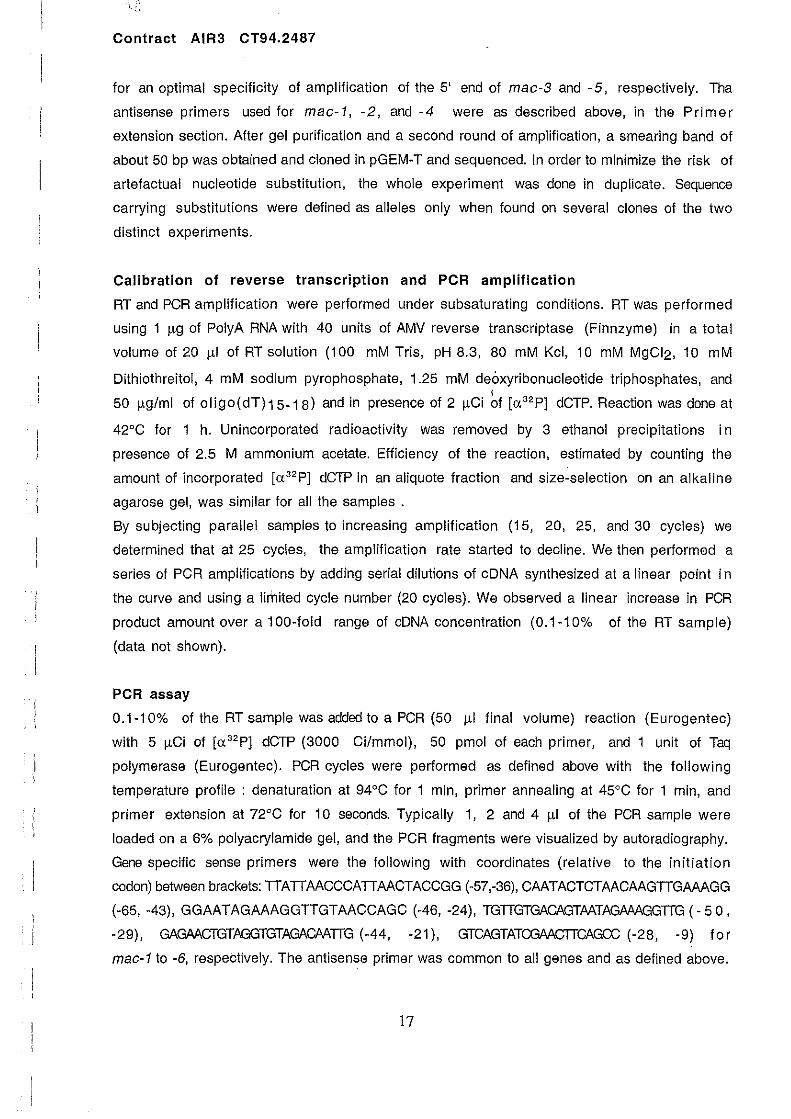
for an optimal specificity of amplification of the 5' end of mac-3 and -5, respectively. Tha
antisense primers used for mac-1, -2, and -4 were as described above, in the Primer
extension section. Alter gel purification and a second round of amplification, a smearing band of
about 50 bp was obtained and cloned in pGEM-T and sequenced. In order to minimize the risk of
artefactual nucleotide substitution, the whole experiment was done in duplicate. Sequence
carrying substitutions were defined as aile les only when found on several clones of the two
distinct experiments.
Calibration of reverse transcription and PCR amplification
RT and PCR amplification were performed under subsaturating conditions. RT was performed
using 1 J.lg of PolyA RNA with 40 units of AMV reverse transcriptase (Finnzyme) in a total
volume of 20 J.l1 of RT solution (100 mM Tris, pH 8.3, 80 mM Kcl, 10 mM MgCI2, 10 mM
Dithiothreitol, 4 mM sodium pyrophosphate, 1.25 mM deoxyribonucleotide triphosphates, and , 50 J.lg/ml of 0Iigo(dT)15.18) and in presence of 2 J.lCi of [a 32 p] dCTP. Reaction was done at
42°C for 1 h. Unincorporated radioactivity was removed by 3 ethanol precipitations in
presence of 2.5 M ammonium acetate. Efficiency of the reaction, estimated by counting the
amount of incorporated [a 32 p] dCTP in an aliquote fraction and size-selection on an alkaline
agarose gel, was similar for ail the samples .
By subjecting parallel samples to increasing amplification (15, 20, 25, and 30 cycles) we
determined that at 25 cycles, the amplification rate started to decline. We then performed a
series of PCR amplifications by adding seriai dilutions of cDNA synthesized at a Iinear point in
the curve and using a limited cycle number (20 cycles). We observed a linear increase in PCR
product amount over a 100-fold range of cDNA concentration (0.1-10% of the RT sample)
(data not shown).
PCR assay
0.1-10% of the RT sample was added to a PCR (50 J.l1 final volume) reaction (Eurogentec)
with 5 J.lCi of [a 32p] dCTP (3000 Cilmmol), 50 pmol of each primer, and 1 unit of Taq
polymerase (Eurogentec). PCR cycles were performed as defined above with the following
temperature profile: denaturation at 94°C for 1 min, primer annealing at 45°C for 1 min, and
primer extension at 72°C for 10 seconds. Typically 1, 2 and 4 J.l1 of the PCR sample were
loaded on a 6% polyacrylamide gel, and the PCR fragments were visualized by autoradiography.
Gene specific sense primers were the following with coordinates (relative to the initiation
codon) between brackets: TTATTAACCCATTAACTACCGG (-57,-36), CAATACTCTAACAAGTTGAAAGG
(-65, -43), GGAATAGAAAGGTTGTAACCAGC (-46, -24), TGTTGTGACAGTAATAGAAAGGTTG (·50,
-29), GAGAACTGTAGGTGTAGACAATTG (-44, -21), GTCAGTATCGAACTTCAGCC (-28, -9) for
mac-1 to -6, respebtively. The antisense primer was common to ail genes and as defined above.
17

Contra ct AIR3 CT94.2487
Primary cell culture and media.
After external cleaning the oysters, were opened and internai cavities was washed with
filtered-sterile sea water; hearts from several individuals were collected and washed in
filtered sea water supplemented with 1 % penicillin/streptomycin solution. Tissue was minced
and further washed 4 to 6 times and homogeneized using a manual homogenizer fitted with a
tellon piston and dispensed in 25 cm2 lIasks (Greiner). Cells were allowed to attach in a
minimal amount of media and after 2 hours additional medium was added and then changed
everyday for the first four days in order to prevent contamination. To this point ce Ils were 6
about 80 to 90% confluent, mainly with individual cells (approximately 10 cells per plate).
Medium was renewed twice a week and cells survived for approximately 2 to 4 weeks without
gross morphology changes. Afterward, morphological changes were observed (Figure 1 - 2 ) ,
with altached cells being increasingly sensitive to medium renewal while mortality greatly
increased. No apparent cellular division was observed im the culture. At this time, a large
percentage of the cells went to suspension. Trypan blue exclusion viability test and
microscopie evaluation showed that these cells were alive.
While several culture media were satisfactorily assayed for the first 2 weeks of culture, the
best results were obtained with filtered sea water supplemented with 20% of oyster
hemolymph (a filtered mixture removed from pericard cavity, abductor muscle, mantie, foot
and gonad) and containing 1 % antibiotic mix.
Primary mussel cell cultures were prepared using 1 year old individuals.The animais were
washed as ab ove and the gonade infiltrated mantle was removed and washed in filtered sea water , supplemented with 1% of penicillin/streptomycin solution. It was then minced and further
washed for 4 to 6 times and was then resuspended in 1 ml of filtered sea water with 0.012 %
collagenase and 0.012% of pronase, and was further incubated for 6 to 8 hours at 4°C. Digested
tissues were then washed 3 times and dispensed in 25 cm2 flasks (Greiner). After 2 hours of
incubation at 25QC, media was renewed. The best results were obtained with the same media as
described for oyster cells. Media was renewed twice a week and cells were kept alive for
several weeks. However individual cell attachement was not as good as observed for oyster
cells. In order to split cells, individual cells were obtained through tissue disaggregation. 1 n
order to obtain an optimal tissue disaggregation, cells were treated with a trypsin solution
(0.12%) diluted into a 35/1000 salt solution lacking calcium salts. Cells thus remained
unattached du ring this treament. However, individual cells remain very sensitive to any
enzymatic treatment, particularly mussel cells, and wh en applied this procedure led to a lower
cellular viability. In addition, among the surviving cells a bigger proportion was unable to
reattach to the dish, even in the presence of cell attachment factors such as poly-L-Iysine.
Treatment with substances known to promote alteration in cell division
18

Contract AIR3 CT94.2487
Treatment with Ethyl Methane Sulfonate (EMS): Primary cell cultures were treated fa r
different time periods (ranging from 1 ta 6 hours) with increasing concentrations of EMS,
ranging from 0.5 ta 32 ml of a concentrated solution of EMS (1.17 g/ml, Cat# M-0880,
Sigma). Concentrations were chosen according ta results previously described for yeast cell
mutagenesis. Cells were rinsed twice with a 5%-sodium thiosulphate solution (ta inhibit EMS)
in sea-water and then incubated with regular cell culture medium at 25QC. Treatment with
Quercitin: Cells were treated for various time periods with quercitin which was previously
dissolved in 0.025% DMSO and then diluted ta its final concentration in cell medium.
Transfection
Cells from oyster heart were plated at 3 million cells per well, in 24 well·plates, and each
experiment were performed in triplicate. Cells were transfected by lipofection using the
methodology recommended by the supplier (DOTAP, Boehringer). The complex was prepared by 1
mixing 5 J.lg of DNA diluted into 50 !-lI of Hepes (20mM) ta 100 J.lI of Hepes containing 30 J.lg of
Dotap. Alter an incubation period of 15 min, the mixture was added to 0.5 ml of culture media
and dispersed onto the cells. The campi ex was removed alter an additional incubation period of
24 hours at 25°C and fresh media was added to the culture. Following another 24 hours
incubation period, transfected cells were assayed through in situ detection of Bgalactosidase
activity and/or by determining the Bgalactosidase activity in the cellular extract.
Identification of the expression vectors: A31acZ, the insect expression vector con tains the lacZ
gene under the control of the actin A3 gene promoter from Bombyx mari (Mounier et al.,
1987).The pmac-1 ta -4 mollusc·specific expression vectors contain either the lacZ
reporter gene or the firefly luciferase gene under the control of different segments of the
promoter regions of Mytilus actin genes. Mammalian expression vectors are under the control
of viral promoters (RSV, CMV and SV40) and they contain the lacZ reporter gene.
Suppression Subtractive Hybridization analysis
For Suppression Subtractive Hybridization analysis, double stranded cDNAs were prepared by
reverse transcription and then digested with Rsa 1 restriction enzyme. The cDNA chosen ta be
the tester cDNA was then ligated ta specific adapters and hybridized in high stringency
conditions with an excess of Rsa 1 digested driver cDNA. Specific tester cDNA fragments were
then selected by PCR amplification with polymerase ADVANTAGE mix as described in the
Subtraction cDNA kit (Clontech) using adapter·specific primers. Two rounds of hybridization
and amplification were performed. Generated PCR products were randomly cJoned in pGEM
vector using the pGEM·T-Easy cloning system (promega).
DNA Sequence analysis
19

Contra ct AIR3 CT94.2487
Partial sequences of cloned PCR products were obtained by double stranded DNA sequencing
using plasmid·specifics primers. Sequence data obtained was then submitted to databank se arch
using the gapped Blast research tools. nBlast and xBlast programs were used for homology
searches in nucleotide and protein data banks, respectively.
Northern and "Reverse-Northern" blct analysis
Cloned cDNA fragments obtained by Suppression Subtractive Hybridization analysis were gel
purilied and then used as probes to check the specilicity of RNA accumulation 01 the respective
genes. "Reverse-Northern" blot was obtained by performing a Southern blot with PCR
amplified double stranded cDNA obtained under optimized conditions. Probes were labeled with
[a32PjdCTP (3000 Ci/mmol, Amersham) using the Prime-It Il kit (Stratagene) and
hybridized overnight in 6X SSPE, 0.5% SOS, 50% Formamide, 50 ~g /ml CalI thymus DNA at
42QC. Membranes were stringently washed in 0.1X SSPE at 65QC du ring 30 min., and , autoradiography perlormed at -70QC with Kodak X-Omat AR film.
Detection of the B-galactosidase activity
For in situ detection, culture media was removed, cells were washed once with sea-water,
lixed in 0.5% glutaraldehyde (note that larvaes were lixed in 2% glutaraldehyde) solution fo r
15 min. and then incubated in PBS with 0.6mM chloroquine at 30°C for 45 min to inhibit
endogenous Iysosomal Bgalactosidase activity. Cell layer or larvae were th en washed 3 times in
PBS and stained with a solution containing 5mM K-ferricCN, 5mM K-IerroCN, 2mM MgCI2,
0.1 % Triton X-100, 0.01 % sodium deoxycholate and 1 mg/ml X-gal at 37QC lor 24 hours.
Cells expressing the Bgalactosidase activity appeared blue stained.
Cell extracts for quantitative assay were prepared according to the manulacturer's
recommendations and were kept at -80QC until processed. Detection was perlormed with the
Luminescent Bgalactosidase Genetic Reporter System Il (Clontech) with 20 ~I of cell extract
using a scintillation counter (Beckman) equiped with a single photon option. Total protein was
determined using the Bio-Rad Protein Assay kit with 10 III of cell extract. Results are
expressed per mg of total protein.
20

Contract AIR3 CT94.2487
Chapter 3. Results
Isolation and characterization of the Mytil us actin gene family
A series of 19 clones were obtained by screening a mussel genomic DNA library with a probe
corresponding to the 5'-end of the actin gene of Bombyx mari. Sub-clones corresponding to 3
clones still hybridizing to the same probe were sequenced and analyzed. An actin encoding open
reading frame was identified in ail three clones (with two clones sharing the same amino acid
sequence) which were referred to as mac-1 and mac-2 genes. mac-1 and mac-2 specifie
primers were used to screen the 16 remaining uncharacterized clones of which 8 were found to
be distinct genes.
A weakly degenerated primer was then designed to hybridize into the actin ORF and was used to
sequence these 8 distinct genes. Actin encoding sequences ,with amino acid substitutions, were
lound in 4 of these clones which were termed mac-3 (2c1ones), mac-4 and mac-5 while the 4
remaining clones were not further characterized. Therefore, the 19 initial clones led to the
identification of 8 mac-1, 3 mac-2, 2 mac-3, 1 mac-4 and 1 mac-5 genes.
Full-Iength sequencing 01 mac-1 to -5 promoter regions was carried out and it determined the
existence 01 a 126-nt-long exon with a distinct actin-related amino acid sequence lor each
gene. In addition the 5'-non-coding region was lound different lor each gene conlirming the
assumption that live distinct mussel actin genes were identilied.
The 5'-non-coding sequences 01 the dillerent actin genes were lound to share no homology.
Different transcription· regulatory elements (enhancer), including GC-box, CArG-box,
CCAAT-box, are similar to the elements known in vertebrate promoter regions. They are not
present or located at the same coordinates in ail the live genes (Fig. 1).
ln addition, sequencing 01 two mac-1 promoter regions revealed the existence 01 mac-1 alleles
which were used as genetic markers in the study 01 the Mytilus species complex.
Amplilication 01 the actin cDNA 5'-end was perlormed by anchored-PCR using an antisense
primer corresponding ta a consensus sequence for the N-terminus 01 the mussel actin genes.
DNA Iragments migrating as 50 to 60 bp were obtained and their sequence corresponded to the
previously identilied genomic clones (data not shown). By contrast, a sequence distinct in both
the leader and the amino acid sequence was identilied and it provided evidence lor an additional
mussel actin gene, here relerred to as mac-5 (Fig.1), lor which we do not have a genomic
clone. Interestingly, the cDNA 5'-end 01 mac-3 was not homologous with any 01 the actin
sequences determined above, thus demonstrating there is a first non-coding exon belore the
sequence encoding the actin N-terminus. Because 01 its peculiar structure, this gene was set
apart and th us is now relered to as mac-6. Southern blot analysis 01 genomic clones using the
leader sequence 01 mac-6 cDNA as a probe did not reveal any positive clone (data not shown).
21
ATG
-1290 -955 Cap ! axon 1 intron 1 +820 1 . ,. ~
C+l ft . mac-l il " -182-159 -108 + 126
- 56
Cap ATG
-1041
t! ! axon 1 intron 1 + 1112
L-...{.' mac-2 ~ " , , "
- 161 - 111 +1 + 126 - 66
ATG Cap 1 exon 1 intron 1
-927 r: +441 ~~'----------~O~----~ .. t=========~/ '
-146 +1 +126
mac-3
- 60
ATG lntron 1
1 axan 1
-8C~.~, ____________________ ~x .. ===========~~15 mac-4
- 833 -42 +1 + 126 +303
ATG 1 exon 1 +45
mac-5 - 34 +1
Splice acceptor
site ~ ATG
.. ,--1 ----tI.'I-' __ ---,1~,'I-' _:LJUt::==ex=o=n=2==]-__ +:...:;~77 -55 -7 -762 -6 +1
exon 1 intron 1 intron 2
mac-6 + 126
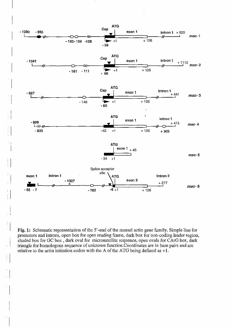
Fig. 1: Schematic representation of the 5' -end of the mussel actin gene family_ Simple line for promoters and introns, open box for open reading frame, dark box for non-coding leader region, shaded box for GC box, dark oval for micro satellite sequence, open ovals for CArG box, dark triangle for homologous sequence of unknown function. Coordinates are in base pairs and are relative to the actin initiation codon with the A of the ATG being defined as + 1.

Contraet AIR3 CT94.2487
Thus, the 55·bp-long cDNA leader sequence corresponds to a 49·bp-long first exon that is not
present in the mac-6 genomic clones plus a 6·bp-long non-coding sequence of the second exon
as indicated on Fig. 1.
Sequence analysis showed no homology between the diflerent actin genes apart fram a motif
located in the leader sequences of each gene except mac-5 and it is also in the intron-1 of mac-
6 (Fig. 1; Fig. 2). This motif is not significantly homologous to any other sequence and its
putative function is unknown.
Analysis revealed that the predicted amino acid sequence for the exon-1 is identical for mac-1
and mac-2 while it diflers by one or several substitutions with the other genes as summarized
on Fig. 3. Conservation of the N-terminal part between the diflerent Mytilus actins (88 to
100%) is not greater than it is with invertebrate or vertebrate actins (Table 1). nd
Downstream, the open reading frame is interrupted alter the 42 amino acid by a stop codon
except in mac-1, itself preceded by a sequence homologou~ to the consensus for a splice donor , site (Fig. 4). Actin introns at position 41/42 are conserved with chordates at the diflerence of
lower invertebrates and arthropods which have few introns with variable positions
(Sheterline et al., 1995). No second exon could be localized in our genomic actin clones
Sequence polymorphism.
RNA and genomic DNA were separately extracted from single individuals. Anchored-PCR was
performed using an antisense primer designed in the actin N-terminal amino acid sequence as
above. In parallel, PCR amplification was performed on genomic DNA using the same antisense
primer as above and sense primers specifie for each actin gene (see Materials and Methods).
DNA fragments amplified from genomic DNA extended fram -116, -152, -380, -227, -96 to
+45 (the A of ATG being defined as +1) for mac-1, -2, -3, -4, and -6, respectively.
Sequencing of the DNA fragments amplified with mac-1 to -4 primers revealed two sequences
corresponding to two alleles for each individual as it is expected for a gene present as one copy
per haploid genome. For mac-6, five distinct sequences were found (data not shown) that
correspond to at least 3 closely related genes per haploid genome. Allelic sequence
polymorphism was found identical on both the cDNA and the genomic DNA, and amounted to
1.6%, 1.3%, 1.3% and 0.4% for mac-1, -2, -3, and -4, respectively, and it did not result in
amino acid substitution. These values are in good agreement with data previously obtained fo r
the intran sequence of mac-1 (Ohresser et al, 1997). By contrast, the percentage of
substitution between the mac-6 sequences amounted to 5% for the intron-1 (4 sequences on
42 bp) and to 8% for the exon-1 (3 sequences on 64 bp). Interestingly, the live mac-6
sequences isolated from a single individual encoded 3 diflerent sequences on the first 15 amine
acids (Fig. 3, var2, var3 and var4) which are distinct from the original sequence isolated
22

Table 1. Amino-acids and nucleotidic palrwise identity score" in the 42 first amino acid region of the actln genes of Mytilus galloprovincialis and other speciesb
•
Invertebrates Vertebrate
Moliuscs
MusseI
mac-l mac-2 mac-6 mac-3 mac-4 mac- A.c.n A.c.rn P.m. B.g. A.s. H.s.s H.S.b 3"
mac-l 100 93 93 93 95 100 98 98 98 100 86 95
mac-2 97 93 93 93 95 100 98 98 98 100 86 95
mac-6 89 89 95 95 88 93 90 90 95 93 88 93
mac-3 89 89 90 98 88 93 90 9p 95 93 88 95
mac-4 89 89 88 97 88 93 90 90 93 93 81 93
mac- ND d ND ND ND ND 95 93 93 93 95 86 93 3"
A.c.n 79 82 78 74 73 ND 98 98 98 100 86 95
A.c.rn 75 77 73 72 70 ND 87 100 95 98 83 95
P.m. 80 81 78 77 74 ND 85 82 95 98 83 93
B.g. 85 84 82.5 82 80 ND 82 80 82.5 98 88 98
A.s. 82 79.5 79 78 76 ND 84 81 82.5 78 86 95
H.s.s 68 67 68 63 64 ND 76 77 69 69 70 88
H.s.b 70 70 68.5 70 68.5 ND 82 84.5 83 77 77 79
GC% 47 47 47 45 44 ND 60 63 59 53 53 69 68
a Noted in percent on the basis of Fig 3 aligment. Note that gap pasi tians between pairs were not considered for identity score. Amino acid identity 18 above the diagonal, and nucleotide identi ty is under the diagonal. Coding sequence are determined from genomic lambda clones, excepted for mac-6 which sequence was deduced from a cDNA.
b For abreviation see fig 3 legend
C The undetermined 27 last amino acid of mac-6 region were considered as conserved
d Not determined

mac-l mac-2
mac-l mac-2
mac-l mac-lbl mac-i(XLi-i) mac-i (XLl-8)
mac-2 mac-2(XLl-2)
-112 -115
-87 -99
-40
-49
CTCCGCCC CTCCGCCC
CATAAATA CATATAAA
AAAGGTTTTAAGACGTG AAAGGTTTTAAGACGTG AAAGGTTTTAAGACGTG AAAGGTTTTAAGACGTG
GAAAGGTTCGC--ACGTG GAAAGGTTCGC--ACGTG
mac-2(XLl-8) GAAAGGTTCGC--ACGTG
mac-3 -57 AGTCAATTGTTGTGACAGTAATAGAAAGGTTGTTACAAGGT mac-3(XLl-2) AGTCAATTGTTGTGACAGTAATAGAAAGGTCGTTACAAGTT mac-3(XLl-5) AGTCAATTGTTGTGACAGTAATAGAAAGGTCGTTACAAGTT
mac-4 -29 AGACAATTGTTGAG mac-4(XLl-22) AGACAATTGTTGAG mac-4(XLl-24) AGACAATTGTTGAG
mac-6 -68 rnac-6 (XLl-2)
mac-6(XLi-CL3) -55 rnac-6(XLi-CL5) mac-6 (XLi-CL6) mac-6 (XL1-CL8)
TGTAATAGGACCAGACAGGTTGCAAAAAATA TGTGATAGGACCAGAAAGGTTGCAATAAATA
GTTGTTTCAGGAATAGAAAGGTTGTAACCAGCT CAGTAA------GGTTGTTACAATCC CAGCAA------GGTTGTAACAATCC CAGCAA------GGTTGTAACAATCC
Fig. 2. Sequence conservation between different alleles of the different actin genes. Theses mot ils are in the untranslated leader sequence 01 the actin RNAs except 10 r mac-6 where it is also present in the intron-1 and lormac-5 in which it was not lound. Nucleotide identity is shown in bold; coordinates are relative to the A 01 the actin initiation codon.

mac-l MCDDD-VAALVVDNGSGMCKAGFAGDDAPRAVFPSIVGRPRHQ 42 mac-2 · .... - .................................... . mac-3 .D.E.- ..... I ............... ················ mac-4 .D.K.- ..... I .............................. . mac-5 · .... -LS ....... . mac-6 varl ... E.- ... M.I ................... ············ mac-6 var2 · .... - ..... IH .. . mac-6 var3 · .... - ..... I ... .
mac-.6 var4 · . . .. . ........ . mac-6 var5 ., .E.- ..... I ....
P.m. ... . E- ................ ············· ....... . A.c. · .... - .................................... . B.b. . .. E.- .................................... .
A.s. • •••• - •••••• o ••••••••••••••••••••••••••••••
H.b .-.E.-I ................... ; ............... . H.a .. . E.ETT ... C .............. ; ................ .
Fig. 3. Amino acid sequence comparison of the actln N-terminal part. A. C.,
Aplysia californica (neurons); P. m., Placopecten mage/lanicus; B. g., Biomphalaria glabrata; A. S., Artemia satina (actin 211). H. a, human a-actin (skeletic) gene; H. b, human B-actin (cytoplasmic) gene.
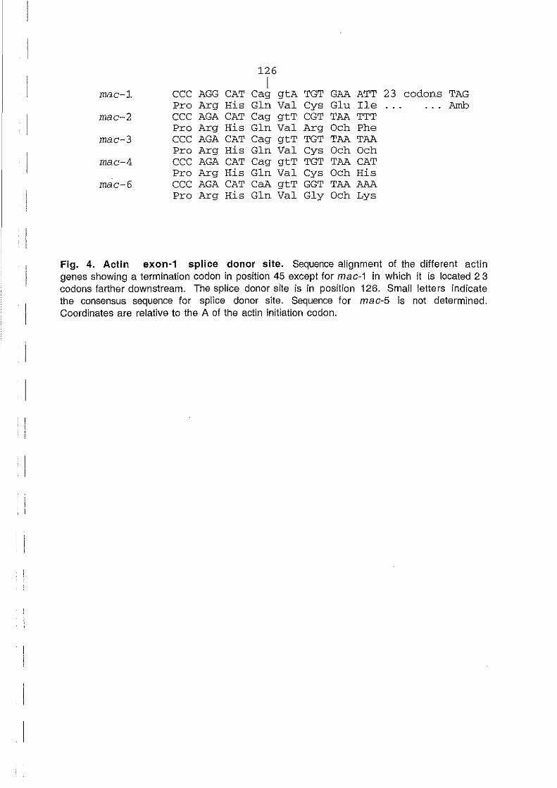
126 1
mac-1 CCC AGG CAT Cag gtA TGT GAA ATT 23 codons TAG Pro Arg His Gin Val Cys Glu Ile Amb
mac-2 CCC AGA CAT Cag gtT CGT TAA TTT Pro Arg His Gin Val Arg Och Phe
mac-3 CCC AGA CAT Cag gtT TGT TAA TAA Pro Arg His Gin Val Cys Och Och
mac-4 CCC AGA CAT Cag gtT TGT TAA CAT Pro Arg His Gin Val Cys Och His
mac-6 CCC AGA CAT CaA gtT GGT TAA AAA Pro Arg His Gin Val Gly Och Lys
Fig. 4. Actin exon-1 splice donor site. Sequence alignment of the different actin genes showing a termination codon in position 45 except for mac-1 in which it is located 23 codons farther downstream. The splice don or site is in position 126. Small letters indicate the consensus sequence for splice donor site. Sequence for mac-5 is not determined. Coordinates are relative to the A of the actin initiation codon.

Contract AIR3 CT94.2487
from the genomic library (Fig. 3, var1). An additional amino acid sequence (Fig. 3, var5) was
identified on a separate individual.
Cap-site determination
ln order to determine the cap-sites 01 mac genes and because 01 the allelic sequence
polymorphism, probes were cloned Irom the genomic DNA 01 the individual that provided RNA.
PCR amplilication was performed on genomic DNA using the same antisense primer as above
and sense primers specific lor each actin gene (see Materials and Methods). DNA Iragments
extending Irom +68 to -135, +68 to -172, and +68 to -178 lor mac-1, -2, and - 3,
respectively, were cloned and in vitro transcribed Irom pBluescript vector. RNase protection
assay was perlormed using uniformly labelled riboprobes that provided 125-, 137- and
132-nt-long undigested lragments (data not shown) which correspond to 56-, 68-, and 64-
nt-long leader sequences riboprobes lor mac-1, -2, and'-3, respectively (Fig. 1). Similar
experiment was carried out lor mac-4 in various conditibns 01 stringency with RNase One 0 r
RNases A plus T1 and provided a protected Iragment corresponding to the exon-1 length (data
not shown). This result does not provide the mac-4 cap-site position but instead it indicates
the location 01 an ArT rich region (a stretch of 17 AIT in position -15 to +2) which appears to
interfer with the RNase assay. In addition this assay could not be done lor mac-6 and mac-5 lor
which we do not have a corresponding genomic sequence.
Gene-specific primer extension using antisense primers starting 1 nucleotide belore the 32
initiation codon was performed in presence 01 [a Pl dCTP. Reaction products were resolved on
denaturing polyacrylamide gel and their size was precisely determined by the co-migration of
a sequencing reaction. Elongation products 01 mac-1, -2, and -3 migrated as 56-, 66-, and
60-nt-long DNA fragments (data not shown) thus providing a cap-site location in agreement
with the value obtained above by RNase protection assay.
Cap-sites are located 20 to 25 bp downstream a putative TATA box (7 AIT). Approximate
location 01 the cap-site position lor mac-4 to -6 was estimated by sequencing the longest
products 01 anchored-PCR reactions performed in parallel. These experiments provided 52 - ,
34-, 55-nt-long leader sequence lor mac-4, -5, and -6, respectively, as schematically
represented on Fig. 1.
Tissue-specifie expression of the actin gene family~
Northern blot analysis revealed a 1.9-kb-long signal lor ail the actin genes. Quantitative PCR
was perlormed on RNA extracted from tissues obtained alter dissection and it revealed some
degree of tissue-specilicity in the expression 01 the distinct actin genes. Comparison 01 the
level 01 expression 01 a single gene in the different tissues revealed that mac-1 and mac-2
patterns are similar although mac-1 is rather expressed in the mantle border while mac-2 is
23

Contract AIR3 CT94.2487
more expressed in the internai part of the mantle. mac-3, mac-4 and mac·6 are more
expressed in the gill while mac-4 is the unique gene to be substantially expressed in the foot.
mac-5 is more expressed in the adductor muscle and in the mantle border. More experiments
are un der way to beller define the tissue-specificity of these genes. In addition, RNA was
prepared from larvaes at different stages and determination of the stage-specificity of each
actin gene is currently underway.
Isolation of a neuronal- and development-specific transcription factors.
PCR amplification performed on mussel cDNA using primers designed in a phylogenetically
conserved region allowed the isolation of short sequences belonging to two distinct POU
proteins. The sequence corresponding to a POU protein was used as a probe to isolate a unique
clone from the genomic Iibrary. Sequencing of this gene revealed an open reading frame
encoding a 432 amino acid long protein. This protein is homologous to the POU proteins of other
organisms in the Homeo and POU domains. It otherwise displays no homology with other known
proteins. Preliminary data indicate that this transcription factor is not able to transactivate
mammalian promoters that are activated by the mammalian POU protein.
Degenerated primers were designed in phylogenetically conserved regions of two genes, snail
and twist, involved in the development of mesoderm. PCR amplification performed on genomic
DNA provided a sequence for both genes that were used as probes to screen the mussel genomic
library.
This work allowed the isolation of 5 clones for each gene which are currently in the process of
sequencing. Characterization of the tissue-specificity of expression of these two genes will be
done in parallel with the POU gene.
A genetic marker for population studies in the marine mussels Mytilus
galloprovincialis Lmk. and M. edulis L.
A novel intron length-polymorphism at the actin gene locus mac-1 is here reported and used as
a genetic marker for population studies in mussels of the genus Mytilus. Two closely related
genes, mac·1a1 and mac-1b1 from a genomic library of M. galloprovincialis were partially
cloned and sequenced. They mainly differed from each other by a 65-bp insertion within their
lirst intron. Polymerase chain reaction (PCR) primers were designed outside the insertion.
The PCR analysis of 166 individual mussels from M. galloprovincia/is and M. edu/is
populations revealed three size-classes of alleles or allelomorphs, two of which were of the
expected sizes for mac-1a1 and mac-1b1. One allelomorph was absent from M. edulis samples,
while it was present at substantial frequencies in M. galloprovincialis populations. The
frequencies of the two other allelomorphs significantly differed between M. galloprovincialis
and M. edulis populations. The comparison of six mac-1 intron sequences over 277 bp showed
24

Contract AIR3 CT94.2487
at once that allelomorphs encompassed alleles dillering Irom one another by substanlial
numbers of mutations, and that identical alleles were present in both M. galloprovincialis and
M. edulis individuals, a probable result of the recent introgression between the two species. A
very detailed report and discussion 01 this work is provided in annexe 1.
Optimization of the primary cell culture system
Most of the work has been locused on oyster heart that proved to be the best cell donor for 0 u r
purpose. Oyster heart proved to be the most convenient source of cells and routinely provided
uncontaminated, well-attached individual cells that lormed a 80 to 90% confluent monolayer.
By contrast, cells Irom mussel tissues and in particular from mantle (a tissue lound to be a
good cell donor) were consistently more dilficult to disaggregate and provided small aggregates
instead 01 individual cells. In addition, a great proportion were Ilagellated cells that wou Id not
attach because 01 their motility. Culture conditions to obtain attached epitheliaHike cells have
not changed lrom those described last year. We tried other' serum substitute such has Ultraser
(Gibco) without any better results in term of cell division or survival. Results obtained with
calf serum supplementation are consistently worse than those obtained with hemolymph, which
remains the best supplement for these cells. These results conlirm previous data (Odintsova et
al, 1994) showing that calf serum can be added up to 2% without affecting cell morphology 0 r
survival, but increasing its concentration is detrimental to the cell survival.
Characterization of a eukaryotic cell type from a culture of oyster embryos
Because embryos conta in undifferenciated cells, it is an attractive source 01 potentially
dividing cells. Attempts were made to obtain dividing cells Irom Crassostrea gigas embryos.
Early stage embryos were obtained through in vitro fertilization, and were collected and
enzymatically dissociated. Later stages such as trocophore and veliger stages were
mechanically and enzymatically treated. Dissociated tissues were th en spread in tissue culture
plates and maintained in culture as described above in order to detect dividing cells. While
many attempts failed to produce dividing cell culture, in one instance we observed apparent
cell division. These ce Ils were isolated and grown in culture at 28QC using a sea-water based
medium supplemented with L 15 and serum substitutes. Its morphology clearly corresponded to
eukaryotic cells (Figure 5) but did not to usual mollusc cells. The growth curves determined
Irom the estimation 01 the number 01 cells on a Malassay counting cell indicated a doubling lime
of approximately 7 days. This culture was kept in culture for 24 weeks and succeslully
sustained cryopreservation in liquid nitrogen. Wh en thawed and placed in culture conditions
these cells resumed their growth pattern. PCR analysis using C. gigas-specilic primer pairs
designed to amplify microsatelitte sequences (our unpublished data) did not allow the detection
01 the DNA markers specilic lor C. gigas. The 18S ribosomal RNA gene of these cells was
partially cloned and sequenced. This gene shows some highly conserved nucleotide sequences
25

Figure 5 Morphology of the unidentified cells isolated from cultures of oyster embryos .
• 1
1
1
Contract AIR3 CT94.2487
among ail known eukaryotes and thus is conveniently used in phylogenetic studies. The
corresponding region of C. gigas was studied in parallel as a positive control. Specifie PCR
primers were designed in a region of maximal conservation among eukaryotes. Forward primer
starts at the 4th nucleotide of Homo sapiens 18S rRNA and the reverse primer was designed in
order to PCR-amplify a 467 bp-Iong DNA fragment. Genomic DNA was prepared from C. gigas
and from the unidentified cells. For both samples a 450 bp-Iong DNA fragment was obtained, as
observed by agarose gel electrophoresis (data not shown). This DNA fragment was partially
sequenced. As expected, the sequence derived from C. gigas shows a higher match with the 18S
rRNA of two oysters, Crassostrea virginiea and Ostrea edulis respectively. Instead, the
sequence from the unidentified ce Ils showed a higher match with Developayella elegans (Leipe
et al, 1996), and with Chaunacanthid sp. (Amaral Zettler, Sogin & Caron, 1997) and with
Perkinsus sp. (GenBank accession number L0737). Chaunaeanthid sp.has been described as a
"Single cell collected off the coast of Bermuda in September 1994", while Perkinsus is we 11-
known unicellular parasite infesting some bivalves includ'ing C. gigas. In addition, histological
procedures routinely used to identify Perkinsus failed to detect this parasite. Thus our results
suggest that this organism is different from any known marine unicellular organisms. More
work is being performed for a more precise identification of this organism.
Reporter gene expression in primary cell culture
We used two different preparation procedures in order to obtain cells expressing different
phenotypes: epithelial-like and cardiomyocyte-like cells. Both cell types were transiently
transfected with the different vectors prepared by participant 1 alter improving the conditions
to introduce DNA into oyster cells and embryos through lipofection and electroporation. We
transfected by lipofection the following Bgal expression vectors: Bgal-mac-1 and pBgal-mac-
2M and their derivatives pBgal-mac-2, pBgal-mac-1 E and pBgal-mac-2E in the two types of
oyster cell primocultures. The Bgal activity was detected in situ 2 days alter transfection by
Xgal chromogenic substrate (blue positive) in ail samples (Table 2) except for non
transfected controls. Few cells (about10·5) express enough Bgal activity to be detected by this
qualitative method and inter-experiment variability was observed. Similar results were
obtained when uSing 2 hour-old cultures alter dissociation with collagenase in 25%
hemolymph-containing sea water or when using 4 day-old cultures produced fro m
mechanically dissociated cells maintained in 2X L15 medium with 2% fetal calf serum.
ln order to increase the sensitivity of the essay and to obtain quantitative results, Bgal was
enzymatically dosed (activity/total protein) in extracts of cells transfected with the
expression vectors used above (Table 2) and with pBgal-mac-3, pBgal-mac-4 and pBgal
mac-5, (promoter coordinates are -964/-1, -893/-1, -899/-1 lor mac-3 to - 5,
respectively, where +1 relers to adenine 01 translation start codon). More Bgalactosidase
activity (mean values) was detected in cells translected with any 01 the èxpression vectors
th an in non translected cells (Fig 6). For both types of primocultures, we observed inter-
26

Table 2. Efficiency of the expression vectors derived from mussel actin genes (mac) in oyster heart primocultures.
Plasmid Promoter œF SV40 Bgal Name enhance expression
b
gene Coordinates a
pBgal-mac-1 mac-1 -1069/-1 lac-z + pBgal-mac- mac-1 -1069/-1 lac-z + + 1E pBgal-mac-2 mac-2 -746/-1 lac-z + pBgal-mac- mac-2 -746/-1 lac-z + + 2E Bgal-mac-2M mac-2 -990/-1 lac-z +
a Relative to the A (+1) of the translation initiation codon. b ln situ detection of the Bgalactosidase activity in transfected cells.

C
Mac1 Mac2 Mac3 Mac4 Mac5 CMV Mac1E Mac2E
Mac2Em
Figure 6: Transient transfection of the different vectors in oyster cells.Primary cultures of oyster cells from both epithelial and cardiomyocyte phenotype were transfected with the different consttucts and the luminiscence catalyzed by the Bgalactosidase activity was detected as described above. Results of triplicate experiments are expressed as percent of the maximal levels of expression obtained for the A3lacZ vector (logarithmic scale), after being corrected for the total protein level of each sample. Identification of the plasmids is indicated under the bars and refers to those shown in Table 3. C for negative control.
Contra ct AIR3 CT94.2487
experiment variability but also an unexpected high intra-experiment variability lor which we
do not have satislactory explanation. Note that the heart cells lorm large clumps in culture
conditions, which are sometime lost during translection. Nevertheless, we conclude Irom these
experiments that we were able to detect Bgalactosidase activity in the translected ce Ils since
the analysis 01 variance shows that our results are signilicantly different Irom the control
(p<O.01).
The expression vector containing the promoter region 01 an actin gene Irom Bombyx mori
(A3IacZ), which we have shown belore to work weil in cells Irom both vertebrate and
invertebrate origin (see report 2), continued to be used as a positive translection control both
in a control cell culture system (SI9, an insect cell line) and in the primary cultures 01 oyster
cells. We also used this system to test the specilicity 01 the mussel expression vectors. 1 n
addition we tried to improve the signal to noise ratio in our translections results by increasing
the sensitivity 01 the assay through the use 01 luminiscence activity, but we lound no
significant improvement suggesting that the limiting lactbr is not the sensitivity 01 the assay
but the signal to noise ratio.
Treatment with drugs to promote alteration of cell division
A number 01 mutagens have previously been used successfully to induce increased cell division
in a number 01 cell systems. The difference with our present cell culture system is that we
have, at this time, no evidence that our cultures are dividing, thus making it more difficult to
increase cell division by chemical treatment. We have tested two different mutagenic agents
previously used with some success in other culture systems: (1) Ethyl Methane Sulfonate
(EMS) which is a chemical routinely used to increase the Irequency 01 mutants in yeast cells,
randomly promoting Ge to AT transversions (see Current Protoco/s in Mo/ecu/ar Bi%gy, unit
13); (2) quercitin which is a Ilavonoid widely distributed in man y edible fruits and
vegetables, and it is one 01 the most potent mutagens lound in bracken lern. Recent reports
suggest that it may act in promoting mutagenesis and cell translormation, in particular when
cells are both translected with an oncogene and treated with this Ilavonoid. In our system, both
substances either promoted high cell mortality or they lailed to induce cell division in the
cultures.
Transient transfection in axenic larvaes of oyster (Crasso5trea glga5)
Artificial lecundation, is a commonly used technique that involves in vitro mixture 01 the two
types 01 gametes. For this purpose, 3 lemale oyster were picked alter gonad inspection. Gonads
were striped and rinsed in 2 liters 01 liltered and UV-irradiated sea water. The same was done
27

Table 3. cDNAs differenctially expressed between oyster cell culture (C) and heart tissue (H)
cDNAs # Related gene d Identity score ,d Putative function
Cl
C2
Cl4
Cl5
Cl6
Cl7
Cl8
Cl9
C20
C21
eIF_4A 90% mammalian Translation regulation
to actin 99% oyster Cytoskeleton compound
Plastin 64% mammalian Cytoskeleton compound
Astacin 48% crustacean endopeptidase
Rho-GDI 1 56% mammalian MIO inactivation
Ribophorin 1 64% mammalian Protein glycosylation
AIF-I 59% mammalian Allograff reject in mammalian 1
cytochrome P-450 48% mammalian detoxification
EGF-like 34% C. elegens ligand-receptor interaction?
Hl ADF
H2toH3 MLC
38% yeast
78% scallop
Cytoskeleton compound
Cytoskeleton compound
H4
H5
H6
H7
Collagen
PTPase
COI
NADHubi
42% mammalian Extracellular matrix
34% mammalian Phosphorilating membranar receptor
60% echinodenn Mitochondrial respiration
47% mussel Mitochondrial respiration
Reverse Northern
Heart/Cells" Size
++/+++
+/++++
+/++++
-/++++
+/++++
1.8
1.3
3
2
1.7
+/+++ 1.3
+/++++
+/++++
+/++++
+/++++
++++/+
+/++
3.3
1.6
lA
1.7
1.5
1.7
+++++/++ 1.2
+
++++++/+ lA
+
+/+++ 1.6
+/+ 0.7
+/++
ND'
1.6
H8 16S rRNA 100% oyster Mitochondrial tranlation ND
a Ratio of RNA accumulation between heart and cens cultured for 1 week in medium containing 80%
hemolymph. b Not determined. C Sequences identification based upon the identity score. d See text for
details

Contract AIR3 CT94.2487
with a mature male gonad. Soth gamete suspensions were filtered through 150 (.Lm to eliminate
debris. Suspensions were allowed to rest 30 min. for sperm activation and chorion brake down,
and th en mixed until each oocyte stayed su.rrounded by about 10 spermatozoids, and it was
allowed to proceed for one hour. The eggs were then pelleted (2000 rpml 5min) and
resuspended on 500 (.LI. 20 (.Lg of DNA (A3IacZ) were added to 200 000 eggs and this
suspension incubated for 5 min. in ice. Cells were then electroporated (1 pulse, 600V) in a
Sn< electroporator. Following electroporation, eggs were diluted 100 eggs/ml in sea water
and egg development was allowed to proceed for 40 hours at RTo. Larvae were th en fixed and
expression of betagalactosidase was detected by histochemistry as follows: Larvaes were fixed
in 1.25% Glutaraldehyde solution for 15 min, incubated in PSS with 0.6mM Chloroquine
solution at 30°C for 45 min., and then washed three times in PBS buffer and stained with 5mM
K-ferric Cn, 5mM K-ferro Cn, 2mM MgCI2, 0.1% Triton X-l00, 0.01% Sodium deoxycholate
and lmg/ml X-gal solution at 37°C for 24 hours. Data sho"w that both trochophore and veliger ;
larvaes were found expressing the characteristic blue staining indicative of Bgalactosidase
expression while no staining was observed in control cultures. Our preliminary results
showed a 1 %-efficiency in term of transfected larvae. Ali larvaes were used at this stage so we
have no information about the survival rate of electroporated larvaes.
Cloning of genes differentially expressed between whole heart and he a rt·
derived primary cell culture.
Messenger RNA was isolated from both intact oyster heart tissue and from heart primary cell
culture. Two subtractions of cDNA libraries were conducted using a commercially available kit
(Clontech). These libraries were selected so that they would be enriched in genes
preferentially expressed either in the donor tissues (heart) or in primary cell culture.
Differentiai screeening of each library allowed the identification of clones overexpressed in
each condition. Alignment of the partial sequences of the isolated cDNAs indicated significant
homology with sequences in the data bank. 13 out of 32 initial clones were identified as actin
genes, 8 more genes shared significant homology clones in the database (see Table 3) and the
remainder showed no homology with any known gene. Since these clones were ail obtained from
subtraction library, they are ail presumably expressed at high level under cell culture
condition. Indeed, reverse Northern analysis confirmed that the isolated clones corresponded to
genes with a higher RNA steady state level in cell culture condition and th us they correspond to
genes moderately or highly expressed under cell culture conditions. This analysis indicated that
the subtraction library was successful. The approximate cell to heart ratio of expression of
these genes was estimated by comparing signal intensity between cells and whole tissue (Table
3) .
28

Table 4. Comparison of the RNA steady state level for the genes preferentially
expressed in heart primary cell culture when maintained in different culture media.
CDNAs Related Size Seawater 2x Leibovitz medium
# gene Kb 80%H b 25%H 2%S" 10%S 10%S+0.5%L d
#1 #2 #3 #4 #5
Cl eIF-4A 1.8 +++ + + + +
C3 actin 1.3 ++++ ++++ ++ ++ +++
C16 Astacin 2 +++++ +++ + + ++
C17 Rbo-GD! 1 1.7 ++++ ++ ++ ++ ++
1.3 +++ + + + +
C20 PA50 ++++ ++++ +++ +++, +++
C21 EGF-like 1.7 ++++ ++ + + ++
Hl ADF 1.5 + + +++ ++ +
H4 Collagen lA ++ + + +++ ++
aEstimated from Reverse Northem. ~emolymph. CPetal calf serum. dLactalbunùn hydrolysate.
Contraet AIR3 CT94.2487
Actin related cDNAs isolated Irom SSHc contained sequences with high identity scores (near
99%) with actin Irom C. gigas but also with vertebrate and invertebrate actin sequences. These
partial cDNAs are either internai to the ORF (11 clones) or overlap the 5' untranslated region
(UTR) (1 clone) or the 3' UTR (1 clone). The 5'UTR overlapping clone show sequence identity
in the non-coding sequence with a previously reported C. gigas actin cDNA while the 3' UTR
overlapping clone (ex) shows no match with this cDNA. So these 13 actin clones represent
most Iikely transe ri pts Irom several different actin genes. When an aclin coding sequence was
used ta probe reverse Northern blot, a 1.3 kb-Iong band was obtained. More actin RNA was
detected in cells cultured with both hemolyph containing culture medium (Table 4) when
compared to whole heart tissue (Tables 3 and 4). In addition, a small increase 01 RNA level was
observed when lactalbumine hydrolysate was added ta the medium (Table 4).
29

Contract AIR3 CT94.2487
Chapter 4. Discussion
• The actin gene family.
Five aetin genomic clones were isolated from a mus sel genomic library made in the phage vector
Lambda Fix. Sequence analysis revealed an exon carrying a 42 amino acid-Iong open reading
frame homologous to the N-terminus of the actin proteins of other organisms.
The S'-end gene structure. Analysis of the nucleotide sequence revealed an exon de!imited
by a sequence eorresponding to the consensus for a splice donor site and a promoter-like region
containing a putative T ATAA box, and motifs homologous to the transcription enhancers of other
eukaryotic organisms.
The most significant motif, the GC box that is the target of "the sp 1 transcrition factor, is present , on mac-l, -2 and -4. Another element, the SRE element, is the target of factors belonging to the
Serum Response Transcription Factors (SRF) family. These enhancers are commonly found in
the promo ter region of actin genes (Chen and Schwattz, 1996) and have been shown to participate
to the early regulation of the alpha-actin gene in mammals. Interaction between SRF and other
factors suggest a rnechanism through which cells can coordinate intrinsec information on cell
specificity with the response to external regulatory signaIs and a mechanism which can control
cell-type-specifie expression and differentiation in bath a positive and negative manner
(Gruenberg et al., 1995).
Downstream, the open reading frame is interrupted after the 42nd amino acid by a stop codon
except in mac-l, itself preceded by a sequence homologous to the consensus for a splice donor
site (Fig. 4). Aetin introns at position 41/42 are conserved with ehordates at the difference of
lower invertebrates and arthropods which have few introns with variable pqsitions (Sheterline et
al., 1995). No second exon eould be localized in our genomic actin clones:
PCR amplification of the actin gene S'-end (RACE-PCR) allowed the identification of an
additional actin gene refered to as mac-5 for which we have no genomic clone. The short N
terminal amino acid sequence and the short leader sequence clearly belong to a gene that is
different of the previously identified mussel genes.
Two groups of actin genes. Analysis of the products of PCR amplification carried out on
individuals revealed that five actin genes, mac-1 to -5, share the common following features of (i)
being present as a single copy per haploid genome, (ii) displaying nucleotide sequence
polymorphism at a rate of about 1 % and (3i) eneoding a unique protein sequence.
RACE-PCR provided evidence for an additional non-eoding exon in one of the actin genes,
termed mac-6. Its 49-nt-Iong first exon appears to be located far upstream since it was not found
30

1
Contract AIR3 CT94.2487
in any of the genomic clones. This gene was found at least as 3 copies per haploid genome and it
displayed quite a higher polymorphism than the other actin genes. In addition, at least five distinct
protein sequences were identified for this gene in M. galloprovincialis. Such an amino acid
sequence variability strikingly contrasts with the other mussel actins in which a unique sequence
has been found. It can be hypothesized that this unusual sequence polymorphism results of the
presence of multiple copies of this gene per genome, since it can be hypothesized that a single
functional copy of a gene can complement for defective copies. This process would thus allow the
accumulation of mutations in non- or less functional copies of the gene without the usual negative
feedback by the pressure of selection. Properties of the mussel actin gene family is summarized
on Fig. 7.
The N-terminus of the actin protein. Actin sequences are more highly conserved that:n
almost any other proteins. Comparison of the actin sequence of a large number of organisms led
to the finding that 13% of position in the amino acids are inVariant and that 25 % of positions have
substitutions of only one other amino acids (nearly always a conservative change). In addition, in
14% of positions this substitution occurs in only one actin sequence. For most positions amino
acids are identical in the majority of actin sequences and in 248 positions 95% of the actins have
the same amino acid. In this context, the N-tenninal part is the most variable of the whole actin
protein. This domain is acidic and it is involved in myosin activation and it is close or part of the
binding site for a number of other actin-binding proteins (for review, Sheterline et al., 1995)
The predicted protein sequences of the actin clones coyer the first 42 residues except for mac-5 for
which only the first 15 amino acids were determined. The actin sequence is more homologous to
the human B-actin (cytoplasmic) than to the human skeletal a-actin as it is expected for
invertebrate actins (Sheterline et al., 1995). The actin N-terminal region contains acidics residues
between position 1 and 7 which are involved in the binding to myosin and tropomyosin. This site,
in most of the actins including 5 out of 6 mus sel actins, is made of a series of 3 or 4 contiguous
acidic residues (glutamic or aspartic acid). Interestingly, mac-4 is the first actin reported to carry a
lysine, a basie residue, in the myosin binding site (see Fig. 3). Whether this may induce a
functional effect is not known, although it is important to underline the relatively high probability
of apparition of a lysine in that context since a single nucleotide substitution is sufficient to change
an glutamic acid in a lysine.
In addition, mac-5 sequence carnes a Serine (hydrophilic) in position 7 instead of the usual
alanine (hydrophobie). mac-6 sequence polymorphism goes from 0 to 3 amino acid substitutions
on the first 15 residues and it is noteworthy that the substitution in position 12 is unique since an
aspartic acid (pKa: 3.8) is replaced with a histidine (pKa: 6). More data will be required to
detennine whether the observed protein sequence polymorphism is a mere result of the presence
31

Fig. 7. Structure ·of the Sl-·ëndofthe Adin Gelle FamilY.ofMvtilus . . . ..
~roup A: single copy genes
promoter leader exon-1
42aa 1
42aa
1
42aa
1
42 aa
1
15 aa
Intron-1 ------------
mac-1
mac-2
mac-3
mac-4
mac-5
1copyperh<iPloidgeno~e ..
Ai.mi~ùepiotein séqué~bé ri/.per .•• gérlEl'
1 Group B: multi-copy gene
exon-1 intron-1 exon-2 intron-2
~ 42aa ~ _/ ~-=. ========::fl""""""-- mac-6
Individual Population
3 copies per haploid genome 5 live protein sequences identilied 3 protein sequences per individual
Contract AIR3 CT94.2487
of tbis gene as a multi-copy or if instead the polymorpbism is observed because it provides
adaptative advantages.
• Tissue culture from oyster larvaes
Our laboratory was not primarily involved in tbis part of the programme. Nevertheless, It was
tempting to try to transform the undifferentiated tissues present in larvaes, despite the technical
difficuIties encountered. As mentionned above, it has been easier to obtain oyster than mus sel
larvaes and thus we attempted tissue culture and carcinogenesis on tbis organism. Two weeks old
axenic larvaes were treated with EMS or with Dibenzo-carbazole and placed in culture media.
They survived surprisingly weil for days and then would sediment on the floor plate. Tissue
would come out of the shell. Cells attached to the floor and after days would start to produce large
spheric refringent cells. The most intriguing part of this seriés of experiments was the finding of a , biofilin that developped in presence of various antibiotics and did not seem to interfer with the
surviving tissue. Chemical transformation was also attempted on primary cell cultures (see results
section) but did not succeed in inducing cellular division.
Lack of human ressources and cell culture facility, hampered thls work, that could not be
pursued. Nevertheless, we believe sorne aspects of tbis work should be more profoundly
explored, especiaIly the intriguing and possibly positive interaction between the contaminating
prokaryotes and the cell culture.
• Isolation of developmentally regulated genes.
Although, blue mus sel is a c1assical model in biology, study of more fundamental mechanisms
suffered of the insufficient knowledge of its molecular biology. Soon after our first publication , came out, we hadcontacts with research teams focusing on developmental biology for
collaboration. Tbis is the main reason why we isolated the homologue of a protein involved in the
development of mammalian nervous system and why we more recently isolated two families of
genes related to twist and snail, wbich are involved in mesoderm development in vertebrates.
More demands are coming about other development-specific genes. This programme thus
contributes to the progress of a dynamic scientific field. This interesting aspect promises to be the
most immediate outcome of our programme.
• Characteristics of differentially expressed genes in oyster cell culture and
whole heart tissue
Possible functions for the oyster genes identified by differential screening can be extrapolated
from those ofhomologous genes extracted from the data bank and are summarized as follows:
32
Contract AIR3 CT94.2487
DNA specifie of heart derived primary eell culture. Actins, which are proteins of about
380 arnino acids, are encoded by a family of genes (6 or more in mussel) differential!y expressed
in al! tissues. These proteins are components of muscle fibers and microfilaments of cytoskeleton.
Except for vertebrates and arthropods, identity of different actins genes cannot be discriminated
by their arnino-acid sequence.
Plastinlfimbrin genes code for 70 KDa actin-crosslink proteins of about 600 aminoacids (aa)
implicated in the organization of the actin skeleton. Astacin family of metalloendopeptidase are
extracel!ular proteins which al! contain aN-terminal 200 aa-Iong protease domain with a zinc
binding motif. Proposed functions of these proteases include activation of growth factor,
degradation of polypeptides and processing of extracellu1ar proteins. While crayfish astacin, a
digestive enzyme secreted in digestive organ, and the fish choriolysins, implicated in hard chorion
and egg envelope digestion, contain only the protease domain, other members of this family
including Bone Morphogenetie Protein-1 (BMP-I) contain ddditionai c-terminal domains. BMP-1
is a metalloprotease capable of inducing ectopic bone formation, whieh in humans, has a domain
structure similar to that of the Drosophila dorsal-ventral patteming gene-product tolloid.
A common feature of proteins that include epidermal growth factor-like motifs is their
involvement in extracellular functions such as adhesive and ligand-receptor interaction.
Rybridization pattem obtained with G.gigas clones of astacin and EGF domain are very similar
and are probably relevant of the same RNA containing both protease and EGF domains.
Translation initiation factor elF-4A, a protein of 47 kD has been showned to carry a RNA helicase
activity and it is implicated in regulating cel! growth by selectively modulate the translation of
different mRNAs based on their differing properties. Allograff inflammatory factor-1 (AlF 1), a
smal1 protein (17 kD), is expressed in macrophage of transplanted human heart and also in a rat
model of chronic cardiac rejection. These proteins con tain a calcium-binding domain and are
related to calmodulin.
RHO GDP-dissociation inhibitor 1 (RHO GD! 1) is a 204 aa-Iong protein shown to inactivate
RRO protein, which among other functions as been reported to be involved in cytoskeleton
reorganization and cellular transformation. Ribophorin l is a 67 KD subunit of N
oligosaccharyltransferase catalyzing the transfer of a high mannose oligosaccharide onto
asparagine within a consensus motif in newly synthesized proteins.
Cytochrome P450 belongs to a farnily of heme protein enzymes. These enzymes are widely
distributed in animal tissues, plants and microorganisms and catalyze the monooxygenation of a
vast variety of hydrophobie substances. They play an important role in the detoxification of
drugs, mutagenes, and carcinogens.
eDNAs specifie of heart tissue. Myosin interacting with actin filament is responsible of
muscle contraction and cytoskeleton rriovements. Myosin is composed from 2 heavy chains and 4
33

Contra ct AIR3 CT94.2487
light chains. Associated with each myosin head are 2 light chains, one essential light chain and
one regulatory light chain with closely related sequences responsible for regulating the interaction
of the myosin head with actin.
Actin depolymerizing factors (ADF) are proteins of 140-150 aa. These genes are differentially
expressed and phosphorylated in different tissues, and play specifie roles in regulating actin
filament dynamics in vivo. Collagen proteins have been shown to form the major part of the
extracellular matrix in many tissues. The transmembrane receptor protein tyrosine phosphatase
(PTPase) sigma contain 2 intracellular PTPase domains and an extracellular region consisting of
Ig-like and fibronectin type III-like domains, and is implicated in intracelIular phosphorylation.
16S rRNA is a ribosomal RNA primarily transcribed from a large mitochondrial polyadenylated
RNA. Cytochrome oxidase l (COI, 517 aa) and NADH-ubiquinone oxidoreductase chain 5 (441
aa in mus sel) are mitochondria encoded pro teins and components of the respiratory chain.
As expected, these results confirm a differential expression of genes between whole heart and
heart-derived primary cultures, indicating that both celIs typ~s are adjusting to their environment.
In both systems, genes involved in cytoskeleton reorganization were identified although not with
the sarne specificity of accumulation. Interestingly, cells in culture overexpressed specifie factors
previously related to different stress conditions (RRO-GDI, AIF-I and cytochrome P450) as welI
as to development and transformation (RRO-GDI and Astacin-like protein), suggesting that cells
are reacling to their new environment.
This response can be modulated by different celI culture condition as can be seen in Table 4, and
is probably related to the different phenotypes observed after one week in culture.
As can also be seen in Table 4 the use of different concentration s of hemolymph, fetal calf serum
and lactoalburnine hydrolysate, which are common additives to invertebrate cell culture media, can
significantly affect the level of expression and/or the transcription of several genes in these celIs.
In conclusion, our results show beyond any doubt that marine molIusc celIs not only survive in
culture but are able to response to exterual stimuli by adapting their transcription machinery to
their needs.
34

Contract AIR3 CT94.2487
References
AIi·Verma, T., Westphalen, P., Myllys, V., and M. Sandholm. 1997. Binding of Staphylococcus
aureus to milk fat globules increases resistance to penicillin-G. J. Dairy res. 64(2): 235-
260.
Amaral Zettler, L.A., Sogin M.L., and D.A. Caron. 1997. Phylogenetic relationships between the
Acantharea and the Polycystinea: a molecular perspective on Haeckel's Radiolaria. Proc Natl.
Acad. Sc. U.S.A., 94, 11411-11416.
Ausubel. 1996. Current Protocols in Molecular Biology. John Wiley and Sons, Inc.
Bayne, C. J. 1976. Culture of molluscan organs: a review. Invertebr.Tissue Culture. ,
Bayne, C. J., Owczarzak, and J. R. Allen. 1978. Molluscan (Biomphalaria) cell line : serology,
karyotype, behavioral and enzyme electrophoretic characterization. J. Invertebr. Pathol.
32:35-39.
Chen C. Y., and R. Schwartz. 1996. Recruitment of the tinman homolog Nkx-2.5 by serum
response factor activates cardiac alpha-actin gene transcription. Mol. Cell. Biol., 16:6372-
6384.
Chomczynski, P., and N. Sacchi. 1987. Single-step method of RNA isolation by acid
guanidinium thiocyanate-phenol-chloroform extraction. Anal. Biochem. 162:156-159.
Dalton, H. M., Poulsen, L. K., Halasz, P., Angles, M. L., Goodman, E. E., and K. C. Marshall.
1994. Substratum-induced morphological changes in a marine bacterium and their relevance
to biofilm structure. J. Bacteriol., 176(22):6900-6906.
Delsert C., M. L. Cancela, M. Ohresser; and E. Lopes. 1996. Annual Report #1 "Development
of Mussel Specifie Expression Vectors suitable for transgenic organisms: implication in the
establishment of mollusc continuous cell lines". European Contract AIR3.CT94.2487, EC (DG
XIV).
Delsert C., M. L. Cancela, M. Ohresser, and E. Lopes. 1997. Annual Report #2 "Development of
Mussel Specifie Expression Vectors suitable for transgenic organisms: implication in the
35

Contract AIR3 CT94.2487
establishment of mollusc continuous cell lines" European Contract AIR3.CT94.2487, EC (DG
XIV).
Eiston R, and M.T. Wilkinson. 1985. Pathology, management and diagnosis of oyster velar
virus disease. Aquaculture 48: 189-210
Farley CA, Banfield W.G., Kosnic G, and M.T. Foster. 1972. Oyster herpes-type virus. Science
178: 759-760
Farley, C. A. 1976. Ultrastructural observations on epizootic neoplasia and Iytic infection in
bivalve molluscs. Prog. exp. Tumor Res., 20: 283-294.
Flynn A, and C.G. proud 1996. The role of elF4 in cell proliferation. Cancer SUry. 27 : 293-
310
Grueneberg, D. A., Simon, K. J., Brennan, K., and M. Gilman. 1995. Sequence-specific
targeting of nuclear signal transduction pathway by homeodomain proteins. Mol. Cell. Biol.,
15:3318-3326.
Hansen, E. L. 1974. A cell line from freshwater snail Blomphalaria glabrata. Int. Res. Comm.
Syst., 2:1703.
Jahn, L., Sadoshima, J., Greene, A., Parker, C., Morgan, K. G. , and S. Izumo. 1996. Conditional
differentition of heart- and smooth muscle-derived cells transformed by a temperature
sensitive mutant of SV40 T antigen. J. Cell Sci., 109: 397-407.
James, S.G., Holmstrom, C. and S. Kjellerberg. 1996 Purification and characterization of a
novel antibacterial protein from the marine bacterium D2. Appl. Environ. Microbiol. 1 996.
62:2783-2788.
Kelleher D.J., and R. Gilmore. 1997. DAD1, the defender against apoptotic cell death, is a
subunit of mammalian oligosaccharyltransferase. Proc Natl. Acad. Sc. U.S.A. 13: 94 (10):
4994-4999
Laucker, G. 1983. Disease of mollusca : Bivalvia. in Kinne 0 ed. Disease of marine animal
vol.ll, Hamburg: Biologische Anstalt Helgland: 479-89
36

Contract AIR3 CT94.2487
Leffers H, Nielsen MS el al .. 1993. Identification of two human Rho GDP dissociation inhibitor
proteins whose overexpression leads to disruption of the actin cytoskeleton. Exp. Cell Res.
209(2): 165-174
Leipe D.D., Tong S.M., Goggin C.L., Siemenda S.B., Pieniazek N.J., and M.L. Sogin. 1996. 16s-
like rDNA sequences from Deve/opaye/la elegans, Labyrinthuloides haliotidis and
Proteromonas lacerlae confirm that the stramenopiles are a primary heterotrophic group.
Eur. J. Protistol. 32: 449-458.
Lopes, E., Ohresser, M., Delsert, C., and M.L. Cancela. 1996. An in sect expression vector
capable of directing expression of the lacZ reporter gene in cells from marine invertebrate as
weil as vertebrate organisms. In Proceedings of the X Annual Meeting of the Portuguese
Biochemistry Society, October 31 to November 2th, Braga, Portugal.
Lopes, E., Ohresser, M., Delsert, C., and M.L. Cancela.199,7. Expression of Lac Z reporter gene
under the control of an insect-derived actin promoter in larvae and primary cell cultures of
oyster (Crassostrea glgas.) - ln Proceedings of the III International Symposium on Research
for Aquaculture : Fundamental and applied aspects. European Society for Comparative
Physiology and Biochemistry. August 24-27, Barcelona, Spain.
Nicolas, J.L., Comps, M., and N. Cochennec. 1992. Herpes-like virus infecting Pacifie oyster
larvae Crassostrea gigas. Bull Eur Ass Fish Pathol 12(1): 11-13
Prassler, J., Stocker, S., Marriott, G. et al, (1997) "Interaction of a Dictyostelium member
of the plastin/fimbrin family with actin filaments and actin-myosin complexes." Mol. Biol
Cell, 8: 1 83-95
Merviel, P., Degeorges, A., Salat-Baroux, J., and F. Calvo. 1994. Normal human endometrial
cells in culture: characteriztion and immortalization of epithelial and stromal cells by SV40
large T antigen. Biol. Cell, 80:187-193.
Mix, M. C. 1976. A review of the cellular proliferative disorders of oysters (Ostrea lurida)
from Yaquina Bay, Oregon. Prog. exp. Tumor Res., 20:275-282.
Odintsova, N. A., Ermak, A. V., and L. G. Tsal. 1994. Substrate selection for long-term
cultivation of marine invertebrate cells. Comp. Biochem. Physiol., 107 A (4):613-619.
37
1
1
Contract AIR3 CT94.2487
Ohresser, M., Borsa, P., and C. Delsert. 1997. Intron-Iength polymorphism at the actin gene
locus mac-1: a genetic marker for population studies in the marine mussels My ti/us
galloprovincialis Lmk. and M. edulis. Mol. Mar. Biol. Biotech., 6(2): 123-130.
Sheterline, P., Clay ton, J. , and J. C. Sparrow. 1995. Actin. Protein Profile. 2(1):1-68.
Stocker W, Grams F., Baumann U. et al. 1995.The metzincins-topological and sequential
relations between the astacins, adamalysins, serralysins, and matrixins (collagenases) define
a superfamily of zinc-peptidases Protein Sei. 4:5: 823-840
Utans, U., Quist,W.C., Mc Manus, B.M., et al. 1996. Allograft inflammatory factory-1. A
cytokine-responsive macrophage molecule expressed in transplanted human hearts.
Transplantation, 61 (9): 1387 -92
Warbrick, E. and D. Glover. 1994. A drosophila gene enaoding a DEAD box RNA helicase can
su press 1055 of Wee1/mik1 function in Schizosaccharomyces pombe. Mol. Gen. Genet.245:
654-657.
38
Contract AIR3 CT94.2487
Chapter 5. Finalization of tasks
• Programme Proposai
ln the proposai for this research programme we proposed to develop the molecular biology of
mussel in order to help with the transformation of mussel primary cell cultures in continuous
cell lines.
The workplan included the foliowing experiments:
1· Construction of a mussel genomic library and isolation of one or more genes.
2· Construction of one or more expression vectors:
3· Establishment of primary cell cultures for My/ilus.
4- Expression of oncogenes in primary cell cultures.
5- Isolation of genetic markers.
6- Attempts to isolate mussel continuous celi lines.
• Achievements
1- Construction of a mussel genomic library and isolation of one or more
genes.
The construction of the genomic library was suecessful since it produced a library with an
unusually high titer and a good representation of the genome of Mytllus galloprovincialis. This
is indeed proven by our success in isolating various genes from this library including the actin
gene family, a neuron-specifie gene (POU) and genes involved in the process of development
(snail and twist).
Regarding the isolation of the actin genes we were primarily interested in the eloning of the
upstream region of theses genes. We isolated a number of clones which allowed the
identification of five distinct actin genes although they were not ail charaeterized. This indeed
might be the reason why we uncovered mac5 only later on as a cDNA fragment and thus why we
did not isolate a mac-5 genomic clone.
Because of the probe we used, we were successful in isolating clones eontaining the actin
promoter regions except for mac-6 for which we isolated the region framing the !irst eoding
exon but not the promoter region beeause of an unexpected 5' non-coding exon farther
upstream.
This first task was thus very successfuliy completed.
39

Contract AIR3 CT94.2487
2- Construction of one or more expression vectors.
Expression vectors were build with the promoter region of the first four actin genes and also
with the mac-6 gene before we discovered we did not have the promoter region for this gene.
Nevertheless, the region of mac-6 we have cloned in a vector is still potentially a promoter
since an actin gene has been reported in Bombyx mori with two promoter regions separated by
a non-coding exon. Indeed, this region contains transcription regulatory elements. Two series
of expression vectors were built with or witout the enhancer of SV40, a strong transcription
activator in mammalian cells.
The Bgalactosidase reporter gene was inserted in these expression vectors. In addition, the
most efficient luciferase reporter gene and the SV40 large T antigen, an oncogene, have been
inserted in the mac-4 expression vector.
3- Establishment of primary cell cultures for Mytilus.
Parameters for primary cell culture were studied for mussel and oyster. Oyster was found to
be easier to manipulate and to routinely provide good quality primary cell cultures. Despite
our attempts to ameliorate the culture medium with different nutrients and/or drugs and the
culture conditions, we were not able to observe dividing cells. We th us believe that more
research is needed to beUer define cell physiology and metabolism in order to understand at the
basic level the absence of cell division in primary cultures.
5- Isolation of genetic markers.
While sequencing a number of clones of the mac-1 gene we uncovered sequence variability in
the intron-1 region. This short region turned out to be a genetic marker that was used to
analyse the Mytilus populations. My ti/us is a complex group of quasi-species (M. edu/is, M.
galloprovincialis, M. trossu/us) that are closely related but still are distinct. These quasi
species sometime share the same geographic area and it is thus most usefull to have a
molecular marker available. Indeed, this marker permits a c1ear distinction between edulis
and galloprovincialis populations. A study based upon this marker has been pUblished and more
work is being carried out on larger geographic area.
4- and 6- Expression of oncogenes ln primary cell cultures and to get
continuous cell lines.
40

Contract AIR3 CT94.2487
Experiments 4 and 6 are here discussed together since they are closely linked. At the difference
of other biological systems (mammals, fish, batracians) no spontaneous cell transformation
was ever reported in mollusc primary cell cultures. As initially mentionned in the programme
proposai this part was the most hasardous of the whole project but it deservde to be tried for
its major impact inncase of success. Here we can point out some of the reasons why we did not
succeed in transforming cells.
First, in order to test our ability to transfect primary cell culture, we transfected by
lipofection or electroporation, the different expression vectors carrying the Bgalactosidase
gene as a reporter gene. Detection assay was performed in situ and it revealed the presence of a
few blue-stained cells showing that the different expression vectors are functional but that
transfection efficiency was limited. Conclusions were essentially the same wh en a luminescent
assay was carried out instead of the in situ staining.
Nevertheless, because in theory a unique transformation event would be suffi cie nt to provide a
continuous cell li ne, an expression vector carrying the SV4'o large T antigen (an oncogene) was
lipolected in primary cell cultures; this assay did not succeed in inducing cell transformation.
Therefore, beside increasing the transfection efficiency it might be necessary to try other
oncogenes il possible cloned from molluscs.
As an alternative approach, oyster larvaes were used to as a source 01 dividing cells based upon
the lact they contain non-fully differenciated cells. Young axenic oyster larvaes were placed,
after incubation in a carcinogenic drug (EMS), in cell culture media where they survived fo r
up to a week. They usually attach to the floor plate and a layer 01 cells most likely
corresponding to the velum develops. This gives way to a brown tissue that survives for weeks.
One problem we encountered was the presence of bacteria, some being inside 01 certain cells,
that did not seem to be altering this cell culture. This observation raises the question of the
relation of mollusc cells with the environment, including for some cells the possibility of
symbiosis with bacteria.
We believe that more research is needed to beller define cell physiology and metabolism in
order to refocus this type of approach. In that prospect, an experiment was performed to
identify genes that participated in the cell response to the culture conditions. Subtraction
library was successful in cloning and identifying a number of genes overexpressed in cell
culture. This work is in progress and it should contribute to our understanding of the cell
adaptation and thus to the m.etabolism pathways affected in cell culture.
41

Contract AIR3 CT94.2487
Experiments 4 and 6 are here discussed together since they are closely Iinked. At the difference
01 other biological systems (mammals, lish, batracians) no spontaneous cell translormation
was ever reported in mollusc primary cell cultures. As initially mentionned in the programme
proposai this part was the most hasardous 01 the whole project but it deservde to be tried 10 r
its major impact inncase 01 success. Here we can point out some 01 the reasons why we did not
succeed in translorming cells.
First, in order to test our ability to translect primary cell culture, we translected by
lipofection or electroporation, the different expression vectors carrying the Bgalactosidase
gene as a reporter gene. Detection assay was performed in situ and it revealed the presence 01 a
lew blue-stained cells showing that the dillerent expression vectors are lunctional but that
translection efficiency was limited. Conclusions were essentially the same when a luminescent
assay was carried out instead 01 the in situ staining.
Nevertheless, because in theory a unique translormation event would be sufficient to provide a
continuous cell line, an expression vector carrying the SV40 large T antigen (an oncogene) was
lipolected in primary cell cultures; this assay did not succeed in inducing cell translormation.
Therelore, beside increasing the translection efficiency it might be necessary to try other
oncogenes il possible cloned Irom molluscs.
As an alternative approach, oyster larvaes were used to as a source 01 dividing ce Ils based upon
the lact they contain non-Iully differenciated cells. Young axenic oyster larvaes were placed,
alter incubation in a carcinogenic drug (EMS), in cell culture media where they survived lor
up to a week. They usually at\ach to the Iloor plate and a layer 01 cells most likely
corresponding to the velum develops. This gives way to a brown tissue that survives lor weeks.
One problem we encountered was the presence of bacteria, some being inside 01 certain ce Ils,
that did not seem to be altering this cell culture. This observation raises the question of the
relation 01 mollusc cells with the environment, including lor some cells the possibility of
symbiosis with bacteria.
We believe that more research is needed to better deline cell physiology and metabolism in
order to relocus this type 01 approach. In that prospect, an experiment was perlormed to
identify genes that participated in the cell response to the culture conditions. Subtraction
library was successful in cloning and identifying a number 01 genes overexpressed in cell
culture. This work is in progress and it should contribute to our understanding 01 the cell
adaptation and thus to the metabolism pathways affected in cell culture.
Coordinator
Claude Delsert
Partiei pa nt
Leonor Calcela da Fonseca
41

Exploitation Report for the Programme AIR3 CT94-2487
Description of the results and innovative aspects.
Keywords: Molluscs. Mussel. Actin gene family. Expression vectors. Subtraction library
The practical outcomes of this programme are:
1. A highly representative genomic DNA library of M. gal/oprovincialis. 6
This library was initially made of 1.2 10 independent recombinants. The master library which was built in the vector Lambda Fix 2 (Stratagene, San Diego, USA) was amplified
9
only once and it contains 3 10 particule forming units per ml. The average insert-size was estimated to be 14 kb. This library is the first mussel genomic DNA library.
2. Sequence of the S'-end of the members of the mussel actin gene family.
- The promoter, the exon-1 and part of the intron-1 regions of 4 actin genes (mac--1 to -4) were cloned and analyzed. i
- The first non-coding and coding exons, part of the upstream and downstream region of the intron 1 and 2 of one actin gene (mac-6).
- The 5'-half of the exon-1 and the short leader sequence of one actin gene (mac-5).
Ali these sequences will be submitted to the genbank.
3. A series of mussel-specific expression vectors.
- p.mac-1 to -4 B galactosidase vectors are under the controle of the actin 1 to 4 promoters and contain the B-galactosidase reporter gene.
- p.mac- e. 1 to -4 B galactosidase are similar to the above vectors but contain in addition the SV40 enhancer elements.
- p.mac- 4 T Ag vector contains the SV40 large T antigen, a viral oncogene widely used to transform mammalian cells.
- p.mac- 1 to 4 Luciferase vectors con tains the Luciferase reporter gene under the controle of the actin 1 to 4 promoters.
These vectors are the first mollusc-specific expression vectors developped.
4. A genetic marker usable in population genetics.
An intron length-polymorphism at the actin gene locus mac-1 was identified and used as a genetic marker for population studies in mussels of the genus Mytilus. Two closely related genes, mac-1a 1 and mac-1 b1 from a genomic library of M. galloprovincialis were partially cloned and sequenced. They mainly differed from each other by a 65 - b P insertion within their first intron. Polymerase chain reaction (PCR) primers were designed outside the insertion. The PCR analysis of 166 individual mussels from M.

galloprovincia/is and M. edulis populations revealed three size-classes of alleles 0 r allelomorphs, two of which were of the expected sizes for mac-laI and mac-lbl. o-e allelomorph was absent from M. edulis samples, while it was present at substantial frequencies in M. galloprovincia/is populations. The frequencies of the two other allelomorphs significantly differed between M. galloprovincia/is and M. edu/is populations. This marker therefore allows a clear distinction between the two quasi-species M. edu/is and M. galfoprovincia/is.
5. Oevelopment of an expression system for mollusc primary cell culture.
Transfection parameters were characterized for mollusc primary cell culture and embryo. Expression was obtained with both homologous (mussel-derived) and heterologous (insect-derived) expression vectors.
6. Isolation of a eukaryotic cell dividing in culture conditions.
While preparing primary cell culture from oyster larvaes, a dividing eukaryotic cell culture was isolated. Analysis 01 partial sequence of the 18S ribosomal RNA revealed these ce Ils correspond to an oyster inlecting parasite. Tl;1is culture is preserved in liquid nitrogen and it will provide the molecular tools needed to study and diagnose this parasite in the wild.
7. A subtractive cONA library from oyster. A catalogue 01 genes over-expressed in whole heart tissues or in heart-derived cell culture was obtained that represent a series 01 molecular tools to a beller understanding 01 the biology 01 these cells.
Description of the practical applications.
Results and tools provided by this programme have no immediate economical applications. Nevertheless, the practical outcomes mentionned above constitute a signilicant contribution to the knowledge 01 the biology 01 molluscs.
It is noteworthy to mention the scientilic fields that already benelit or are likely to benelit of the results 01 this work:
- As mentionned above the isolated POU protein gene led to a collaboration with a lab specialized in molecular neurobiology (University 01 Hamburg).
- twist and snail, two genes involved in mesoderm development are being studied in collaboration with a lab specialized in the phylogeny 01 development (CNRS, G il-s u r-Yvette).
- The identification 01 a number 01 nuclear receptors and the many more clones waiting for sequencing show the importance 01 their participation into the physiological regulation in mussel. The impact 01 the understanding 01 their ro le in other biological systems show the potential benelit their study could bring to aquaculture because 01 their involvment in growth, development and reproduction.
- A Iist 01 oyster genes overexpressed in cell culture condition that will help to deline the adaptation 01 cells . A collaboration on this project is under way with the Center for Marine Biotechnology at the UMBI in Baltimore (USA).

- Molecular tools to detect a single-cell organism dividing in culture conditions which is a putative oyster parasite. This study will be pursued as a collaboration with a marine pathology laboratory at the University of Porto.
- Mollusc-specific expression vectors are now available for more work on cell transfection of primary cell culture or mollusc larvaes. They have been carefully designed in order to facilitate their utilization. Convenient restriction sites were placed alter the promoter and before the transcription termination site to facilitate the introduction of new protein coding sequences. In addition, we are currently defining their tendency in term of tissue-specificity, in order to
. optimize their expression.
Exploitation Plan
As mentionned above, this basic research programme was not intended to directly provide economical applications. Il indeed intended to provide tools for the development of practical applications. Accumulation of knowledge about the molecula·r biology of molluscs and the construction of series of mussel-specific expression vectors constitute important tools for further Research and Development efforts. Hopefully, these tools will help with the obtention of cell cultures provided a serious effort is maintained in that direction.

Publications
- Ohresser, 'M., Borsa, P., and C. Oelsert. 1997. Intron-Iength.'· j)'olymorphism at the actin gene locus mac .. 1: a genetic marker lor population studies il1' the marine mussels Mytilus galloprovincialis Lmk. and M. edulis. Mol. Mar. Biol. Biotech., 6(2):123-130.
- Ohresser, M., and C. Oelsert. Tissue- and development-specilic expression 01 the actin gene lami,ly 01 the marine mussel. Manuscript in preparation.
- Lapes, E., Ohresser, M., Oelsert, C., and M.L. Cancela. Transient expression 01 molluscspecific expression vectors in oyster primary cell cultures. Manuscript in preparation.
- Lopes E., Ohresser M., and M.L. Cancela. 1998. "Identification of genes differentially expressed in primary cultures of heart-derived oyster cells (Crassostrea gigas)". Manuscript in preparation.
- Lapes E., Ohresser M., and M.L. Cancela. 1998. Identification of a a new parasite form from cj'ster embryos in Ria Formosa, portugal, and development of specific molecular tools for its study and diagnosis. )". Manuscript in preparation.
Supplementary investment
This programme was not carried out in the initially intended convenient material conditions. This work was made possible by the generous help Irom several laboratories, although it is not possible to precisely quantify this help.
- We are indebted to F. Bonhomme and to A. Gérard lor providing lab space and lunding through the URM 16, a link between the Laboratoire Génome et Populations (Dr. F. Bonhomme, UPR 9060 CNRS - Université de Montpellier 2) and the Laboratoire Génétique, Aquaculture et Pathologie (Dr. A. Gérard, Station IFREMER, La Tremblade).
- We are indebted to M. Dorée and N. Morin (Dr. M. Dorée, Laboratoire CRBM CNRS, Montpellier) for mate rial support ail along this programme.
- We are grateful to Y. Naciri and M. Comps (Station IFREMER, Palavas-Ies-Flôts) fo r their help and support during the course 01 this work.
- We are gratelul to the center lor Marine sciences at the University 01 Algarve fo r providing space to implement the Marine Molecular bBology Laboratory.
- We are indebted ta P. Gavaia (University of Algarve) for help with oyster and mussel depuration and to C. Massapina (INIP, Olhao) for initial help with lertilization techniques.
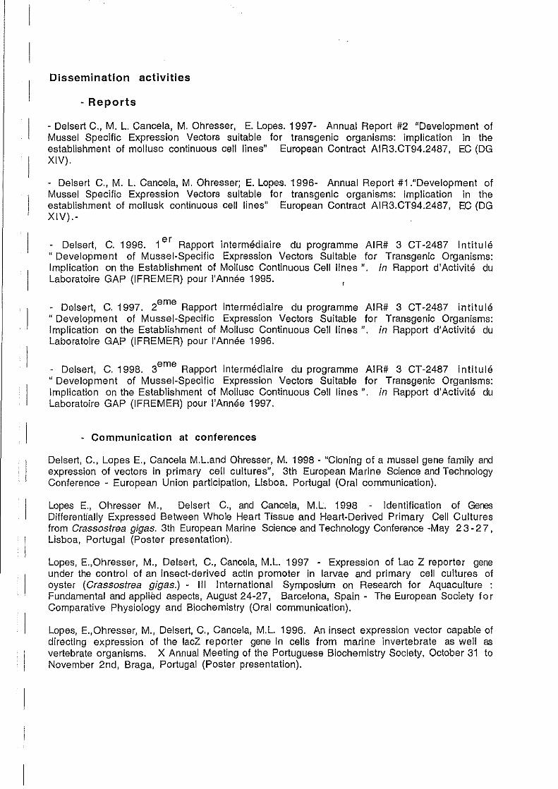
1
1
Dissemination activities
- Reports
- Delsert C., M. L. Cancela, M. Ohresser, E. Lopes. 1997- Annual Report #2 "Development of Mussel Specifie Expression Vectors suitable for transgenic organisms: implication in the establishment of mollusc continuous cell lines" European Contract AIR3.CT94.2487, EC (DG XIV).
- Delsert C., M. L. Cancela, M. Ohresser; E. Lopes. 1996- Annual Report #1 ."Development of Mussel Specifie Expression Vectors suitable for transgenic organisms: implication in the establishment of mollusk continuous cell lines" European Contract AIR3.CT94.2487, EC (DG XIV).-
- Delsert, C. 1996. 1 er Rapport intermédiaire du programme AIR# 3 CT-2487 intitulé "Development of Mussel-Specific Expression Vectors Suitable for Transgenic Organisms: Implication on the Establishment of Mollusc Continuous Cell lines ". in Rapport d'Activité du Laboratoire GAP (IFREMER) pour l'Année 1995.
- Delsert, C. 1997. 2eme
Rapport intermédiaire du programme AIR# 3 CT -2487 intitu lé "Development of Mussel-Specific Expression Vectors Suitable for Transgenic Organisms: Implication on the Establishment of Mollusc Continuous Cell lines ". in Rapport d'Activité du Laboratoire GAP (IFREMER) pour l'Année 1996.
- Delsert, C. 1998. 3eme
Rapport intermédiaire du programme AI R# 3 CT-2487 intitulé "Development of Mussel-Specific Expression Vectors Suitable for Transgenic Organisms: Implication on the Establishment of Mollusc Continuous Cell lines ". in Rapport d'Activité du Laboratoire GAP (IFREMER) pour l'Année 1997.
- Communication al conferences
Delsert, C., Lopes E., Cancela M.L.and Ohresser, M. 1998 - "Cloning of a mussel gene family and expression of vectors in primary cell cultures", 3th European Marine Science and Technology Conference - European Union participation, Lisboa. Portugal (Oral communication).
Lopes E., Ohresser M., Delsert C., and Cancela, M.L. 1998 - Identification of Genes Differentially Expressed Between Whole Heart Tissue and Heart-Derived Primary Cell Cultures from Crassostrea gigas. 3th European Marine Science and Technology Conference -May 23 - 2 7 , Lisboa, Portugal (Poster presentation).
Lopes, E.,Ohresser, M., Delsert, C., Cancela, M.L. 1997 - Expression of Lac Z reporter gene under the control of an insect-derived actin promoter in larvae and primary cell cultures of oyster (Crassostrea gigas.) - 111 International Symposium on Research for Aquaculture : Fundamental and applièd aspects, August 24-27, Barcelona, Spain - The European Society 10 r Comparative Physiology and Biochemistry (Oral communication).
Lopes, E.,Ohresser, M., Delsert, C., Cancela, M.L. 1996. An insect expression vector capable of directing expression 01 the lacZ reporter gene in ce Ils from marine invertebrate as weil as vertebrate organisms. X Annual Meeting 01 the Portuguese Biochemistry Society, October 31 to November 2nd, Braga, Portugal (Poster presentation).