Embed Size (px)
Citation preview

Determining the Origin of Fe3+-induced BODIPY
Probe Fluorescence in Aqueous Solution
A Major Qualifying Project Report
Submitted to the Faculty of
WORCESTER POLYTECHNIC INSTITUTE
In partial fulfillment of the requirements for the
Degree of Bachelor of Science
By:
________________________________
Paige A. Lamica
April 26, 2017
Approved by
____________________
Prof. Shawn C. Burdette, Ph.D.
Project Advisor

i
Abstract
Iron is the most physiologically abundant transition metal with numerous applications to
bodily systems and disease states. Too little iron will affect oxygen metabolism, a critical
function of free iron, while excess iron has been associated with diseases including hepatitis,
hemochromatosis, and neurological degeneration. The medical importance of iron homeostasis
warrants research into developing accurate aqueous chemosensors. Sensors created with
BODIPY fluorophores have been studied heavily in recent years for this purpose. Many
published studies of these sensors identify iron binding to the probe as the source of the
fluorescent signal. The purpose of this project was to synthesize and characterize two BODIPY
iron sensors to demonstrate Fe3+ binding is not necessary to induce a fluorescence response. Fe3+
precipitates in water to produce iron (III) hydroxide, indicating that the fluorescence seen in
published literature may not be caused by iron binding to the receptor of BODIPY. The
compounds created in this MQP do not have a strong binding affinity, therefore it was
hypothesized that if the molecule still fluoresces, this is due to other causes and can be used
comparatively as a control when studying BODIPY based molecules that have strong Fe3+
recognition units.

ii
Acknowledgements
I would like thank the Burdette research lab group for their support and guidance of this MQP:
Shawn Burdette, Prem Basa, Chelsea Barr and Jingjing Yan. I would also like to thank WPI for
its commitment to undergraduate research.

iii
Table of Contents
Abstract …………………………………………………………………………………….…….i
Acknowledgements ………………………………………………………………………….......ii
Table of Contents …………………………………………………………………………….....iii
List of Figures …………………………………………………………………………….……..iv
List of Schemes …………………………………………………………………………………..v
1. Introduction ……………………………………………………………………………...1
2. Background ……………………………………………………………………….……..2
2.1 Clinical Significance of Iron Metal Ions ...……………………………………….2
2.2 Chemosensors ………………………………………………………………….…3
2.3 BODIPY Fluorophores ……………………………………………………….…..3
2.4 Existing Research ………………………………………………………….….….4
3 Methodology …………………………………………..……………………………..………6
3.1 General Synthetic Procedures ………...…………………………………….…….6
3.2 Hydroxy Aldehyde …..……………………………………………………….…...6
3.3 Hydroxy Bodipy ……………………………………………………………….….7
3.4 Methoxy Aldehyde ………………………………………………………….…….8
3.5 Methoxy Bodipy ……………………………………………………………….…9
4 Conclusions & Future Research ……………………………………………………..........10
5 References ...………………………………………………………………….........………..12
6 Supplementary Information …………………………………………………..…………..14

iv
List of Figures
Figure 1: Chemosensor Mechanics …………………………..………………………………….3
Figure 2: BODIPY-based Chemosensor Example ………………………………………………4
Figure 3: Iron-specific BODIPY sensor …………………………………………………..….….4
Figure 4: Cellular Imaging of BODIPY Sensor Florescence Function ………………….……....5
SF 1: Hydroxy Aldehyde 1H NMR ……………………………………………………………..14
SF 2: Hydroxy Aldehyde 13C NMR …………………………………………………………….15
SF 3: Hydroxy Aldehyde LC-MS ……………………………………………………………….16
SF 4: Hydroxy Aldehyde Transmittance ………………………………………………………..17
SF 5: Hydroxy BODIPY 1H NMR ……………………………………………………………...18
SF 6: Hydroxy BODIPY 13C NMR ……………………………………………………………..19
SF 7: Hydroxy BODIPY LC-MS ……………………………………………………….………20
SF 8: Hydroxy BODIPY Transmittance ………………………………………………………...21
SF 9: Methoxy Derivative 1H NMR …………………………………………………………….22
SF 10: Methoxy Aldehyde 1H NMR ……………………………………………………………23
SF 11: Methoxy Aldehyde 13C NMR …………………………………………………………...24
SF 12: Methoxy Aldehyde LC-MS ……………………………………………………………..25
SF 13: Methoxy Aldehyde Transmittance ………………………………………………………26

v
SF 14: Methoxy BODIPY 1H NMR …………………………………………………………….27
SF 15: Methoxy BODIPY 13C NMR ……………………………………………………………28
SF 16: Methoxy BODIPY LC-MS ……………………………………………………………...29
SF 17: Methoxy BODIPY Transmittance ………………………………………………………30

vi
List of Schemes
Scheme 1: Hydroxy Compound Synthesis ……………………………………...…….…………6
Scheme 2: Methoxy Compound Synthesis ………………………………………………...…….8

1
1. Introduction
Iron is the most physiologically abundant transition metal and has roles in numerous
cellular functions.1 Humans as well as other mammals and even bacteria require iron for
biological processes including oxygen metabolism, electron transfer, transcription and
reproduction.2-5 Due to iron’s abundance and versatility in the body, it also plays a role in many
disease states. Levels of iron are normally maintained by homeostasis, however, iron
concentration can fluctuate in the body resulting in illness. Too little iron in the body can weaken
the immune system and cause blood clotting disorders, while excess iron in the body has been
associated with the development of cancer, hepatitis and hemochromatosis.6, 7 Additionally,
much effort has been recently devoted to the study of iron’s potential roles in neurodegenerative
diseases such as Parkinson’s and Alzheimer’s.8-11
The clinical significance of iron homeostasis warrants research into developing accurate
chemosensors which can detect Fe3+ under aqueous conditions. A chemosensor is a molecule
which contains a receptor site that binds to the target analyte, in this case Fe3+, resulting in a
response from the signaling unit of the molecule. One molecule which is particularly useful for
this application is BODIPY, a fluorophore which has been researched extensively in recent
years.12, 13 Some published research cites studies of these sensors in methanol which then
extrapolate the results to water-based systems.
The Burdette research group has synthesized several BODIPY-based sensors, and this
MQP is a continuation of that research.1, 14 More specifically, this project hypothesized that these
BODIPY-based sensors for iron may not be working as reported in aqueous solution due to the
fact that Fe3+ will precipitate in water to form iron (III) hydroxide. To study this, two BODIPY-

2
based sensors with a weak binding affinity for iron were synthesized. Both hydroxy and methoxy
BODIPY derivatives were synthesized and characterized by 1H NMR, 13C NMR, LC-MS, and
FT-IR. In future research, these sensors will be studied using UV-vis and fluorescence studies
and compared to the published fluorescence data of iron-specific BODIPY sensors.
2. Background
It is important to understand the clinical significance of iron homeostasis, its biological
abundance and why we need to research how to accurately detect levels of iron in solution.
BODIPY-based iron sensors will be introduced as a way to detect iron in organic solutions, and
potential downfalls of these sensors in aqueous solution will be discussed.
2.1 Clinical Significance of Iron Metal Ions
Iron is the most physiologically abundant transition metal and has roles in numerous
biological processes including oxygen metabolism, electron transfer, transcription and
reproduction.1-5 Research into iron homeostasis has uncovered that iron imbalances in the body
play roles in many different diseases including anemia, blood clotting disorders, hepatitis,
hemochromatosis and some cancers.6,7 Additionally, recent research has suggested that there
may be a link between iron and neurodegenerative diseases such as Parkinson’s and Alzheimer’s
[8-11]. It is thought that ferrous iron reacts with hydrogen peroxide via the Fenton Reaction to
form hydroxide and also hydroxyl free radicals (OH).15 These hydroxyl free radicals are very
reactive and suspected to play a role in the development and progression of neurodegeneration.16-
18 Due to iron’s abundance and complex roles in biological processes and disease states, it
warrants research into developing accurate chemosensors which can function in aqueous
solution.

3
2.2 Chemosensors
A chemosensor is a molecule which contains a signaling unit, spacer, and receptor site.
The target being sensed, in this case Fe3+, is known as the analyte. When the analyte binds to the
specific receptor site, the signaling unit responds. In the case of this MQP, the signaling unit
responds by fluorescing.
Figure 1: Chemosensor Mechanics. A receptor site binds to the desired analyte resulting in a
response from the signaling unit. This response results in the molecule “sensing” the presence of
the analyte.
2.2 BODIPY Fluorophores
BODIPY, also known as boron dipyrromethene, is an organic dye which possesses many
unique photophysical properties. Due to this, it is a great fluorophore to be used as the signaling
unit in a chemosensor. When iron binds to the receptor unit of the molecule, it allows for
electron flow through the iron which, in turn, results in the visible BODIPY fluorescence.

4
Figure 2: BODIPY-based Chemosensor Example. The sensor without Fe3+ present induces
minimal fluorescence response, however, upon addition of the analyte, the sensor binds to iron
and the signaling unit fluoresces.19
2.3 Existing Research
A published study of BODIPY-based iron sensors was used to make hypotheses which
partially formed the basis of this MQP. The study in question used a BODIPY sensor which was
responsive to iron to demonstrate the ability for the molecule to act as a turn-on fluorescence
probe.19
Figure 3: Iron-specific BODIPY sensor. This molecule has a strong binding affinity for iron
when compared to the molecules synthesized in this MQP, therefore it can be used
comparatively to assess differences in fluorescence.19

5
This sensor was then used in cells to show the effect on fluorescence in cells with just sensor and
with sensor and iron present.19
Figure 4: Cellular Imaging of BODIPY Sensor Florescence Function. The top row represents
cells treated with sensor and iron, while the bottom row represents cells treated with just sensor.
The difference from column 1 to column 2 is 1hr incubation time.19
This figure provoked further inquiry into understanding how the iron probes were analyzed in
cells. The top row of cells visually appeared to be of higher confluency, while the bottom row of
cells appeared visually blurry. The fluorescence displayed in the bottom row of cells, meant to
act as a control of just sensor, indicates that iron binding may potentially not be required for
these BODIPY-based sensors to fluoresce. Fluorescence may be caused by something else.
Determining the true cause of BODIPY-sensor florescence is the long-term objective of this
project.

6
3. Methodology
3.1 General Synthetic Procedures
All materials were obtained in the highest purity available from Fisher, Acros Organic and Alfa
Aesar and used without further purification. Solvents were purged with argon and dried using a
Seca Solvent Purification System. 1H and 13C NMR were recorded using a 500 MHz Bruker-
Biospin NMR instrument, and chemical shifts are reported in ppm on the δ scale relative to
tetramethylsilane (TMS). FT-IR spectra were recorded using Bruker Optics Vertex 70 with MIR
source as neat crystalline powdered samples and Spectrum 100 Version 10.4.2 (PerkinElmer)
fitted with diamond ATR as oils. Melting-point information was obtained using Hydrothermal
Mel-Temp instrument. Full syntheses are shown in Scheme 1 and Scheme 2.
Scheme 1: Hydroxy Compound Synthesis
3.2 Hydroxy Aldehyde
4-[bis(2-hydroxyethyl)amino]benzaldehyde (2) N-phenyldiethanolamine (2.0 g, 11.0 mmol)
was combined with phosphorus oxychloride (8.43 g, 55.0 mmol) in DMF (20 mL). After 24 h in
a 60 ° C oil bath, the reaction mixture was applied over ice and neutralized to pH 7 with
saturated NaHCO3. The product was extracted using saturated NaCl solution and ethyl acetate (3
(1) (2) (3)

7
× 50 mL). The combined extracts were dried over NaSO4. Solvent removal under reduced
pressure yielded yellow oil. The resultant oil was purified using column chromatography with
1:1 hexanes: ether as the eluent and yielded a white solid (1.17 g, 50.4 % yield); mp =80-82 ° C.
1H NMR (500 MHz, CDCl3) δ 9.79 (s, 1 H), 7.79 (d, J = 9.0 Hz, 2 H), 6.76 (d, J = 9.0 Hz, 2 H),
3.86 (t, J = 7.0 Hz, 4 H), 3.70 (t, J = 7.0 Hz, 4 H). 13C NMR (500 MHz, CDCl3) δ 190.4, 151.2,
132.5, 111.5, 53.5, 40.2. FT-IR (neat, cm-1) 2963.8, 2822.2, 2747.0, 2324.5, 2163.9, 1665.8,
1588.9, 1559.9, 1521.2, 1461.1, 1433.0, 1403.0, 1360.9, 1313.0, 1284.2, 1254.3, 1240.9, 1215.6,
1194.1, 1163.2, 1124.3, 1032.1, 1000.8, 961.5, 837.7, 817.1, 806.1, 784.2, 747.3, 712.9, 637.2.
HRMS (+ESI) calculated (C11H15NO3) for 209.2 and observed 248.0 [M+K+].
3.3 Hydroxy Bodipy
[[2,2-[[4-[(3,5-dimethyl-1H-pyrrol-2-yl- κN)(3,5-dimethyl-2H-pyrrol-2-ylidene-
κN)methyl]phenyl]imino]bis[ethanolato]](1-)] difluoro-, (T-4)-boron (3) 2, 4-dimethylpyrrole
(0.43 g, 48 mmol) was combined with compound 2 (0.43 g, 21.6 mmol) in 30 mL DCM. After
initial mixing, 4 drops trifluoroacetic acid (TFA) was added and left to stir under nitrogen for 24
h at room temperature. DDQ (0.48 g, 2.13 mmol) was then mixed with DCM (20 mL) and added
1 mL/min to the reaction mixture. The reaction was stirred at room temperature for 24 h and then
triethyl amine (8 mL) was added and left to stir at room temperature. After 30 minutes,
borontrifluoride ethelate (6.6 mL) was added and the reaction stirred for 24 h at room
temperature. The product was extracted using saturated NaCl solution and DCM (3 × 75 mL).
The combined extracts were dried over NaSO4. Solvent removal under reduced pressure yielded
black oil. The resultant oil was purified using column chromatography with 1:1 hexanes: ether as
the eluent and yielded a red solid (536 mg, 27.6 % yield); mp = 148-150 ° C. 1H NMR (500
MHz, CDCl3) δ 7.12 (d, J = 8.8 Hz, 2 H), 5.98 (s, 2 H), 3.80 (t, J = 7.1 Hz, 4 H), 3.69 (t, J = 6.9

8
Hz, 4 H), 2.55 (s, 6 H), 1.47(s, 7 H). 13C NMR (500 MHz, CDCl3) δ 155.3, 146.9, 143.3, 142.4,
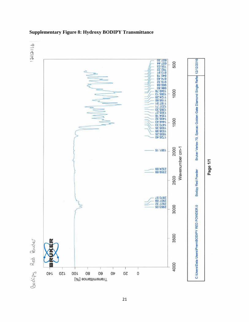
132.3, 129.7, 124.1, 121.3, 112.4, 53.7, 40.5, 29.9, 14.9, 14.8. FT-IR (neat, cm-1) 2963.1,
2927.3, 2907.7, 2870.6, 2359.7, 2324.7, 1981.2, 1726.4, 1606.3, 1539.0, 1506.4, 1470.3, 1444.4,
1406.3, 1354.2, 1305.3, 1265.3, 1237.7, 1187.9, 1156.6, 1124.3, 1085.1, 1049.8, 986.8, 969.7,
919.3, 874.4, 840.8, 813.9, 762.2, 705.0, 657.4, 607.3. HRMS (+ESI) calculated (C13H19NO3) for
237.2 and observed 238.1 [H+].
Scheme 2: Methoxy Compound Synthesis
3.4 Methoxy Aldehyde
4-[bis(2-methoxyethyl)amino]benzaldehyde (5) N-phenyldiethanolamine (2.0 g, 11 mmol) was
added to THF (30 mL), stirring under nitrogen. Sodium hydride (2.2 g, 91 mmol) was added to
(1) (4) (5)
(6)

9
the reaction mixture slowly and then refluxed at room temperature for 30 minutes, 40 ° C for 30
minutes and then brought back to room temperature. Methyl iodide (3.43 mL) was added to the
reaction mixture slowly and allowed to stir overnight at room temperature. The product
(compound 4) was extracted using saturated NaCl solution and DCM (3 × 50 mL). The
combined extracts were dried over NaSO4. Solvent removal under reduced pressure yielded
yellow oil. The resultant oil was purified using silica column chromatography with 1:1 hexanes:
ether as the eluent and yielded a clear oil. This compound was then used as a starting material for
the aldehyde. Compound 4 (0.15 g, 0.72 mmol) was added to DMF (4 mL). Phosphorus
oxychloride (0.55 g, 3.59 mmol) was added to the reaction mixture slowly. After 24 h in a 60 ° C
oil bath, the reaction mixture was applied over ice and neutralized to pH 7 with saturated
NaHCO3. The product was extracted using saturated NaCl solution and ethyl acetate (3 × 50 mL).
The combined extracts were dried over NaSO4. Solvent removal under reduced pressure yielded
brown oil. The resultant oil was purified using column chromatography with 1:1 hexanes: ether
as the eluent and yielded a yellow oil (0.39 g, 24.4 % yield); 1H NMR (500 MHz, CDCl3) δ 9.73
(s, 1 H), 7.71 (d, J = 9.3 Hz, 2 H), 6.75 (d, J = 9.4 Hz, 2 H), 3.66 (t, J = 5.9 Hz, 4 H), 3.58 (t, J =
5.8 Hz, 4 H), 3.35 (s, 6 H). 13C NMR (500 MHz, CDCl3) δ 190.2, 152.8, 132.3, 125.5, 111.2,
70.0, 59.2, 51.2. FT-IR (neat, cm-1) 2924.9, 2879.0, 2812.6, 2733.9, 2152.8, 2028.4, 1666.8,
1590.2, 1555.8, 1522.7, 1452.4, 1434.1, 1399.7, 1356.7, 1314.3, 1279.1, 1237.9, 1197.3, 1165.0,
1108.0, 1009.9, 1000.1, 959.7, 908.9, 813.8, 729.8, 710.4. HRMS (+ESI) calculated
(C11H15NO3) for 209.2 and observed 248.0 [M+K+].
3.5 Methoxy Bodipy
[[2,2-[[4-[(3,5-dimethyl-1H-pyrrol-2-yl- κN)(3,5-dimethyl-2H-pyrrol-2-ylidene-κN)methyl]
phenyl]imino]bis[methoxyethanolato]](1-)] difluoro-, (T-4)-boron (6) 2,4-dimethylpyrrole

10
(0.34 g, 3.58 mmol) was added to compound 4 (0.39 g, 1.63 mmol) in DCM (25 mL). After
initial mixing, 3 drops trifluoroacetic acid was added and left to stir under nitrogen for 24 h at
room temperature. DDQ (0.38 g, 1.68 mmol) was then mixed with DCM (20 mL) and added 1
mL/min to the reaction mixture. The reaction was stirred at room temperature for 24 h and then
triethyl amine (6 mL) was added and left to stir at room temperature. After 30 minutes,
borontrifluoride ethelate (2.98 g, 21 mmol) was added and the reaction stirred for 24 h at room
temperature. The product was extracted using saturated NaCl solution and DCM (3 × 75 mL).
The combined extracts were dried over NaSO4. Solvent removal under reduced pressure yielded
black oil. The resultant oil was purified using silica column chromatography with 1:1 hexanes:
ether as the eluent and yielded a red solid (trace yield); mp = 110-114 ° C. 1H NMR (500 MHz,
CDCl3) δ 7.07 (d, J = 8.9 Hz, 2 H), 8.86 (d, J = 8.9 Hz, 2 H), 6.07 (s, 2 H), 3.56 (m, 8 H), 3.31 (s,
6 H). 13C NMR (500 MHz, CDCl3) δ 154.9, 148.7, 143.4, 132.4, 129.2, 122.4, 121.1, 112.4, 70.2,
59.3, 51.2, 29.9, 14.9. FT-IR (neat, cm-1) 3098.7, 2957.7, 2923.0, 2884.1, 2824.9, 2738.0,
2339.4, 1727.5, 1668.4, 1606.8, 1540.3, 1525.8, 1499.8, 1468.4, 1396.0, 1359.9, 1305.6, 1295.1,
1265.5, 1232.7, 1188.2, 1156.9, 1106.4, 1061.8, 1009.3, 970.1, 902.2, 813.7, 761.8, 703.8, 647.8.
HRMS (+ESI) calculated (BC25F2H32N3O2) for 455.3 and observed 455.3 and 456.2 [H+].
Conclusions & Future Research
This research resulted in the synthesis and characterization of two BODIPY sensors
which do not have a strong binding affinity for iron. To further this research, these sensors
should be studied using UV-vis, titration experiments with iron, and fluorescence studies.
Additionally, these sensors should be compared side-by-side with another published BODIPY
sensor which is known to bind to iron in both organic solvent and aqueous solvent to
demonstrate differences and similarities in fluorescence response. Finally, these sensors should

11
be used in cells and cellular images should be taken which show florescence under varying
sensor and Fe3+ conditions. If the sensors created in this MQP, which should not bind iron, show
similar fluorescence responses when compared to the BODIPY sensors which should bind iron,
that may provide evidence that iron binding is not necessary to induce a fluorescence response.

12
References
1. Kennedy, D. P.; Kormos, C. M.; Burdette, S. C. FerriBRIGHT: A Rationally Designed
Fluorescent Probe for Redox Active Metals, J. Am. Chem. Soc. 2009, 131, 8578-8586.
2. Dai, S.; Schwendtmayer, C.; Schurmann, P.; Ramaswamy, S.; Eklund, H. Redox Signaling in
Chloroplasts: Cleavage of Disulfides by an Iron-Sulfur Cluster. Science. 2000, 287, 655−658.
3. Atkinson, A.; Winge, D. R. Metal Acquisition and Availability in the Mitochondria. Chem.
Rev. 2009, 109, 4708−4721.
4. Kaplan, C. D.; Kaplan, J. Iron Acquisition and Transcriptional Regulation. Chem. Rev. 2009,
109, 4536−4552.
5. Theil, E. C.; Goss, D. J. Living with Iron (and Oxygen): Questions and Answers about Iron
Homeostasis. Chem. Rev. 2009,109, 4568−4579.
6. S.C. Huang, Y.J. Yang, C.N. Cheng, et al. The etiology and treatment outcome of iron
deficiency and iron deficiency anemia in children. J Pediatr Hematol Oncol, 2010, 32, 282–285
7. Weinberg, E. D. The Role of Iron in Cancer. Eur. J. Cancer Prev. 1996, 5, 19−36.
8. Halliwell, B. Reactive Oxygen Species and the Central Nervous System. J. Neurochem. 1992,
59, 1609−1623.
9. Dornelles, A.; Garcia, V.; Lima, M. M.; Vedana, G.; Alcalde, L.; Bogo, M.; Schroder, N.
Mrna Expression of Proteins Involved in Iron Homeostasis in Brain Regions Is Altered by Age
and by Iron Overloading in the Neonatal Period. Neurochem. Res. 2010, 35, 564−571.
10. Gaeta, A.; Hider, R. C. The Crucial Role of Metal Ions in Neurodegeneration: The Basis for
a Promising Therapeutic Strategy. Br. J. Pharmacol. 2005, 146, 1041−1059.
11. Molina-Holgado, F.; Hider, R.; Gaeta, A.; Williams, R.; Francis, P. Metals Ions and
Neurodegeneration. Biometals. Neurochem. 2007, 20, 639−654.

13
12. Qu, X.; Liu, Q.; Ji, X.; Chen, H.; Zhou, Z.; Shen, Z. Enhancing the Stokes’ Shift of Bodipy
Dyes Via through-Bond Energy Transfer and Its Application for Fe3+-Detection in Live Cell
Imaging. Chem. Commun. 2012, 48, 4600−4602.
13. Sahoo, S. K.; Sharma, D.; Bera, R. K.; Crisponi, G.; Callan, J. F. Iron(III) Selective
Molecular and Supramolecular Fluorescent Probes. Chem. Soc. Rev. 2012, 41, 7195−7227.
14. FerriCast: a macrocyclic photocage for Fe3+. Daniel P. Kennedy, Christopher D. Incarvito,
and Shawn C. Burdette. Inorganic chemistry, 2010, 49(3), 91
15. Sunahara, H.; Urano, Y.; Kojima, H.; Nagano, T., Design and Synthesis of a Library of
BODIPY-Based Environmental Polarity Sensors Utilizing Photoinduced Electron-Transfer-
Controlled Fluorescence ON/OFF Switching. J. Am. Chem. Soc. 2007, 129, 5597-5604.
16. Turfan, B.; Akkaya, E. U., Modulation of Boradiazaindacene Emission by Cation-Mediated
Oxidative PET. Org. Lett. 2002, 4, 2857-2859.
17. McCusker, J. K.; Walda, K. N.; Dunn, R. C.; Magde, D.; Hendrickson, D. N., Subpicosecond
1 MLCT-5 T2 Intersystem Crossing of Low-Spin Polypyridyl Ferrous Complexes. J. Am. Chem.
Soc. 1993, 115, 298-307.
18. Toftlund, H.; Yde-Andersen, S., Acta. Chem. Scand. J Inorg Biochem. 1981, 35, 575-585.
19. Novel BODIPY-Based Fluorescence Turn-on Sensor for Fe3+ and Its Bioimaging
Application in Living Cells. Binglin Sui, Simon Tang, Taihong Liu, Bosung Kim, and Kevin D.
Belfield. ACS Applied Materials & Interfaces. 2014, 6 (21), 18408-18412

14
Supplementary Information
Supplementary Figure 1: Hydroxy Aldehyde 1H NMR

15
Supplementary Figure 2: Hydroxy Aldehyde 13C NMR

16
Supplementary Figure 3: Hydroxy Aldehyde LC-MS

17
Supplementary Figure 4: Hydroxy Aldehyde Transmittance

18
Supplementary Figure 5: Hydroxy BODIPY 1H NMR

19
Supplementary Figure 6: Hydroxy BODIPY 13C NMR

20
Supplementary Figure 7: Hydroxy BODIPY LC-MS
21
Supplementary Figure 8: Hydroxy BODIPY Transmittance

22
Supplementary Figure 9: Methoxy Derivative 1H NMR

23
Supplementary Figure 10: Methoxy Aldehyde 1H NMR

24
Supplementary Figure 11: Methoxy Aldehyde 13C NMR

25
Supplementary Figure 12: Methoxy Aldehyde LC-MS

26
Supplementary Figure 13: Methoxy Aldehyde Transmittance

27
Supplementary Figure 14: Methoxy BODIPY 1H NMR

28
Supplementary Figure 14: Methoxy BODIPY 13C NMR

29
Supplementary Figure 15: Methoxy BODIPY LC-MS

30
Supplementary Figure 16: Methoxy BODIPY Transmittance