Embed Size (px)
Citation preview

CHIRAL BODIPY DYES & PHOTOSENSITIZERS FOR
PHOTODYNAMIC THERAPY AND DYE SENSITIZED SOLAR
CELLS
A DISSERTATION SUBMITTED TO
MATERIALS SCIENCE AND NANOTECHNOLOGY PROGRAM
OF THE GRADUATE SCHOOL OF ENGINEERING AND SCIENCE
OF BILKENT UNIVERSITY
IN PARTIAL FULFILLMENT OF THE REQUIREMENTS
FOR THE DEGREE OF
DOCTOR OF PHILOSOPHY
By
YUSUF ÇAKMAK
February, 2013

ii
I certify that I have read this thesis and that in my opinion it is fully adequate, in
scope and in quality, as a thesis of the degree of Doctor of Philosophy.
………………………………….
Prof. Dr. Engin U. Akkaya (Advisor)
I certify that I have read this thesis and that in my opinion it is fully adequate, in
scope and in quality, as a thesis of the degree of Doctor of Philosophy.
………………………………….
Assoc. Prof. Dr. Mehmet Bayındır
I certify that I have read this thesis and that in my opinion it is fully adequate, in
scope and in quality, as a thesis of the degree of Doctor of Philosophy.
………………………………….
Assist. Prof. Dr. Serdar Atılgan
iii
I certify that I have read this thesis and that in my opinion it is fully adequate, in
scope and in quality, as a thesis of the degree of Doctor of Philosophy.
………………………………….
Assist. Prof. Dr. Özgür Altan Bozdemir
I certify that I have read this thesis and that in my opinion it is fully adequate, in
scope and in quality, as a thesis of the degree of Doctor of Philosophy.
………………………………….
Assist. Prof. Dr. Tamer Uyar
Approved for the Graduate School of Engineering and Science:
………………………………….
Prof. Dr. Levent Onural
Director of the Graduate School

iv
ABSTRACT
CHIRAL BODIPY DYES & PHOTOSENSITIZERS FOR
PHOTODYNAMIC THERAPY AND DYE-SENSITIZED SOLAR
CELLS
Yusuf Çakmak
PhD in Materials Science and Nanotechnology
Supervisor: Prof. Dr. Engin U. Akkaya
February, 2013
Bodipy is a molecule with many superior properties. After its discovery in
1968, most of the features were not recognized until mid 1990s. Thereafter, many
research papers and patents have been produced and the number of publications and
citations is still on the rise today. An important fraction of the research done with this
fluorophore is in chemosensing field to probe various analytes including anions,
cations and even biomolecules. However, in this research we have focused on
different areas of subjects and tried to find novel applications for these dyes. First,
we designed orthogonal bodipy dimers for efficient triplet photosensitization without
heavy atoms in contrast to most other sensitizers and efficient singlet oxygen
generation was achieved (Φ∆=0.51). In the second project, calix[4]arene molecules
were designed and synthesized as carriers for photodynamic therapy, potentially
behaving as a molecular basket carrying the agents to the tumor tissues. Later, we
focused on obtaining axial chiral molecules by using solely bodipy dyes, and we
were able to obtain enantiopure fragments were separated by using chiral HPLC.
These rare molecules are desirable for modern biological labeling and advanced
optoelectronic devices. Finally, we designed bodipy dyes for dye sensitized solar
cells by adapting relevant functional groups, and following synthesis work, we
constructed cells to assess the design parameters via measuring the electrical output
results.
Keywords: Photodynamic therapy, triplet photosensitization, axial chirality, dye
sensitized solar cell, singlet oxygen

v
ÖZET
KİRAL BODIPY BOYALARI & FOTODİNAMİK TERAPİ VE BOYA
DUYARLILAŞTIRILMIŞ GÜNEŞ PİLLERİ İÇİN
FOTODUYARLILAŞTIRICILAR
Yusuf Çakmak
Malzeme Bilimi ve Nanoteknoloji, Doktora
Tez Yöneticisi: Prof. Dr. Engin U. Akkaya
Şubat, 2013
Bodipy üstün özelliklere sahip olan bir moleküldür. 1968 yılında
keşfedilmesinden sonra 1990’lı yılların ortalarına kadar özelliklerinin büyük bir
kısmı bilinmiyordu. Sonrasında ise, oldukça fazla sayıda bilimsel makale ve patent
üretilmiştir, bugün bile bu boya ile ilgili makale sayısı ve atıf sayısı hızlı artmaktadır.
Bu madde ile yapılan çalışmaların büyük kısmı anyonları, katyonları ve hatta
biyomolekülleri algılama da kullanılmaktadır. Fakat, bu araştırmada biz bodipy
molekülünün en iyi olmadığı fakat iyi olma potansiyeli olan diğer alanlara
yoğunlaştık ve bodipy’i bu alanlara uygulamaya çalıştık. İlk olarak bir çoğunda
olduğunun aksine ağır atom içermeyen dik bodipy dimerleri dizayn ettik ve etkili
singlet oksijen üretimi elde ettik (ΦΔ=0.51). Bu konu altındaki ikinci projede
kaliks[4]aren moleküllerini, fotodinamik terapi ajanları taşıyacak ve moleküler sepet
gibi davranan hedefli bir ünite olarak tasarladık. Sonra, sadece bodipy
moleküllerinden oluşan eksensel kiralite özelliği olan moleküllere yoğunlaştık ve
enantiyosaf parçaları kiral HPLC kullanarak ayırmayı başardık. Bu nadir görülen
maddeler modern biyolojik etiketlemede ve gelişmiş optoelektronik aygıtlar için
talep edilmektedir. Son olarak, başarısı kanıtlanmış fonksiyonel grupları bodipy
molekülüne uygulayarak boya duyarlılaştırılmış güneş pilleri için boyar madde
tasarladık ve sentez ve pil yapımı aşamalarından sonra elektrik çıktı sonuçlarını
ölçerek dizayn parametrelerini değerlendirdik.
Anahtar kelimeler: Fotodinamik terapi, triplet fotoduyarlılaştırma, eksensel kiralite,
boya duyarlılaştırılmış güneş pili, singlet oksijen

vi
ACKNOWLEDGEMENT
I would like to thank to TÜBİTAK (The Scientific and Technological
Research Council of Turkey) for giving me the opportunity of very beneficial abroad
scholarship programme
I would like to thank my supervisor Prof. Dr. Engin U. Akkaya whose
encouragement and support made this study possible. I also want to express my
sincere appreciation to him for his guidance, teaching, and understanding during this
research.
I would like to thank Safacan Kölemen, Ziya Köstereli, Tuğrul Nalbantoğlu,
Tuğçe Durgut for their patience, friendship and help during the projects which we
worked with.
I want to thank our present group members Ruslan Guliyev, Onur
Büyükçakır, Tuğba Özdemir, Fazlı Sözmen, Bilal Uyar, Ahmet Atılgan, Tuba Yaşar,
Yiğit Altay, Bilal Kılıç, Esra Tanrıverdi, Nisa Yeşilgül, Ahmet Bekdemir, İlke
Şimşek, the past members Gökhan Barın, Sencer &Hande Boyacı Selçuk and the
other Akkaya group members for their valuable friendships, wonderful
collaborations, and great ambiance in the laboratory. It was a great experience to
work with them.
Also special thanks to Özgür Altan Bozdemir, Mahmut Deniz Yılmaz, Ali Coşkun,
Serdar Atılgan and Erhan Deniz for their recommendations, comments and beautiful
friendships starting from the very beginning of the research life.
I would like to express my sincere gratitude to Yavuz Dede, Muhammed
Büyüktemiz, Şule Erten-Ela and Bora Bilgiç for their devoted help for the various
subjects that make my thesis better.
My very special thanks go to my close friends and Mehmet Alp, Defne,
Nilay&Osman, Alper, My Father, My Mother and the special person of my life
Sündüs.

vii
LIST OF ABBREVIATIONS
Bodipy : 4,4-difluoro-4-bora-3a,4a-diaza-s-indacene
CASSCF : Complete active-space self consistent field methodology
cheno : Chenodeoxycholic acid
DSSC : Dye-sensitized solar cell
DS-TR : Doubly Substituted-Tetra radicalic
DFT : Density functional theory
ISC : Intersystem crossing
FRET : Förster Resonance Energy Transfer
HOMO : Highest Occupied Molecular Orbital
LUMO : Lowest Unoccupied Molecular Orbital
MALDI : Matrix-Assisted Laser Desorption/Ionization
MS : Mass Spectrometry
NMR : Nuclear Magnetic Resonance
NOON : Natural Orbitals and Occupation Numbers
PDT : Photodynamic Therapy
PS : Photosensitizer
SS : Singly substituted
SOMO : Singly occupied molecular orbital
TLC : Thin Layer Chromotography
viii
TABLE OF CONTENTS
1 INTRODUCTION ............................................................................................... 1
2 BACKGROUND ................................................................................................. 5
2.1 Photodynamic Therapy .................................................................................. 5
2.1.1 General Information ............................................................................... 5
2.1.2 Development of PDT ............................................................................. 6
2.1.3 The Mechanism and Methods of PDT ................................................... 8
2.1.4 Mechanism of Singlet Oxygen Production64........................................ 15
2.1.5 Molecular Orbital Diagram of Triplet and Singlet Molecular Dioxygen65 ......................................................................................................... 17
2.2 Triplet Photosensitization ............................................................................ 19
2.2.1 Heavy Atom Effect on Triplet Photosensitization ............................... 21
2.2.2 Triplet PSs with low-lying n-π* transitions .......................................... 21
2.2.3 Exciton Coupling Behaviour of Chromophores ................................... 22
2.2.4 Utilizing Spin Converter for Triplet State Photosensitization ............. 22
2.2.5 Methods to Detect Triplet Excited States............................................. 22
2.2.6 Triplet PSs recently studied ................................................................. 23
2.2.7 Triplet PSs without Heavy Atom ......................................................... 26
2.2.8 Employment of Triplet PSs .................................................................. 32
2.3 Asymmetry in Luminophores ...................................................................... 39
2.3.1 Optical Spectroscopy ........................................................................... 40
2.3.2 CD Spectroscopy .................................................................................. 41
2.3.3 Atropisomerism .................................................................................... 44
2.3.4 Bodipy Asymmetry .............................................................................. 50
2.4 Dye Sensitized Solar Cell ............................................................................ 52
2.4.1 General Information ............................................................................. 52
2.4.2 Main types of organic dye based DSSCs ............................................. 54

ix
2.4.3 n-Type Semiconductor TiO2-based Type-I DSSCs ............................. 54
2.4.4 Solar Cell Photovoltaic Parameters112 .................................................. 56
3 Novel Sensitization in PDT with Bodipy Dyes.................................................. 60
3.1 Theory-Guided Access to Efficient Photosensitizers by Heavy Atom Free Orthogonal Bodipy Dimers .................................................................................... 60
3.1.1 Introduction .......................................................................................... 60
3.1.2 Theoretical Investigation ...................................................................... 61
3.1.3 Design Principles ................................................................................. 62
3.1.4 Synthesis Methodology ........................................................................ 66
3.1.5 Singlet Oxygen Production .................................................................. 68
3.1.6 Cytotoxicity Experiments .................................................................... 70
3.1.7 Extended Conjugation of an Bis-BODIPY Derivative ........................ 73
3.1.8 Conclusion ........................................................................................... 80
3.1.9 Experimental Section ........................................................................... 81
3.2 PEGylated Calix[4]arene as a Carrier for a Bodipy-based Photosensitizer 93
3.2.1 Introduction .......................................................................................... 93
3.2.2 Design and Synthesis ........................................................................... 94
3.2.3 Results and Discussion ....................................................................... 100
3.2.4 Conclusion ......................................................................................... 104
3.2.5 Experimental Details .......................................................................... 105
4 Axial Chiral Bodipys ....................................................................................... 110
4.1 Introduction ............................................................................................... 110
4.2 Design And Synthesis ............................................................................... 111
4.3 Separation And Photophysical & Circular Dichroism Characterization Of Enantiomers.......................................................................................................... 114
4.4 Conclusion ................................................................................................. 118
4.5 Experimental Details ................................................................................. 118
5 Thiophene Based PS’s For Dssc ...................................................................... 122

x
5.1 Introduction ............................................................................................... 122
5.2 Design and Synthesis: ............................................................................... 125
5.3 Results and Discussion .............................................................................. 127
5.4 Conclusion ................................................................................................. 135
5.5 Experimental Details ................................................................................. 136
5.5.1 Device Fabrication: ............................................................................ 137
5.5.2 Synthesis ............................................................................................ 138
6 Conclusion ....................................................................................................... 140
7 Appendix .......................................................................................................... 154
7.1 Theory-Guided Access to Efficient Photosensitizers by Heavy Atom Free Orthogonal Bodipy Dimers .................................................................................. 154
7.1.1 1H and 13C NMR Spectra ................................................................... 154
7.1.2 Mass Spectra ...................................................................................... 160
7.1.3 Size distribution analysis ................................................................... 161
7.1.4 Photophysical Measurements ............................................................. 162
7.2 PEGylated Calix[4]arene as a Carrier for a Bodipy-based Photosensitizer 163
7.2.1 1H and 13C NMR Spectra ................................................................. 163
7.2.2 Mass Spectra ...................................................................................... 168
7.3 Axial Chiral Bodipys ................................................................................. 171
7.3.1 1H and 13C NMR Spectra ................................................................... 171
7.3.2 Mass Spectra ...................................................................................... 175
7.3.3 Absorbance and Emission Spectra ..................................................... 177
7.4 Thiophene Based Ps’s For Dssc ................................................................ 178
7.4.1 1H and 13C NMR Spectra ................................................................... 178
7.4.2 Mass Spectra ...................................................................................... 181

xi
LIST OF FIGURES
Figure 1.Web citation report of the number of research papers and the citations when
searched in the topic part “bodipy” .............................................................................. 1
Figure 2. Modified Jablonski diagram for singlet oxygen production ......................... 6
Figure 3. Structure of Haematoporphyrin .................................................................... 7
Figure 4. Biosynthesis of protoporphyrin in mammals starting from aminolevunilic
acid ............................................................................................................................. 14
Figure 5. Some biomolecules’ reaction with singlet oxygen and its products64 ........ 17
Figure 6. Molecular orbital diagram of molecular dioxygen with its ground and
excited states65 ............................................................................................................ 19
Figure 7. Fluorescein and its halogenated derivatives as triplet PSs75 ....................... 24
Figure 8. Diiodinated Bodipy for PDT76 .................................................................... 24
Figure 9. Aza BODIPY derivative for PDT application ............................................ 25
Figure 10. Naphtalene diimide molecule used for triplet photosensitization79 .......... 26
Figure 11. Simplified Jablonski diagram showing the photopysics of C60-
chromophore hybrids as heavy atom free triplet PSs ................................................. 27
Figure 12. Fullerene-bodipy dyads as efficient heavy-atom-free organic triplet
photosensitizers80 ....................................................................................................... 28
Figure 13. Heavy atom free small organic molecules as triplet PSs absorbing at UV
region.......................................................................................................................... 29
Figure 14. di(Perylene Bisimide)s as PSs for singlet oxygen generation83................ 31
Figure 15. Exciton coupling behaviour of bis-bodipy triplet photosensitizers. ......... 32
Figure 16. Mechanism for the water splitting to produce H2. .................................... 33
Figure 17. Molecular Structures of the Platinum(II) Terpyridyl π-Conjugated ......... 34
Figure 18. TTA upconversion process in modified Jablonski diagram ..................... 35
Figure 19. Structure of the Bodipy PS ....................................................................... 36
Figure 20. Proposed reaction mechanism of visible-light-induced aerobic
hydroxylation reaction92 ............................................................................................. 37
Figure 21. Ratiometric O2 sensor ............................................................................... 39
Figure 22. The combined effect of electronic and magnetic transition moments,
which is the origin of chirality98 ................................................................................ 41

xii
Figure 23. The electric vector of left circularly polarized light following an
anticlockwise path. It rotates per wavelength along the axis of light propagation98.. 42
Figure 24. The ellipcity, θ ........................................................................................ 44
Figure 25. Axial chiral molecules .............................................................................. 45
Figure 26. Chirality induced to phtalocyanine molecule by binaphtyl99 ................... 47
Figure 27. Structure of optically active fluorescent binaphtol appended bodipy100 .. 49
Figure 28. a) decrease of bodipy fluorescence with the addition of
diisopropylethylamine in acetonitrile. b) Plots for the quenching of chiral hydroxyl
binaphtol bodipy with S- and R- Phenylethylamine (PEA) in acetonitrile100 ............ 50
Figure 29. Boron asymmetry in bodipy molecule3 .................................................... 51
Figure 30. Circular Dichroism (CD) spectra .............................................................. 52
Figure 31. Working scheme and principles of n-type TiO2 DSSC (Type I)113 .......... 55
Figure 32. Equations showing the mechanism of photocurrent via n-type TiO2 based
DSSC (Type I)113 ....................................................................................................... 56
Figure 33. Structure of Bodipy (4,4-difluoro-4-bora-3a,4a-diaza-s-indacene) .......... 62
Figure 34. Frontier orbital plots and natural orbital occupation numbers (NOON) for
the dimer OB3.. .......................................................................................................... 63
Figure 35. Conceptual frontier MO diagram.............................................................. 65
Figure 36. Relative energies (eV) of equilibrium structures of S0, S1, and T1 states of
OB3. Vertical transitions computed at CAS(6e in 6o)/cc-pVDZ level. .................... 66
Figure 37. Synthesis of the target photosensitizers. ................................................... 67
Figure 38. Structures of the dimeric Bodipys.. .......................................................... 67
Figure 39. Singlet oxygen phosphorescence .............................................................. 69
Figure 40. Photocytotoxicity of the sensitizer OB3. .................................................. 71
Figure 41. Photocytotoxic activity of the dimeric Bodipy OB3. ............................... 73
Figure 42. Synthesis scheme of OB8 ......................................................................... 74
Figure 43. Bodipy monomers and dimers used for the computational calculations .. 76
Figure 44. Selected natural orbitals and occupation numbers for bis-9, bis-10 and
bis-10a ....................................................................................................................... 78
Figure 45. Comparative singlet oxygen generation experiment.. .............................. 88
Figure 46. Decrease in absorbance of DPBF in dichloromethane ............................. 88

xiii
Figure 47. Decrease in absorbance of DPBF in dichloromethane in the presence of
compound OB6 in medium. ....................................................................................... 89
Figure 48. Decrease in absorbance of DPBF in dichloromethane in the presence of
compound OB7 in medium. ....................................................................................... 89
Figure 49. Decrease in absorbance of DPBF in dichloromethane in the presence of
compound methylene blue in medium. ..................................................................... 90
Figure 50. Singlet oxygen generation experiment in aqueous solution.. ................... 90
Figure 51. Frontier molecular orbitals. HOMO (left) LUMO (right), of the parent
Bodipy core at B3LYP/6-31G(d,p) level of theory. Surfaces are plotted at 0.2 a.u. . 91
Figure 52. Frontier orbital plots and natural orbital occupation numbers (NOON) of
S1 of OB6 at CAS(6e in 6o)/cc-pVDZ level. Surfaces are plotted at 0.2 a.u. It is clear
that all the four orbitals in both states retain characteristics of BODIPY frontier
orbitals and S1 is a tetra-radical. ................................................................................. 92
Figure 54. Design of the final CB10 molecule. ......................................................... 95
Figure 55. Synthesis of the calix[4]arene derivative to be reacted with Bodipy ....... 97
Figure 56. Synthesis of the diiodinated Bodipy derivative ........................................ 97
Figure 57. Synthesis scheme of the final CB10 photosensitzer ................................. 98
Figure 58. Comparative 1H NMR Study of the final compound CB10 and CB9 ...... 99
Figure 59. Change in absorbance spectrum of 1,3-Diphenylisobenzofuran (DPBF) in
the presence of 46 nM CB10 in IPA; first 15 min dark then 60 min irradiation with
725 nm LED array (left). Normalized absorbance vs. time graph of DPBF; control
experiment without (black line) and with (red line) CB10 (right). .......................... 101
Figure 60. Structure of 2'-(Anthracene-9,10-diylbis(methylene))dimalonic acid
(ADMDA) which is used to track 1O2 production in aqueous media. ..................... 102
Figure 61. Change in absorbance spectrum of 2'-(Anthracene-9,10-
diylbis(methylene))dimalonic acid (ADMDA) in the presence of 2.3 µM of CB10 in
PBS with pH 7.4 ....................................................................................................... 103
Figure 62. Synthesis scheme for Orthogonal Bodipy Oligomers ............................ 113
Figure 63. Structures of AB5 (left) and AB7 (right). ............................................... 114
Figure 64. Absorbance and Emission Spectra of the dimer AB5 and trimer AB7 .. 115
Figure 65. HPLC chromatogram of dimer AB5 ...................................................... 116
Figure 66. CD spectrum of dimer AB5 .................................................................... 116

xiv
Figure 67. HPLC chromatogram (up) and CD spectrum (below) of trimer AB7 .... 117
Figure 68. Some of bodipy PSs synthesized recently in literature4 ......................... 123
Figure 69. Novel designed PSs for liquid electrolyte DSSC in this research .......... 124
Figure 70. Synthesis scheme of the precursor molecules ........................................ 126
Figure 71. Synthesis of the final PSs TB6 and TB7 ................................................ 126
Figure 72. Absorbance Spectra of the compounds synthesized. TB5 in MeOH, TB6
and TB7 in DCM. .................................................................................................... 128
Figure 73. Cyclic Voltammetry Measurements of the TB5 (red), TB6 (green) and
TB7 (blue). ............................................................................................................... 128
Figure 74. Energy Level Diagrams of TB5, TB6 and TB7 from the electrochemical
data in Table 6. ......................................................................................................... 130
Figure 75. HOMO (left) and LUMO (right) orbitals of the dyeTB6 ....................... 130
Figure 76. HOMO (up) and LUMO (down) orbitals of the dyeTB7 ....................... 132
Figure 77. Current vs. voltage graphs of the photosensitizers ................................. 133
Figure 78. Incident photon to current conversion efficiencies as a function of
wavelengthfor the liquid electrolyte based DSSCs .................................................. 134
Figure 79. Size distribution of compound micellar compound OB3.. ..................... 161
Figure 80. Absorbance spectra of the synthesized molecules OB3, OB6 and OB7 in
CHCl3. ...................................................................................................................... 162
Figure 81. Fluorescence Spectra of the synthesized molecules in CHCl3 ............... 162
Figure 82. Excitation Spectra of compounds OB3, OB6 and OB7 in CHCl3.
Emission data were collected at 527 nm for OB3, at 588 nm for OB6 and 605 nm for
OB7. ......................................................................................................................... 163

xv
LIST OF TABLES
Table 1. spectroscopic and pysicochemical properties of clinically approved
photosensitizers41 ....................................................................................................... 13
Table 2. Comparative spectroscopic properties of Bodipy compounds. .................... 70
Table 3. Photophysical properties of the synthesized Bodipy compounds ................ 74
Table 4. Selected Parameters for Modified BODIPY Cores and Associated
Orthogonal Bis-BODIPYs ......................................................................................... 77
Table 5. Photopysical Properties of AB5 and AB7.................................................. 115
Table 6. Redox Potentials and EHOMO and ELUMO Levels of Bodipy Derivatives. ... 129
Table 7. Photovoltaic Performance of TiO2 based Dye Sensitized Solar Cells. ..... 135

1
CHAPTER 1
1 INTRODUCTION
It is very well known that bodipy dye is an important tool with wide
application areas in biology, chemistry and physics and intersection of these
sciences. When searched bodipy as a topic totally 6385 articles is found in Web of
Science Citation report. The first publications have been published in 1990 and the
second one in 1993, and now the publication number and the citation number
increases efficiently. Hence, it means that this type of dyes will be popular also in
the future studies at least a few decades.
Figure 1.Web citation report of the number of research papers and the citations when searched in the topic part “Bodipy”.
Although the first bodipy molecule synthesized is back to 1968 by Treibs and
Kreuzer after this date to mid 1990s the significance has not been recognized. After
this date there has been a dramatical increase in the patent number also with the
increase in the number of research fields that bodipy has been applied to. These
include; biological labeling, electroluminescent devices, tunable laser dyes,
candidate for solid-state solar concentrators, fluorescent switches and fluorophores

2
in sensors and labels. In addition, in recent years, these dyes were studied in
photodynamic therapy and dye sensitized or polymer solar cell concepts too. These
latter subjects have been studied after 2005 and our group has been working on these
extensively.
What make these dyes special are their superior properties compared to its
analogues. The properties are robustness against light and chemicals, high molar
extinction coefficients, high fluorescence quantum yields, low triplet-state
formation, high emission peak intensities with narrow bands, good solubility in
common organic solvents, resistance towards self-aggregation in solution, visible
region excitation and emission, and nanosecond fluorescence lifetime.1 Another
important thing is its ease of functionalization, can be tuned well according to the
type of application. For instance, low triplet state yield can be increased by
incorporation of heavy atoms. Due to its rich chemistry around the core, many
research groups have been studying the use of these dyes for the purpose of their
application. Our aim is parallel to this thought and we aimed to employ this
excellent dye accordingly in three fields of research; in triplet-state
photosensitization and photodynamic therapy (PDT), in atropisomeric chiral dyes
and in dye-sensitizer-solar-cell fields.
To begin with by starting from the Chapter 3, we tried to put some novelty to
the triplet-state photosensitizers (PS) used up to now. Triplet state PSs was mainly
used in photodynamic therapy concept but it has found application also in other
fields like photoinduced catalytic H2 production from water. It has been known that
attaching heavy atoms like bromine and iodine to a molecule increases its spin-orbit
coupling and leads to increased population of triplet states and this strategy has been
employed in various bodipy dyes also and successful results has been obtained. On
the other hand, mechanism of triplet state population is an attractive field since it has
not been exploited much before (e.g. the mechanism of singlet oxygen production of
metal-free porphyrin is not known yet, although it is a biomolecule). Therefore, we
have designed a molecule and inspected its photophysical properties including

3
triplet-state population efficiency and observed that orthogonally arranged two
bodipy molecules can produce singlet oxygen upon reaction with ground state triplet
oxygen upon irradiation with light through population of its triplet excited state
without having a heavy atom! A series of dimeric orthogonal bodipys has been
synthesized, their mechanism of intersystem crossing has been evaluated, and their
successes in producing singlet oxygen hence killing the malignant cancerous cells in
vitro were studied thanks to the collaboration with the active scientists in the field.2
In the second study in this field, we designed a photosensitizer this time synthesized
with heavy atoms, where calix[4]arene molecule has been used as a carrier scaffold.
Although, we have not utilized this molecule in vitro assays, the mode of design and
novelty in the synthesis of the molecule is thought to be useful in more elegant PSs
including a targeting group for the malignant tissues in the body.
In Chapter 4, I have been focused on the axial chirality concept which has
been studied very limited with bodipy dyes. In this project, we have been developed
the first atropisomeric bodipy dyes formed solely from bodipy units. Fluoregenic
chiral molecules have been desired for applications such as the determination of the
enantiomeric purity of chiral biomolecules like amines, amino acids, and alcohols. In
these assays, binaphtol based compounds have been used in real applications and in
order to widen the range of the compounds used and shifting the working
wavelength from UV to visible region, Bodipy fluorophores have been employed. In
an elegant study by Ziessel et al., asymmetric chiral molecules have been
synthesized by employing four different ligands around the boron atom.3 In our
project, we achieved several chiral molecules and two of them have been studied
extensively by separating them in chiral high-performance liquid chromatography,
and we could observed sets of enantiomers that are able to rotate the direction of the
incoming polarized light to different directions in circular dichroism spectroscopy.
The detailed scope of this project has been given in Chapter 4.
In the last project, I have been involved in dye-sensitized solar cell (DSSC)
concept. The first DSSC has been proposed in 1991 and up to now it has been
4
studied extensively by several research labs including large global companies but the
desired efficiency has not been attained compared to the efficiencies of silicon based
solar cells. Due to the high cost of production and difficult purification steps silicon
solar cells’ proliferation seems not probable in the future. Hence, as cheap and easy
synthetic methods make DSSCs or polymer solar cells an important candidate for
future energy platforms in condition that the overall efficiency has been increased to
closer values for monocrystalline solar cells. During my research period, I have been
involved in DSSC applications by using bodipy as a photosensitizer dye and we
achieved the highest efficiency in DSSC concept with bodipy dyes with 2.46%.4
Although this value is low compared to the recently found efficiency of 12.3% with
porphyrin dyes co-sensitized with another dye5, bodipy dyes believed to be a future
prospecting dye due to its advanced properties stated above. In addition, we should
consider the progress in porphyrins because in the first application of these
molecules on DSSCs in 1993 the efficiency have been found to be 2.6% and any
significant development could be done to 2005 and after this date it was increased up
to 7.1% in 2007.6 In the project I have been involved recently, we attached
thiophene units as anchoring groups to TiO2 and used novel highly bulky donor
groups in order to inhibit dye aggregation on semiconductor TiO2 surface which is
known to decrease the of photon-to-electricity conversion efficiency. Thiophene
molecule is used frequently in DSSC concept due to its high π electron conjugation
ability and conductivity so it was attached to the 8- position of the bodipy which is
known to be the most convenient position for electron injection to the semiconductor
oxide conduction band.
5
CHAPTER 2
2 BACKGROUND
2.1 Photodynamic Therapy
2.1.1 General Information
Photodynamic therapy (PDT) is a method which has been found about a
century ago in order to cure carcinogenic tissues. It is an alternative method to
traditional chemotherapy and radiotherapy. It is well-known that latter two methods
are highly toxic and result in side effects throughout the body. Photodynamic
therapy, however, is a method which uses harmless components while curing. In
PDT, there are three components: light, photosensitizer and singlet oxygen. Unlike
to radiotherapy in which highly energetic light is used, undisruptive low energetic
visible/near infrared is used. The second component is the photosensitizer (PS), this
is an organic molecule and it is an important parameter of the method. PS is used to
form singlet oxygen from the ground state triplet oxygen available in the region of
interest which causes cell damage in the region of illumination by reacting with.
Hence oxygen is the other component. During therapy, photosensitizer is excited
with light and an electron passes to the forbidden triplet state in some special
conditions (intersystem crossing) and interacts and transfers energy to ground state
molecular triplet oxygen to yield singlet oxygen (Figure 2, details of this mechanism
is in section 2.2).

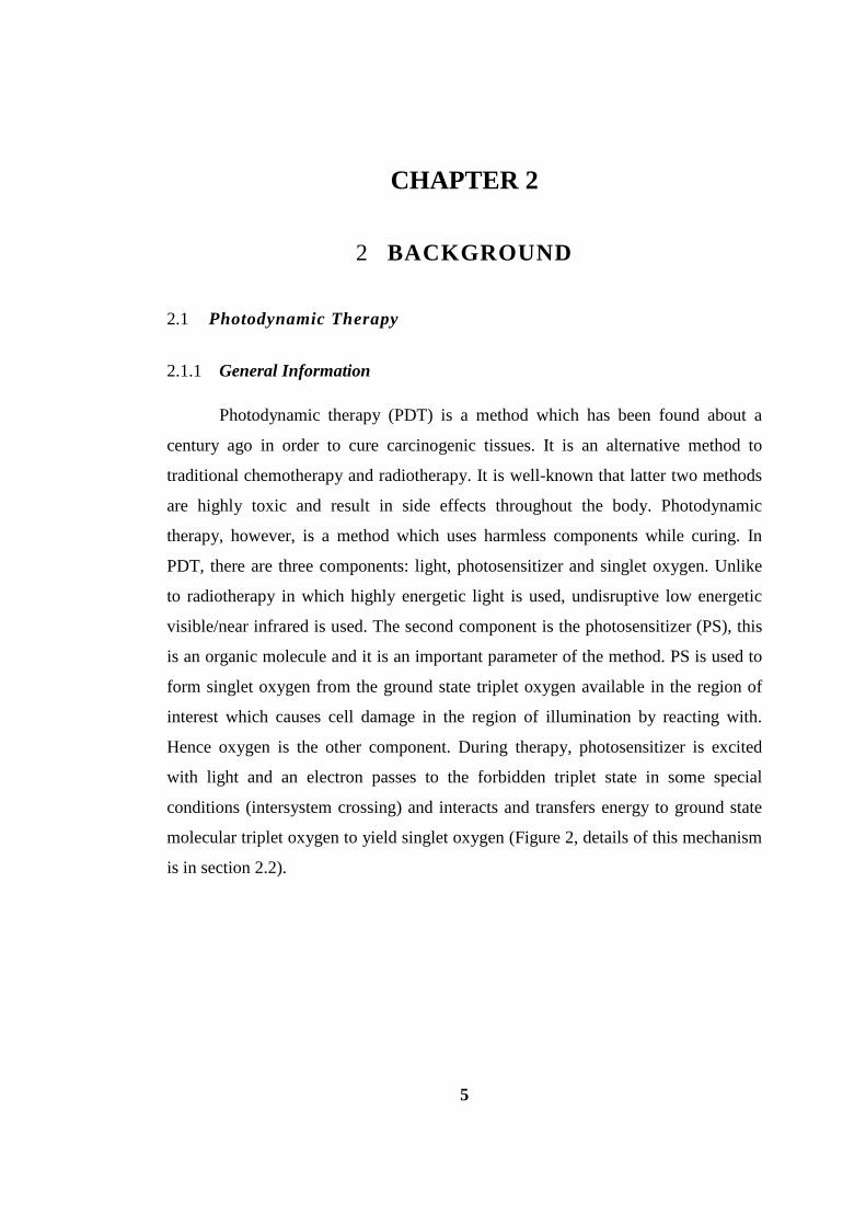
6
Figure 2. Modified Jablonski diagram for singlet oxygen production.
2.1.2 Development of PDT
The history of photodynamic therapy goes to more than 100 years ago;
researchers found that light and certain chemicals can induce cell death. In 1900,
Oscar Raab, a medical student found that in the presence of acridine and light has a
killing effect to infusoria, a small aquatic creature.7 Three years later, Herman Von
Tappeiner and A. Jezionek used eosin and light and named this action as
photodynamic action. Today, the most accepted, widespread photosensitizer is
porphyrin and W. Hausmann reported one of the first studies with and used
haematoporphyrin (See Figure 2) and light on paramecium and red blood cells and
saw that it killed both.8 In 1913, Friedrich Meyer-Betz used porphyrins on his own
body to see the activity and in light exposed areas, he reported pain and swelling.9
7
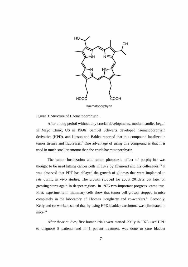
Figure 3. Structure of Haematoporphyrin.
After a long period without any crucial developments, modern studies begun
in Mayo Clinic, US in 1960s. Samuel Schwartz developed haematoporphyrin
derivative (HPD), and Lipson and Baldes reported that this compound localizes in
tumor tissues and fluoresces.7 One advantage of using this compound is that it is
used in much smaller amount than the crude haemotoporphyrin.
The tumor localization and tumor phototoxic effect of porphyrins was
thought to be used killing cancer cells in 1972 by Diamond and his colleagues.10 It
was observed that PDT has delayed the growth of gliomas that were implanted to
rats during in vivo studies. The growth stopped for about 20 days but later on
growing starts again in deeper regions. In 1975 two important progress came true.
First, experiments in mammary cells show that tumor cell growth stopped in mice
completely in the laboratory of Thomas Dougherty and co-workers.11 Secondly,
Kelly and co-workers stated that by using HPD bladder carcinoma was eliminated in
mice.12
After those studies, first human trials were started. Kelly in 1976 used HPD
to diagnose 5 patients and in 1 patient treatment was done to cure bladder

8
carcinoma.13 It was shown that tumor growth delayed and also tumor necrosis
observed in this patient. In addition, Dougherty used PDT in 25 patients with total
113 primary or secondary tumors and 98 of them were eliminated by using HPD
again as a photosensitizer.14 After these two types of carcinogenic tumors, Y. Hayata
and co-workers tried PDT on lung tumors and observed tumor growth delay in most
of the patients, however total curing was observed in one of fourteen patients.15
Growth of PDT action to other cancers continued in 1984, in which
McCaughan and colleagues treated patients with oesophageal cancer16, Balchum and
co-workers used to cure lung cancer.17 Hayata and his colleagues tried to cure gastric
carcinoma one year later. After all these preliminary trials of PDT, it was noted that
this method is efficient in early-stage patients that is inoperable due to other reasons.
Besides the cancer types noted, many other carcinogenic tissues have been tested
with PDT action. These are breast cancer, gynaecological tumors, intraocular
tumors, brain tumors, head and neck tumors, colorectal cancer, cutaneous
malignancies, intraperitoneal tumors, mesothelioma, cholangiocarcinoma and
pancreatic cancer. After all these experiments it was noted that PDT has only limited
success due to the reasons such as specificity and the effectiveness of the
photosensitizer. Hence, more powerful in terms of both specificity and strength
should have been developed and under clinical trials.
2.1.3 The Mechanism and Methods of PDT
It is told before that PDT is a method applied only in the region of interest
e.g. cancerous tissue. And it is not harmful to the other parts of the body because
half life of the singlet oxygen in biological systems is <0.04 μs and the singlet
oxygen is active only in the region of a radius of <0.02 μm.18 The photodynamic
action may be controllable, it may depend on various factors; type of the
photosensitizer, its intracellular and extracellular localization, the amount of PS
introduced, light’s exposure amount and its fluence rate, the amount of oxygen in the
region and finally the amount of time between the introduction of the drug to the
body and exposing the light.7

9
The mechanism of photodynamic action is very important and it has been
investigated deeply in order to broaden the applicability and to increase the respect
and universality of the method. Up to date, it has been proposed three ways of
killing of tumor cells; direct killing (necrosis), shutting down the tumor vasculature
so that tumor infarction yields and finally opening ways of immune system to
facilitate killing of tumor cells (apoptosis).7 These three ways should not be thought
as independent since each of them may influence the other and the importance and
the way that affect the other will probably be seen in future studies.
The first way of killing tumor cells is direct killing by photodynamic activity.
It is observed that number of tumor cells decrease with this way, however, it is not
enough always to wipe out all the illness. M. Korbelik and his colleagues observed
that PS accumulation may vary in tumor tissues.19 Intravenously injected PS may not
always go to the parts of the cancerous cells which are distant from the vasculature.
Another issue related with the direct killing of cells is the amount of oxygen needed
during therapy. The level of oxygen amount is not always enough may be owing to
two reasons; the first reason is during therapy oxygen is consumed and secondly,
because of the effect of PDT on the microvasculature fresh oxygen cannot be
reached to the region anymore. Both of these effects have been reported.20,21
Although shutting down the microvessels have an affirmative effect in long term, it
may affect direct killing. A solution to this problem may be decreasing the fluence
rate of the light and applying therapy in different times with decrased dose in order
to keep the oxygen amount on a tolerable level.7
One type of indirect killing of tumor cells is by inducing vascular damage via
PDT. Increasing amount of tumor cells is dependent on the available nutrients, and
so targeting the vascular system to the tumor tissue is an important tool to utilize.
There has been enough number of studies showing PDT causes vascular
damage22,23,24, resulting in delay of tumor growth by hypoxia and anoxia25,26,27 which
both refer to the reduced level of oxygen where the latter is an extreme version.
After PDT treatment, it has been shown in various in vivo studies that, first
10
vasoconstriction takes place and after a period of three hours thrombus formation
occurs, which results in tumor growth depletion. In 1989, Henderson and colleagues
showed that by using Photofrin (an approved photosensitizer being used in clinic)
tumor cells eradicated by oxygen depletion in tumor tissues by shutting down tumor
vasculature in a fibrosarcoma mouse model.28 Hence, by targeting tumor vasculature
it is possible to delay tumor growth.
Another indirect treatment of PDT is calling out the immune system
elements (lymphocytes, leukocytes and macrophages) on work into the treated
tissue. The inflammation starts with various components such as clotting cascades,
acute-phase proteins, proteinases, peroxidases, reactive oxygen species.29 W. deVree
observed neutrophil attraction to the PDT activated tissue so that tumor growth was
delayed.30 Korbelik and colleagues in 1996 compared the tumor growth in normal
tissue and in immunodeficient tissue in order to understand the long term effects of
PDT.31 In short term they observed that PDT effect is similar, however, in long
period of time tumor growing is very fast in immunodeficient tissue compared to the
regular one. Therefore, immune response is an important parameter while fighting
with cancerous cells.
There is an interesting study on immunology studies after PDT; in 1999
Korbelik and colleagues have shown that tumor-sensitized immune cells have been
found on lymphoid sites which are far to the region where PDT applied.32 Another
related study was performed by Henderson group; they recovered the tumor-cell
lysate from the PDT applied tissue and they used this as a vaccine and applied to a
mice, and showed that tumor growing prevention. 33 This study is important for the
topic of induction of tumor-specific immunity. It has been postulated that this type
of vaccination is more effective than producing lysates from tumors that were
irradiatied with UV or ionizing irradiation. These studies designate that PDT may
have a potential to be used as a systemic immune therapy. The studies related with
anti-tumor immunity and PDT have been reviewed recently34 and it has been
emphasized that while other methods like chemotherapy, surgery and radiotherapy

11
are mostly immunosuppressive, PDT can cause acute inflammation, express heat-
shock proteins, calling leukocytes to tumor region and increase the presentation of
tumor-derived antigens to T cells. Studies related with the immunogenic properties
of PDT is ongoing and it forms as an important part of the study.
In order to PDT to be effective tumor localization is an important issue.
Hence, photosensitizers are under study and a PS named MV6401 (a
pyropheophorbide derivative) has been developed which especially localizes in
tumor vasculature.35 Delivery of the PS depends on vascular permeability and
interstitial diffusion into the cell which may change with size, charge, configuration,
hydrophilic and hydrophobic properties of the molecule and also to the physiological
properties of the blood vessels.
Another issue which affects the PDT efficacy is the time between
introduction of the drug to the body and irradiation. If the interval is short like 15
minutes drug generally holds on the vasculature, however in longer times like 4
hours drug localizes on the tumor tissues by using PS MV6401. If irradiation starts
in a short time vascular stasis and thrombus formation starts, hence indirect killing
of tumors achieved. With most cancer types and PS in the market, generally longer
period of times (24-72 hours) have been waited in order to accumulate in
extravascular compartment. These indicate that application of light in different time
periods affects the mechanisms of PDT.
In 2002, a different approach has been applied and found to be superior to
the methods above. The drug has been given to the body more than one time at
different time intervals. By this way both vasculatures and tumor cells were
activated. This fractionated dose of PSs enabled to obtain long term tumor growth
control.24b
Targeting the tumor region is one of the important task in PDT and increased
concentration of photosensitizers in tumor cells have been observed and called
passive targeting. However, the exact reason for this complex phenomenon is not
12
completely understood. It is reported that probably it is related to high vascular
permeability of PS, and their affinity for proliferating endothelium and minimum
lymphatic drainage in tumor cells. In order to attain an effective delivery agent,
conjugated antibodies have also been utilized in order to direct these to tumor
associated antigens or vascular antigens (e.g. ED-B domain).36 In 1990s some
photosensitizers have been developed for targeting mitochondria, plasma membrane,
nuclei and lysosomes.37 It has been reported that mitochondria is an important
organelle to target for PDT38,39 and these type of delivery agents are important when
combining the strengths of two major areas; PDT and therapeutics studying on
targeting subcellular regions.
The efforts for the commercialization of PDT have resulted partially in 1993
for Photofrin in Canada for the first time for the prophylactic treatment of the
bladder cancer. This drug is the partially purified version of the haematoporphyrin
(HPD) which was utilized in 1970s. However, Photofrin is not very pure; it contains
more than 60 types of porphyrin derivatives. This impurity also complicates its
reproduction easily. The absorption maximum of this compound is 630 nm which is
tolerable but it has low extinction coefficient (1,170 M-1 cm-1). Due to this high drug
dose and fluence rate should be used during therapy. Moreover, it has a long lasting
skin photosensitivity.7 According to the studies reported by Orenstein and
colleagues, Photofrin has low normal to tumor cell accumulation in C26 colon
carcinoma bearing mice.40 Hence, patients should avoid sunlight for 4-6 weeks after
therapy in order to prevent skin photosensitization. Still, Photofrin is the most
widespread drug used in PDT. It has been approved in the Netherlands and France
for lung cancer, in Germany for early-stage lung cancer, in the United States for
advanced oesophageal cancer and in Japan for early-stage oesophageal, gastric and
cervical cancer (Table 1).

13
Table 1. spectroscopic and pysicochemical properties of clinically approved photosensitizers41.
Photosensitizer λmax abs (nm) ε (M-1 cm-1) λmax emiss
(nm)
ΦFL ΦΔ LogPo/w
Porfirmer Sodium (Photofrin®) 630 3000 NA NA 0.25 3.96
Protoporphyrin IX(Levulan®) 635 5000 630 0.011 0.54 NA
Temoporfin (Foscan®) 650(EtOH) 39000 655 NA 0.31 9.24
Verteporfin (Visudyne®) 692(PBS) 31200 694 0.049 0.82 7.76
Talaporfin(Laserphyrin®) 654(PBS) 40000 660 NA 0.77 -1.92
Ce6 (Photolon®) 663(PBS) 38000 662 0.18 0.75 0.78
The limitation with Photofrin makes scientists to work on more efficient
photosensitizers. More ideally the new PS should absorb at longer wavelength in
order to achieve deeper skin penetration of light, more target specific and less skin
photosensitive. In this way, a drug called Foscan has been developed which is very
efficient compared to previous and approved for head and neck cancers. This
chlorine derivative PS requires using very low dosage (0.1 mg/ kg of body weight).42
However, due to its being reactive, some complications have been reported.43 Also,
for actinic keratosis and basal-cell carcinoma 5-aminolevulinic acid and 5-ALA
methyl ester have been approved. The structure of 5-ALA is unusual since it is not
an absorbing dye, but it is an endogenous porphyrin which is the first compound in
porphyrin synthesis pathway in the body for synthesis of heme in mammals and
chlorophyll in plants (Figure 4).
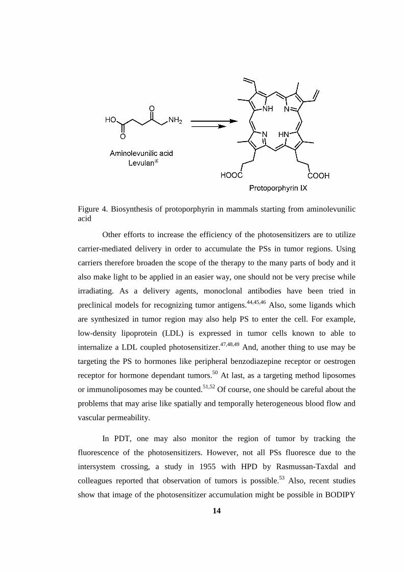
14
Figure 4. Biosynthesis of protoporphyrin in mammals starting from aminolevunilic acid
Other efforts to increase the efficiency of the photosensitizers are to utilize
carrier-mediated delivery in order to accumulate the PSs in tumor regions. Using
carriers therefore broaden the scope of the therapy to the many parts of body and it
also make light to be applied in an easier way, one should not be very precise while
irradiating. As a delivery agents, monoclonal antibodies have been tried in
preclinical models for recognizing tumor antigens.44,45,46 Also, some ligands which
are synthesized in tumor region may also help PS to enter the cell. For example,
low-density lipoprotein (LDL) is expressed in tumor cells known to able to
internalize a LDL coupled photosensitizer.47,48,49 And, another thing to use may be
targeting the PS to hormones like peripheral benzodiazepine receptor or oestrogen
receptor for hormone dependant tumors.50 At last, as a targeting method liposomes
or immunoliposomes may be counted.51,52 Of course, one should be careful about the
problems that may arise like spatially and temporally heterogeneous blood flow and
vascular permeability.
In PDT, one may also monitor the region of tumor by tracking the
fluorescence of the photosensitizers. However, not all PSs fluoresce due to the
intersystem crossing, a study in 1955 with HPD by Rasmussan-Taxdal and
colleagues reported that observation of tumors is possible.53 Also, recent studies
show that image of the photosensitizer accumulation might be possible in BODIPY

15
based system by conjugating PS with a fluorophore.54 An alternative way of
employing PDT is to deliver membrane-impermeable drugs to the cytosol by a
method called photochemical internalization.55
Besides curing cancerous diseases PDT also employed in other disorders.
The most popular ones are age related macular degeneration and other eye problems
related to the neo-vascularization.56 Verteporfin has been approved starting from
1999 in many countries in order to cure this type of problems. Also, PDT has been
used to treat arteries with intimal hyperplasia57 and after stent implantation in
cardiovascular part of medicine.58 In dermatology field, psoriasis and scleroderma
have been treated with PDT.59,60 In rheumatology area, arthritis has been tested.61
Finally, PDT has been used to kill microorganisms also.62,63
2.1.4 Mechanism of Singlet Oxygen Production64
Biochemical reactions in living things which take place with light can be
splitted to two main categories. The first is the photosynthesis and vision processes.
These have been a nearly totally investigated topic and known very well since they
are both vital topics of living on earth. The second thing is the photodynamic effect.
In this topic, a photosensitizer is used and this is not a biologically known object and
in the body, and other biological things may behave to these differently. Just because
of this its mechanism is an interesting subject and have been investigated by
different disciplines. The mechanism of photodynamic effect can be investigated in
four parts: In vivo interactions (1), excitation (2), reaction of excited species (3) and
reactions of reactive secondary products in vivo (4). In the in vivo interactions case
pharmacokinetics (i.e. photosensitizer localization in tissues and cells, targeting and
clearance rate) and structure of the photosensitizer and activity relationship play an
important role. For the excitation case several photophysical characteristics (i.e.
absorption properties, energies, quantum yields, lifetimes and singlet oxygen
quantum yields) are investigated. Upon excitation with light, three main reactions of
excited species occur: electron transfer (TYPE I, results in radicalic reactions like
crosslinking of proteins), energy transfer (TYPE II; singlet oxygen produced) and
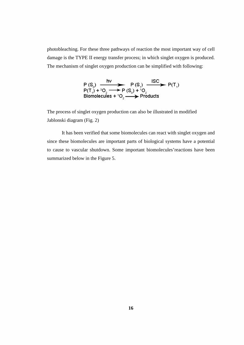
16
photobleaching. For these three pathways of reaction the most important way of cell
damage is the TYPE II energy transfer process; in which singlet oxygen is produced.
The mechanism of singlet oxygen production can be simplified with following:
The process of singlet oxygen production can also be illustrated in modified
Jablonski diagram (Fig. 2)
It has been verified that some biomolecules can react with singlet oxygen and
since these biomolecules are important parts of biological systems have a potential
to cause to vascular shutdown. Some important biomolecules’reactions have been
summarized below in the Figure 5.

17
Figure 5. Some biomolecules’ reaction with singlet oxygen and its products64.
2.1.5 Molecular Orbital Diagram of Triplet and Singlet Molecular Dioxygen65
Dioxygen molecule is an exception to the most of the molecules because of
its being in triplet state, while most others are singlet. This is possible in molecular
orbital theory when degenerate MOs exist. The lowest energy configuration of
dioxygen molecule is [N2] (π*) 2, where [N2] is the electronic configuration of the N2

18
molecule. Hund’s rule application to this molecule yields a triplet due to energy
minimization and electrons are in parallel. The representation is shown in Figure 6.
As the name implies, there are three sublevels which are equal in energy.
Two of them can be viewed easily, the first one is two up parallel arrows α(1)α(2) as
in the Figure 6. Other one is the two parallel down arrows β(1) β(2), while the arrow
indicates each electron’s spin projection along a given fixed axis. The third triplet
level is difficult to represent since it is the combination of two states. And both of
these states have zero spin angular momentum, but the total spin angular momentum
is non zero for the triplet shown as: α(1)β(2) + β(1)α(2). A similar combination
stands for singlet when combination is α(1)β(2) − β(1)α(2).
Hence, the ground triplet state oxygen molecule is denoted as 3Σ and
according to the Pauli’s exclusion principle the two electrons in the HOMO should
be in different orbitals as in the Figure 6. The corresponding triplet sublevels are
α(1)α(2), β(1)β(2) and α(1) β(2)+β(1)α(2). The excited states of oxygen is also
shown with the configuration of [N2] (π*)2, however in this case with increased
energy. The first excited state is 1∆g and often called as singlet oxygen, where two
electrons are in the same orbital in different spins. In this case we have a double
degenerate singlet states and it is denoted as α(1)β(2)−β(1)α(2).* In the second
excited state which is higher in energy (1Σg) corresponds to two electrons with
different spins and in different orbitals with again same combination
α(1)β(2)−β(1)α(2). As a result, the absorption and emission take place in oxygen
molecule is a singlet-triplet or triplet-singlet transition.
In general, most of the illustrations of the excited state electron transitions
are shown as two orbitals have been involved in the process, namely HOMO and
LUMO. However, in reality this is not the case because many orbitals may involve
* In this case the spin functions should be thought not strictly as px * and py
*. It should thought as linear combinations of these two which have well-defined orbital angular momenta.

19
in excitation, deexcitation process, and excited states are described as the
superposition of these configurations. In some cases different phenomena like
double excitations* may contribute.65
Figure 6. Molecular orbital diagram of molecular dioxygen with its ground and excited states65.
2.2 Triplet Photosensitization
When a fluorophore absorbs light and it excites to singlet excited state, and
in general case it deactivates via S1 to S0 transition (fluorescence).65 However,
sometimes with ISC triplet state can be populated (S1 to Tn). So, in order to get
successful candidates for triplet PSs, efficient ISC should be attained. The number of
studies with triplet photosensitizers and the factors and relationship between the
molecular structure and efficiency is limited.66 The application areas with triplet PSs
is diverse, e.g. electroluminescence, phosphorescent bioimaging,67 PDT68,
* Simultaneous excitations of two electrons

20
photoinitiated polymerization and photoredox reactions.69 Fluorescent molecules
generally used for their luminescence property, however, triplet PSs generally used
for initiating another reaction, and the product is used for purpose.70 The two main
reactions carried with triplet PSs are singlet oxygen production from ground state
stable triplet molecular oxygen and triplet-triplet annihilation upconversion reaction.
In the latter subject triplet PSs are used for triplet-triplet energy transfer donor.
Designing a triplet PS is known to be a challenge especially it does not
contain a heavy atom. The main reason for this fact is the covered and unexplored
facts of intersystem crossing. For example, the photophysical difference between
antracene and 9,10-diphenyantracene is interesting. Anthracene has a φF of 0.27 in
EtOH however, DPA has 0.95 in same conditions. Similiarly, antracene has a φISC of
0.73 which is smaller than 0.05 in DPA.71
One reason for the rare examples of molecules that undergo ISC is because
of their including quantum mechanically forbidden steps. However, in some case
ISC is allowed. When a chromophore is excited, S1 state is populated then a non-
radiative transition from S1 state to Tn (n≥1) should take place. For this transition to
occur two spin states should involve in the process. Magnetic torques should be
exerted on the magnetic moment (µs) vector of an electron spin, or as a result of the
spin angular momentum (µs) coupling to the orbital angular momentum (µL). In
organic molecules in most of the time coupling of the electron spin with the orbital
angular momentum (i.e. spin-orbit coupling) is the case for ISC. Spin-orbit coupling
is defined and can be understood by thinking the motion of the electron around the
nucleus in a Bohr-like orbit. There are two types of rotations and these cause a
magnetic field. First, electron rotates in its own spin and the second it rotates around
the nucleus. These two types of magnetic fields interact with each other yielding
spin-orbit coupling.65

21
2.2.1 Heavy Atom Effect on Triplet Photosensitization
Atoms with a high number of atomic weights can effect the spin-orbit
coupling in order to increase intersystem crossing. Spin-orbit coupling efficiency has
a dependence of Z4.* The effect of internal heavy atom can be explained with the
electron’s movement around the nuclei. The movement around the nuclei is
accelerated when it gets close to the more positively charged nuclei, inducing
increased orbital magnetic torque (µL). As a result, the coupling of the spin (µS) and
orbital (µL) angular momentum increases. Hence, ISC is increased with the heavy
atoms when they are close to the center of molecule. This strategy is an important
task when designing a triplet photosensitizer.72 Some heavy atoms used are Ir, Pt,
Ru, Os, Re, Rh, I and Br. Various metal complexes are also used for triplet PSs but
they have generally have low absorbance in the visible region which limits their
utilization.72
2.2.2 Triplet PSs with low-lying n-π* transitions
For a transition to occur from singlet state to the triplet state there are some
restrictions. It is similar to the selection rules of electronic transitions. El Sayed’s
rule is generally accepted with these restrictions.72 El Sayed’s rule is related with the
magnitude of rate constant of intersystem crossing denoted as 1kISC. This rate is
increases when there is an orbital change between transitions. For example transition
from 1(n-π*) orbital to 3(π-π*) orbital is faster than 1(π-π*) - 3(π-π*) transition.
Because of these statements most aromatic chromophores do not display intersystem
crossing behavior and show intense fluorescence.
* Where Z is the atomic weight

22
2.2.3 Exciton Coupling Behaviour of Chromophores
Exciton coupling is a way of populating to triplet state from singlet excited
state. It occurs when almost similar chromophores link together without having a π
conjugation, and their singlet orbitals split into two and the one in lower energy has
increased probability to pass to the triplet state because of minimized energy
difference between states.73
2.2.4 Utilizing Spin Converter for Triplet State Photosensitization
Designing and synthesizing a triplet photosensitizer without using heavy
atom is a difficult task. The principles of ISC and organic molecular structure is not
a very well known subject. Hence, all the studies done in this topic without using a
heavy atom, especially in the visible region is highly important. One strategy used in
this context is using a spin converter. In literature the only molecule used as a spin
converter is the fullerene.74 It is an intrinsic triplet state photosensitizer. In all the
cases within this topic, fullerene molecule is used as an excitation energy transfer
(EET) acceptor molecule and another molecule is used as an donor. Upon excitation
from the donor molecule* intersystem crossing take place with the help of fullerene
moiety.
2.2.5 Methods to Detect Triplet Excited States
Detecting a triplet state of a molecule is not a straightforward process like in
singlet excited states. Some of the triplet PSs are phosphorescent but some of them
not. Time-resolved transient absorption (TA) spectroscopy method is used to get
information about triplet population.72
* Which is a poor triplet photosensitizer

23
This method is based on the pulse-probe (or pump probe) method. There are
two light sources have been used, the first is the typical xenon lamp for tracking
absorption change and the other is a pulsed laser source for pump purpose.
2.2.6 Triplet PSs recently studied
In the literature it is possible to see a growing publication rate of synthesized
triplet PSs, However the rate is not as much as fluorescent molecules. The PSs
studied can be broadly separated into two parts, the ones with heavy atom and
without heavy atom. It is generally accepted that designing a PS without heavy
atoms is much difficult.72 The PSs with heavy atom can also be divided into two
parts: (i) transition metal complexes (ii) iodo or bromo substituted organic
chromophores. Both of these have many examples in the literature, and due to the
limitations of transition metal complexes halogenated organic molecules have been
used more frequently. The most famous of these include xanthene based ones and
bodipys. Recently, studies on naphthalene diimides were also conducted. Rose
Bengal, Eosin Blue, Erythrosin B and Eosin Yellow are xanthene based PSs (Figure
7).75 These molecules have been known for a long time mainly for staining in
microscopy. However, one drawback for these types of structures is their limited
functionalization leads to restricting the absorption wavelength to visible region.
Due to this problem derivativable organic fluorophores have been studied more in
this topic. Most successful candidates are bodipy and perylene which was already
noted.
Bodipy derivatives have the advantage of ease functionalization and many
PSs in the range of 500-800 nm absorbances have been synthesized. Upon
halogenation high singlet oxygen quantum yields have been attained(see Figure 6).76

24
Figure 7. Fluorescein and its halogenated derivatives as triplet PSs75.
Figure 8. Diiodinated Bodipy for PDT76.
Since absorbance around 500 nm is not enough for PDT application due to
the limited penetration depth of light at this wavelength, near-IR absorbing bodipy
molecules are more important. Recently, Ramaiah et al. have synthesized an
azabodipy derivative (Figure 7) absorbing at 666 nm.77 Their characterization of Φ∆
and ΦT showed that these molecules can be used for PDT applications with values
0.70 and 0.78, respectively. In order to learn more about the excited state properties

25
of these dyes they have analyzed with nanosecond flash photolysis (transient
absorption). They observed two transient absorption peaks at 320 and 510 nm with
excitation with 355 nm laser pulses. The band at 510 nm is suspected for triplet
excited state formation since its quenching with dissolved oxygen (see Figure 9).
The triplet quantum yield of this dye was determined using triplet-triplet energy
transfer to β-carotene using a tris(bipyridyl)ruthenium(II) complex as a reference.
For this calculation transient absorption at 320 nm was tracked and 0.78 yield with
at lifetime of 1.6 µs. This observed triplet quantum yield has been the highest
reported for aza-bodipy derivatives so far at the year it was published.
Figure 9. Aza BODIPY derivative for PDT application and its transient absorption spectrum. There are two transient absorption peaks at 320 and 510 nm. The inset graphic shows the first-order decay of the 510 nm absorption.78
In the recent study by Zhao group79 triplet excited state studies was
conducted with naphthalene diimide (NDI) molecule. Since these compounds mainly
absorb in the UV range, electron donating amino groups was attached and
absorbance maxima at 526 nm was observed. Although this wavelength is not
sufficient for PDT, it may have a potential to be used in triplet-triplet annihilation
upconversion studies or in photocatalysis reactions. Actually, long triplet state

26
lifetime has been recorded (τT = 51.7 µs) and high upconversion quantum yield has
been observed (see Figure 10).
Figure 10. Naphtalene diimide molecule used for triplet photosensitization.79
2.2.7 Triplet PSs without Heavy Atom
It was already noted that designing a PS without a heavy atom is a
complicated process. One way of realizing this strategy is to use spin convertors.
Spin convertor is such a molecule that has a high quantum yield of intersystem
crossing without any heavy atom. In the literature rare examples of spin convertors
have been reported one of which is the C60 fullerene molecule.74 This molecule has
an absorption maximum at 335 nm and has with very low molar absorptivity in the
visible range and near IR range (around 700 M-1 cm-1) and almost quantitative
intersystem crossing quantum yield. Moreover, it has a low S1 state energy level
(1.72 eV) so that a wide range of organic chromophores can make energy transfer to
C60 molecule*. To use fullerene molecule as a spin convertor dyes should be
attached around the fullerene and energy transfer from dye to spin convertor yields
intersystem crossing. Actually, the term energy transfer should be used with care in
this concept because in typical Forster energy transfer, emission wavelength from
* Dyes have an absorption maxima from UV range to about 700 nm are convenient

27
the donor should be compatible with the absorption of the acceptor moiety (spectral
overlap). A simplified version of Jablonski diagram for C60-chromophore triplet
sensitizers is shown below (Figure 9).80
Figure 11. Simplified Jablonski diagram showing the photopysics of C60-
chromophore hybrids as heavy atom free triplet PSs.
One successful example to fullerene-chromophore triplet PS is by using the
bodipy chromophore.80 In this study bodipy units absorbing at 600-650 nm range
have been attached to the C60 units, and with the help of transient absorption
spectroscopy they have observed ping-pong type reaction. First, bodipy core has
been excited around 600 nm and energy transfer occurs to the fullerene and
intersystem crossing to the triplet state of fullerene took place and this time triplet-
triplet EnT from fullerene to antenna bodipy realized. As a result long-lived triplet
state lifetime and high singlet oxygen quantum yield have been observed (τT= 123.2
ms and Φ∆=0.85). The studies with this strategy to use triplet PSs are new and
expected to be utilized more in the future.

28
Figure 12. Fullerene-bodipy dyads as efficient heavy-atom-free organic triplet photosensitizers.80
Other than spin convertors some small organic molecules absorbing at UV
region also do sensitize to triplet state. Since their absorbance are at UV region their
treatment to applications such as PDT is not seem possible, but their mechanism of
ISC is an important issue while designing triplet photosensitizers. A list of several
molecules of this type have been show in Figure 13. One if them is pyrene molecule
which has been used widely for fluorescence with a fluorescence quantum yield of
0.65, however one cannot discount its intersystem crossing quantum yield of 0.35. In
the pyrene, there is a special occasion of delayed fluorescence. It take place when
luminescence rate is very slow. In delayed fluorescence T1 to S1 state reverse
intersystem crossing take place due to this abnormal situation lifetime of emission is
very long (e.g. τF= 450 ns in cyclohexane which is ten folds of typical bodipy
fluorophore’s). As in other small organic triplet PSs their modification in the core
results diminished ISC rates. For example, when pyrene’s core is substituted with π
framework, the emission is turned to be a prompt fluorescence with τF of a few
nanoseconds.81

29
Some aromatic ketones shown in Figure 13 also shows high intersystem
crossing quantum yields. As it was already noted, low lying 1(n-π*) transition to 3(π-
π*) transition is fast, so this effect can be explained by obeying this rule.
Figure 13. Heavy atom free small organic molecules as triplet PSs absorbing at UV region.
Along with these heavy atom free triplet photosensitizers, another important
class of organic molecules, also very important for life on earth, have been utilized
for the same function; porphyrins. Their usage in this concept is very early however,
their mechanism of triplet photosensitization has not been explained up to now very
well. In a study reported, it has been suggested that vibration of the molecule with
excited state intramolecular proton transfer aids for intersystem crossing.82 Still
porphyrins absorbing strongly (e.g. high molar absorptivity) in the near IR region are
needed for their wider utilization in PDT.
One of rare examples of triplet PSs without heavy atoms is the
di(perylenebisimide)s. It has been synthesized recently and their singlet oxygen
efficiencies were investigated with Φ∆ of 0.67 and 0.59 for two types molecules.83
The near IR absorbances are 671 and 655 nm. The reason for successful intersystem
30
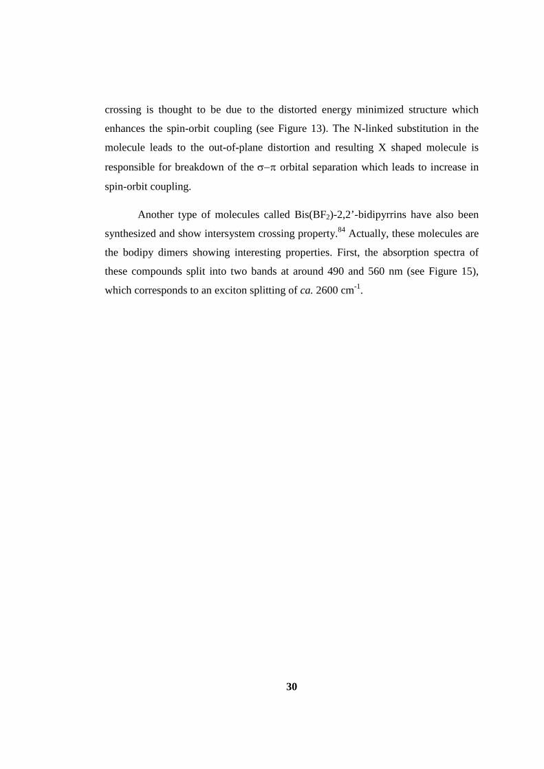
crossing is thought to be due to the distorted energy minimized structure which
enhances the spin-orbit coupling (see Figure 13). The N-linked substitution in the
molecule leads to the out-of-plane distortion and resulting X shaped molecule is
responsible for breakdown of the σ−π orbital separation which leads to increase in
spin-orbit coupling.
Another type of molecules called Bis(BF2)-2,2’-bidipyrrins have also been
synthesized and show intersystem crossing property.84 Actually, these molecules are
the bodipy dimers showing interesting properties. First, the absorption spectra of
these compounds split into two bands at around 490 and 560 nm (see Figure 15),
which corresponds to an exciton splitting of ca. 2600 cm-1.
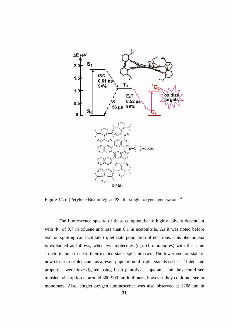
31
Figure 14. di(Perylene Bisimide)s as PSs for singlet oxygen generation.83
The fluorescence spectra of these compounds are highly solvent dependant
with Φfl of 0.7 in toluene and less than 0.1 in acetonitrile. As it was noted before
exciton splitting can facilitate triplet state population of electrons. This phenomena
is explained as follows; when two molecules (e.g. chromophores) with the same
structure come to near, their excited states split into two. The lower exciton state is
now closer to triplet state; as a result population of triplet state is easier. Triplet state
properties were investigated using flash photolysis apparatus and they could see
transient absorption at around 800-900 nm in dimers, however they could not see in
monomers. Also, singlet oxygen luminescence was also observed at 1268 nm in

32
aerated solution upon excitation at 490 nm. The singlet oxygen quantum yield of
these dimer dyes have been quantified by using the singlet oxygen emission and
found at most 0.4 in toluene, 0.5 in dichloromethane and <<0.1 in acetonitrile.
Figure 15. Exciton coupling behaviour of bis-bodipy triplet photosensitizers. The graph on the right shows absorbance of monomer (solid line) and dimer (dashed).84 2.2.8 Employment of Triplet PSs
Triplet PSs have been used in various applications including
electroluminescence, photodynamic therapy, photoinduced catalytic H2 production,
triplet-triplet annihilation upconversion, photoredox synthetic organic reactions and
luminescent oxygen sensing. Transition metal based PSs have been used generally
for electroluminescence and will not be discussed here more. And PDT also will not
be discussed here since enough information has been given in the early part of
background.
2.2.8.1 Photoinduced catalytic H2 production
Hydrogen gas production from water (water splitting) is thought to be an
important research topic for years.85 In this topic there are three main components;
photosensitizer, sacrificial reductant (SR) and water reduction catalyst (WRC).86 The
mechanism of water splitting is shown in Figure 16. First, it is needed a PS with a
visible light harvesting ability and triplet excited state lifetime is also important. In

33
the mechanism, first PS is excited with light, and electron transfer occurs from SR to
PS. After, PS transfers its electron to WRC to reduce water to obtain H2.*
Figure 16. Mechanism for the water splitting to produce H2. PS is for photosensitizer, SR is for sacrificial reductant and WRC stands for water reduction catalyst.86 In an example to this research, Castellano and co-workers designed a
platinum complex as a photosensitizer and they observed H2 production from three
different types of series.87 They have used triethanolamine as a sacrificial reductant
and cobaloxime as a catalyst for water reduction. The structures of platinum PSs are
shown below in Figure 17 and the rate of H2 production is larger as the length of R
group is longer due to the increased visible light harvesting ability of the extended π
unit.
* H+ is reduced to H2

34
Figure 17. Molecular Structures of the Platinum(II) Terpyridyl π-Conjugated Arylacetylide PSs.87
2.2.8.2 Triplet Triplet Annihilation Upconversion(TTA UC)
Due to its potential application in photovoltaics, photocatalysis and optical
materials etc. upconversion is a hot topic.88 Some of the techniques used in
upconversion are by using rare earth metal nanoparticles, two-photon absorbing dyes
and TTA UC. TTA UC is based on a triplet photosensitizer and a triplet acceptor
often in a fluid matrix.89 First, a PS is excited with light and singlet excited state is
populated and via intersystem crossing triplet excited state of PS is populated, then a
triplet acceptor molecule accepts energy transfer from the excited triplet of PS*,
finally by annihilation singlet excited state of the triplet acceptor is populated; as a
result fluorescence emission from the triplet acceptor’s singlet excited state is
observed (see Figure 18).70 In order for this process take place triplet states of donor
and acceptor should be matched to each other.
In literature a recent study exemplifies this type of upconversion where well-
known bodipy molecule with heavy atoms is used as a triplet PS and perylene
molecule for triplet acceptor.90 As a result of the study, fluorescence emission form
* To which is called triplet triplet energy transfer

35
perylene unit at 445-518 nm range has been observed upon excitation with 532 nm
laser; hence laser emission is upconverted. The long lived triplet excited state of
bodipy (τT= 95.2 µs) is a desired property for increased quantum yield of
upconversion, where they could observe 0.165. The structure of the PS, triplet
acceptor and upconverted emission is also shown in Figure 19.
Figure 18. TTA upconversion process in modified Jablonski diagram for bodipy photosensitizer (BI-1) and perylene acceptor (Py). 1BI-1* (FC) is the Franck- Condon state of excited singlet state of bodipy. 1BI-1* is the excited singlet state of bodipy. 3BI-1* triplet excited state of bodipy. Other states are similar to this. IC is internal conversion and ISC is intersystem crossing.90

36
Figure 19. Structure of the bodipy PS, perylene triplet acceptor. Right emission photographs (a) sensitizers alone (b) when triplet acceptor perylene added, upconversion emission is seen. In toluene excited at 532 nm.90
2.2.8.3 PSs in Photoredox reactions
To find green and sustainable methods for effective synthesis of important
chemicals is an important task in research area.91 Visible-light-induced photoredox
catalysis is one of them and due to high natural abundance, cleanliness,
sustainability and environmental friendly it is an important reaction.
Recently emerged this type of reaction has been accomplished for aerobic
oxidative hydroxylation of arylboronic acids to aryl alcohols (Fig. 20).92 They have
utilized Ruthenium complex as a catalyst and in combination with visible light and
air (as the source of oxygen) they could achieve excellent yields. Besides, a
nonmetal organic catalyst, Acid Red 87 was also used. In the reaction first PS is
excited with visible light and this excited PS converts oxygen in the air to
superoxide radical anion which reacts with the arylboronic acid to yield arylalcohol.

37
Figure 20. Proposed reaction mechanism of visible-light-induced aerobic hydroxylation reaction92
Visible light induced redox reactions is not limited to this reaction, other
reactions include intermolecular [3+2] cycloaddition of cyclopropylamines with
olefins93, C-C bond formation by aerobic oxidation94 or aerobic cross
dehydrogenative coupling reactions95.
2.2.8.4 O2 sensing
O2 sensing is important for biological and environmental science but also for
chemical science. Hence research in this subject has been developed and mainly
transition metal complexes have been used. The reason for their utilization is their
phosphorescence is quenched with O2 hence the presence of oxygen can be tracked
in that way. Conventional O2 sensing complexes are Pt(II) or Pd(II) porphyrin based
ones.
Other type of sensing strategy of oxygen is using ratiometric sensing. In this
route, one fluorescent and one phosphorescent group is used separately or in one
molecule. In the presence of oxygen phosphorescence is quenched however,
fluorescence is not so that the amount of oxygen concentration can be tracked in that
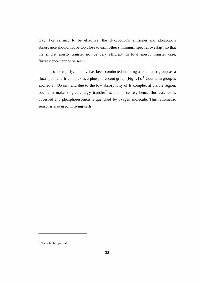
38
way. For sensing to be effective, the fluorophor’s emission and phosphor’s
absorbance should not be too close to each other (minimum spectral overlap), so that
the singlet energy transfer not be very efficient. In total energy transfer case,
fluorescence cannot be seen.
To exemplify, a study has been conducted utilizing a coumarin group as a
fluorophor and Ir complex as a phosphorescent group (Fig. 21).96 Coumarin group is
excited at 405 nm, and due to the low absorptivity of Ir complex at visible region,
coumarin make singlet energy transfer* to the Ir center, hence fluorescence is
observed and phosphorescence is quenched by oxygen molecule. This ratiometric
sensor is also used in living cells.
* Not total but partial

39
Figure 21. Ratiometric O2 sensor (on top) a) Emission Intensity of aerated (black dash) and Argon purged (red line) in CH3CN b) the diffent emission colors due to the different O2 partial pressure.96
2.3 Asymmetry in Luminophores
Asymmetry is an important topic in biology. For example knowing the
stereochemical structure of the potential drug is essential because the potency,
selectivity of one enantiomer can be different from the other enantiomer. Solution
based sensor systems are important while learning these information during drug-
receptor interactions.
Basically, isomers are divided into two main categories and stereoisomers is
one of them. The other type structural isomerism is out of our topic. Stereoisomers
also has two parts; enantiomers and diastereomers. Enantiomers are characterized by
their nonsuperimposability, while diastereomers are not related with their mirror
40
images. For instance, enantiomers are their mirror images and they are not
superimposable to each other, however, diastereomers are not mirror images of each
other.
The simplest source of chirality in an organic molecule is the carbon atom
with four different substituents. Lactic acid is the oldest example of chirality.97 Both
enantiomers have been found in nature. (−)-lactic acid is found in certain
fermentation products of sugars, in some species mixed with its enantiomer. (+)-
lactic acid is found in muscle fluid and it forms after the biochemical reaction of
muscle work. Most properties of matter do not change with reflection, so two
enantiomers’ are identical in many aspects like solubility, density, refractive index,
IR, Raman, UV, NMR and very similar X-ray diffraction pattern. Only their
chiroptical properties such as optical rotation, ortical rotatory dispersion and circular
dicroism different in terms of sign but not magnitude.97 Some of the fermentation
products of lactic acid are mixture of two enantiomers, so they are (±)- or racemic
lactic acid. However, in nature these racemic mixtures are unusual since most of the
chiral products are either single enantiomer or one enantiomer is predominant.
2.3.1 Optical Spectroscopy
Optical spectroscopy is mainly based on the interaction between the atoms or
molecules with incident electromagnetic radiation.98 In 1860, it was postulated by
Scottish physicist James Clerk Maxwell that oscillating electric field generates an
oscillating magnetic field and reverse is also true. So, a sinusoidal electromagnetic
field takes place when electric field and magnetic field oscillate perpendicular to
each other and to the direction of propagation. It was described by Albert Einstein
that electromagnetic radiation has both wave and particle properties and in quantum
mechanical terms as a particle, referred as a photon which has no mass or charge. In
the long wavelength region of the electromagnetic spectrum which is greater than
1000 nm, an oscillating electromagnetic wave interacts with nuclei and causes
molecular vibrations and detected by infrared spectroscopy. In the shorter
wavelengths region, e.g. UV-Visible, the electromagnetic radiation can no more

41
interact with the nuclei significantly, however, its oscillation can still affect the
surrounding electrons which are polarized to the direction of oscillating electron
field, resulting in an electronic transition from ground state configuration to the
excited state configuration. Information about this process is measured by using UV-
Visible absorption spectroscopy.
Figure 22. The combined effect of electronic and magnetic transition moments, which is the origin of chirality.98
In organic molecules, an absorption phenomenon is often related with the
type of bond or a structural unit in the molecule. For example, for the case of
saturated organic compounds wavelengths shorter than 200 nm is needed to cause
electronic transitions. Current UV-vis spectrometers which are working at room
temperature are generally restricted to above 200 nm because in lower wavelengths
strong absorption takes place by oxygen and ozone formation. Due to this limitation,
this type of spectroscopy mainly applied to π bond including organic molecules or
inorganic metal complexes.98
2.3.2 CD Spectroscopy
Optical data is the analysis of absorption or emission of a photon of light
with specific wavelength.98 During analysis redistribution of charge in a molecule
takes place which is based on a transition moment coupling with the ground and
excited electronic states and as a result a new stationary excited state occurs. When
electromagnetic radiation is exerted on a molecule the electric field of

42
electromagnetic radiation cause linear displacement of the electronic charge* and
magnetic field part cause a circulation of charge.† When there is no mirror plane or
centre of symmetry these two motions combine to cause a helical redistribution of
the electron density. The incident photons are chiral because they are composed of
left and right circularly polarized light, hence their interaction with chiral molecules
are different in terms of different electronic excitation of electronic transitions. For
organic molecules, carbon atom with four different substituents is said to be chiral
centre. Stereoisomers of a chiral molecule will prefer to absorb either left circularly
polarized light or right circularly polarized light according to its handedness of
helical electronic redistribution for a particular transition. After this absorption a
differential absorption take place due to unequal absorption of left or right circularly
polarized light and this affect is often called Cotton effect which forms the basis of
CD spectroscopy.98
Figure 23. The electric vector of left circularly polarized light following an anticlockwise path. It rotates per wavelength along the axis of light propagation98
* which is called electric dipole transition moment
† Called magnetic dipole transition moment
43
Today, CD intensity is usually given as the differential absorbance of left
(AL) and right circularly polarized (AR) light and it can be expressed as:
∆A = AL − AR
The absorbance value can also be given as molar absorptivity:
∆ε = εL − εR = ∆Α/ cl
Where εL and εR are molar extinction coefficients, c is the molar
concentration and l is the pathlength in cm.
Chemists and biochemists often use absorbance and molar extinction
coefficients, however, due to the historical background of CD spectroscopy a new
term ellipcity (θ ) is used.
Plane polarized light can be thought as a superposition of left and right
circularly polarized light with equal amplitude and phase, then a projection of these
combined amplitudes of these two components which are perpendicular to the axis
of light propagation yields a straight line. If this linear polarized beam passes
through an optically active sample, the amplitude of the more strongly absorbed
component will be smaller than the other component which is absorbed less. As a
result, an ellipsoid figure compose when the amplitudes of these combined. The
degree of ellipcity is defined as the tangent of the ratio of the minor to major
elliptical axes. Linearly polarized light has 0⁰ of ellipticity, the full left and right
circularly polarized light has +45⁰ and −45⁰ ellipticity, respectively.

44
Figure 24. When left and right circularly polarized light have different intensities: IR and IL, they combine to form an ellipsoid not an linearly polarized beam. The ellipcity, θ is the angle between the long and short axes of the ellipse.98
The conversion between ellipticity and relative absorbance is as follows;
∆A = θ / 32.982
Molar ellipticity [θ]M can be derived similar to the molar absortivity calculations
according to the Beer-Lambert law. The units of molar ellipticity are deg.cm2.dmol-
1 or deg.M-1.m-1 and it is calculated as:
[θ]M = 100θ / cl
And finally relationship between molar extinction coefficient and ellipcity is:
∆ε = [θ]M / 3298.2
2.3.3 Atropisomerism
As told before most of the chiral molecules have a chiral stereogenic center.
Carbon atom is the most abundant atom which induces chirality to the molecules.
However, nitrogen atom and boron atom based chirality can also be found in
literature. Another type of chirality can be found in some molecules without having
a chiral center as told. These are called axial chirality and can be defined as the type
of stereoisomerism resulting from the non-planar arrangement of four groups in
pairs about a chirality axis. The examples of these types of structures are allenes and
atropisomerism of ortho-substituted biphenyls. The two different configurations of
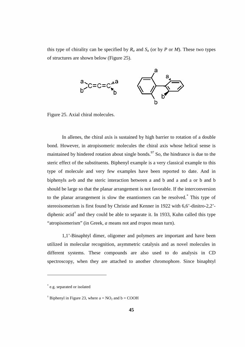
45
this type of chirality can be specified by Ra and Sa (or by P or M). These two types
of structures are shown below (Figure 25).
Figure 25. Axial chiral molecules.
In allenes, the chiral axis is sustained by high barrier to rotation of a double
bond. However, in atropisomeric molecules the chiral axis whose helical sense is
maintained by hindered rotation about single bonds.97 So, the hindrance is due to the
steric effect of the substituents. Biphenyl example is a very classical example to this
type of molecule and very few examples have been reported to date. And in
biphenyls a≠b and the steric interaction between a and b and a and a or b and b
should be large so that the planar arrangement is not favorable. If the interconversion
to the planar arrangement is slow the enantiomers can be resolved.* This type of
stereoisomerism is first found by Christie and Kenner in 1922 with 6,6’-dinitro-2,2’-
diphenic acid† and they could be able to separate it. In 1933, Kuhn called this type
“atropisomerism” (in Greek, a means not and tropos mean turn).
1,1’-Binaphtyl dimer, oligomer and polymers are important and have been
utilized in molecular recognition, asymmetric catalysis and as novel molecules in
different systems. These compounds are also used to do analysis in CD
spectroscopy, when they are attached to another chromophore. Since binaphtyl
* e.g. separated or isolated
† Biphenyl in Figure 23, where a = NO2 and b = COOH

46
moiety is not absorbing in the visible region, the chromophore attached should
strongly absorb at that region. The other properties that this chromophore should
have are large, symmetrical and achiral. In an example phtalocyanines have been
used to get use this molecule in chirality concept (Figure 26).99

47
Figure 26. Chirality induced to phtalocyanine molecule by binaphtyl.99
As can be seen in Fig. 26 that the CD signal at 709 nm is splitted into two
from different enantiomers and originated from phtalocyanine molecule.
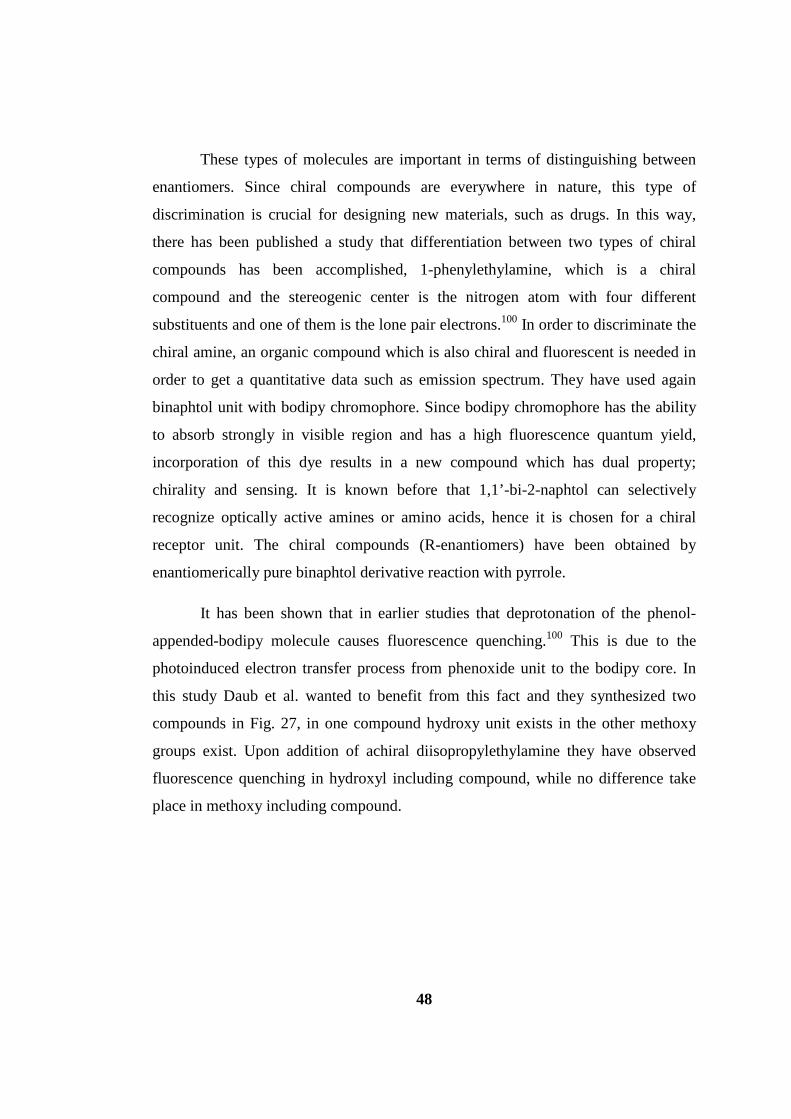
48
These types of molecules are important in terms of distinguishing between
enantiomers. Since chiral compounds are everywhere in nature, this type of
discrimination is crucial for designing new materials, such as drugs. In this way,
there has been published a study that differentiation between two types of chiral
compounds has been accomplished, 1-phenylethylamine, which is a chiral
compound and the stereogenic center is the nitrogen atom with four different
substituents and one of them is the lone pair electrons.100 In order to discriminate the
chiral amine, an organic compound which is also chiral and fluorescent is needed in
order to get a quantitative data such as emission spectrum. They have used again
binaphtol unit with bodipy chromophore. Since bodipy chromophore has the ability
to absorb strongly in visible region and has a high fluorescence quantum yield,
incorporation of this dye results in a new compound which has dual property;
chirality and sensing. It is known before that 1,1’-bi-2-naphtol can selectively
recognize optically active amines or amino acids, hence it is chosen for a chiral
receptor unit. The chiral compounds (R-enantiomers) have been obtained by
enantiomerically pure binaphtol derivative reaction with pyrrole.
It has been shown that in earlier studies that deprotonation of the phenol-
appended-bodipy molecule causes fluorescence quenching.100 This is due to the
photoinduced electron transfer process from phenoxide unit to the bodipy core. In
this study Daub et al. wanted to benefit from this fact and they synthesized two
compounds in Fig. 27, in one compound hydroxy unit exists in the other methoxy
groups exist. Upon addition of achiral diisopropylethylamine they have observed
fluorescence quenching in hydroxyl including compound, while no difference take
place in methoxy including compound.
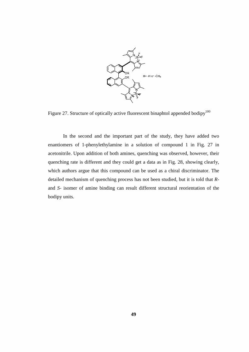
49
Figure 27. Structure of optically active fluorescent binaphtol appended bodipy100
In the second and the important part of the study, they have added two
enantiomers of 1-phenylethylamine in a solution of compound 1 in Fig. 27 in
acetonitrile. Upon addition of both amines, quenching was observed, however, their
quenching rate is different and they could get a data as in Fig. 28, showing clearly,
which authors argue that this compound can be used as a chiral discriminator. The
detailed mechanism of quenching process has not been studied, but it is told that R-
and S- isomer of amine binding can result different structural reorientation of the
bodipy units.
50
a)
b)
Figure 28. a) decrease of bodipy fluorescence with the addition of diisopropylethylamine in acetonitrile. b) Plots for the quenching of chiral hydroxyl binaphtol bodipy with S- and R- Phenylethylamine (PEA) in acetonitrile.100
2.3.4 Bodipy Asymmetry
There is a demand of luminophores for the use in modern biological labeling
and advanced optoelectronic devices, therefore chemists has an intention to design
and engineer new type of organic dyes to use in these applications.101 Bodipy is an
important luminophore and it is used frequently in advanced applications including
sensors102, energy transfer cassettes103, biological labeling104, or photovoltaics4. Its
core is also open to modification which is possible to modify its optical and physical
properties. It was notified before that most known chiral organic fluorophores are
binaphtol modules and they have used in chiral recognition processes like
determining enantiomeric purity of chiral amino acids, amines or alcohols.105

51
Figure 29. Boron asymmetry in bodipy molecule.3
In order to utilize bodipy core for these applications Ziessel et al. have
engineered asymmetric bodipy units by modifying the boron center, e.g. the
stereogenic center is the boron atom (Figure 29).3 The number of boron complexes
which are stable and asymmetric is less due to the ease of dissociation and inversion
processes. The stability of boron is ensured with N-donor chelate which was
expressed that it is related to the N-B bond strength.106 The few examples of
enantiomerically pure bodipys known were obtained with decoration of the central
core with asymmetric carbon atom100, 107 and this research is the first decoration of
the boron center.
During designing process following requirements were needed; (i) lateral
differentiation of the dipyrromethene core; (ii) attaching a polar group in order to
associate intramolecularly with BF unit; (iii) using polyaromatic units for inhibiting
rotation around boron atom and easing resolution in chiral HPLC. The
antisymmetric core has been accomplished by oxidizing one of the methyl units to
aldehyde with DDQ. By this way, it was also made possible that aldehyde proton
associate with F atom, so that rotation of F is not possible. Reacting with
naphtylmagnesium bromide also led to structure sterically hindered for rotation. The
nature of F-H association has also been verified with X-ray crystallography. As a
result, optically active bodipy is synthesized and separated with chiral HPLC and
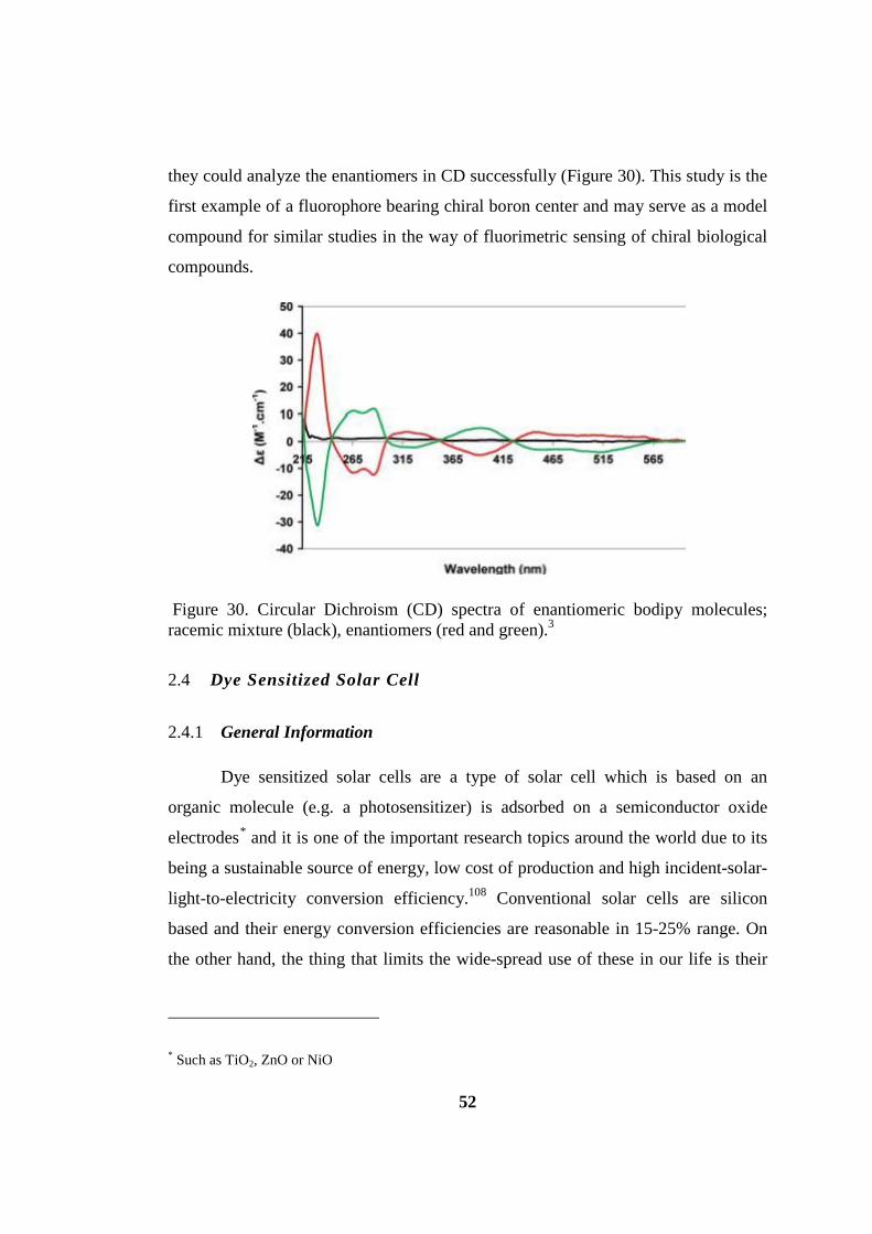
52
they could analyze the enantiomers in CD successfully (Figure 30). This study is the
first example of a fluorophore bearing chiral boron center and may serve as a model
compound for similar studies in the way of fluorimetric sensing of chiral biological
compounds.
Figure 30. Circular Dichroism (CD) spectra of enantiomeric bodipy molecules; racemic mixture (black), enantiomers (red and green).3
2.4 Dye Sensitized Solar Cell
2.4.1 General Information
Dye sensitized solar cells are a type of solar cell which is based on an
organic molecule (e.g. a photosensitizer) is adsorbed on a semiconductor oxide
electrodes* and it is one of the important research topics around the world due to its
being a sustainable source of energy, low cost of production and high incident-solar-
light-to-electricity conversion efficiency.108 Conventional solar cells are silicon
based and their energy conversion efficiencies are reasonable in 15-25% range. On
the other hand, the thing that limits the wide-spread use of these in our life is their
* Such as TiO2, ZnO or NiO

53
requirement of high-purity silicon and skilled manufacturing techniques, which
means that they need high amount of electricity produced from fossil fuels.108
The first publication of dye sensitized solar cells has been published by
Gratzel and O’Regan by using polypyridyl ruthenium (II) complex dyes adsorbed on
a nanocrystalline n-type TiO2 electrode.109 Most DSSCs are constructed with a dye
adsorbed on a wide band gap of a semicrystalline oxide, a liquid electrolyte usually
contains I−/I3− and a platinum-coated counter electrode. To date highest conversion
efficiencies with Ru dyes have been obtained with N3110, N719111 and Black dye112,
owing to their intense and wide range of absorption of sunlight. The efficiency
measurements have been done under air mass AM 1.5 simulated sunlight (100 mW
cm-2).
Like ruthenium based DSSCs, organic dye based DSSCs have also been
studied intensely. Their advantages are (i) prepared and purified with lower cost, (ii)
higher molar extinction coefficients than ruthenium dyes and have wide range of
absorption capability (iii) many alternative structural groups can be used, so that
control of photophysical, electrochemical and three-dimensional structure on
semiconductor is possible (iv) no resource limitation since the structure is not based
on rare metals such as ruthenium.113 Some of the dye types synthesized up to date to
be used in DSSC concept are coumarin dyes, polyene dyes, hemicyanine dyes,
bodipy dyes, perylene and porphyrin dyes.
There is much effort to get high conversion efficiencies with organic dye
DSSCs but still they remain inferior compared to Ru dyes. Some reasons for this
situation are; (i) relatively narrow absorption bands of visible light (ii) tendency to
p-stack on the semiconductor surface of these dyes which leads to decrease in the
amount of injected electrons from dye to the conduction band of semiconductor due
to intermolecular energy transfer (iii) relatively low open-circuit photovoltage (Voc)
values due to the faster charge recombination between the injected electrons in

54
semiconductor and triiodide (I3−)* (iv) the stability of organic dyes are generally
lower, which may be due to the radicals formed upon strong irradiation prone to
decomposition.113
2.4.2 Main types of organic dye based DSSCs
Although many studies have been done in this concept because of the
importance of energy is well-known, in recent years improvement of cell efficiencies
slowed down. In order surpass this situation scientists try new solar cell types and up
to now following cell types have been studied:113
[1] n-type semiconductor TiO2-based Type-I DSSC
[2] n-type semiconductor TiO2-based Type-II DSSC
[3] new TiO2 DSSCs with a pyridine group as the anchoring group to
semiconductor
[4] n-type semiconductor ZnO-based Type-I DSSC
[5] p-type semiconductor NiO-based Type-I DSSC
[6] tandem DSSCs with TiO2 as the photoanode and NiO as the photocathode
Among these cell types most studied one is the first type and highest
efficiency with 10.1% have been achieved.114 Hence, focus will be on this type.
The detailed mechanism, differences and operational principles of these cells
can be found in the recent informative review by Ooyama and Harima.113 However,
basic information about n-type TiO2 based Type-I DSSC will be outlined below.
2.4.3 n-Type Semiconductor TiO2-based Type-I DSSCs
* Related to the electron lifetime which is smaller in organic based DSSCs

55
Schematic representation of n-TiO2-based type I DSSC is shown below and
it is mainly composed of a photoanode and a cathode* which sandwiches an
electrolyte solution. It is composed of five materials: 1) Flourine-doped tin oxide
(FTO) 2) a nanocrstalline TiO2 thin film as a semiconductor 3) an organic dye 4) a
redox mediator electrolyte solution 5) platinum-coated glass substrate.
Figure 31. Working scheme and principles of n-type TiO2 DSSC (Type I)113
From three main parts photoanode is prepared first by applying
nanocrystalline TiO2 particles to the FTO glass then this material is dipped into the
solution of dye to adsorb to the TiO2 surface. Cathode part is composed from
platinum-coated glass substrate and electrolyte is usually a solution of I−/I3−.
The generation of photocurrent is occur as follows; first organic dye† is
excited with sunlight and electron from HOMO of the dye excited to the LUMO,
which injects to the conduction band of TiO2‡, an electron from the I− in the
electrolyte solution go to the cationic dye’s HOMO so that cationic dye returns to its
neutral state and the injected electron move through interconnected TiO2
* e.g. a photoinert counter electrode
† also called photosensitizer
‡ or ZnO depends on the semiconductor type used

56
nanoparticles and reaches to the FTO glass and then to the counter electrode
through external circuit (Pt-coated glass). Afterwards, the electron reduces I3− to
regenerate I− and the circuit is completed then. The mechanism of these processes is
shown below in Fig. 32.
Figure 32. Equations showing the mechanism of photocurrent via n-type TiO2 based DSSC (Type I)113
In the mechanism told above there are some problematic issues to deal with.
These undesirable side processes originate from (i) when electron injected from
LUMO of the dye to the conduction band of TiO2 it reduces the oxidized dye again
to its neutral state (recombination) or I3− to yield I− (dark current). Also,
radiationless relaxation from excited state of the dye to the ground state also takes
place. In order to get a high photocurrent value electron injection and regeneration
should be kinetically more favorable than dark current and recombination processes.
2.4.4 Solar Cell Photovoltaic Parameters113
Mainly the performances of DSSCs have been evaluated according to six
parameters: IPCE, photocurrent/voltage curves (J/V curves), open circuit voltage
(Voc), Short circuit photocurrent density (Jsc), fill factor (ff), solar energy-to-
electricity conversion yield (η). In addition to these, dye stability under solar
illumination is also an important factor. These six important parameters will be
explained below.
57
2.4.4.1 Incident-Photon-to-Current Conversion Efficiency (IPCE)
IPCE is also called as external quantum efficiency (EQE) and the definition
is the number of electrons flowing through the external circuit from anode to
cathode which is divided by the number of incident photons from sun light. The
equation is:
IPCE (%) = 1240 (𝑒𝑉 𝑛𝑚)𝐽𝑝ℎ(𝑚𝐴 𝑐𝑚−2)
𝜆(𝑛𝑚)𝐼(𝑚𝑊 𝑐𝑚−2)
where Jph is the short circuit photocurrent density which is generated by the
monochromatic light, λ is the wavelength of the monochromatic light and I is the
intensity of the monochromatic light.
Another method for determining the IPCE value is according to the following
equation:
𝐼𝑃𝐶𝐸 (𝜆) = 𝐿𝐻𝐸 (𝜆).Φ𝑖𝑛𝑗. 𝜂𝑐𝑜𝑙𝑙
In this equation LHE is the light harvesting efficiency of the dye at
wavelength λ and it is determined by
𝐿𝐻𝐸 (𝜆) = 1 − 10−𝐴
Quantum efficiency of the injected electrons to the TiO2 is also an important
parameter, and ηcoll is the collection of the injected electrons in the FTO glass.
As a result, IPCE is directly related to the absorption assets of the dye,
loading of the adsorbed dye amount on TiO2, electron injection efficiency of the dye
LUMO to the conduction band of TiO2 and efficiency of the collected electrons in
the external circuit. The maximum efficiency of the IPCE (%) is 80-85 % and the
total efficiency is not achievable due to the reflection and absorption losses of the
FTO glass. IPCE of the DSSC is generally given as a spectrum of IPCE versus
excitation wavelength.

58
2.4.4.2 Photocurrent/Voltage Curves (J/V curves)
Under standard AM 1.5 sunlight simulation (100 mW.cm-2) measuring the
DSSC performance is easily analyzed with J/V curve. And four imperative
parameters are Voc, Jsc, ff and η which are frequently used to analyze the DSSC
performance will be explicated below.
2.4.4.3 Open-Circuit Photovoltage (Voc)
The definition of this important parameter is the difference between the
electrical potentials of the two compartments of the cell upon illumination with
sunlight when the circuit is open. The maximum value of this parameter is the
difference between the energy level of the conduction band of TiO2 (Ecb)* and the
redox potential of the electrolyte Er (I−/I3−)†. This corresponds to a value
approximately 0.8-0.9 V. However, this value decreases in real applications because
of the dark current and recombination events.
2.4.4.4 Short-Circuit Photocurrent Density (Jsc)
This value corresponds to the photocurrent per unit area (mA.cm-2) when the
DSSC under irradiation is short circuited. Open cicrcuit voltage (Voc) is the value
between the Ef of the electron in TiO2 and the redox potential of the electrolyte.
However, Jsc is related with the communication between TiO2 and dye adsorbed and
the absorption capability of the dye. As a result, Jsc value is dependant to (i) ability
of the dye molecules absorb sunlight over a wide range of the electromagnetic
spectrum (ii) electron-injection ability of the dye from HOMO to the CB of the
semiconductor oxide (iii) reduction efficiency of the cationic dye molecules by
I−after injection. Consequently, photophysical and electrochemical properties of the
dye molecules and molecular structures of the dye are highly important in order to
get high Jsc value. Hence, the interaction between dye molecules and TiO2 surface is
* It is -0.5 to -0.4 V vs. NHE (normal hydrogen electrode)
† 0.4 V vs. NHE

59
also important and orientation of the dyes on semiconductor is also crucial. Jsc value
can be found by integrating the IPCE spectrum according to the following equation:
𝐽𝑠𝑐 = 𝑒� 𝐼𝑃𝐶𝐸(𝜆). 𝐼𝑠(𝜆).𝑑𝜆
Here Is(λ) is the photon flux at wavelength λ while irradiated with AM 1.5.
2.4.4.5 Fill Factor (ff)
This term is defined as the maximum power output (JmpVmp) divided by the
product of Jsc and Voc. The ff is determined according to the J/V curve and it is
found according to the maximum area of the rectangle. So, it is obvious that
maximum value of fill factor is unity but various resistances in the cell like electron
transport resistance at the cathode reduce this value.
2.4.4.6 Solar Energy-to-Electricity Conversion Yield (η)
This term is defined as the ratio of the maximum output electrical power of
the DSSC to the energy of incident sunlight (I0) and it is calculated according to the
following equation:
𝜼(%) = 𝑱𝒔𝒄(𝒎𝑨. 𝒄𝒎−𝟐).𝑽𝒐𝒄(𝑽).𝒇𝒇
𝑰𝟎(𝒎𝑾. 𝒄𝒎−𝟐)
In order to get a high value of η Voc, Jsc and ff of the cell should be
optimized. This can be attained by optimization of the electrochemical and
photopysical properties of the dye, and control of the dye molecules’ orientation and
arrangment on the semiconductor layer.
60
CHAPTER 3
3 NOVEL SENSITIZATION IN PDT WITH
BODIPY DYES
First part of this chapter was published in the following publications:
Cakmak, Y.; Kolemen, S.; Duman, S.; Dede, Y.; Dolen, Y.; Kilic, B.; Kostereli, Z.; Yildirim, L. T.; Dogan, A. L.; Guc, D.; Akkaya, E. U. Angew Chem Int Ed 2011, 50, 11937-11941.
Duman, S.; Cakmak, Y.; Kolemen, S.; Akkaya, E. U.; Dede, Y. J Org Chem 2012, 77 (10), 4516-4527 (Featured).
3.1 Theory-Guided Access to Efficient Photosensitizers by Heavy Atom
Free Orthogonal Bodipy Dimers
3.1.1 Introduction
Photodynamic therapy (PDT) is a treatment modality for certain malignant
(skin, head and neck, gastrointestinal, gynecological cancers), premalignant (actinic
keratosis), and nonmalignant (psoriasis) indications.115 Broader acceptance by the
medical community and applicability is hampered, at least in part, by the less than
optimal photophysical characteristics of the porphyrin derivatives. This situation
sparked a worldwide search for novel sensitizers leading to new compounds, some
holding more promise than others.116 The primary cytotoxic agent involved in the
photodynamic action is singlet oxygen (1Δg), the efficient generation of which is
linked invariably to the intersystem crossing (ISC) efficiency of the excited state of
the sensitizer. Most organic dyes have low triplet quantum yields, and in many
recent candidates for photodynamic sensitizers, heavy atoms are incorporated into
the structure as a strategy to improve spin–orbit coupling leading to facilitated
intersystem crossing.78 While this approach seems fail-safe, incorporation of heavy
atoms such as bromine, iodine, selenium, and certain lanthanides very often leads to
increased “dark toxicity”.117 Unlike traditional chemotherapy agents, in principle,

61
photodynamic therapy sensitizers themselves can be nontoxic, either at cellular or
organ levels, even at relatively high concentrations. We have been interested in
trying to find alternative ways of achieving increased intersystem crossing without
the use of heavy atoms to minimize dark toxicity and to investigate the intersystem
crossing mechanism, turning our attention to the excited state properties of the
sensitizers.
3.1.2 Theoretical Investigation
Designing efficient photoinduced 1O2 generators requires that any existing
operative fluorescence cycle of the fluorophore, which is through the S0 →S1→S0
states, has to be perturbed so as to minimize or shut down the S1→S0 deactivation,
and switch to the triplet surface once S1 is accessed (intersystem crossing). A general
design principle for a favorable S1→T1 transition from an electronic structure
viewpoint would in principle require the structural and electronic compatibility of
the S1 and T1 states to surpass that of the S1–S0 pair.
Once multiple electronic states come into play, quantum mechanical
calculations providing a detailed understanding of the electronic structure are
extremely helpful. Multi-configurational self-consistent field (MCSCF) techniques
are the state-of-the-art computational chemistry approaches, when near degeneracies
and excited states are considered. These methods are not accurate for ± 2–3
kcalmol−1, but they are very helpful while understanding the photopysics taking
place. Therefore, a popular variant of MCSCF techniques was used; the complete
active space SCF (CASSCF) method in combination with relatively large basis sets
and different active spaces. Details of CASSCF calculations are provided in the
computational methods part (Section 3.1.5.8).
Calculations on the parent Bodipy (4,4-difluoro-4-bora-3a,4a-diaza-s-
indacene, Fig. 33) showed that natural orbital occupancies of S1 state do describe an
open-shell singlet with essentially double (>1.9) or zero (<0.1) electrons for all
orbitals except HOMO and LUMO that are singly occupied (simple single electron
excitation taking place in a fluorophore upon excitation). Since most of the

62
fluorophores have an excited state comprising of only two orbitals with single
occupancy leads to low triplet quantum yields, one should design a dye which is
different than the simple HOMO → LUMO transition. Among multiply excited
configurations, doubly substituted ones are particularly important in enhancing S1-T1
coupling as shown by the seminal work of Salem and Rowland118 and the following
work by Michl.119 Thus, the substitutions should invoke simultaneous two electron
excitation from the ground state, guiding us to search for molecules with a pair of
degenerate or near-degenerate occupied frontier orbitals correlating with a similar
virtual (unoccupied) pair and pointing out a design principle of orthogonal dimeric
chromophores. This foundation has two important features: (i) Orthogonal
placement prevents mixing of the π-systems of the two subunits leaving us with two
essentially undisturbed chromophore cores. (ii) Upon irradiation, both cores are
almost equally likely to undergo a HOMO → LUMO electron transfer (referring to
the original monomer MOs) yielding an excited state mainly comprised of double
substitutions.
Figure 33. Structure of Bodipy (4,4-difluoro-4-bora-3a,4a-diaza-s-indacene)
3.1.3 Design Principles
Our previous involvement with Bodipy dyes led us to seek ways to
implement these design considerations using these dyes. Bodipy dyes are
exceptional fluorophores with amazing versatility120 and rich chemistry.121 Heavy
atom functionalized Bodipy dyes showed some promise as potential sensitizers for
photodynamic activity.122 It is also interesting to note that a few years ago, dimeric
(albeit non-orthogonal) Bodipy derivatives were reported123 with peculiar properties
linked to exciton coupling between the chromophores. We believe orthogonality to
be an important distinction in our design. The calculations on the orthogonal 8-2’
dimer OB3 clearly show (Fig. 34) the essentially unperturbed Bodipy HOMO and
LUMO as a result of lack of mixing of orthogonal π-frameworks.
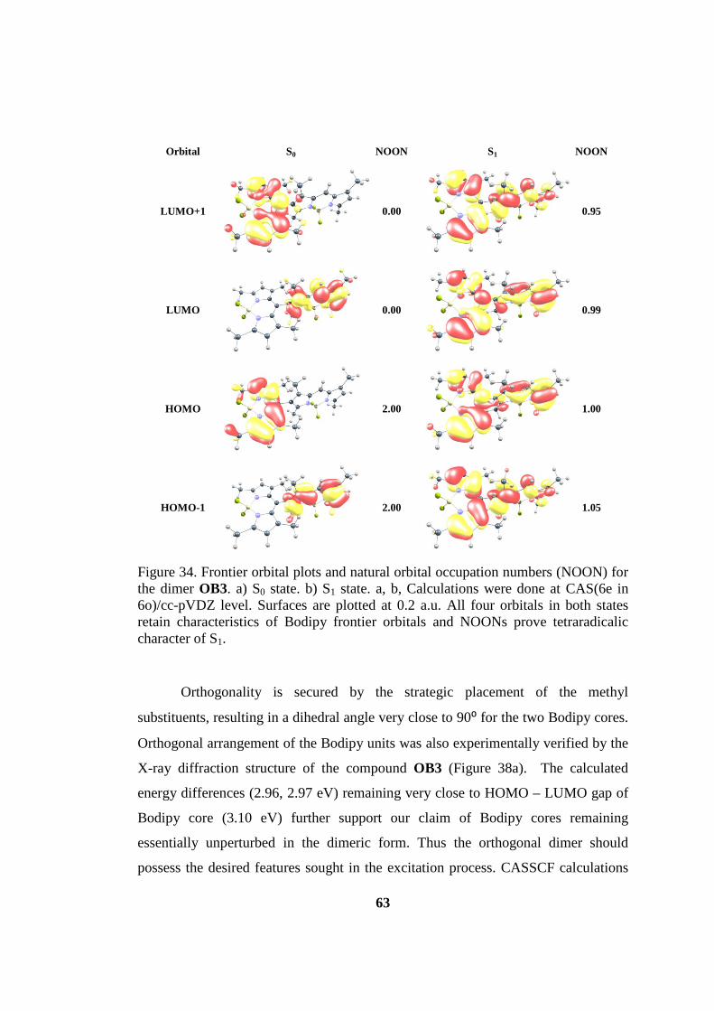
63
Orbital S0 NOON S1 NOON
LUMO+1
0.00
0.95
LUMO
0.00
0.99
HOMO
2.00
1.00
HOMO-1
2.00
1.05
Figure 34. Frontier orbital plots and natural orbital occupation numbers (NOON) for the dimer OB3. a) S0 state. b) S1 state. a, b, Calculations were done at CAS(6e in 6o)/cc-pVDZ level. Surfaces are plotted at 0.2 a.u. All four orbitals in both states retain characteristics of Bodipy frontier orbitals and NOONs prove tetraradicalic character of S1.
Orthogonality is secured by the strategic placement of the methyl
substituents, resulting in a dihedral angle very close to 90⁰ for the two Bodipy cores.
Orthogonal arrangement of the Bodipy units was also experimentally verified by the
X-ray diffraction structure of the compound OB3 (Figure 38a). The calculated
energy differences (2.96, 2.97 eV) remaining very close to HOMO – LUMO gap of
Bodipy core (3.10 eV) further support our claim of Bodipy cores remaining
essentially unperturbed in the dimeric form. Thus the orthogonal dimer should
possess the desired features sought in the excitation process. CASSCF calculations
64
on the orthogonal dimer OB3 show that there is a huge amount of electronic
reorganization upon S0 → S1 transition. S1 with an equilibrium geometry very close
to S0 lies 80 kcal/mol above the ground state.
Computations of the S0 ↔ S1 vertical transitions show that only 15 kcal/mol
of this difference is due to structural deformation, whereas electronic reorganization
is more than four times as costly (65 kcal/mol). Thus, interestingly electron
movements are more important than the structural change upon excitation. This
implies that the electronic structure of S1 is rather unusual. Natural orbital
occupation numbers (NOON) show four odd electrons in four very similar orbitals
(Fig. 34). This situation from now on called as tetra-radicalic. Thus, on excitation,
both Bodipy units are essentially equally likely to undergo the inherent (Bodipy)
HOMO → LUMO transition from HOMO-1 to LUMO and HOMO to LUMO+1 of
Figure 34. In addition, orbitals optimized for S1 state support the involvement of
both cores via their clearly visible delocalized character. Thus S1 state could be
predicted to be comprised of a linear combination of doubly substituted
configurations (with respect to the closed shell reference) and in line with this
expectation the CASSCF wavefunction is almost totally made up of doubly
substituted configurations, an unexpectedly large value for any chromophore ( Fig.
35, and following discussion). And one of five doubly substituted excitations cause
tetra radicalic state formation.

65
Figure 35. Conceptual frontier MO diagram describing the building up of S1 states of
the dimers OB3 and OB6 and dominance of doubly substituted configurations. Only
the leading configurations are shown and minus signs to restrain antisymmetry in the
S1 states are intentionally excluded for clarity. As a result of spatial orthogonal
arrangement HOMO and LUMO of both Bodipy cores are essentially kept in their
original form (only slightly modified) in the dimers. Thus upon irradiation all the
five configurations are almost equally probable and this picture is verified by
CASSCF calculations. Each of the five configurations making up S1 are termed as
doubly substituted (DS) with respect to the closed shell reference S0 state of the
dimer.
The dominance of the S1 wavefunction by double substitutions is in stark
contrast with S0 of orthogonal dimer as the latter is ca. 90% dominated by the
reference configuration. These findings suggest a severe mismatch between the S1
and S0 states and an enhanced hop to the T1 state lying 1.7 eV lower (at 1.8 eV with
respect to the ground state). Essentially the same electronic structure fingerprints are
observed for 8,8’ dimer OB6 (Fig. 36). Repeated calculations with different active
space–basis set combinations without exception, showed a picture dominated by
doubly substituted configurations and four singly occupied molecular orbitals in the
S1 state.
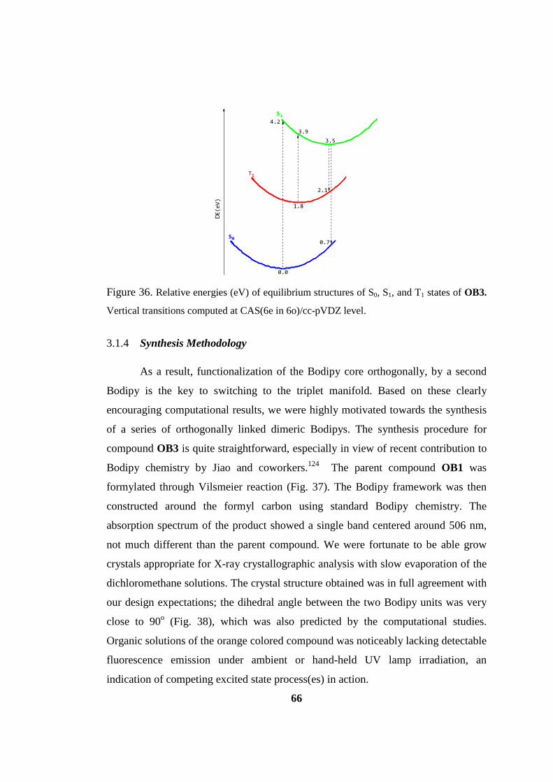
66
0.0
0.7
1.8
3.5
4.2
DE(e
V)
2.1
3.9
S1
T1
S0
Figure 36. Relative energies (eV) of equilibrium structures of S0, S1, and T1 states of OB3.
Vertical transitions computed at CAS(6e in 6o)/cc-pVDZ level.
3.1.4 Synthesis Methodology
As a result, functionalization of the Bodipy core orthogonally, by a second
Bodipy is the key to switching to the triplet manifold. Based on these clearly
encouraging computational results, we were highly motivated towards the synthesis
of a series of orthogonally linked dimeric Bodipys. The synthesis procedure for
compound OB3 is quite straightforward, especially in view of recent contribution to
Bodipy chemistry by Jiao and coworkers.124 The parent compound OB1 was
formylated through Vilsmeier reaction (Fig. 37). The Bodipy framework was then
constructed around the formyl carbon using standard Bodipy chemistry. The
absorption spectrum of the product showed a single band centered around 506 nm,
not much different than the parent compound. We were fortunate to be able grow
crystals appropriate for X-ray crystallographic analysis with slow evaporation of the
dichloromethane solutions. The crystal structure obtained was in full agreement with
our design expectations; the dihedral angle between the two Bodipy units was very
close to 90o (Fig. 38), which was also predicted by the computational studies.
Organic solutions of the orange colored compound was noticeably lacking detectable
fluorescence emission under ambient or hand-held UV lamp irradiation, an
indication of competing excited state process(es) in action.

67
Figure 37. Synthesis of the target photosensitizers. a) POCl3, DMF, ClCH2CH2Cl, 500C b) 2,4-Dimethylpyrrole, TFA, CH2Cl2,p-chloranil, Et3N, BF3.OEt2 c) TFA, CH2Cl2, p-chloranil, Et3N, BF3.OEt2.
Figure 38. Structures of the dimeric Bodipys. a) X-ray diffraction structure of the orthogonal 8,2’ dimer OB3. b) Optimized geometry of orthogonal 8,8’ dimer OB6 at CAS(6e in 6o)/cc-pVDZ level. Dihedral angle between the two Bodipy units is very close (± 0.5o) to 90o in both dimers.
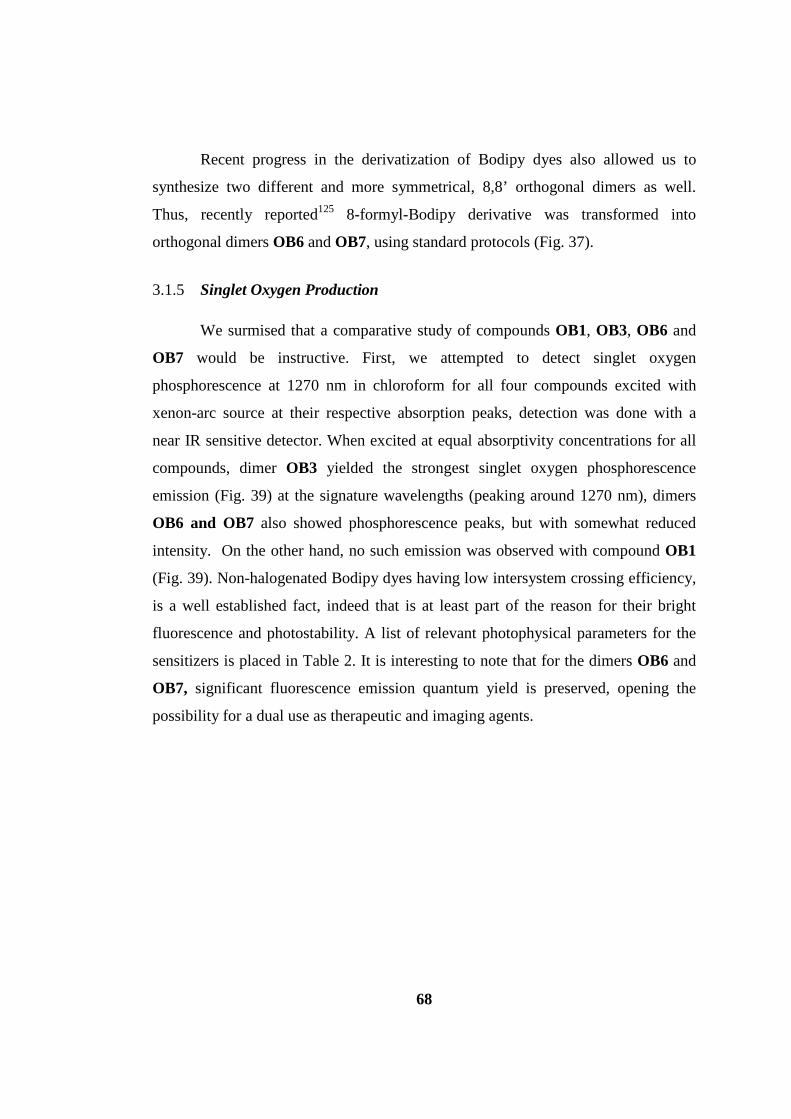
68
Recent progress in the derivatization of Bodipy dyes also allowed us to
synthesize two different and more symmetrical, 8,8’ orthogonal dimers as well.
Thus, recently reported125 8-formyl-Bodipy derivative was transformed into
orthogonal dimers OB6 and OB7, using standard protocols (Fig. 37).
3.1.5 Singlet Oxygen Production
We surmised that a comparative study of compounds OB1, OB3, OB6 and
OB7 would be instructive. First, we attempted to detect singlet oxygen
phosphorescence at 1270 nm in chloroform for all four compounds excited with
xenon-arc source at their respective absorption peaks, detection was done with a
near IR sensitive detector. When excited at equal absorptivity concentrations for all
compounds, dimer OB3 yielded the strongest singlet oxygen phosphorescence
emission (Fig. 39) at the signature wavelengths (peaking around 1270 nm), dimers
OB6 and OB7 also showed phosphorescence peaks, but with somewhat reduced
intensity. On the other hand, no such emission was observed with compound OB1
(Fig. 39). Non-halogenated Bodipy dyes having low intersystem crossing efficiency,
is a well established fact, indeed that is at least part of the reason for their bright
fluorescence and photostability. A list of relevant photophysical parameters for the
sensitizers is placed in Table 2. It is interesting to note that for the dimers OB6 and
OB7, significant fluorescence emission quantum yield is preserved, opening the
possibility for a dual use as therapeutic and imaging agents.
69
Figure 39. Singlet oxygen phosphorescence with sensitization from Bodipy derivatives: OB3 (blue), OB6 (black) and OB7 (red), OB1 (green) in CHCl3 at equal absorbances (0.2) at the peak wavelength of their respective absorbances.
While integral areas under the phosphorescence emission peak is a measure
of singlet oxygen quantum yield, we opted for more quantitative assessment of
singlet oxygen quantum yields using 1,3-diphenylisobenzofuran (DPBF) as a trap
molecule and with Methylene Blue (MB) as the reference compound (Methylene
Blue has a singlet oxygen quantum yield of 0.57 under the conditions of the study in
dichloromethane126). Excitation of the dyes was carried out by irradiation at the
respective absorbance peak wavelengths with monochromatized light source, at a
fluence rate around 30 μW/cm-2. The quantum yield for singlet oxygen generation
for OB3 is 0.51 in dichloromethane, much higher than all non-halogenated Bodipy
dyes and many other organic chromophores and photosensitizers, under comparable
conditions. The other Bodipy dimers OB6 and OB7 also showed respectable singlet
oxygen quantum yields of 0.46 and 0.21, respectively (See experimental part for
details).

70
Table 2. Comparative spectroscopic properties of Bodipy compounds.
Compound λabs[a] [nm] λems
[a] [nm] Φf τf[a] [ns] Φ∆
[e]
OB1 509 514 0.80[d] 5.2 -
OB3 514 527 0.03[a,b] 2.5 0.51
OB6 515 588 0.31[a,c] 10.9 0.46
OB7 542 605 0.49[a,c] 5.0 0.21
[a] in CHCl3, [b] in reference to Fluorescein in 0.1 M NaOH solution excited at 496
nm, [c] in reference to Rhodamine 6G in EtOH excited at 480 nm, [d] in EtOH [e]
Singlet oxygen quantum yield was determined with respect to Methylene Blue (0.57
in Dichloromethane)126.
3.1.6 Cytotoxicity Experiments
For further demonstration of singlet generation capacity and
photocytotoxicity of the most active dimeric dye (compound OB3), we carried out a
cell culture assay with cancer cell lines. To that end, we prepared a micellar
formulation of the 8,2’-orthogonal dimeric dye using Cremophor-EL (experimental
section).122 The size distribution of the micelles was determined using
electrophoretic light scattering (Appendix part for graph). The size distribution
reveals a median size of 100 nm for the micellar constructs. Micelle embedded dye
retained high levels of singlet oxygen generation capacity as revealed by another
singlet oxygen trapping experiment, this time in aqueous media, using a water
soluble anthracene derivative (2,2'-(Anthracene-9,10-diylbis(methylene))dimalonic
acid (For the structure of this compound and its use see Section 3.2).
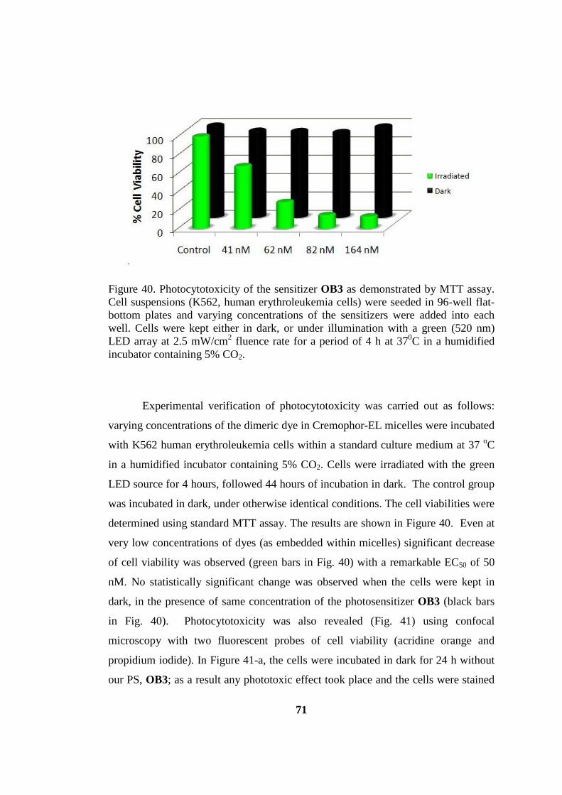
71
Figure 40. Photocytotoxicity of the sensitizer OB3 as demonstrated by MTT assay. Cell suspensions (K562, human erythroleukemia cells) were seeded in 96-well flat-bottom plates and varying concentrations of the sensitizers were added into each well. Cells were kept either in dark, or under illumination with a green (520 nm) LED array at 2.5 mW/cm2 fluence rate for a period of 4 h at 370C in a humidified incubator containing 5% CO2.
Experimental verification of photocytotoxicity was carried out as follows:
varying concentrations of the dimeric dye in Cremophor-EL micelles were incubated
with K562 human erythroleukemia cells within a standard culture medium at 37 oC
in a humidified incubator containing 5% CO2. Cells were irradiated with the green
LED source for 4 hours, followed 44 hours of incubation in dark. The control group
was incubated in dark, under otherwise identical conditions. The cell viabilities were
determined using standard MTT assay. The results are shown in Figure 40. Even at
very low concentrations of dyes (as embedded within micelles) significant decrease
of cell viability was observed (green bars in Fig. 40) with a remarkable EC50 of 50
nM. No statistically significant change was observed when the cells were kept in
dark, in the presence of same concentration of the photosensitizer OB3 (black bars
in Fig. 40). Photocytotoxicity was also revealed (Fig. 41) using confocal
microscopy with two fluorescent probes of cell viability (acridine orange and
propidium iodide). In Figure 41-a, the cells were incubated in dark for 24 h without
our PS, OB3; as a result any phototoxic effect took place and the cells were stained

72
with acridine orange only (merge-green), the indication of cells are alive. In b, OB3
was added to the well and incubated for 24h in dark, again any cytotoxicity fulfilled.
In c row in Figure 41, the effect of light to cells was investigated without addition of
OB3, again resulted with survival of cells. However, in d cells were incubated with
OB3 for 20h after illumination with light for 4h and after 24h, the phototoxic affect
was revealed since the cells were stained with propidium iodide (merge-red). Thus,
using confocal microscopy also, the activity of our micellar PS working well has
been proved.

73
Figure 41. Photocytotoxic activity of the dimeric Bodipy OB3 visualized via confocal microscopy. Cell suspensions (K562, human erythroleukemia cells) were seeded in 24-well plates. a) Cells in Control 1 (upper plates) wells were incubated in dark for 24 hours; b) Cells in Control 2 wells (second row plates) were incubated with 500 μl/well Cremophor-EL solubilized sensitizer OB3 (at a final concentration of 164 nM) and kept in dark for 24 hours in the same incubator, c) Cells in Control 3 well were illuminated for 4 h without the sensitizer and incubated for a further 20 h in dark at the incubator. d) Cells in Control 4 (bottom row of plates) well were illuminated for 4 h after addition of 164 nM dye OB3 and incubated for a further 20 h in dark at the incubator. Cells in wells were collected after 24 h and imaged at 40x magnification. Live cells are preferentially stained with acridine orange (AO, green) (a), whereas dead cells are preferentially stained with the dye propidium iodide (PI, red) due to increased cellular permeability (b).
3.1.7 Extended Conjugation of an Bis-BODIPY Derivative
The above results were motivating, particularly for utilizing many
chromophores in heavy atom free ISC by only dimerizing them in an orthogonal

74
fashion. Consequently, we aimed to surpass the accomplishments of OB3 and OB6
by synthesizing modified orthogonal bis-BODIPYs that will operate at the red end of
the spectrum and, hence, potentially be better suited for photodynamic applications.
The well-known modification to red shift λabs, i.e., decrease the energy gap between
frontier orbitals is, extending127 the π-system of the chromophore core. For this
purpose, synthesis of bis-8,2-distyrylBODIPY (OB8) derivative shown in Figure 42
was performed.
Figure 42. Synthesis scheme of OB8
Spectroscopic properties of OB8 was investigated, and an emission at 680
nm together with a negligible triplet quantum yield were surprisingly observed,
contrary to the expectations followed from the computed structural orthogonality
(interplane angle = 90.1°). 1O2 phosphorescence measurements (Table 3) around
1270 nm also confirmed that this red-absorbing dye essentially did not undergo the
presumed ISC to the triplet manifold.
Table 3. Photophysical properties of the synthesized Bodipy compounds
Compound λabsa / nm εmax/M-1
cm-1 λems
a / nm Φf τ a / ns ΦΔ
OB1 509 89,200 514 0.80d 5.2 -
OB3 514 119,400 527 0.025a,b 2.5 0.51
OB8 661 125,300 680 0.06a,c 2.2 0.06 a) in CHCl3 b) with respect to Fluorescein in 0.1 M NaOH solution excited at 496 nm c)
with respect to cresyl violet perchlorate in MeOH excited at 610 nm d) in EtOH
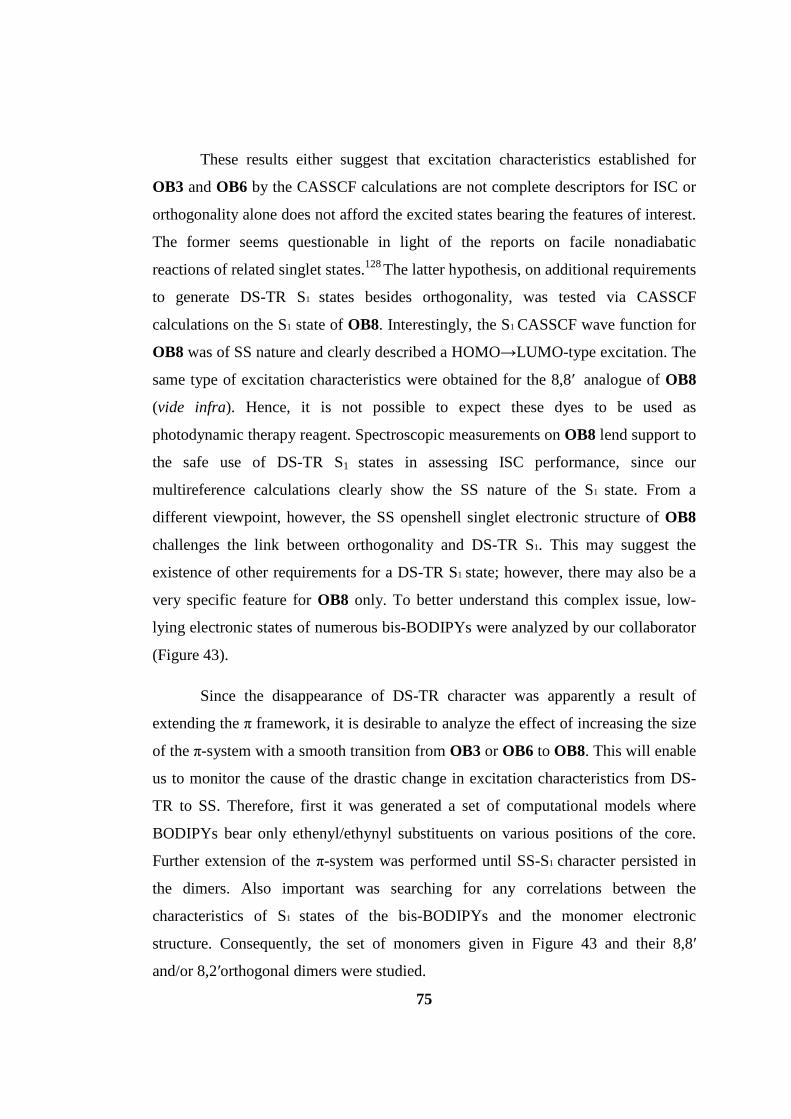
75
These results either suggest that excitation characteristics established for
OB3 and OB6 by the CASSCF calculations are not complete descriptors for ISC or
orthogonality alone does not afford the excited states bearing the features of interest.
The former seems questionable in light of the reports on facile nonadiabatic
reactions of related singlet states.128 The latter hypothesis, on additional requirements
to generate DS-TR S1 states besides orthogonality, was tested via CASSCF
calculations on the S1 state of OB8. Interestingly, the S1 CASSCF wave function for
OB8 was of SS nature and clearly described a HOMO→LUMO-type excitation. The
same type of excitation characteristics were obtained for the 8,8′ analogue of OB8
(vide infra). Hence, it is not possible to expect these dyes to be used as
photodynamic therapy reagent. Spectroscopic measurements on OB8 lend support to
the safe use of DS-TR S1 states in assessing ISC performance, since our
multireference calculations clearly show the SS nature of the S1 state. From a
different viewpoint, however, the SS openshell singlet electronic structure of OB8
challenges the link between orthogonality and DS-TR S1. This may suggest the
existence of other requirements for a DS-TR S1 state; however, there may also be a
very specific feature for OB8 only. To better understand this complex issue, low-
lying electronic states of numerous bis-BODIPYs were analyzed by our collaborator
(Figure 43).
Since the disappearance of DS-TR character was apparently a result of
extending the π framework, it is desirable to analyze the effect of increasing the size
of the π-system with a smooth transition from OB3 or OB6 to OB8. This will enable
us to monitor the cause of the drastic change in excitation characteristics from DS-
TR to SS. Therefore, first it was generated a set of computational models where
BODIPYs bear only ethenyl/ethynyl substituents on various positions of the core.
Further extension of the π-system was performed until SS-S1 character persisted in
the dimers. Also important was searching for any correlations between the
characteristics of S1 states of the bis-BODIPYs and the monomer electronic
structure. Consequently, the set of monomers given in Figure 43 and their 8,8′
and/or 8,2′orthogonal dimers were studied.

76
Figure 43. Bodipy monomers and dimers used for the computational calculations.
Extending the conjugated π-systems of the monomers yielded four major sets
of BODIPYs possessing the following substituents neighboring the core shown in
Figure 43; any substituents (4); ethenyl and/or ethynyl (5 to 8); butadienyl (9); and
styryl (10, 11). All these models preserved an essentially planar geometry within the
monomer, and their dimers were computed to be spatially orthogonal. Table 4 shows
frontier orbital energies of ground states of all the monomers and dimers as well as
the relative energies of S1 and T1 states of the orthogonal dimers. Also, the
information about their excited state character (doubly substituted or singly
substituted) was also given.

77
Table 4. Selected Parameters for Modified BODIPY Cores and Associated Orthogonal Bis-BODIPYs.
aPrime (‘) indicates 8,2’ coupling motif of the monomers,
and absence of the prime (‘) shows 8,8’ binding. bDS-TR: Doubly substituted tetraradical.
SS: Singly substituted HOMO→LUMO-type open-shell singlet state. cRelative energies of
T1 and S1 states with respect to the ground state S0 of the dimer at the CAS(6,6) level.
Excitation characteristics of the S1 state (DS-TR or SS) of bis-BODIPYs are
also shown. It is clear from the frontier orbital energy differences that, orthogonality
dictates formation of two pairs of near degenerate orbitals in the dimer that
effectively preserve monomeric character, as supported by orbital isodensity plots.
Extension of the π-system lowered the HOMO−LUMO gaps as anticipated, and
interestingly, beyond the very first double bond added to the chromophore core; i.e.,
for set (9 to 11), the DS-TR character of S1 state disappears, hence population of
triplet state is not possible. In such cases, S1 MOs were no longer delocalized on both
chromophore cores (Scheme 44), which is also the case for OB8; as a result they
78
have a simple HOMO-LUMO transition character as found in most organic
fluorophores.
Figure 44. Selected natural orbitals and occupation numbers for bis-9, bis-10 and bis-10a
The lowering of the HOMO−LUMO gap and disappearance of DS-TR
character of S1 do correlate; however, it is illusory to conclude that DS-TR fades
with decreased HOMO−LUMO gaps since the energy gap decrease itself is caused
by a structural perturbation that extends the π-system. Thus, the fundamental reason
of switching from DS-TR to SS type S1 states needs further investigation (vide infra).
Inspection of the relative energetic positioning of T1 and S1 states does not provide an
answer since T1 is roughly equally separated from S0 and S1 states for all species and
there is no significant pattern in favor of the DS-TR-S1 or SS-S1 possessing species.
Incorporation of functional groups in order to donate or withdraw electron density as
in the series of species 5, 9, 10, and 11 do result in deviations (from core BODIPY)
in the anticipated directions for frontier MO levels but do not switch the excitation
characteristics of S1. Extension of the π-system shutting down the DS-TR S1 state
seems to be one net conclusion that could be drawn, despite the survival of near-
degenerate monomer based orbitals for all the dimers considered. Nonetheless, from
the four sets of models with increasing sizes of conjugated π-systems, the only
groups with a DS-TR S1 state were (4 and 5 to 8), whose π-system extension is

79
truncated at the very first C=C double bond. Apparently, one is only allowed to
slightly modify S0−S1 energy difference without losing the DS-type excitation motif,
and unfortunately, this does not result in the desired amount of HOMO−LUMO gap
lowering needed for PDT to be applied due to skin penetration reasons. It is
important to note that relative energies with the CASSCF method may not be
quantitatively reliable and perturbation treatments on the CASSCF wave function
are necessary for energy differences of satisfactory quality. Unfortunately, MP2
corrections to this size of molecules are unfeasible and the scope of the current work
is identifying the key electronic structure details of the orthogonal dimers that
apparently undergo ISC, not a quantitatively accurate prediction of relative energies.
Such a high level treatment may be available in the future, yet we were able to
complete CAS-MP2 calculations for bis-4 and bis-4′ which are the species of
utmost interest. CAS-MP2 calculations also suggest that the DS-TR states are above
the closed shell S0 at 2.60 and 2.85 eV and the next singlet state is at 4.58 and 4.68
eV for bis-4 and bis-4′, respectively. Although CAS-MP2 treatment decreases the
relative spacing between higher excited states with respect to CASSCF, the DS-TR
S1 state is still well separated from S0 and S2. Thus, there is little doubt that DS-TR
character is the correct nature of the first excited singlet state. The issue with
quantitative reliability may be even more critical depending on the system of interest
as nicely demonstrated by the recent work of Martinez et al.128e Fortunately, our
experiments show that probing for DS-TR or SS character in the S1 state is
successfully working for the systems under investigation in assessment of ISC
performance. Our TD-DFT calculations suggest that 8, the species bearing the
lowest HOMO−LUMO gap with a DS-TR S1 for the dimer, may have an absorption
maximum close to but lower than 600 nm when estimated errors for TD-DFT are
considered. Thus, it is questionable that π-extension would carry the features of bis-
BODIPYs, desired for effective ISC, to longer wavelengths.

80
3.1.8 Conclusion
Detailed multireference quantum chemical analyses were performed by our
collaborator on several electronic states of orthogonal bis-BODIPYs. These
parameters are highly important for us before synthesizing new compounds for PDT
applications. These studies show how important to go with the successful
computational methods. Essentially isolated monomeric states in the dimers dictated
formation of a pair of (near) degenerate S0 frontier orbitals, which yielded four
SOMOs (singly occupied molecular orbitals) in the S1 state. Domination of S1 wave
function by doubly substituted configurations was utilized as an electronic structure
descriptor in assessing ISC efficiency. Formation of DS-TR S1 states was not only
tied to orthogonality but also to a certain degree of truncation in the π-framework of
the substituents. Bis-BODIPYs with extended π-systems possess destabilized DS-TR
states, as exchange coupling among unpaired electrons of same spin is significant
only at small distances. An increase in S1−T1 spin orbit coupling constant from
monomer to dimer correlates with presence of the DS-TR motif in S1 state of dimer.
Comparisons with iodine incorporated bis-BODIPYs revealed that facile 1O2
production by orthogonal bis-BODIPYs without heavy atoms is via the specific
electronic structure fingerprint, DS configurations, that are closely related to
zwitterionic configurations which are well known to effectively couple with
triplets.129 Spectroscopic properties of a newly synthesized near-IR absorbing bis-
BODIPY are in excellent agreement with the results of CASSCF calculations.
Shifting the absorption wavelength to the red region is achieved with π-extension of
bis-BODIPYs; however, ISC properties are adversely affected for such dimers.
Modifications in the chromophore cores are likely to be a more promising research
direction. It is concluded that DS-TR is a valuable descriptor in assessing the ISC
performance of S1 states for heavy-atom-free bis-chromophores. Calculated DS-TR
motif present in the S1 wave function correlates with, if not explains, the ability of
the orthogonal bis-BODIPY systems to photosensitize 1O2. Quantitatively more
reliable quantum chemical/dynamical calculations can provide more insight into the

81
behavior of the excited states. Principles to design DS-TR S1 states in other bis-
chromophores should be established to get desired PS.
Thus, our work demonstrated for the first time a practical example of excited
state design leading to efficient cytotoxic singlet oxygen generation with a potential
in photodynamic therapy. With a sound theoretical framework in design, it is only
natural to expect other photosensitizers to emerge with similar line of reasoning. In
this particular proof-of-principle work, excitation outside the therapeutic window is
needed, but that issue can be easily addressed by the use of upconverting or
persistent luminescence nanoparticles as secondary excitation sources.
3.1.9 Experimental Section
3.1.9.1 Cremophor-EL solubilisation
Photosensitizer OB3 (5 x 10⁻⁵ moles) was dissolved in THF (1mL) treated with
Cremophor EL (CrEL, 150 mg) and sonicated for 30 mins. THF was removed under
reduced pressure, the remaining oil was dissolved in phosphate-buffered saline
(PBS) (5mL), filtered through a 0.2 μm membrane filter and made up to 10 mL with
PBS (1X , pH= 7.4).
3.1.9.2 Cell Culture and MTT Assay
K562 human erythroleukemia cells (ATCC) were cultured in 25 cm3 culture flasks
containing RPMI 1640 medium supplemented with heat inactivated 10 % fetal
bovine serum, 2 mM L-glutamine,100 units.mL-1 penicillin G and 100 μg.mL-1
streptomycin at 370 ⁰C in a humidified incubator containing 5% CO2. Micellar
preparation of the sensitizer was dissolved in RPMI 1640 medium and test
concentrations were prepared daily. The methlythiazolyltetrazolium (MTT) assay
was used to evaluate cell viability. Briefly, 50 μL cell suspensions containing 4 x 10⁴
K562 cells were seeded in 96-well flat-bottom plates (Costar, Cambridge, MA) and
varying concentrations of Cremophor-EL solubilized dye OB3 (Final
concentrations= 41 nM - 164 nM) were added in 50 μL into each well. All dye
concentrations were tested in triplicate for three times. Then, cells were kept either
82
in dark or under illumination with a green (520 nm) LED array at 2.5 mW/cm2
fluence rate for a period of 4 h at 370⁰C in a humidified incubator containing 5%
CO2. In order to evaluate the cytotoxicity of dye, 4 h irradiated plates were kept in
dark for a further 44 h and then MTT solution was added for measuring cell
viability. In addition, plates kept in dark for 4 h were also incubated for an additional
44 h for control. After 48 h, 25 μL of MTT solution (1.0 mg/mL final concentration)
(Sigma Chemical Co., St. Louis, MO) were added to each well, and the plates were
incubated for a further 4 h. The formazan precipitate was solubilized by adding 80
μL lysing buffer (pH=4.7) composed of 23% SDS (sodium dodecyl sulfate)
dissolved in a solution of 45% N,N- dimethylformamide. After an overnight
incubation at 370⁰C, the absorbances were read at 570 nm using a microplate reader
(Spectramax Plus, Molecular Devices, Sunnyvale, California, USA). Cells incubated
in culture medium alone served as a control for cell viability (nontreated wells)
either in irradiated plate or in plate that was kept in dark. Cell viability (%) was
calculated as OD of treated wells/OD of nontreated cells x 100.
3.1.9.3 Preparation of stock solutions for fluorescence microscopy
A stock solution of propidium iodide (PI) (Sigma P-4170) was prepared in
distilled water, and used at a concentration of 0.5 mg/mL. Acridine orange (AO) was
dissolved in phosphate buffered saline at a concentration of 100 μg/mL. One tablet
of phosphate buffered saline (PBS) (Amresco) was dissolved in the distilled water to
yield a pH=7.4 buffered solution.
3.1.9.4 Confocal microscopy
Confocal microscopic analysis was also performed to evaluate cell viability.
Briefly, 1500 μL cell suspensions containing 2 x 10⁵ K562 cells/well were seeded in
24-well plates. Cells in Control 1 well were incubated in dark for 24 hours at 370⁰C
in a humidified incubator containing 5% CO2. Cells in Control 2 well were
incubated with 500 μL/well Cremophor-EL solubilized dye OB3 (Final dye
concentration = 164 nM) and kept in dark for 24 hours in the same incubator. Cells
in Control 3 well were illuminated for 4 h without dye and incubated for a further 20

83
h in dark at the incubator. Cells in Control 4 well were illuminated for 4 h after
addition of 164 nM dye OB3 and incubated for a further 20 h in dark at the
incubator. All conditions were studied in duplicate. Cells in wells were collected
after 24 h and confocal microscopy images were acquired.
3.1.9.5 General Experiment Details
General: 1H NMR and 13C NMR spectra were recorded on Bruker DPX-400
(operating at 400 MHz for 1H NMR and 100 MHz for 13C NMR) in CDCl3 solvent
with tetramethylsilane as internal standard. All spectra were recorded at 25oC and
coupling constants (J values) are given in Hz. Chemical shifts are given in parts per
million (ppm). Absorption spectra were performed by using a Varian Cary-100 and
Varian Cary 5000 UV-VIS-NIR absorption spectrophotometer. Fluorescence
measurements were conducted on a Varian Eclipse spectrofluometer. The
Fluorescence decay measurements were carried out with the Horiba Jobin-Yvon
Time-Resolved Fluorometer, Fluorolog FL-1057. The instrument response function
was measured with an aqueous Ludox solution. Singlet oxygen phosphorescence
around 1270 nm was determined by using Horiba Jobin-Yvon Fluoremeter with
Hamamatsu NIR PMT module, model H-10330-75. The decays were analyzed with
a multiexponential fitting function by iterative reconvolution and chi-square
minimization. Mass spectra were recorded with Agilent Technologies 6224 TOF
LC/MS and 6530 Accurate Mass Q-TOF LC/MS. Irradiation of photosensitizers
OB3, OB6, OB7 and Methylene Blue (MB) was accomplished by monochromatic
light system composed of Spectral Products CM 110 1/8m monochromator, ASB-
XE-175 Xenon light source, Newport Multi Function Optical Meter model 1835-C.
Electrophoretic Light Scattering was performed with Malvern NanoZS zeta
potential. Reactions were monitored by thin layer chromatography using Merck TLC
Silica gel 60 F254. Silica gel column chromatography was performed over Merck
Silica gel 60 (particle size: 0.040-0.063 mm, 230-400 mesh ASTM). 4,4-Difluoro-8-
formyl-1,3,5,7-tetramethyl-4-bora-3a,4a-diaza-s-indacene (OB4)125 and 4,4-
Difluoro-8-formyl-2,6-diethyl-1,3,5,7-tetramethyl-4-bora-3a,4a-diaza-s-indacene
(OB5)125 were synthesized according to literature. All other reagents and solvents

84
were purchased from suppliers and used without further purification. Micelle has
been prepared with compound OB3 according to the literature.122 In singlet oxygen
measurements 1,3-Diphenylisobenzofuran was used as a singlet oxygen trap in
organic solvent measurements and was purchased from supplier. 2,2'-(Anthracene-
9,10-diylbis(methylene))dimalonic acid was used a singlet oxygen trap in aqueous
solvent and was synthesized according to the literature.130
3.1.9.6 Synthetic Details
Compound OB1
250 mL 1,2-dichloroethane was degassed with N2. 2,4-dimethyl pyrrole (1 mL,
11.37 mmol), triethylorthoformate (0.95 mL, 5.69 mmol) and POCl3 (0.58 mL, 6.25
mmol) were added to degassed solvent. Reaction was allowed to stir for 2 hours at
room temperature. Then 11.55 mL NEt3 and 11.55 mL BF3.OEt2 were added. After 1
hour the reaction was washed with water (3x250 mL), the organic layer separated,
dried on NaSO4 and evaporated in vacuo. Column chromatography with CHCl3
yielded the pure product as reddish solid (400 mg, 28 %). 1H NMR (400 MHz,
CDCl3): δH 7.09 (1H, s, ArH), 6.10 (2H, s, ArH), 2.60 (6H, s, CH3), 2.29 (6H, s,
CH3); 13C NMR (100 MHz, CDCl3): δC 156.8, 141.3, 133.5, 120.2, 119.1, 14.8, 11.2
ppm.
Compound OB2
4 mL of DMF and 4 mL of POCl3 was stirred in an ice bath for 5 min under argon.
Then warmed to room temperature and waited for 30 minutes. To this mixture
compound OB1 (100 mg, 0.328 mmol) was added in dichloroethane (40 mL). The
temperature raised to 50 ⁰C and stirred for 2 hours. The reaction medium was cooled
and poured into 150 mL NaHCO3 solution slowly under cool conditions. After
cooling it was stirred for further 30 min, then washed with water 3 times. The
organic layers was combined and dried over NaSO4 and evaporated to dryness. The
compound has been purified by column chromatography by using chloroform as an
eluant and the compound has been got pure as reddish solid (95 %). 1H NMR (400
85
MHz, CDCl3): δH 10.02 (1H, s, CHO), 7.20 (1H, s, ArH), 6.20 (1H, s, ArH), 2.77
(3H, s, CH3), 2.58 (3H, s, CH3), 2.48 (3H, s, CH3), 2.28 ppm (3H, s, CH3); 13C NMR
(100 MHz, CDCl3): δC 185.5, 163.5, 157.2, 145.4, 140.6, 136.4, 131.1, 125.8, 122.0,
121.5, 15.2, 12.9, 11.5, 10.4 ppm; MS HRMS (TOF- ESI): m/z calcd for
C14H15BF2N2O+: 275.1246 [M+H]+ ; found: 275.1212[M+H]+, Δ = 12.3 ppm.
Compound OB3
In N2 bubbled 250 mL dichloromethane, 2,4-Dimethylpyrrole (50 μL, 0.478 mmol)
and OB2 (60 mg, 0.217 mmol) were mixed. 1 drop of trifluoroacetic acid has been
added. The reaction mixture stirred at room temperature overnight. Then p-chloranil
(49.2 mg, 0.217 mmol) was added to the reaction. After stirring 1 hour at room
temperature, NEt3 (2 mL) and BF3.OEt2 (2 mL) was added and stirred again for 1
hour. The reaction was finished by extracting with water (3x250 mL). Column
chromatography with CHCl3 yielded the pure product (20 %). 1H NMR (400 MHz,
CDCl3): δH 7.18 (1H, s, ArH), 6.15 (1H, s, ArH), 6.02 (2H, s, ArH), 2.61 (3H, s,
CH3), 2.59 (3H, s, CH3), 2.57 (3H, s, CH3), 2.54 (3H, s, CH3), 2.40 (3H, s, CH3),
2.30 (3H, s, CH3), 2.12 (3H, s, CH3), 1.72 ppm (3H, s, CH3); 13C NMR (100 MHz,
CDCl3): δC 160.3, 155.8, 151.5, 143.7, 142.5, 136.6, 134.7, 133.4, 132.3, 131.7,
124.2, 121.3, 120.7, 120.5, 14.9, 14.7, 13.9, 12.7, 9.8, 8.7 ppm; MS HRMS (TOF-
ESI): m/z calcd for C26H28B2F4N4+: 493.2436 [M+H]+ ; found: 493.2417 [M+H]+,
Δ = 3.9 ppm. Extinction coefficient (ε); 119,400 cm-1mol-1L in CHCl3.
Compound OB6
In N2 bubbled 100 mL dichloromethane 2,4-Dimethylpyrrole (16.2 μL, 0.12 mmol)
and OB4 (20 mg, 0.06 mmol) were mixed. 1 drop of trifluoroacetic acid has been
added. The reaction mixture stirred at room temperature overnight. Then p-chloranil
(14.8 mg, 0.06 mmol) was added to the reaction. After stirring 1 hour at room
temperature, NEt3 (1 mL) and BF3.OEt2 (1 mL) was added and stirred again for 1
hour. The reaction was finished by extracting with water (3x100 mL). Column
chromatography with CHCl3 yielded the pure product (16 %). 1H NMR (400 MHz,
CDCl3): δH 6.05 (4H, s, ArH), 2.61 (12H, s, CH3), 1.60 (3H, s, CH3); 13C NMR (100
86
MHz, CDCl3): δC 157.2, 142.6, 121.6, 14.8, 14.3 ppm; MS HRMS (TOF-APCI):
m/z calcd for C26H28B2F4N4: 493.2358 [M-H]- ; 493.2385 found: [M-H]-, Δ = 5.5
ppm.
Compound OB7
In N2 bubbled 100 mL dichloromethane 2,4-Dimethylpyrrole (14.8 μL, 0.143 mmol)
and OB5 (18 mg, 0.065 mmol) were mixed. 1 drop of trifluoroacetic acid has been
added. The reaction mixture stirred at room temperature overnight. Then p-chloranil
(16 mg, 0.065 mmol) was added to the reaction. After stirring 1 hour at room
temperature, NEt3 (1 mL) and BF3.OEt2 (1 mL) was added and stirred again for 1
hour. The reaction was finished by extracting with water (3x100 mL). Column
chromatography with CHCl3 yielded the pure product (9 %). 1H NMR (400 MHz,
CDCl3): δH 2.54 (12H, s, CH3), 2.36 (8H, q, J = 11.3 Hz, CH2,), 1.82 (12H, s, CH3),
1.10 (12H, s, CH3); 13C NMR (100 MHz, CDCl3): δC 155.1, 137.8, 134.8, 133.2,
129.9, 17.1, 14.7, 12.8, 11.6 ppm; MS HRMS (TOF-APCI): m/z calcd for
C34H43B2F4N4: 605.3610 [M-H]- ; found: 605.3618 [M-H]-, Δ = 1.3 ppm.
Compound OB8
Compound OB3 (0.121 mmol, 60.0 mg) and 4-methoxybenzaldehyde (0.728 mmol,
99.2 mg) were added to a 100 mL round-bottomed flask containing 50 mL of
benzene, and to this solution were added piperidine (0.6 mL) and acetic acid (0.6
mL). The mixture was heated under reflux using a Dean−Stark trap, and the reaction
was monitored by TLC (CHCl3). When all of the starting material had been
consumed, the mixture was cooled to room temperature and solvent was evaporated.
Water (100 mL) was added to the residue, and the product was extracted into the
chloroform (3 × 100 mL). The organic phase dried over Na2SO4 and evaporated, and
the residue was purified by silica gel column chromatography using CHCl3 as the
eluant. Product was separated as a blue solid in 30% yield (35 mg, 0.0363 mmol). 1H
NMR (400 MHz, CDCl3): δH 7.68 (3H, d, J = 18.8 Hz), 7.65−7.55 (m, 8H), 7.40 (d,
2H, J = 8.9 Hz), 7.35 (d, 1H, J = 15.2 Hz), 7.22 (d, 2H, J = 15.2 Hz), 7.10 (s, 1H),
7.02−6.93 (m, 6H), 6.86−6.77 (m, 3H), 6.66 (s, 2H), 3.95−3.85 (9H, m), 3.80 (3H,
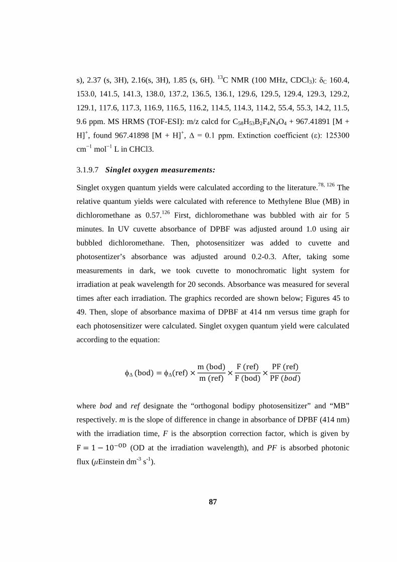
87
s), 2.37 (s, 3H), 2.16(s, 3H), 1.85 (s, 6H). 13C NMR (100 MHz, CDCl3): δC 160.4,
153.0, 141.5, 141.3, 138.0, 137.2, 136.5, 136.1, 129.6, 129.5, 129.4, 129.3, 129.2,
129.1, 117.6, 117.3, 116.9, 116.5, 116.2, 114.5, 114.3, 114.2, 55.4, 55.3, 14.2, 11.5,
9.6 ppm. MS HRMS (TOF-ESI): m/z calcd for C58H53B2F4N4O4 + 967.41891 [M +
H]+, found 967.41898 [M + H]+, Δ = 0.1 ppm. Extinction coefficient (ε): 125300
cm−1 mol−1 L in CHCl3.
3.1.9.7 Singlet oxygen measurements:
Singlet oxygen quantum yields were calculated according to the literature.78, 126 The
relative quantum yields were calculated with reference to Methylene Blue (MB) in
dichloromethane as 0.57.126 First, dichloromethane was bubbled with air for 5
minutes. In UV cuvette absorbance of DPBF was adjusted around 1.0 using air
bubbled dichloromethane. Then, photosensitizer was added to cuvette and
photosentizer’s absorbance was adjusted around 0.2-0.3. After, taking some
measurements in dark, we took cuvette to monochromatic light system for
irradiation at peak wavelength for 20 seconds. Absorbance was measured for several
times after each irradiation. The graphics recorded are shown below; Figures 45 to
49. Then, slope of absorbance maxima of DPBF at 414 nm versus time graph for
each photosensitizer were calculated. Singlet oxygen quantum yield were calculated
according to the equation:
φΔ (bod) = φΔ(ref) ×m (bod)m (ref)
×F (ref)F (bod)
×PF (ref)
PF (𝑏𝑜𝑑)
where bod and ref designate the “orthogonal bodipy photosensitizer” and “MB”
respectively. m is the slope of difference in change in absorbance of DPBF (414 nm)
with the irradiation time, F is the absorption correction factor, which is given by
F = 1 − 10−OD (OD at the irradiation wavelength), and PF is absorbed photonic
flux (μEinstein dm-3 s-1).

88
Figure 45. Comparative singlet oxygen generation experiment. Absorbance decrease of DPBF at 414 nm with time in dichloromethane in the presence of Bodipy photosensitizers: OB3 (black), OB6 (blue), OB7 (red) and reference photosensitizer methylene blue (green).
Figure 46. Decrease in absorbance of DPBF in dichloromethane in the presence of compound OB3 in medium. Details are given in singlet oxygen measurements part.

89
Figure 47. Decrease in absorbance of DPBF in dichloromethane in the presence of compound OB6 in medium.
Figure 48. Decrease in absorbance of DPBF in dichloromethane in the presence of compound OB7 in medium.
90
Figure 49. Decrease in absorbance of DPBF in dichloromethane in the presence of compound methylene blue in medium.
Figure 50. Singlet oxygen generation experiment in aqueous solution. Decrease in Absorbance spectrum of trap molecule (anthracene derivative) in the presence of 1.69 μM
compound OB3 in phosphate buffer saline (PBS).

91
3.1.9.8 Computational Details
Geometries were fully optimized with the ab initio Complete Active Space
Self Consistent Field (CASSCF) methodology and B3LYP Density Functional
employing CEP-31G (SBKJC) and cc-pVDZ basis sets. Harmonic vibrational
frequencies were computed to assure that the species did not possess any imaginary
frequencies and hence correspond to true minima. After a number of trial and error
the active space in the CASSCF calculations were established as 6 electrons
distributed over 6 valence orbitals (6,6) for the 8,2’ dimer OB3 and 4 electrons
distributed over 4 valence orbitals (4,4) for the 8,8’ dimer OB6 and. Enlarged active
spaces up to 12,12 also reproduced the key features presented in the main text. For
the sake of consistency a 6 electrons in 6 orbitals CAS is used for both dimers.
Geometries of excited states T1 and S1 were also fully optimized. Natural orbitals of
the CASSCF wavefunction and configuration state function coefficients were
analyzed to understand the nature of the electronic state as being a single electron
HOMO → LUMO type or multiply substituted type. Whereas Bodipy core S1 is a
clean HOMO → LUMO dominated ππ* state, S1 states of OB3 and OB6 are
drastically different and showed a large degree of multi-reference character.
Gaussian 03 suite was mainly used for the DFT calculations, CASSCF calculations
were mainly performed with MOLCAS, and GAMESS-US programs (References
for these computational can be found at ref. 129).
Figure 51. Frontier molecular orbitals. HOMO (left) LUMO (right), of the parent Bodipy core at B3LYP/6-31G(d,p) level of theory. Surfaces are plotted at 0.2 a.u.
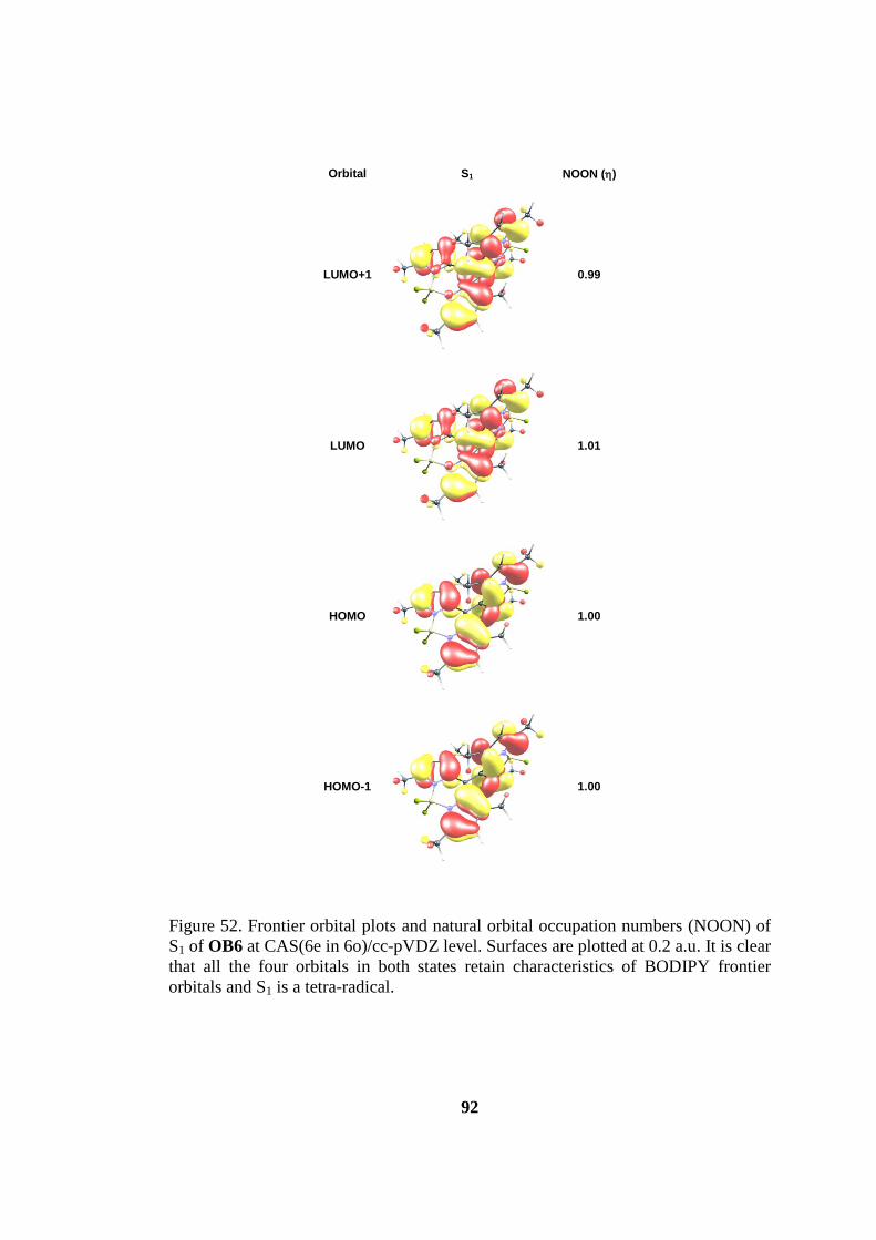
92
Orbital S1 NOON (η)
LUMO+1
0.99
LUMO
1.01
HOMO
1.00
HOMO-1
1.00
Figure 52. Frontier orbital plots and natural orbital occupation numbers (NOON) of S1 of OB6 at CAS(6e in 6o)/cc-pVDZ level. Surfaces are plotted at 0.2 a.u. It is clear that all the four orbitals in both states retain characteristics of BODIPY frontier orbitals and S1 is a tetra-radical.
93
3.2 PEGylated Calix[4]arene as a Carrier for a Bodipy-based
Photosensitizer
3.2.1 Introduction
Photodynamic therapy (PDT) is a non-invasive or moderately invasive
alternative to more traditional therapeutic methodologies such as surgery,
chemotherapy and radiation therapy.7 Photodynamic therapy has a built in selectivity
as the cytotoxic action is contingent upon the presence of the three components at
the same spatiotemporal coordinates, namely, light, sensitizer and dissolved oxygen.
Consequently, by controlling the region of irradiation, some selectivity can be
achieved.
Recent years witnessed a growing interest in the out-of-the-box proposals for
generating and controlling the photodynamic action.2, 131 At the same time, novel
chromophore families were introduced as potential sensitizers.80 Bodipy dyes
emerged as a promising class in part, due to our contribution highlighting the
chemistry and singlet oxygen generation capacity of these compounds.129, 132 Bodipy
dyes were previously recognized as chemically and photochemically stable
chromophores, and part of this stability was ascribed to the inaccessibility of the
triplet manifold, thus reducing the chances of potential degradative interactions with
ambient oxygen. However, Bodipys can be transformed into efficient
photosensitizers by the incorporation of heavy atoms or by increasing the
degeneracy of the excited state frontier orbitals.2, 133
Another matter of concern is the wavelength of irradiation. Current practice
of PDT makes use of porphyrins or closely related derivatives.134 These dyes have
relatively weak absorptions in the red end of the visible spectrum where mammalian
tissues are most transparent. Therefore, the ever existing drive to obtain dyes with
strong absorptions in the red and near IR region of the spectrum is understandable.
The absorption bands of the Bodipy dyes are highly tunable. Especially with styryl
substitution, one can move the absorption peak anywhere from 500 nm to 850 nm.135

94
By utilizing this strong property of this dye we come up with a new type of scaffold
carrying bodipy units and also enabling it to absorb near IR light.
On the other hand, calix[4]arene is a useful tool for organic chemists due to
its hydrophobic core and its intrinsic geometry and shape. Up to date, there is a few
example of calix[4]arene molecule have been used with bodipy. In one of the
example, where Akkaya et al. have synthesized136 a calixarene bodipy unit for pH
sensing purpose by taking advantage of the photoinduced electron transfer (PeT)
process very well known for bodipy type of structures. In this molecule, authors
simply monoformylated tetrahydroxycalix[4]arene and bodipy is synthesized around
the formyl unit and they could obtained the one bodipy unit is attached to one
calix[4]arene unit. It is also important to note that since phenyl of calix[4]arene is
orthogonal to the bodipy core, π conjugation is faded, hence the absorption spectrum
of the bodipy is not altered. Our synthesis is the improved version of this study in
terms of nearly everything; absorbance, application, synthetic route and number of
bodipy units attached to the calix[4]arene scaffold.
3.2.2 Design and Synthesis
The basic principle in our design is to incorporate maximum number of
bodipy units to the calix[4]arene backbone. While achieving this we aimed to put
forward a novel type of molecule which has not been synthesized before in order to
stimulate for other potential applications.
In the design of our PS (Fig. 54), we have judiciously attached two bodipy
chromophores to the calix[4]arene scaffold. The number of bodipy units designed is
an indication of our aim to enhance phototoxic effect. The second important
parameter of PDT is the absorption wavelength of the PS as told before. Hence, we
have thought the bond between calix[4]arene and bodipy should have a π-bond
character, therefore we benefit from this strategy to extend the π-conjugation of the
bodipy core which causes bathochromic shift of the absorbance wavelength of it.
And this is the first time that this strategy has been applied for bodipy with any type

95
of calixarene. Third, we have designed to attach four decyl units to the lower rim of
the calix[4]arene. The aim for this type of attachment is to increase their solubility in
common organic solvents and hence to facilitate the synthesis and to make the one
side of the molecule hydrophobic for molecule to go inside the cells by diffusing
through cell membrane by interacting with hydrophobic moieties there. The fourth
design parameter is to make the molecule water soluble, in order to accomplish this,
we have attached two poly(ethyleneglycol) polymers to the bodipy. By this way, the
other important parameter for this type of applications has been accomplished;
amphiphilicity. As a fifth property, in order to increase the π- conjugation further,
ethenyl-4-(N,N)-dimethylaminobenzene units were attached to the bodipy core. By
this way, maximum absorbance wavelength of around 700 nm is secured. Finally,
appropriate to the application we have been trying this molecule to use it for; we
have attached heavy iodine atoms to the periphery of the bodipy core. Heavy atom
induced spin-orbit coupling causes increase in the rate of intersystem crossing for
efficient production of singlet oxygen, one of the basic necessity of PDT.
Figure 53. Design of the final CB10 molecule.*
* Note that image is schematic and red alkyl chains are decyl groups

96
The synthesis of the final compound have been accomplished in eleven steps
and eight of it is consecutive and the synthesis scheme is shown in Figures 55-57. In
order to get dialdehyde calix[4]arene CB6 molecule, we needed to go for five steps
(Fig. 55). In the first step CB1 was reacted with benzoyl chloride to get CB2 as a
protecting group for de-tert-butylation step. By using AlCl3 and toluene we
successfully de-tert-butylated two of the four tert-butyl groups of calix[4]arene CB2
to yield CB3. Then, for deprotection of the calixarene from benzoyl groups, we
needed to do hydrolysis with NaOH and EtOH. Then, alkylation reaction with 1-
bromodecane to yield, CB5. In the fiftht step, by using hexamethyhlenetetraamine
and TFA at elevated temperature diformylation of the CB5 is accomplished to get
CB6, which is then reacted with pre-prepared diiodobodipy to get CB8.
CB7 was prepared in three steps (Fig. 56). In the first step propargyl bromide
was reacted with 4-hydroxybenzaldehyde. Product was reacted with 2,4-
dimethylpyrrole with typical bodipy procedure yielded alkynyl bodipy derivative,
which then diiodinated with iodine and iodic acid to get CB7. With typical
Knoevenagel reaction procedure CB8 is achieved (Fig. 57), and this compound is
purified without performing column chromatography, but precipitated with hexane
after dissolved in a minimum amount of CHCl3. Then a second Knoevenagel
reaction was performed on CB8 to get CB9. Finally, with pre-prepared PEG-N3
click reaction was carried out to yield CB10, and purified with column
chromatography.

97
Figure 54. Synthesis of the calix[4]arene derivative to be reacted with Bodipy.
Figure 55. Synthesis of the diiodinated Bodipy derivative.

98
Figure 56. Synthesis scheme of the final CB10 photosensitzer
The characterization of the compounds has been made by 1H NMR, 13C
NMR and ESI or MALDI mass spectroscopy. Due to the large molecular weights of
compounds CB9 and CB10 the characterization was hard to get it done. But, by
comparative 1H NMR analysis, the structure of the final compound CB10 was
verified (Fig. 58).

99
Figure 57. Comparative 1H NMR Study of the final compound CB10 and CB9.
1H NMR spectrum of compound CB10 with respect to CB9 has been shown
in Fig. 58. After final click reaction some alterations took place in limited amounts
of nuclei, these will be shown schematically in Figures 58 and will be explained
(vide infra). In aliphatic region there are three differences between molecules CB9
and CB10 which is the proof of the success of the final click reaction. First,
disappearance of the acetylenic proton at 2.61 ppm is due to the click reaction which
indicates that click reaction is accomplished (proton a in Fig. 58). Second, the
appearance of the methylene proton of PEG group linked directly to the triazole at
4.58 ppm is verified (proton d). And third, shifting of the methylene bridge protons
which connects the triazole unit to phenyl group of Bodipy 4.89 ppm to 5.25 ppm
is another indication (shift of b to c in Fig 58). This kind of peak shift is also
observed in a similar compound synthesized by Erbas et al.133 In the aromatic region
100
of the comparison (Fig. 58-right) appearance of triazole proton at 7.90 ppm is easily
seen (proton e). The other protons in this region do not differentiate much since
electronic structure and geometric shape of the compound changes a little or not.
3.2.3 Results and Discussion
Subsequent to synthesis of the final compound CB10 we devoted our time to
establish the photodynamic activity experiments. At this time, since our compound
is amphiphilic we designed two experiments in organic and in aqueous media. As a
universal singlet oxygen scavenger compound for studies in organic media we used
1,3-diphenylisobenzofuran molecule (DPBF). This compound has a maximum
absorbance at around 410 nm in various solvents and ahead of reacting with singlet
oxygen which is produced by the PS in action, the intensity of the absorbance peak
decreases, and a new peak in UV region starts to appear. And we focused on the
decrease of the intensity at 410 nm.
Initially, we performed control experiments with DPBF, light and bubbled air
(5 min) in isopropanol as a solvent, but not add our PS; CB10. The photodynamic
experiments have been started with recording absorbance spectrum for 15 min in
dark by withdrawing light irradiation. Subsequently, for 60 min irradiation with
LED light array of 725 nm for 5 min time intervals have been done. The black line
in Fig.x successfully shows that there is no decrease in the absorbance of trap
compound, specify that no cyctotoxic 1O2 formation.

101
Figure 58. Change in absorbance spectrum of 1,3-Diphenylisobenzofuran (DPBF) in the presence of 46 nM CB10 in IPA; first 15 min dark then 60 min irradiation with 725 nm LED array (left). Normalized absorbance vs. time graph of DPBF; control experiment without (black line) and with (red line) CB10 (right).
In the second experiment in isopropanol, we have now CB10 (46 nM)
besides the other reagents in control experiment. In this case, also 15 min dark
toxicity experiment has been held, and there is no 1O2 formation and the absorbance
remains the same (red curve in Fig. 59). Afterward, the photodynamic activity has
been tested by irradiating with 725 nm light for 60 min with 5 min time intervals
again. Successful decrease of the absorbance of scavenger with time is the evidence
for 1O2 formation.

102
Figure 59. Structure of 2'-(Anthracene-9,10-diylbis(methylene))dimalonic acid (ADMDA) which is used to track 1O2 production in aqueous media.
Later on, we proceeded to the singlet oxygen ability in aqueous medium.
Nonetheless, in this solvent we need a water soluble trap molecule since DPBF is
not soluble in highly polar solvents. And we synthesized an anthracene derivative
substituted with malonic acid groups* (Figure 60) to track the 1O2 formation.
Through reaction with 1O2 dioxygen bridge forms and the absorbance signal around
380 nm of the original compound decreases. As a control experiment in PBS
solution (with pH 7.4), we recorded absorbance of the trap first with dark (15 min)
and 60 min irradiation As it is seen well (Figure 61) (right, black curve) there is a
minimum decrease in the absorbance.
* Entire name: 2,2'-(anthracene-9,10-diylbis(methylene))dimalonic acid
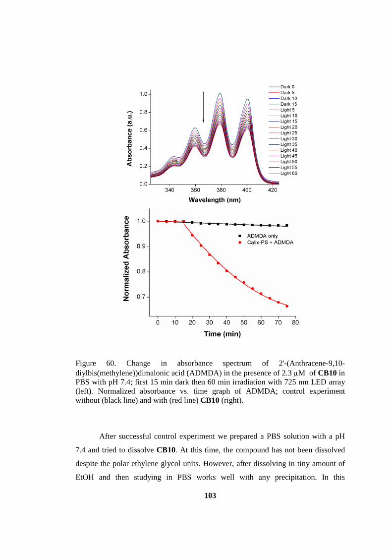
103
Figure 60. Change in absorbance spectrum of 2'-(Anthracene-9,10-diylbis(methylene))dimalonic acid (ADMDA) in the presence of 2.3 µM of CB10 in PBS with pH 7.4; first 15 min dark then 60 min irradiation with 725 nm LED array (left). Normalized absorbance vs. time graph of ADMDA; control experiment without (black line) and with (red line) CB10 (right).
After successful control experiment we prepared a PBS solution with a pH
7.4 and tried to dissolve CB10. At this time, the compound has not been dissolved
despite the polar ethylene glycol units. However, after dissolving in tiny amount of
EtOH and then studying in PBS works well with any precipitation. In this
104
photoactivity experiment in PBS, 2.3 µM CB10 was used. Due to short lifetime of 1O2 in aqua (2 µs) we were obliged to use 50 fold of CB10 in organic media. The
same protocol has also been applied to this experiment; 15 min dark absorbance has
been recorded with no change in absorbance and 60 min irradiation with 725 nm
LED resulted in successful decrease in the absorbance (Figure 61).
As a result, benefiting from the amphiphilic nature of the calix-bodipy-PS we
could successfully observed the singlet oxygen formation and photodynamic activity
in both organic (isopropyl alcohol) and aqueous (10% EtOH in PBS) media.
3.2.4 Conclusion
At the very beginning of this project, our aim is to get a carrier molecule (e.g.
claix[4]arene) substituted with four bodipy units. Moreover, we thought to link this
molecule to an antibody of a cancer tissue to recognize it and behave as a targeting
group. However, it is obvious that it is difficult to characterize the complex structure
of the antibody and react it with our designed PS. In addition, the synthetic
difficulties also valid for the calix[4]arene molecule and we had difficulties to
functionalize the core unit to yield multiple functional groups (e.g. aldehyde) to react
with bodipy. In the end, we come up with decyl units. Although its selectivity in
biological media is low, it is very well known that its hydrophobic nature is useful
for interacting with cell membrane and diffuse into cells for strong photodynamic
effect since nucleus are the primary site of cell destruction. In addition, amphiphilic
nature of the PS imitates the biological molecules. As a result, an accessible
molecule which is made of facile synthetic routes is the truth of life, at least in an
organic chemistry laboratory.
Another problematic part of this study was the low yields that hampered our
aim to use this photosensitizer in vitro and/or in vivo cytotoxicity experiments.
Specifically, the two Knoevenagel condensation reactions prior to final reaction was
completed with low yields (10-20%). In addition, eight consecutive totally eleven

105
step reactions sequence is another factor that limits the amount of compound
synthesized.
To conclude, though a variety of troubles we faced, we could finally come up
with an amphiphilic photosensitizer with dual photoactivity in both organic and
aqueous media and the novel type of directly π-conjugated bodipy calix[4]arene
scaffold and its unique structure may have a potential to be used for other
applications.
3.2.5 Experimental Details
3.2.5.1 General:
1H NMR and 13C NMR spectra were recorded on Bruker DPX-400
(operating at 400 MHz for 1H NMR and 100 MHz for 13C NMR) in CDCl3 solvent
with tetramethylsilane as internal standard. All spectra were recorded at 25oC and
coupling constants (J values) are given in Hz. Chemical shifts are given in parts per
million (ppm). Absorption spectra were performed by using a Varian Cary-100 and
Varian Cary 5000 UV-VIS-NIR absorption spectrophotometers. Fluorescence
measurements were conducted on a Varian Eclipse spectrofluometer. Mass spectra
were recorded with Agilent Technologies 6224 TOF LC/MS and 6530 Accurate
Mass Q-TOF LC/MS. And MALDI spectrums were recorded in Izmir Institute of
Technology, Chemistry Department with Bruker MALDI-TOF-TOF. Reactions
were monitored by thin layer chromatography using Merck TLC Silica gel 60 F254.
Silica gel column chromatography was performed over Merck Silica gel 60 (particle
size: 0.040-0.063 mm, 230-400 mesh ASTM). All other reagents and solvents were
purchased from suppliers and used without further purification. In singlet oxygen
measurements 1,3-Diphenylisobenzofuran was used as a singlet oxygen trap in
organic solvent measurements and was purchased from supplier. 2,2'-(Anthracene-
9,10-diylbis(methylene))dimalonic acid was used a singlet oxygen trap in aqueous
solvent and was synthesized according to the literature.130 Compound CB4137 and
CB7133 were synthesized according to the literature.

106
3.2.5.2 Synthesis of Compounds
Calix[4]arene-25,26,27,28-tetradecyloxy (CB5)
NaH (60% dispersion in oil – 2.23 g, 55.7 mmol) was washed with hexane and
filtrate was added to the dry DMF (40 mL) with care. Then Calix[4]arene-OH (CB4)
(1.79 g, 3.34 mmol) and 1-Bromodecane (7.38 mL, 33.4 mmol) were added to the
solution. The reaction mixture was heated at 60⁰ C for 12 hours. After reaction it
was allowed to cool down. Ice/water was added (100 mL) and the mixture was
extracted with dichloromethane (3×50 mL). Organic layer was washed with water
(3×50 mL), aqueous ammonium chloride solution (2×50 mL) and brine (50 mL) and
dried with K2CO3. Then organic layer was evaporated in low pressure and yellow
solution was obtained. Upon waiting overnight the compound precipitated in MeOH.
The compound was filtrated and washed with MeOH to get the pure product as a
white solid (2.4 g, 60%). 1H NMR : 7.09 (s, 4H; ArH), 6.21 (t, 2H, J = 7.5 Hz; ArH),
6.09 (d, 4H, J = 7.5 Hz; ArH), 4.45 (d, 4H, J = 13.2 Hz; CH2), 4.05 (t, 4H, J = 8.3;
OCH2), 3.72 (t, 4H, J = 6.5; OCH2), 3.11 (d, 4H; J = 13.3), 1.95-2.01 (m, 4H; CH2),
1.81-1.95 (m, 4H; CH2), 1.51-1.65 (m, 4H; CH2), 1.15-1.50 (m, 70H; CH2, CH3),
1.00-0.85 (m, 12H; CH2CH3); 13C NMR : 155.5, 155.3, 144.2, 135.9, 133.6, 127.2,
125.6, 122.0, 75.1, 75.0, 34.1, 32.0, 31.8, 31.3, 30.6, 30.4, 30.2, 29.9, 29.8, 29.5,
26,7, 26.1, 22.8, 14.1. HRMS (TOF-ESI): m/z calcd for C76H120O4Na: 1119.9084
[M+Na]+; found: 1119.9138 [M+Na]+, Δ = 4.82 ppm.
Calix[4]arene-11-23-dialdehyde-25,26,27,28-tetradecyloxy (CB6)
A mixture of CB5 (1 g, 0.91 mmol), hexamethylenetetraamine (4.52 g, 27.34 mmol)
and Trifluoroacetic acid (20 mL) was stirred at 125⁰ C for 4 h in a screw-capped
vial, then the reaction mixture was cooled to room temperature. Diluted with
aqueous 1 M HCl (150 mL) and CH2Cl2 (50 mL) and it was stirred for 3 h
vigorously. The organic layer was taken and extracted with dichloromethane (50
mL). Then dried with K2CO3 and evaporated in vacuo. The compound 3 was
obtained pure after column chromatography with chloroform (1.044 g, 99%). 1H
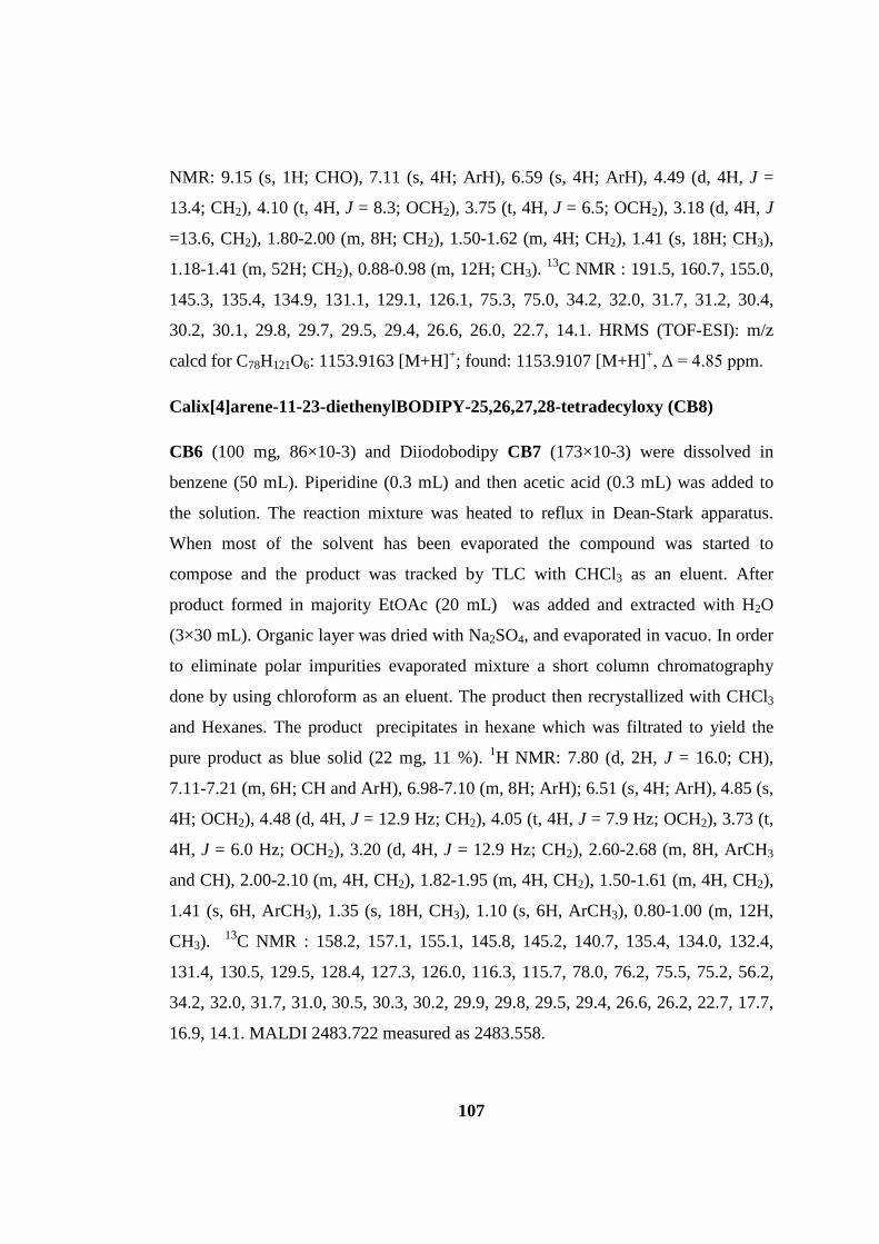
107
NMR: 9.15 (s, 1H; CHO), 7.11 (s, 4H; ArH), 6.59 (s, 4H; ArH), 4.49 (d, 4H, J =
13.4; CH2), 4.10 (t, 4H, J = 8.3; OCH2), 3.75 (t, 4H, J = 6.5; OCH2), 3.18 (d, 4H, J
=13.6, CH2), 1.80-2.00 (m, 8H; CH2), 1.50-1.62 (m, 4H; CH2), 1.41 (s, 18H; CH3),
1.18-1.41 (m, 52H; CH2), 0.88-0.98 (m, 12H; CH3). 13C NMR : 191.5, 160.7, 155.0,
145.3, 135.4, 134.9, 131.1, 129.1, 126.1, 75.3, 75.0, 34.2, 32.0, 31.7, 31.2, 30.4,
30.2, 30.1, 29.8, 29.7, 29.5, 29.4, 26.6, 26.0, 22.7, 14.1. HRMS (TOF-ESI): m/z
calcd for C78H121O6: 1153.9163 [M+H]+; found: 1153.9107 [M+H]+, Δ = 4.85 ppm.
Calix[4]arene-11-23-diethenylBODIPY-25,26,27,28-tetradecyloxy (CB8)
CB6 (100 mg, 86×10-3) and Diiodobodipy CB7 (173×10-3) were dissolved in
benzene (50 mL). Piperidine (0.3 mL) and then acetic acid (0.3 mL) was added to
the solution. The reaction mixture was heated to reflux in Dean-Stark apparatus.
When most of the solvent has been evaporated the compound was started to
compose and the product was tracked by TLC with CHCl3 as an eluent. After
product formed in majority EtOAc (20 mL) was added and extracted with H2O
(3×30 mL). Organic layer was dried with Na2SO4, and evaporated in vacuo. In order
to eliminate polar impurities evaporated mixture a short column chromatography
done by using chloroform as an eluent. The product then recrystallized with CHCl3
and Hexanes. The product precipitates in hexane which was filtrated to yield the
pure product as blue solid (22 mg, 11 %). 1H NMR: 7.80 (d, 2H, J = 16.0; CH),
7.11-7.21 (m, 6H; CH and ArH), 6.98-7.10 (m, 8H; ArH); 6.51 (s, 4H; ArH), 4.85 (s,
4H; OCH2), 4.48 (d, 4H, J = 12.9 Hz; CH2), 4.05 (t, 4H, J = 7.9 Hz; OCH2), 3.73 (t,
4H, J = 6.0 Hz; OCH2), 3.20 (d, 4H, J = 12.9 Hz; CH2), 2.60-2.68 (m, 8H, ArCH3
and CH), 2.00-2.10 (m, 4H, CH2), 1.82-1.95 (m, 4H, CH2), 1.50-1.61 (m, 4H, CH2),
1.41 (s, 6H, ArCH3), 1.35 (s, 18H, CH3), 1.10 (s, 6H, ArCH3), 0.80-1.00 (m, 12H,
CH3). 13C NMR : 158.2, 157.1, 155.1, 145.8, 145.2, 140.7, 135.4, 134.0, 132.4,
131.4, 130.5, 129.5, 128.4, 127.3, 126.0, 116.3, 115.7, 78.0, 76.2, 75.5, 75.2, 56.2,
34.2, 32.0, 31.7, 31.0, 30.5, 30.3, 30.2, 29.9, 29.8, 29.5, 29.4, 26.6, 26.2, 22.7, 17.7,
16.9, 14.1. MALDI 2483.722 measured as 2483.558.

108
Calix[4]arene-11-23-di(ethenyldimethylaminobenzene)ethenylBODIPY-
25,26,27,28-tetradecyloxy (CB9)
CB8 (50 mg, 0.021 mmol) and N,N-Dimethylaminobenzaldehyde (7.46 mg, 0.05
mmol) were dissolved in Benzene (50 mL). Piperidine (0.3 mL) and then acetic acid
(0.3 mL) was added to the solution. Then the reaction mixture was heated to reflux
in Dean-Stark apparatus. When most of the solvent has been evaporated the
compound was started to form and tracked by TLC and CHCl3 used as an eluent.
After product formed in majority EtOAc (20 mL) was added and extracted with H2O
(3×30 mL). Organic layer was dried with Na2SO4, and evaporated in vacuo. The
mixture was obtained as a pure product after column chromatography with CHCl3:
Hexane (3:1) as a green solid(17 mg, 31%). 1H NMR: 8.15 (d, 2H, J = 16.7 Hz;
CH), 7.81 (d, 2H, J = 16.6 Hz; CH), 7.59 (d, 4H, J = 8.8 Hz; ArH), 7.53 (d, 2H, J =
16.8 Hz ; CH), 7.15-7.24 (m, 6H; ArH and CH), 7.00-7.09 (m, 8H; ArH), 6.77 (d,
4H, J = 8.1 Hz; ArH), 6.53-6.62 (m, 4H; ArH), 4.89 (d, 4H, J = 2.4 Hz; CH2), 4.48
(d, 4H, J = 12.8 Hz; CH2), 4.05 (t, 4H, J = 8.1 Hz; CH2), 3.75 (t, 4H, J = 6.6 Hz;
CH2), 3.21 (d, 4H, J = 12.9 Hz; CH2), 3.01 (s, 12H; N(CH3)2), 2.61 (t, 2H, J = 2.4
Hz; CH), 2.00-2.10 (m, 4H; CH2), 1.82-1.95 (m, 4H; CH2), 1.51-1.65 (m, 4H; CH2),
1.48 (s, 6H; ArCH3), 1.20-1.42 (m, 70H; CH3 and CH2), 1.18 (s, 6H; ArCH3), 0.80-
0.97 (m, 12H, CH3). 13C NMR : 158.1, 156.9, 154.9, 145.4, 135.3, 133.9, 130.9,
129.8, 129.3, 128.8, 127.4, 125.9, 115.7, 112.2, 78.1, 76.0, 75.5, 75.2, 68.2, 56.2,
40.4, 38.8, 37.1, 34.1, 32.0, 31.9, 31.8, 31.7, 31.0, 30.5, 30.4, 30.2, 30.1, 30.0, 29.9,
29.8, 29.7, 29.5, 29.4, 26.6, 26.2, 23.8, 22.7, 17.7, 17.6, 14.1. MALDI 2639.972
measured as 2639.604.
Calix[4]arene-Photosensitizer (CB10)
CB9 (17 mg, 6.44×10⁻³ mmol) and PEG2000-N3 (25.76 mg, 0.0129 mmol) were
dissolved in dry DMF (5 mL). Argon was bubbled through the solution for 10
minutes and CuI (2.94 mg, 15.4×10⁻³ mmol) was added to the deaerated solution.
The reaction mixture was stirred at 60⁰ C overnight. Then 5 mL of CHCl3 was added
and CuI was filtrated. Then solution was evaporated and compound was separated

109
through column chromatography in MeOH: CHCl3 (7:93) as green solid. 1H NMR:
8.15 (d, 2H, J = 16.2 Hz; CH), 7.90 (s, 2H; CH), 7.85 (d, 2H, J = 16.6 Hz; CH), 7.61
(d, 4H, J = 8.1 Hz; ArH), 7.53 (d, 2H, J = 15.6 Hz ; CH), 7.21 (d, 2H, J = 17.1 Hz,
CH), 7.11-7.17 (m, 4H, ArH), 6.72-6.81 (m, 4H, ArH), 6.58-6.70 (m, 4H, ArH),
5.22-5.28 (m, 4H; OCH2), 4.58 (t, 4H, J = 3.8 Hz; NCH2), 4.48 (d, 4H, J = 12.0 Hz;
CH2), 4.03 (t, 4H, J = 7.9 Hz; OCH2), 3.91 (t, 4H, J = 4.9 Hz; OCH2), 3.35-3.88 (m,
364 H, OCH2), 3.23 (d, 4H, J = 11.0 Hz; CH2), 3.05 (s, 12H; N(CH3)2), 1.83-2.11
(m, 8H; CH2), 1.51-1.63 (m, 4H; CH2), 1.47 (s, 6H; ArCH3), 1.23-1.43 (m, 70 H;
CH2), 0.87-0.95 (m, 12H, CH3). MALDI calculated as 6736.998, found 6728.428.

110
CHAPTER 4
4 AXIAL CHIRAL BODIPYS
4.1 Introduction
Chirality is an important tool for organic chemists especially studying on
biology related topics. Because most biological compounds including amino acids
and sugars are chiral, and the importance of this topic has been understood very
recently compared to the human life on earth. In 20th century the importance of
drugs and their role in human health has been recognized in almost all of the
developed societies, and a huge amount of money has been invested to
pharmaceutical industry. Each year numerous drugs have been put up for sale and
most of these are chiral. The effect of racemic mixtures of drugs on health was seen
to be lethal by awful examples. Taking into account of the significance, it is evident
that chiral organic dyes are extremely vital in several purposes.
Fluorescence enantioselective sensing is one of the most up-to-date and
improvable topic among the optical sensing studies due to its potential as a simple
detection tool in chiral assays. This detection is quite important for drug discovery,
biological labeling, catalysts, and understanding the mechanism of molecular
recognition in biological systems.138 There are number of fluorescent sensors for
recognition of chiral amines, carboxylic acids, alcohols and amino acids.138
Specially biologically important hydroxyacids and amino acids have been attracting
much interest.139 Fluorescence enantioselective sensing can be mainly achieved by
two designs. One is the fluorophore-spacer-receptor type probe (PET type) and the
second is the fluorophore-receptor integrated probe (internal charge transfer (ICT)
type). In both design, one should introduce chirality on receptor (binding site) and
fluorescence enhancement or quenching have to be seen only with one enantiopure
derivative of a chiral molecule. Accordingly, there are many fluorophores have been
employed for this purpose such as binaphtol, cyclodextrin, calixarene, cyclic urea

111
and crown ethers.139a Most widely applied chirality in these compounds is
atropisomerism. Atropisomeric compounds are chiral molecules devoid of chiral
centers. In this class of compounds, there is a plane or axis of chirality in which
chirality exists due to the restricted rotation about single bond. Allenes, cumulenes
and biphenyls are good example for atropisomeric molecules.
Bodipy compounds are now important elements of luminophores family due
to their excellent properties two of which are ease functionalization and tunability of
the absorbance and emission wavelengths. Several attempts to use these compounds
in the topic of chirality have been done.3, 100, 107 In these examples, a stereogenic
center with carbon or boron center exists or bodipy unit is attached to a chiral
binaphtol. In the binaphtol case chirality is induced by the binaphtol due to its axial
chirality and bodipy is attached as a fluorescent probe, and this compound have been
employed for demonstrating different fluorescence characteristics with one
enantiomer of a chiral amine* compared to the other one.100 In this project, we have
designed and synthesized various orthogonal bodipy oligomers some of which
showing atropisomeric character, hence chiral and investigated their enantiomers by
separating using chiral HPLC and verified their helicity using circular dichroism
spectra. This study is believed to be used in the chiral assays in the future as the first
atropisomeric molecules comprise of solely bodipy dyes.
4.2 Design and Synthesis
Previously, we have synthesized a series of dimer bodipy compounds
through 2,8’ and 8,8’ positions, which are shown to exhibit efficient intersystem
crossing thus they are efficient singlet oxygen generators. Then, we could use these
molecules in PDT concept to kill cancerous cells in vitro in small concentrations
upon irradiation with light and in the presence of oxygen. Afterwards, we went
further in the synthesis utilizing the same strategy to yield a set of orthogonal bodipy
* specifically 1-phenylethylamine.

112
oligomers and recognized that some of these structures have a potential to be used as
chiral indicators through universal reputation of the bodipy chromophore.
In the first step we formylated 8H Bodipy synthesized from 2,4-
dimethylpyrrole, by using classical formulating agents POCl3 and DMF in 1,2-
dicholoroethane to get AB2 in Figure 62. Then typical Bodipy reaction has been
accomplished to get AB3 in 20% to 30% yield. In this set of bodipy oligomers only
these two reactions have been done and the yields are around 25% in bodipy
reactions and above 90% in formylation steps. In the third step, after formylation we
have two types of structures which can be separated easily by using column
chromatography, AB4 and AB5. AB5 is the monoformylated product and AB4 is the
diformylated product. At some stage after monoformylation of the AB3, there are
two sites for the second formylation and POCl3-DMF complex picks the bodipy
which is not formylated, due to the electron poorer structure of the formylated
bodipy and yielding AB4. This type of picking has also been verified by the study of
Zhu et al. since they could synthesize the 2,6-diformyl bodipy.140 They could not
diformylate the bodipy core in one step, but after monoformylation they isolated and
consecutive formylation gave the di- product. AB4 then converted into orthogonal
tetramer AB6 in one step. Monoformyl AB5 then first converted to chiral AB7 and
then to AB8 after monoformylation.

113
Figure 61. Synthesis scheme for Orthogonal Bodipy Oligomers. i) POCl3, DMF, ClCH2CH2Cl; ii) 2,4-Dimethylpyrrole, TFA, DDQ, DCM, NEt3, BF3.OEt2.
Upon mono formylation of the 2,8’-dimer bodipy we achieved AB5 which
ensures one of the chirality requirements of the compound that was synthesized by
Ziessel et al.; lateral differentiation of the bodipy core.3 After, 3D analysis this
compound’s comprising a chiral axis has been verified (Figure 63). It is seen
explicitly that the angle between two bodipy units is almost 900. AB7 which consists
of three bodipy units also has a chiral axis (Figure 63-right). Hence, these
compounds have a potential to be used as chiral indicators. It is essential to note that
two novel compounds, AB5 and AB7 are 50%-50% racemic mixture of enantiomers.
And it is impossible to separate them using classical column chromatography.
Hence, we have performed HPLC separation by using cellulose based chiral
columns.

114
Figure 62. Structures of AB5 (left) and AB7 (right).
4.3 Seperation And Photophysical & Circular Dichroism
Characterization Of Enantiomers
We have synthesized five novel orthogonal bodipy oligomers; two dimeric,
two trimeric and one tetrameric. After analysis of their 3D structures we realized that
four of these five molecules have a potential to be used as chiral indicators. Only
compound AB8 is a diastereomeric structure. Photophysical of these compounds
also examined and it is seen that they have very similar absorption and emission
characteristics. All of the synthesized compounds absorb in 499-508 nm region in
chloroform. And as expected they all have low fluorescence quantum yield due to
the enhanced intersystem crossing property of this type of structures verified in our
previous publication.2 The normalized absorbance and emission spectra of AB5
(dimer) and AB7 (trimer) have been shown in Figure x. The absorbance spectra of
these two compounds are very similar and the maxima are 504 and 515 nm for dimer
and trimer, respectively. However, their fluorescence spectra have some differences
but the common point is their fluorescence quantum yields, both are low; 0.078 and
0.075 for dimer and trimer respectively. Other photophysical parameters like
fluorescence lifetime and molar absorptivity have been demonstrated in the Table 5.

115
Figure 63. Absorbance and Emission Spectra of the dimer AB5 and trimer AB7.
Table 5. Photopysical Properties of AB5 and AB7.
Dye λabs [nm] εmax [M-1cm-1] λems [nm] Φf [%] τf [ns]
AB5 504 78000 541 0.78 3.14
AB7 515 162000 565 0.75 3.72
All of the chiral compounds are synthesized as 50%-50% racemic mixtures.
And in order to analyze the enantiomers we need to separate with HPLC by using
chiral column. We focused on two compounds; AB5 and AB7 and we used a
cellulose based chiral column. The separation of dimer aldehyde AB5 is
straightforward by using 5% isopropylalcohol in heptane and recorded the HPLC
spectrum is shown in Figure 64 and the integrated area of two peaks are equivalent,
thus the enantiomers have been separated successfully.
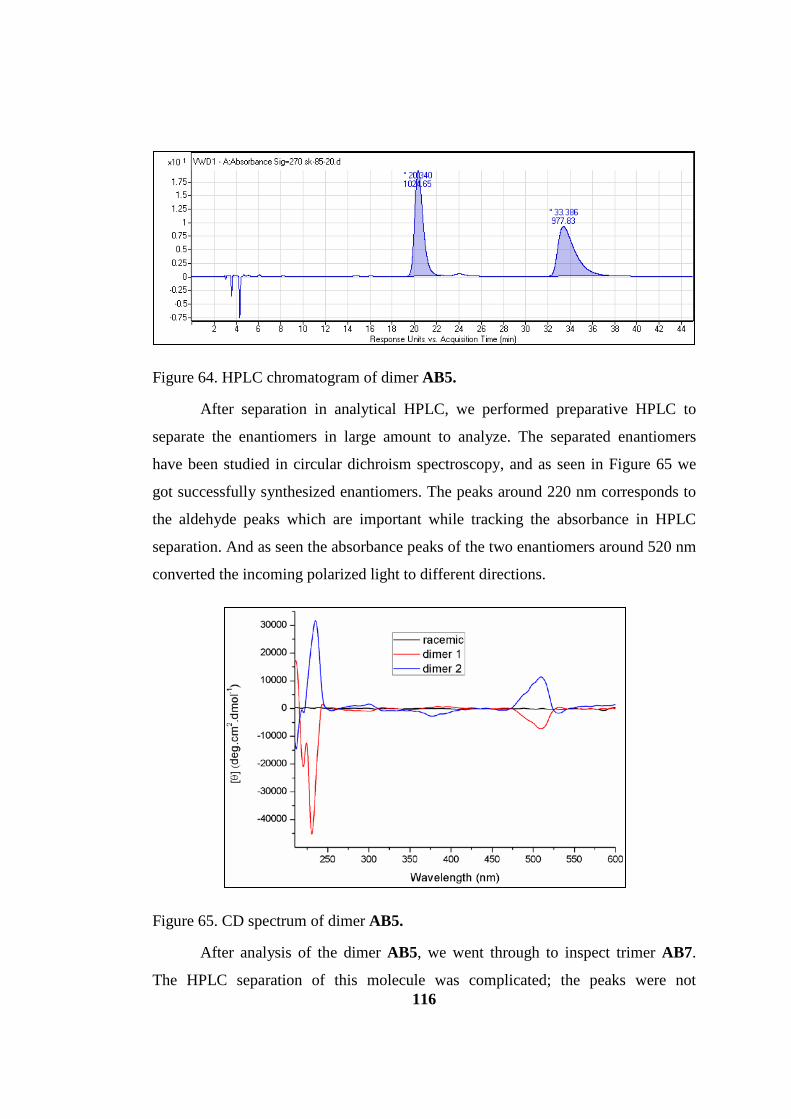
116
Figure 64. HPLC chromatogram of dimer AB5.
After separation in analytical HPLC, we performed preparative HPLC to
separate the enantiomers in large amount to analyze. The separated enantiomers
have been studied in circular dichroism spectroscopy, and as seen in Figure 65 we
got successfully synthesized enantiomers. The peaks around 220 nm corresponds to
the aldehyde peaks which are important while tracking the absorbance in HPLC
separation. And as seen the absorbance peaks of the two enantiomers around 520 nm
converted the incoming polarized light to different directions.
Figure 65. CD spectrum of dimer AB5.
After analysis of the dimer AB5, we went through to inspect trimer AB7.
The HPLC separation of this molecule was complicated; the peaks were not

117
separated 100% though we performed nonpolar solvent system (2% IPA in heptanes
- Fig. 66). The rationale behind this fact is the absence of a polar group (e.g.
aldehyde) which facilitates the separation. However, we could pick the most pure
fractions of the two peaks and analyzed the CD spectrum. This time aldehyde peak
around 220 nm has been disappeared and it is unambiguous that the two enantiomers
also have been separated well and we could see the absorbances of the two
enantiomers around 520 nm which also rotate the incoming polarized light to
different directions.
Figure 66. HPLC chromatogram (up) and CD spectrum (below) of trimer AB7.

118
4.4 Conclusion
Orthogonal bodipys synthesized for chiral organic fluorophore applications
are essential in three ways. First, their structure have a chiral axis which make them
chiral molecules without having a chiral stereogenic center and since this type of
atropisomeric compounds are very rare and most of these type of structures are
binaphtol based, bodipy based chiral assays may have a potential to be used in wide
application areas. More, this study is the first of atropisomeric bodipy comprises of
solely bodipy structure and nearly all of atropisomeric organic molecules are based
on biphenyls. Second, the synthesis is straightforward with two types of reactions
and facile purification steps it is possible to get chiral dyes. Third, since binaphtol
based compound generally absorb in UV region, these compounds have a substantial
gain since they absorb in the visible region. Although their fluorescence quantum
yields are low, several chiral organic and/or bioorganic compounds can be probed by
tracking the absorbance maxima, in condition that the reaction take place in the or
near to the bodipy core (e.g. aldehyde group in AB5). We believe these building
blocks will find a convenient application area in modern biological labeling and
advanced optoelectronic devices.
4.5 Experimental details
General: 1H NMR and 13C NMR spectra were recorded on Bruker DPX-400
(operating at 400 MHz for 1H NMR and 100 MHz for 13C NMR) in CDCl3 with
tetramethylsilane (TMS) as internal standard. All spectra were recorded at 25oC and
coupling constants (J values) are given in Hz. Chemical shifts were reported in parts
per million (ppm). Absorption spectra were performed by using a Varian Cary-100.
Fluorescence measurements were conducted on a Varian Eclipse Spectrofluometer.
The fluorescence decay measurements were carried out with the Horiba Jobin-Yvon
Time-Resolved Fluorometer, Fluorolog FL-1057. The instrument response function
was measured with an aqueous ludox solution. Mass spectra were recorded with
Agilent Technologies 6224 TOF LC/MS and 6530 Accurate Mass Q-TOF LC/MS.

119
High performance liquid chromatography (HPLC) separations were performed by
using Agilent Technologies HPLC-1200 Series with multi-wavelength detector
(MWD) and Agilent Technologies Preparative HPLC-1200 Series with diode array
detector (DAD). Circular dichroism (CD) measurements were conducted with Jasco
J-815 CD Spectrometer. Reactions were monitored by thin layer chromatography
using Merck TLC Silica gel 60 F254. Silica gel column chromatography was
performed over Merck Silica gel 60 (particle size: 0.040-0.063 mm, 230-400 mesh
ASTM). Compounds (AB1), (AB2), and (AB3) were synthesized according to our
pervious study.2 All other reagents and solvents were purchased from suppliers and
used without further purification.
Synthesis of compound AB4 and AB5
1 mL of DMF and 1 mL of POCl3 was stirred in an ice bath for 5 min under argon.
Then warmed to room temperature and waited for 30 minutes. To this mixture
compound (AB3) (200 mg, 0.405 mmol) was added in dichloroethane (60 mL). The
temperature was raised to 500C and stirred for 2 hours. The reaction medium was
cooled and poured into 150 mL of ice cooled NaHCO3 solution slowly. After that it
was stirred for further 30 min, then washed with water 3 times. The organic layers
was combined and dried over Na2SO4 and evaporated to dryness. AB5 and AB4 has
been purified by column chromatography by using 2% MeOH in CHCl3 as an eluent
and the compounds has been got pure as reddish solid (70% AB5 and 25% AB4).
AB5:
1H NMR (400 MHz, CDCl3): δH 10.05 (1H, s, CHO), 7.21 (1H, s, ArH), 6.20 (1H, d,
J= 3.64 Hz, ArH), 2.83 (3H, s, CH3), 2.62 (3H, s, CH3), 2.60 (3H, s, CH3), 2.41 (3H,
s, CH3), 2.35 (3H, s, CH3), 2.12 (3H, s, CH3), 2.02 (3H, s, CH3), 1.75 (3H, s, CH3); 13C
NMR (100 MHz, CDCl3): δC 185.8, 161.9, 161.6, 156.7, 150.1, 146.6, 144.5,
142.2, 135.8, 135.6, 135.2, 134.7, 132.1, 129.9, 126.4, 124.2, 122.9, 121.0, 120.8,
15.1, 14.9, 14.4, 13.1, 12.6, 11.2, 10.8, 9.8 ppm. MS HRMS (TOF-ESI): m/z calcd:
522.239; found: 522.6327 [M-H]−, Δ = 4.4 ppm.

120
AB4:
1H NMR (400 MHz, CDCl3): δH 10.11 (1H, s, CHO), 10.06 (1H, s, CHO), 7.45 (1H,
s, ArH), 6.23 (1H, s, ArH), 2.86 (3H, s, CH3), 2.82 (3H, s, CH3), 2.66 (3H, s, CH3),
2.61 (3H, s, CH3), 2.47 (3H, s, CH3), 2.21 (3H, s, CH3), 2.03 (3H, s, CH3), 1.76 (3H,
s, CH3); MS HRMS (TOF-ESI): m/z calcd: 549.2262; found: 549.2330 [M-H]−, Δ =
12.4 ppm.
Synthesis of compound AB7
In N2 bubbled 250 mL dichloromethane, 2,4-Dimethylpyrrole (145 μL, 1.408 mmol)
and (AB5) (336.6 mg, 0.646 mmol) were mixed. 2 drops of trifluoroacetic acid has
been added. The reaction mixture stirred at room temperature overnight. Then p-
chloranil (159 mg, 0.646 mmol) was added to the reaction. After stirring 1 hour at
room temperature, NEt3 (5 mL) and BF3.OEt2 (5 mL) were added and stirred again
for additional 1 hour. The reaction was finished by extracting with water (3x250
mL). Column chromatography with CH2Cl2 yielded the pure product (20 %). 1H
NMR (400 MHz, CDCl3): δH 7.18 (1H, s, ArH), 6.18 (1H, s, ArH), 6.12 (1H, s,
ArH), 6.00-6.02 (2H, m, ArH), 2.53 (3H, s, CH3), 2.52 (3H, s, CH3), 2.51 (6H, s,
CH3), 2.41 (3H, s, CH3), 2.38 (3H, s, CH3), 2.25 (3H, s, CH3), 2.13 (3H, s, CH3),
1.75 (3H, s, CH3), 1.71 (6H, s, CH3), 1.53 (3H, s, CH3); 13C NMR (100 MHz,
CDCl3): δC 161.4, 159.0, 156.1, 155.5, 150.9, 150.3, 144.8, 144.2, 142.5, 142.0,
137.6, 135.6, 135.1, 134.4, 133.5, 133.0, 132.1, 131.9, 131.7, 131.0, 130.8, 128.8,
125.7, 123.4, 122.6, 121.5, 121.1, 120.8, 120.7, 31.9, 29.7, 22.7, 15.0, 14.8, 14.6,
14.1, 13.8, 13.7, 12.7, 12.5, 11.5, 11.3, 9.7 ppm; MS HRMS (TOF-ESI): m/z calcd:
740.358; found: 740.3585 [M-H]-, Δ = 0.68 ppm.
Synthesis of compound AB6
In N2 bubbled 250 mL dichloromethane, 2,4-Dimethylpyrrole (99 μL, 0.56 mmol)
and (AB4) (120 mg, 0.218 mmol) were mixed. 2 drops of trifluoroacetic acid has
been added. The reaction mixture stirred at room temperature overnight. Then p-
chloranil (107 mg, 0.436 mmol) was added to the reaction. After stirring 1 hour at
121
room temperature, NEt3 (2 mL) and BF3.OEt2 (2 mL) were added and stirred again
for additional 1 hour. The reaction was finished by extracting with water (3x100
mL). Column chromatography with 2% MeOH in CHCl3 yielded the pure product
(20 %). 1H NMR (400 MHz, CDCl3): δH 7.29 (1H, s, ArH), 6.15 (1H, s, ArH), 6.05
(1H, s, ArH), 6.04 (1H, s, ArH), 6.02 (1H, s, ArH), 5.99 (1H, s, ArH), 2.62 (3H, s,
CH3), 2.57 (6H, s, CH3), 2.55 (6H, s, CH3), 2.43 (9H, s, CH3), 2.17 (6H, s, CH3),
1.81 (3H, s, CH3), 1.76 (3H, s, CH3), 1.71 (9H, s, CH3), 1.60 (3H, s, CH3); MS
HRMS (TOF-ESI): m/z calcd: 985.4643; found: 985.4712.3585 [M-H]-, Δ = 7.0
ppm.
Synthesis of compound AB8
3 mL of DMF and 3 mL of POCl3 was stirred in an ice bath for 5 min under argon.
Then warmed to room temperature and waited for 30 minutes. To this mixture
compound (AB7) (70 mg, 0.094 mmol) was added in dichloroethane (30 mL). The
temperature was raised to 500C and stirred for 2 hours. The reaction medium was
cooled and poured into 150 mL of ice cooled NaHCO3 solution slowly. After that it
was stirred for further 30 min, then washed with water 3 times. The organic layers
was combined and dried over Na2SO4 and evaporated to dryness. AB8 was purified
by column chromatography by using 2% MeOH in CHCl3 as an eluent two times
due to separate from other diastereomer and the compounds has been got pure as
reddish solid with 11% yield(7.7 mg, 0.01 mmol).
1H NMR (400 MHz, CDCl3): δH 10.05 (1H, s, CHO), 7.18 (1H, s, ArH), 6.10-6.20
(3H, m, ArH), 2.83(3H, s, CH3), 2.63(3H, s, CH3), 2.62(3H, s, CH3), 2.59(3H, s,
CH3), 2.42(3H, s, CH3), 2.40(3H, s, CH3), 2.30(3H, s, CH3), 2.12(3H, s, CH3),
2.04(3H, s, CH3), 1.76(3H, s, CH3), 1.75(3H, s, CH3), 1.55(3H, s, CH3); 13C NMR
(100 MHz, CDCl3): δC 186.2, 161.7, 161.6, 160.3, 157.0, 150.2, 149.5, 146.1, 145.7,
144.4, 142.4, 136.7, 136.0, 135.3, 135.2, 134.7, 133.5, 132.0, 124.0, 123.1, 121.0,
120.6, 15.1, 15.0, 14.4, 14.2, 14.1, 13.1, 12.7, 12.5, 11.5, 11.4, 10.7, 9.7 ppm; MS
HRMS (TOF-ESI): m/z calcd: 985.4643; found: 985.4712.3585 [M-H]-, Δ = 7.0
ppm.

122
CHAPTER 5
The discussions on this chapter are partially based on the following publication:
Kolemen, S.; Bozdemir, O. A.; Cakmak, Y.; Barin, G.; Erten-Ela, S.; Marszalek, M.; Yum, J. H.; Zakeeruddin, S. M.; Nazeeruddin, M. K.; Gratzel, M.; Akkaya, E. U., Chem Sci 2011, 2 (5), 949-954.
5 THIOPHENE BASED PS’S FOR DSSC
5.1 Introduction
The Great East Japan Earthquake happened on March 11, 2011 and it caused
a disfunction of the nuclear power plant and made people to search for alternative
source of energy in most of the countries in the world. The dye sensitized solar cell
(DSSC) concept is a viable alternative to traditional semiconductor based
photovoltaic constructs.109 Practical potential is also being realized in the form of
rapid commercialization.141 One of the primary issues, which may benefit from
rational design, is the choice of the sensitizer dye. Near IR sensitizability and
panchromaticity are important and challenging goals and these can be attained in
principle by designing appropriate organic dyes.142 We are particularly interested in a
class of dyes known as Bodipy dyes.120, 143 Bodipy dyes are important fluorophores
with a multitude of potential applications. Recent developments in Bodipy
chemistry allow diverse modification on the core structure.144 Through these
modifications, many characteristics of the parent chromophore can be altered in the
desired direction, for example, it is possible to shift absorption wavelength from 500
nm to 800 nm with simple chemical transformations.145 Solubility and aggregation
characteristics of the dyes can also be modulated as needed.144 These interesting
characteristics of bodipy made people direct to itself in many fields. One of those
fields is the DSSC concept, or more generally solar cells, since polymer solar cells
(bulk heterojunction) also started to construct an important brick of the wall.146In a

123
fresh study by Ziessel et al. they could observe an efficiency of 4.7% by using these
types of cells using thiophene appended bodipy structures.
Figure 67. Some of bodipy PSs synthesized recently in literature4
The first rationally designed example of a Bodipy based sensitizer was
reported a few years ago,147 followed by a few more recent articles including both
liquid electrolyte and solid state based DSSCs.4, 148 It is clear that optimal solar cell
performance of a photosensitizer is dependent on a large number of parameters;

124
however, absorption range, anchoring groups and the direction of electronic
reorganization on excitation should be among the most important ones. Taking into
account of these parameters we have designed and studied seven different bodipy
based compounds and constructed cells and evaluated the outputs.4 Six of these
compounds are in Figure 68. In each one of them, we tried to investigate the role of
at least one parameter in solar cell performance. The most successful candidate for a
solar cell is the PS1; it is formed from 2-methylpyrrole in case of 2,4-
dimethylpyrrole so that electron localization to the phenyl ring in the meso position
is more feasible. However, we should mention that a different type of cell was
constructed special to this dye. In order to inhibit aggregation cheno moiety was
used (see abbreviations). Other strategies like putting the anchoring group to 2-
position of the dye or incorporating the dye core with iodine atoms did not work.
Hence, in our design here we set up novel DSSC candidates (Fig. 69) where we have
used novel donor group and a novel anchoring group which have been successful in
other DSSC systems but not tried with Bodipy.
Figure 68. Novel designed PSs for liquid electrolyte DSSC in this research.
125
5.2 Design and Synthesis:
Thiophene is known to be used frequently in DSSC concept because of its
electron transfer ability. Since electron delocalization through the p system is a very
important factor in dye sensitized solar cells we designed a bodipy dye, thiophene
carboxylic acid unit is attached to the meso (8-) position of the bodipy core, where it
was calculated before that electron injection is more feasible.149 Directly attaching of
the thiophene to the core also thought to impart efficiency of electron injection, also.
As in the successful DSSC application in PS1 in Figure 68, we have used 2-
methylpyrrole instead of 2,4-dimethylpyrrole to increase p electron conjugation from
bodipy to thiophene to anchoring unit. As an anchoring group carboxylic acid was
used due to the synthetic problems we faced while trying to synthesize cyanoacetic
acid moiety, which is also known for efficient binding to the TiO2 surface and
increases electron withdrawing ability to the electron injecting unit. Electron donor
part is also an important parameter in DSSC concept, efficiently using these groups
have a substantial role in increased cell efficiencies. In this design, we altered our
mostly used diphenylaminophenyl group with bis-dimethylfluorenyl amino group.
This group has been utilized in some literature reports and due to its bulky structure
it has been widely proposed as donor groups. It was also suggested that using bulky
structures increases stability of oxidized dye molecules. Hence, first time in bodipy
dyes we have designed and synthesized bis-dimethylfluorenyl amino donor groups
for DSSC application.
In the synthesis part (Fig. 70), we first iodinated the fluorene molecule to get
TB1 in 90% yield. In the second reaction we methylate the 2-iodofluorene by using
a modified procedure from literature with MeI and KOMe in DMF. Then, in the
third step we synthesized N,N-Bis(9,9-dimethylfluoren-2-yl)aniline according to the
Ullman reaction conditions by using a variety of reagents at high temperatures.
Then, in order to synthesize TB4 aldehyde which is needed for Knoevenagel
reaction we performed typical Vilsmeier formylation reaction in CHCl3. After
synthesis of the donor part we moved for synthesis of the bodipy dye core. 5-formyl-
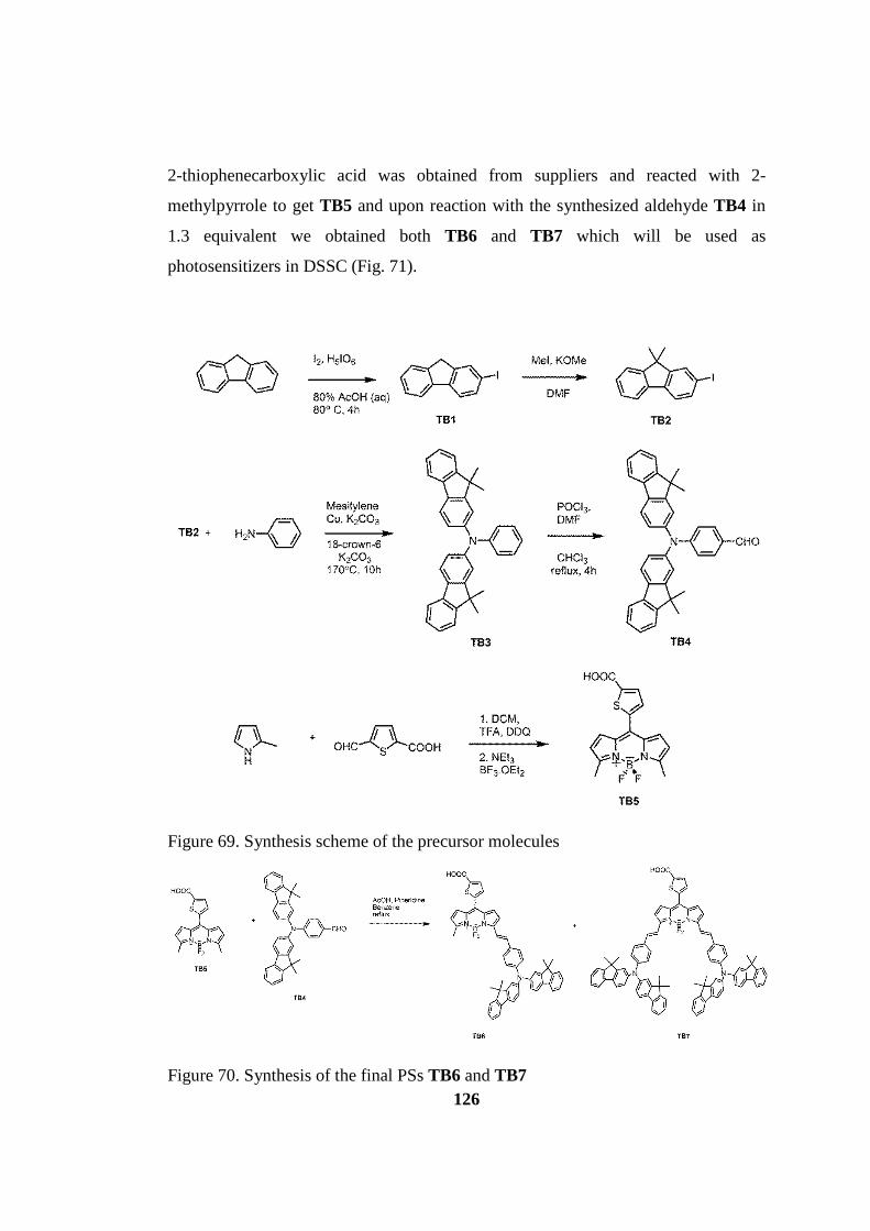
126
2-thiophenecarboxylic acid was obtained from suppliers and reacted with 2-
methylpyrrole to get TB5 and upon reaction with the synthesized aldehyde TB4 in
1.3 equivalent we obtained both TB6 and TB7 which will be used as
photosensitizers in DSSC (Fig. 71).
Figure 69. Synthesis scheme of the precursor molecules
Figure 70. Synthesis of the final PSs TB6 and TB7

127
5.3 Results and Discussion
After synthesis of the PSs TB6 and TB7 along with the sole Bodipy TB5 we
have performed photophysical analysis and CV measurements in order to determine
if these units are suitable for constructing a cell with TiO2 and redox couple I−/I3−.
Absorbance spectra of these dyes have been investigated in Figure 72, and found
that TB5 has a narrow absorption spectrum with λmax at 520 nm which is
characteristic for bodipys and molar absorptivity of this compound is 43700 in
MeOH. This compound is barely soluble in DCM or CHCl3. TB6 has a broader
absorption spectrum with absorption maximum at 665 nm in DCM, also fluorene
moieties also absorb strongly in UV region of the spectrum at around 370 nm. Molar
absorptivity of this compound is 38800. Due to fluorene moieties which inhibit π−π
stacking of the compound increases solubility, we could dissolve it in DCM. Last
PS, TB7 has a strongly red shifted absorbance maximum at 773 nm in DCM. This
compound is promising in this respect because PSs absorbing in the near IR end of
the spectrum is highly desired due to strong sunlight illumination at those
wavelength region. For example, PS1 in Figure 68, which has the highest efficiency
in Bodipy DSSC has an absorption maximum at 724 nm. The molar absorptivity of
TB7 compound is 53700 at its maximum. All the three compounds have very weak
emission which is due to the charge transfer events caused mainly from
thiophenecarboxylic acid group.

128
Figure 71. Absorbance Spectra of the compounds synthesized. TB5 in MeOH, TB6 and TB7 in DCM.
Figure 72. Cyclic Voltammetry Measurements of the TB5 (red), TB6 (green) and TB7 (blue).

129
Table 6. Redox Potentials and EHOMO and ELUMO Levels of Bodipy Derivatives.
Eoxidationa
(Volt)
Ereductionb
(Volt)
Eferrocenec
(Volt)
EHOMOd
(eV)
ELUMOe
(eV)
EBand Gapf
TB5 0.54 -0.70 0.58 4.84 3.52 1.32
TB6 0.87 -0.72 0.57 5.10 3.51 1.59
TB7 0.83 -0.63 0.61 5.02 3.56 1.46 a Oxidation potential of Bodipy Derivatives, bReduction potential of Bodipy Derivatives, cPotential of ferrocene, internal reference electrode dHOMO energy level of Bodipy Derivatives, eLUMO energy level of Bodipy Derivatives fEnergy Band Gap of Bodipy Derivatives
Cyclic Voltammetry of the recently synthesized compounds have been
shown in Figure 73. And the table formed from this data has been given also in
Table 6 shows explicitly the oxidation reduction bands of these compounds and the
HOMO-LUMO band levels and the band gaps. Energy level of the HOMO and
LUMO is very important in DSSC concept. It has been given information about this
topic in Background part. To summarize upon photoexcitation of the dye molecule
an electron excited from HOMO electron orbital to the LUMO and then electron
injection from LUMO to the conduction band of the TiO2 happens. Hence, LUMO
energy level should be higher in energy than the conduction band of TiO2. The
energy level of the conduction band is given 4.2 eV4. In all three PSs; TB5, TB6,
TB7, their LUMO level are convenient for electron injection with 3.51-3.56 eV
values which are higher in energy according to the Figure 74. HOMO levels of the
bodipy dyes should be also considered since their energy level should be lower than
the redox couple energy level (I−/ I3−) which is given also as 4.9 eV. As we look at
the Table 6, we observe that only TB5 is not convenient for reducing of the bodipy
dye by the redox couple. On the other hand, TB6 and TB7 are fit to the desired
energy level with 5.10 and 5.02 eV, respectively. However, it was emphasized in a
recent review by Ooyama and Harima that, the energy level difference between
these levels should be at least 0.2-0.3 eV for efficient electron regeneration.113 In our
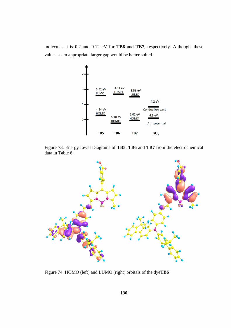
130
molecules it is 0.2 and 0.12 eV for TB6 and TB7, respectively. Although, these
values seem appropriate larger gap would be better suited.
Figure 73. Energy Level Diagrams of TB5, TB6 and TB7 from the electrochemical data in Table 6.
Figure 74. HOMO (left) and LUMO (right) orbitals of the dyeTB6
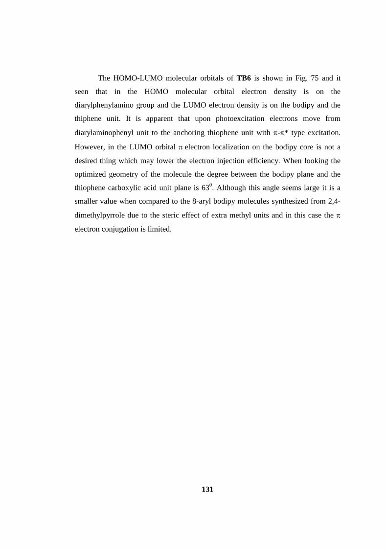
131
The HOMO-LUMO molecular orbitals of TB6 is shown in Fig. 75 and it
seen that in the HOMO molecular orbital electron density is on the
diarylphenylamino group and the LUMO electron density is on the bodipy and the
thiphene unit. It is apparent that upon photoexcitation electrons move from
diarylaminophenyl unit to the anchoring thiophene unit with π-π* type excitation.
However, in the LUMO orbital π electron localization on the bodipy core is not a
desired thing which may lower the electron injection efficiency. When looking the
optimized geometry of the molecule the degree between the bodipy plane and the
thiophene carboxylic acid unit plane is 630. Although this angle seems large it is a
smaller value when compared to the 8-aryl bodipy molecules synthesized from 2,4-
dimethylpyrrole due to the steric effect of extra methyl units and in this case the π
electron conjugation is limited.

132
Figure 75. HOMO (up) and LUMO (down) orbitals of the dyeTB7
In the HOMO-LUMO molecular orbitals of the dye TB7 (Fig. 76), there is a
strange electron localization in the HOMO orbital because the electrons are mainly
in one of the diarylaminophenyl unit. This may imply that one of the electron donor
groups has a low contribution to electron excitation upon sunlight irradiation. This

133
may be due to the angle between the styryl plane and the aminophenyl plane. The
degrees are 200 and 110, and in high angle case p electron contribution is low. When
looking at the LUMO orbital we see a similar picture compared to the TB6. The
angle between the bodipy plane and thiophene plane is also same with 630, and the
localization is both on the bodipy and the thiophene. Also, these optimized
geometries tell us that in the di-Knoevenagel product TB7, large donor groups
(difluorenylaminophenyl) affect their orientation and π electron conjugation is
diminishes, this may lead to steric congestion which may destabilize the molecule.
the design of TB6 is seems more convenient in terms of three dimensional
orientation on TiO2 surface. On the other hand, aggregation is inhibited more
efficiently in TB7. Hence, more elegant design may be by utilizing one of this steric
donor groups and attaching long alkyl chains to the other side of the molecule.
Figure 76. Current vs. voltage graphs of the photosensitizers.
The results indicate above that TB6 molecule seems more convenient in
order to get higher efficiency. After investigation the CV results and computational
calculations of valence molecular orbital energies and electron densities we
constructed the cells by using typical methods as told in experimental section. And
we measured the short-circuit photocurrent density (Jsc), open-circuit photovoltage
(Voc), photocurrent/voltage curve (J/V curve), fill factor (ff) and incident-photon-to-
current conversion efficiency (IPCE) and by the help of these parameters we

134
calculated the solar energy-to-electricity conversion yield (η). Figure 77 shows the
J/V curve of the synthesized three dyes, TB5, TB6 and TB7 and parallel with our
thoughts from CV and computational results TB6 produces the more photocurrent
with 2.61 mAcm-2 under AM 1.5 simulated sunlight (100 mWcm-2). And the cell
constructed from TB6 has open-circuit voltage of 400 mV with fill factor of 0.57.
And the calculated total sunlight-to-electricity conversion yield (η) is 0.59 (Table 7).
This makes this dye finer compared to TB5 and TB7. Also, in the incident-photon-
to-current conversion efficiency (IPCE) spectrum also give us information about the
response of the dye molecules upon illumination with the monochromatic light in a
large range of wavelengths (Figure 78). This spectrum resembles the absorbance of
the dyes, however we see that especially in TB6 and TB7 there is a big amount of
photocurrent produced in the range between 300-400 nm. However, since the
irradiation of sunlight in this range is not strong its reflection to the solarlight
photocurrent is not so important. For TB6 we see effective photocurrent in the range
between 600-700 nm (Fig. 78).
Figure 77. Incident photon to current conversion efficiencies as a function of wavelengthfor the liquid electrolyte based DSSCs.

135
Here we constructed the cell of TB5 as a comparison and we reached low
solar cell parameter values with this dye. For the TB7 DSSC, we acquired similar
values to TB6. Here, the critical parameter is the highly panchromatic nature of
IPCE spectrum (Fig. 78). Although the photocurrent is low it is generated between
500-850 nm. The open-circuit voltage (Voc) is 400 mV, the photocurrent density is
2.12 mAcm-2 and the fill factor is 0.58. The overall sunlight energy-to-electricity
conversion yield (η) is 0.49.
By evaluating the results it should be kept in mind that the optimization of
the cell is an important issue (e.g. electrolyte type, additive (cheno) used) and these
are not the final results for the cells constructed with the dyes TB6 and TB7. The
photocurrent density is expected to increase after improving the cell construction
parameters.
Table 7. Photovoltaic Performance of TiO2 based Dye Sensitized Solar Cells.
Jsc (mAcm-2) Voc (mV) FF η (%)
TB5 1.12 370 0.48 0.19
TB6 2.61 400 0.57 0.59
TB7 2.12 400 0.58 0.49
5.4 Conclusion
After constructing the cells from the dye we synthesized overall efficiency
(η) of 0.59 for AB6 and 0.49 for AB7 has been achieved. However, these results are
under evaluation by optimizing the cell parameters and may increase. The novelty in
this design compared to the publications from our group is using different donor
group and the π electron linker.4, 148-149 As a donor group we have used bis-
dimethylfluorenyl amino groups in the design. This group is the first time used with
bodipy dyes and due to its strerically bulkiness it inhibits the aggregation of the dyes
on the TiO2 nanocrystalline surface. In our two types of dyes these groups sit well
for the TB6, however in TB7 by using these two bulky donor groups make one of

136
the ethenylphenylamino group connected to the bodipy core out-of-plane and
electron density in highest occupied molecular orbital (HOMO) is mainly on one of
the donor groups only. The second parameter we have used in these molecules is the
π electron linker unit, thiophene. This group widely used in various DSSCs due to
their high π electron localization ability. And when we inspected the lowest-
unoccupied molecular orbitals (LUMO) we observed that electron density is moving
successfully from donor groups to the acceptor unit. The main obstacle may be the
electron density is not fully transferred to the acceptor and there is some residual
density is on the bodipy core. This complication may be solved by using
oligothiophene units as a pi electron linker for more efficient electron move.
5.5 Experimental Details
All chemicals and solvents obtained from suppliers were used without further
purification. Reactions were monitored by thin layer chromatography using Merck
TLC Silica gel 60 F254. Chromatography on silica gel was performed over Merck
Silica gel 60 (particle size: 0.040-0.063 mm, 230-400 mesh ASTM). 1H NMR and 13C NMR spectra were recorded at room temperature on Bruker DPX-400 (operating
at 400 MHz for 1H NMR and 100 MHz for 13C NMR) in CDCl3 or DMSO-d6 with
tetramethylsilane (TMS) as internal standard. Coupling constants (J values) are
given in Hz and chemical shifts are reported in parts per million (ppm). Absorption
spectra in solution were acquired using a Varian Cary-100 spectrophotometer. Mass
spectra were recorded on Agilent Technologies 6530 Accurate-Mass Q-TOF
LC/MS. Spectrophotometric grade solvents were used for spectroscopy experiments.
Compound TB1, TB2, TB3 and TB4 were synthesized according to literature.150 CV
measurement was taken by using CH-Instrument 660 B Model Potentiostat
equipment. Solution was prepared in chloroform (10−3 M). A three electrode cell
was used consisting of Glassy carbon working electrode, Pt wire counter electrode
and Ag/AgCl reference electrode, all placed in a glass vessel. Tetrabutylammonium
hexafluorophosphate (TBAPF6), 0.1 M, was used as supporting electrolyte.
Ferrocene was used as internal reference electrode. ELUMO level was calculated by
137
using the formula of ELUMO = − e(E1/2(red) − Efer. + 4.8). The conduction band of
TiO2 layer, E (TiO2-CB) = −4.2 eV.
5.5.1 Device Fabrication:
The construction of the dye sensitized solar cell device requires first cleaning
of the fluorine doped tin oxide (FTO) coated glass substrates in acetone and
isopropanol by using an ultrasonic bath. FTO (SnO2:F, Pilkington TEC-15;
Rsheet:15O/&), electrically conductive oxide-coated glasses were used as
transparent electrodes. TiO2 electrodes consist of an adsorbent mesoporous layer
with 20 nm anatase TiO2 particle size in 7 mm thicknesses and a second light
scattering layer with 400 nm anatase TiO2 particle size of 5 mm thicknesses. Dye
solutions were prepared in chlorobenzene-methanol (1:1) mixture with a
concentration of 0.5 mM BODIPY. TiO2-coated electrodes, after sintering at 4500C
for 30 min and cooling to 1000C, were kept overnight in BODIPY solutions for
adsorption. BODIPY adsorbed TiO2-coated glasses were washed with pure
chlorobenzene and dried in vacuo. Platinized FTO glasses were used as counter
electrode. Platinization of counter electrodes were done by coating of FTO glasses
with 1% solution of hydrogen hexachloroplatinate (Aldrich) in 2-propanol and
annealing at 4000C for 30 min. Cells were prepared in sandwich geometry. Surlyn-
1702 (DuPont) frame was used as a spacer and a thermoplastic sealant between the
two electrodes. Cells prepared in this way were then sealed by heating at 1000C.
Electrolyte was filled into the space created by Surlyn-1702 between the electrodes
under vacuum using a small hole pre-drilled on the counter electrode with the help
of a diamond drill. After filling the electrolyte, a small hole was sealed again using a
piece of Surlyn-1702 and a piece of cover glass. The electrolyte A6986 consisted of
0.6M N-methyl-N-butyl-imidazolium iodide (BMII) + 0.1 M LiI + 0.05 M I2 + 0.5
M 4-tert-butylpyridine (TBP) in acetonitrile/valeronitrile (85/15 v/v). On the other
hand electrolyte Z1040 consisted of 1M LiI, 0.044M I2, 0.25M TBP in
acetonitrile/valeronitrile (85/15 v/v). Active areas of the cells were adjusted to 0.159
cm2 with a special mask. Dye sensitized solar cells were characterized by current-

138
voltage (J −V ) measurement. All current-voltage measurements (J −V ) were done
under 100 mW/cm2 light intensity and AM 1.5 conditions. 450 W Xenon light
source (Oriel) was used to give an irradiance of various intensities. The spectral
output of the lamp was matched in the region of 350–700 nm with the aid of Schott
K113 Tempax sunlight filter. J − V data collection was made by using Keithley
2400 Source-Meter and LabView data acquisition software.
5.5.2 Synthesis
Compound TB5
400 mL argon-degassed CH2Cl2 were added 2-methyl pyrrole (11.5 mmol, 0.93 g)
and 5-formyl-2-thiophenecarboxylic acid (5.46 mmol, 0.85 g). One drop of TFA was
added and the solution was stirred under N2 at room temperature for 1 day. After
addition of p-chloranil (5.46 mmol, 1.34 g) to the reaction mixture, stirring was
continued for 30 min. 5 mL of Et3N and 5 mL of BF3.OEt2 were successively added
and after 30 min, the reaction mixture was washed three times with water (3 x 100
mL) which was then extracted into the DCM (3 x 100 mL) and dried over anhydrous
Na2SO4. The solvent was evaporated and the residue was purified by silica gel
column chromatography using (DCM: MeOH 80:20) as the eluant. Red solid (0.75
g, 40%). 1H NMR (400 MHz, MeOD-d4): δH 7.79 (1H, d, J = 3.8 Hz, ArH), 7.50
(1H, d, J = 3.9 Hz, ArH), 7.12 (2H, d, J= 4.1 Hz, ArH), 6.44 (2H, d, J = 4.4 Hz,
ArH), 2.62 (6H, s, CH3). MS (TOF- ESI): m/z: Calcd: 301.0788, Found: 301.0798
[M-COOH]− , ∆=3.3 ppm. εabs= 43700 (in MeOH)
Compounds TB6 and TB7
TB5 (0.29 mmol, 0.10 g) and TB4 (0.38 mmol, 0.19 g) were added to a 100 mL
round-bottomed flask containing 50 mL benzene and to this solution was added
piperidine (0.3 mL) and acetic acid (0.3 mL). The mixture was heated under reflux
by using a Dean Stark trap and reaction was monitored by TLC (DCM : MeOH
80:20). When all the starting material had been consumed, the mixture is

139
concentrated and directly added to column chromatography by using DCM:MeOH/
80:20 and TB7 was separated as a pure product in green color in 52% yield (0.2 mg,
0.152 mmol). To separate the mono Knoevenagel product TB6 a second column
chromatography was done with DCM:MeOH/ 90:10 and TB6 was separated as a
pure product in 15% yield in deep blue color (35 mg, 0.04 mmol).
TB6:
1H NMR (400 MHz, DMSO-d6): δH 7.80 (2H, d, J = 8.0 Hz, ArH), 7.77 (2H, d, J =
7.3 Hz, ArH), 7.72 (1H, d, J = 16.3 Hz, CH), 7.62 (1H, d, J = 3.8 Hz, ArH), 7.49-
7.60 (5H, m, ArH), 7.43 (1H, d, J=16.3 Hz, CH), 7.25-7.38 (9H, m, ArH), 7.05-7.15
(4H, m, ArH), 6.48 (2H, d, ArH), 2.56 (3H, s, ArCH3), 1.38 (12H, s, CCH3). 13C
NMR (100 MHz, DMSO-d6): δC 163.6, 156.7, 155.4, 153.8, 149.4, 146.4, 139.1,
138.6, 135.2, 133.6, 133.0, 131.3, 129.8, 129.4, 127.6, 127.4, 124.4, 123.2, 122.4,
121.7, 120.2, 119.8, 118.5, 116.2, 47.0, 30.1, 27.1 ppm. MS (TOF- ESI): m/z: Calcd:
833.3059, Found: 833.2955 [M] , ∆=12.5 ppm. εabs= 38800 (in DCM)
TB7:
1H NMR (400 MHz, DMSO-d6): δH 7.76 (4H, d, J = 8.0 Hz, ArH), 7.72 (4H, d, J =
6.8 Hz, ArH), 7.65 (2H, d, J = 16.6 Hz, CH), 7.40-7.50 (12H, m, ArH; CH), 7.20-
7.35 (16H, m, ArH), 7.07 (8H, m, ArH), 1.34 (24H, s, CCH3). 13C NMR (100 MHz,
DMSO-d6): δC 163.4, 155.4, 154.8, 153.7, 149.2, 146.3, 138.5, 137.8, 135.3, 135.2,
132.6, 131.8, 130.0, 129.7, 129.2, 129.0, 127.5, 127.4, 124.3, 123.1, 122.3, 121.7,
120.1, 119.7, 117.9, 116.6, 46.9, 27.1 ppm. MS (TOF-ESI): m/z: Calcd: 1320.5359,
Found: 1320.5085 [M] , ∆=20.7 ppm. εabs= 53700 (in DCM)
140
CHAPTER 6
6 CONCLUSION
Investigation of novel materials is an important tool to respond the problems
like eradicating the cancerous tissues or to solve the energy problem by finding
environmentally friendly devices. In order to achieve these goals the principles of
the materials on earth should be inspected carefully. Chemists with the help of other
fundamental sciences try to develop methods to observe, learn, and apply them to
the materials they study. This thesis is one drop in that ocean which aimed to
increase the wisdom of the people studied to make it done. In this thesis included
three main titles; photodynamic therapy, axial chirality and dye-sensitized solar cell,
and four different research projects.
In the first title of PDT, novel sensitization through intersystem crossing of
the dimer bodipy molecule has been achieved. The novelty lies in the theoretical
design of the excited states and then applying this to a real example of converting
ground state triplet oxygen to the reactive singlet oxygen species with the aim of
destroying malignant tissues. In the early study a bodipy dimer absorbing around
500 nm was synthesized and due to the wavelength limitation of the therapy, shifting
the wavelength to longer part was thought and applied. However, our study failed in
this way because doubly-substituted state forming upon excitation started to
disappear. Hence, it was proposed that an adjustment is needed to realize this
thought e.g. changing the boron difluoride bridge in the core.
Second study in PDT title was about proposing a nanomaterial acting as a
holder for carrying a pharmaceutical to the target. This pharmaceutical is a well-
known bodipy unit with heavy atoms and the essential water soluble units and long
alkyl chains for cell membrane permission. Using both hydrophilic and hydrophobic
structures make these materials amphiphilic which is convenient for studying in
vivo. Due to synthetic complications our first aim has not been totally came true, but

141
the original design and employing calix[4]arene unit with bodipy in a π-conjugated
manner made this project a future prospecting one.
For the concept of axial chirality, we designed oligobodipy molecules which
were attached to each other in an orthogonal fashion. Molecules possessing this
property has been desired mainly in advanced optoelectronic devices and
biomolecule assays. Our molecules forms the first example of atropisomeric type
chiral compounds in which a chiral axis exist around a single bond. A great
percentage of these compounds used are formed from biphenyls and when
considering the superior properties of bodipy dyes, these compounds have a chance
to be utilized in advancing the probing units used.
The final topic was about the DSSCs formed by utilizing the recently
synthesized bodipy dyes for developing the efficiency of the cells constructed
before. Modifying the p electron linker and the donor molecules seems not working
well at least for now. Meso position of the bodipy core is reported to be the most
convenient place for attaching the electron withdrawing and anchoring unit. In our
previous studies we used phenyl units attached to that position, however in this case
thiophene units have been incorporated with carboxylic acid. The angle between the
thiophene and bodipy is 630 which is larger than the phenyl attached one. This may
be one reason for the low efficiency. Bis-dimethylfluorenyl amino groups are widely
used in DSSC due to its steric effect which hampers the aggregation. Using these
dyes in TB7 did not work well because in this molecule this donor groups attached
both 3 and 5 position of the bodipy core due to steric congestion in one of the donor
p electron conjugation seems hampered as it is understood from the HOMO orbitals.
Total solar energy-to-electricity conversion yield (η) was acquired 0.59 for AB6 and
0.49 for AB7, however, these values expected to increase since cell optimization is
still proceeding by our collaborator.
To conclude, with these topics studied it is apparent that bodipy is not a sole
fluorophore but due to its rich chemistry its applications are expected to spread to
most other topic as well.
142
REFERENCES
1. Boens, N.; Leen, V.; Dehaen, W., Fluorescent indicators based on BODIPY. Chem Soc Rev 2012, 41 (3), 1130-1172.
2. Cakmak, Y.; Kolemen, S.; Duman, S.; Dede, Y.; Dolen, Y.; Kilic, B.; Kostereli, Z.; Yildirim, L. T.; Dogan, A. L.; Guc, D.; Akkaya, E. U., Designing Excited States: Theory-Guided Access to Efficient Photosensitizers for Photodynamic Action. Angew Chem Int Edit 2011, 50 (50), 11937-11941.
3. Haefele, A.; Zedde, C.; Retailleau, P.; Ulrich, G.; Ziessel, R., Boron asymmetry in a BODIPY derivative. Org Lett 2010, 12 (8), 1672-5.
4. Kolemen, S.; Bozdemir, O. A.; Cakmak, Y.; Barin, G.; Erten-Ela, S.; Marszalek, M.; Yum, J. H.; Zakeeruddin, S. M.; Nazeeruddin, M. K.; Gratzel, M.; Akkaya, E. U., Optimization of distyryl-Bodipy chromophores for efficient panchromatic sensitization in dye sensitized solar cells. Chem Sci 2011, 2 (5), 949-954.
5. Yella, A.; Lee, H. W.; Tsao, H. N.; Yi, C. Y.; Chandiran, A. K.; Nazeeruddin, M. K.; Diau, E. W. G.; Yeh, C. Y.; Zakeeruddin, S. M.; Gratzel, M., Porphyrin-Sensitized Solar Cells with Cobalt (II/III)-Based Redox Electrolyte Exceed 12 Percent Efficiency. Science 2011, 334 (6056), 629-634.
6. Li, L. L.; Diau, E. W. G., Porphyrin-sensitized solar cells. Chem Soc Rev 2013, 42 (1), 291-304.
7. Dolmans, D.; Fukumura, D.; Jain, R. K., Photodynamic therapy for cancer. Nature Reviews Cancer 2003, 3 (5), 380-387.
8. Hausmann, W., Die sensiblisierende Wirkung des Hematoporphyrins. Biochem. Zeitung 1911, 30, 276-316. 9. Meyer–Betz, F., Untersuchungen uber die biologische
photodynamische Wirkung des Hematoporphyrins und anderer Derivative des Blut und Galenafarbstoffs. Dtsch Arch. Klin. 1913, 112, 476-503.
10. Diamond, I.; Granelli, S. G.; McDonagh, A. F.; Nielsen, S.; Wilson, C. B.; Jaenicke, R., Photodynamic therapy of malignant tumours. Lancet 1972, 2 (7788), 1175-7.
11. Dougherty, T. J.; Grindey, G. B.; Fiel, R.; Weishaupt, K. R.; Boyle, D. G., Photoradiation therapy. II. Cure of animal tumors with hematoporphyrin and light. J Natl Cancer Inst 1975, 55 (1), 115-21.
12. Kelly, J. F.; Snell, M. E.; Berenbaum, M. C., Photodynamic destruction of human bladder carcinoma. British journal of cancer 1975, 31 (2), 237-44.
13. Kelly, J. F.; Snell, M. E., Hematoporphyrin derivative: a possible aid in the diagnosis and therapy of carcinoma of the bladder. The Journal of urology 1976, 115 (2), 150-1.
14. Dougherty, T. J.; Kaufman, J. E.; Goldfarb, A.; Weishaupt, K. R.; Boyle, D.; Mittleman, A., Photoradiation Therapy for the Treatment of Malignant Tumors. 1978.

143
15. Hayata, Y.; Konaka, C.; Takizawa, N.; Kato, H., Hematoporphyin Derivative And Laser Photoradiation In The Treatment Of Lung-Cancer. Chest 1982, 81 (3).
16. McCaughan, J. S.; Hicks, W.; Laufman, L.; May, E.; Roach, R., Palliation Of Esophageal Malignancy With Photoradiation Therapy. Cancer 1984, 54 (12).
17. Balchum, O. J.; Doiron, D. R.; Huth, G. C., Photoradiation Therapy Of Endobronchial Lung Cancers Employing The Photodynamic-Action Of Hematoporphyrin Derivative. Lasers in Surgery and Medicine 1984, 4 (1).
18. Moan, J.; Berg, K., The Photodegradation Of Porphyrins In Cells Can Be Used To Estimate The Lifetime Of Singlet Oxygen. Photochemistry and Photobiology 1991, 53 (4).
19. Korbelik, M.; Krosl, G., Cellular-Levels Of Photosensitizers In Tumors - The Role Of Proximity To The Blood-Supply. British Journal of Cancer 1994, 70 (4).
20. Tromberg, B. J.; Orenstein, A.; Kimel, S.; Barker, S. J.; Hyatt, J.; Nelson, J. S.; Berns, M. W., Invivo Tumor Oxygen-Tension Measurements For The Evaluation Of The Efficiency Of Photodynamic Therapy. Photochemistry and Photobiology 1990, 52 (2).
21. Pogue, B. W.; Braun, R. D.; Lanzen, J. L.; Erickson, C.; Dewhirst, M. W., Analysis of the heterogeneity of pO(2) dynamics during photodynamic therapy with verteporfin. Photochemistry and Photobiology 2001, 74 (5).
22. Star, W. M.; Marijnissen, H. P. A.; Vandenbergblok, A. E.; Versteeg, J. A. C.; Franken, K. A. P.; Reinhold, H. S., Destruction Of Rat Mammary-Tumor And Normal Tissue Microcirculation By Hematoporphyrin Derivative Photoradiation Observed Invivo In Sandwich Observation Chambers. Cancer Research 1986, 46 (5).
23. Fingar, V. H.; Wieman, T. J.; Haydon, P. S., The effects of thrombocytopenia on vessel stasis and macromolecular leakage after photodynamic therapy using photofrin. Photochemistry and Photobiology 1997, 66 (4).
24. (a) Fingar, V. H.; Kik, P. K.; Haydon, P. S.; Cerrito, P. B.; Tseng, M.; Abang, E.; Wieman, T. J., Analysis of acute vascular damage after photodynamic therapy using benzoporphyrin derivative (BPD). British Journal of Cancer 1999, 79 (11-12); (b) Dolmans, D.; Kadambi, A.; Hill, J. S.; Waters, C. A.; Robinson, B. C.; Walker, J. P.; Fukumura, D.; Jain, R. K., Vascular accumulation of a novel photosensitizer, MV6401, causes selective thrombosis in tumor vessels after photodynamic therapy. Cancer Research 2002, 62 (7).
25. Henderson, B. W.; Fingar, V. H., Relationship Of Tumor Hypoxia And Response To Photodynamic Treatment In An Experimental Mouse-Tumor. Cancer Research 1987, 47 (12).
26. Chen, Q.; Chen, H.; Hetzel, F. W., Tumor oxygenation changes post-photodynamic therapy. Photochemistry and Photobiology 1996, 63 (1).
27. Busch, T. M.; Wileyto, E. P.; Emanuele, M. J.; Del Piero, F.; Marconato, L.; Glatstein, E.; Koch, C. J., Photodynamic therapy creates fluence rate-

144
dependent gradients in the intratumoral spatial distribution of oxygen. Cancer Research 2002, 62 (24).
28. Henderson, B. W.; Fingar, V. H., Oxygen Limitation Of Direct Tumor-Cell Kill During Photodynamic Treatment Of A Murine Tumor-Model. Photochemistry and Photobiology 1989, 49 (3).
29. Gollnick, S. O.; Liu, X. N.; Owczarczak, B.; Musser, D. A.; Henderson, B. W., Altered expression of interleukin 6 and interleukin 10 as a result of photodynamic therapy in vivo. Cancer Research 1997, 57 (18).
30. deVree, W. J. A.; Essers, M. C.; deBruijn, H. S.; Star, W. M.; Koster, J. F.; Sluiter, W., Evidence for an important role of neutrophils in the efficacy of photodynamic therapy in vivo. Cancer Research 1996, 56 (13).
31. Korbelik, M.; Krosl, G.; Krosl, J.; Dougherty, G. J., The role of host lymphoid populations in the response of mouse EMT6 tumor to photodynamic therapy. Cancer Research 1996, 56 (24).
32. Korbelik, M.; Dougherty, G. J., Photodynamic therapy-mediated immune response against subcutaneous mouse tumors. Cancer Research 1999, 59 (8).
33. Gollnick, S. O.; Vaughan, L.; Henderson, B. W., Generation of effective antitumor vaccines using photodynamic therapy. Cancer Research 2002, 62 (6).
34. Castano, A.; Mroz, P.; Hamblin, M., Photodynamic therapy and anti-tumour immunity. Nature Reviews Cancer 2006, 6 (7), 535-545.
35. Dolmans, D.; Kadambi, A.; Hill, J. S.; Flores, K. R.; Gerber, J. N.; Walker, J. P.; Rinkes, I.; Jain, R. K.; Fukumura, D., Targeting tumor vasculature and cancer cells in orthotopic breast tumor by fractionated photosensitizer dosing photodynamic therapy. Cancer Research 2002, 62 (15).
36. Ruoslahti, E., Specialization of tumour vasculature. Nature Reviews Cancer 2002, 2 (2).
37. Peng, Q.; Moan, J.; Nesland, J. M., Correlation of subcellular and intratumoral photosensitizer localization with ultrastructural features after photodynamic therapy. Ultrastructural Pathology 1996, 20 (2).
38. Dougherty, T. J.; Gomer, C. J.; Henderson, B. W.; Jori, G.; Kessel, D.; Korbelik, M.; Moan, J.; Peng, Q., Photodynamic therapy. Journal of the National Cancer Institute 1998, 90 (12).
39. McBride, G., Studies expand potential uses of photodynamic therapy. Journal of the National Cancer Institute 2002, 94 (23).
40. Orenstein, A.; Kostenich, G.; Roitman, L.; Shechtman, Y.; Kopolovic, Y.; Ehrenberg, B.; Malik, Z., A comparative study of tissue distribution and photodynamic therapy selectivity of chlorin e(6), Photofrin II and ALA-induced protoporphyrin IX in a colon carcinoma model. British Journal of Cancer 1996, 73 (8).
41. Kamkaew, A.; Lim, S. H.; Lee, H. B.; Kiew, L. V.; Chung, L. Y.; Burgess, K., BODIPY dyes in photodynamic therapy.
42. Ris, H. B.; Altermatt, H. J.; Inderbitzi, R.; Hess, R.; Nachbur, B.; Stewart, J. C. M.; Wang, Q.; Lim, C. K.; Bonnett, R.; Berenbaum, M. C.; Althaus,

145
U., Photodynamic Therapy With Chlorins For Diffuse Malignant Mesothelioma - Initial Clinical-Results. British Journal of Cancer 1991, 64 (6).
43. Grosjean, P.; Savary, J.; Wagnieres, G.; Mizeret, J.; Woodtli, A.; Theumann, J.; Fontolliet, C.; vandenBergh, H.; Monnier, P., Tetra(m-hydroxyphenyl)chlorin clinical photodynamic therapy of early bronchial and oesophageal cancers. Lasers in Medical Science 1996, 11 (4), 227-235.
44. Mew, D.; Wat, C. K.; Towers, G. H. N.; Levy, J. G., Photoimmunotherapy - Treatment Of Animal Tumors With Tumor-Specific Monoclonal Antibody-Hematoporphyrin Conjugates. Journal of Immunology 1983, 130 (3).
45. Goff, B. A.; Bamberg, M.; Hasan, T., Photoimmunotherapy Of Human Ovarian-Carcinoma Cells Exvivo. Cancer Research 1991, 51 (18).
46. Vrouenraets, M. B.; Visser, G. W. M.; Stewart, F. A.; Stigter, M.; Oppelaar, H.; Postmus, P. E.; Snow, G. B.; van Dongen, G., Development of meta-tetrahydroxyphenylchlorin-monoclonal antibody conjugates for photoimmunotherapy. Cancer Research 1999, 59 (7).
47. Allison, B. A.; Pritchard, P. H.; Levy, J. G., Evidence For Low-Density-Lipoprotein Receptor-Mediated Uptake Of Benzoporphyrin Derivative. British Journal of Cancer 1994, 69 (5).
48. Konan, Y. N.; Gurny, R.; Allemann, E., State of the art in the delivery of photosensitizers for photodynamic therapy. Journal of Photochemistry and Photobiology B-Biology 2002, 66 (2).
49. Polo, L.; Valduga, G.; Jori, G.; Reddi, E., Low-density lipoprotein receptors in the uptake of tumour photosensitizers by human and rat transformed fibroblasts. International Journal of Biochemistry & Cell Biology 2002, 34 (1).
50. Swamy, N.; James, D. A.; Mohr, S. C.; Hanson, R. N.; Ray, R., An estradiol-porphyrin conjugate selectively localizes into estrogen receptor-positive breast cancer cells. Bioorganic & Medicinal Chemistry 2002, 10 (10).
51. Zhou, C.; Milanesi, C.; Jori, G., An Ultrastructural Comparative-Evaluation Of Tumors Photosensitized By Porphyrins Administered In Aqueous-Solution, Bound To Liposomes Or To Lipoproteins. Photochemistry and Photobiology 1988, 48 (4).
52. Richter, A. M.; Waterfield, E.; Jain, A. K.; Canaan, A. J.; Allison, B. A.; Levy, J. G., Liposomal Delivery Of A Photosensitizer, Benzoporphyrin Derivative Monoacid Ring-A (Bpd), To Tumor-Tissue In A Mouse-Tumor Model. Photochemistry and Photobiology 1993, 57 (6).
53. Rasmussan-Taxdal, D. S.; Ward, G. E.; Figge, F. H., Fluorescence of human lymphatic and cancer tissues following high doses of intravenous hematoporphyrin. Cancer 1955, 8, 78-81.
54. Erbas-Cakmak, S.; Bozdemir, O. A.; Cakmak, Y.; Akkaya, E. U., Proof of principle for a molecular 1:2 demultiplexer to function as an autonomously switching theranostic device. Chem Sci 2013, 4 (2), 858-862.
55. Berg, K.; Selbo, P. K.; Prasmickaite, L.; Tjelle, T. E.; Sandvig, K.; Moan, D.; Gaudernack, G.; Fodstad, O.; Kjolsrud, S.; Anholt, H.; Rodal, G. H.;

146
Rodal, S. K.; Hogset, A., Photochemical internalization: A novel technology for delivery of macromolecules into cytosol. Cancer Research 1999, 59 (6).
56. Mittra, R. A.; Singerman, L. J., Recent advances in the management of age-related macular degeneration. Optometry and Vision Science 2002, 79 (4).
57. Ortu, P.; Lamuraglia, G. M.; Roberts, W. G.; Flotte, T. J.; Hasan, T., Photodynamic Therapy Of Arteries - A Novel-Approach For Treatment Of Experimental Intimal Hyperplasia. Circulation 1992, 85 (3).
58. Chou, T. M.; Woodburn, K. W.; Cheong, W. F.; Lacy, S. A.; Sudhir, K.; Adelman, D. C.; Wahr, D., Photodynamic therapy: Applications in atherosclerotic vascular disease with motexafin lutetium. Catheterization and Cardiovascular Interventions 2002, 57 (3).
59. Leman, J. A.; Morton, C. A., Photodynamic therapy: applications in dermatology. Expert Opinion on Biological Therapy 2002, 2 (1).
60. Szeimies, R. M.; Landthaler, M.; Karrer, S., Non-oncologic indications for ALA-PDT. Journal of Dermatological Treatment 2002, 13 (1).
61. Trauner, K. B.; Gandour-Edwards, R.; Bamberg, M.; Shortkroff, S.; Sledge, C.; Hasan, T., Photodynamic synovectomy using benzoporphyrin derivative in an antigen-induced arthritis model for rheumatoid arthritis. Photochemistry and Photobiology 1998, 67 (1).
62. Soukos, N. S.; Ximenez-Fyvie, L. A.; Hamblin, M. R.; Socransky, S. S.; Hasan, T., Targeted antimicrobial photochemotherapy. Antimicrobial Agents and Chemotherapy 1998, 42 (10).
63. Hamblin, M. R.; Miller, J. L.; Hasan, T., Effect of charge on the interaction of site-specific photoimmunoconjugates with human ovarian cancer cells. Cancer Research 1996, 56 (22).
64. Bonnett, R., Photosensitizers of the Porphyrin and Phthalocyanine Series for Photodynamic Therapy. Chem Soc Rev 1995, 24 (1), 19-33.
65. Valeur, B.; Berberan-Santos, M. N., Molecular Fluorescence: Principles and Applications. Second ed.; Wiley-VCH Verlag: GmbH: 2012.
66. Kasha, M., Phosphorescence and the role of the triplet state in the electronic excitation of complex molecules. Chem Rev 1947, 41 (2), 401-19.
67. Zhao, Q. A.; Li, F. Y.; Huang, C. H., Phosphorescent chemosensors based on heavy-metal complexes. Chem Soc Rev 2010, 39 (8), 3007-3030.
68. Killoran, J.; Allen, L.; Gallagher, J. F.; Gallagher, W. M.; O'Shea, D. F., Synthesis of BF2 chelates of tetraarylazadipyrromethenes and evidence for their photodynamic therapeutic behaviour. Chem Commun 2002, (17), 1862-1863.
69. Xuan, J.; Xiao, W. J., Visible-Light Photoredox Catalysis. Angew Chem Int Edit 2012, 51 (28), 6828-6838.
70. Singh-Rachford, T. N.; Castellano, F. N., Photon upconversion based on sensitized triplet-triplet annihilation. Coordin Chem Rev 2010, 254 (21-22), 2560-2573.
71. Islangulov, R. R.; Kozlov, D. V.; Castellano, F. N., Low power upconversion using MLCT sensitizers. Chem Commun 2005, (30), 3776-3778.
72. Turro, N. J.; Ramamurthy, V.; Scaiano, J. C., Principles of Molecular Photochemistry: An Introduction. University Science Books: Sausalito, CA, 2009.

147
73. Broring, M.; Kruger, R.; Link, S.; Kleeberg, C.; Kohler, S.; Xie, X.; Ventura, B.; Flamigni, L., Bis(BF(2))-2,2 '-bidipyrrins (BisBODIPYs): Highly fluorescent BODIPY dimers with large Stokes shifts. Chem-Eur J 2008, 14 (10), 2976-2983.
74. Arbogast, J. W.; Darmanyan, A. P.; Foote, C. S.; Rubin, Y.; Diederich, F. N.; Alvarez, M. M.; Anz, S. J.; Whetten, R. L., Photophysical Properties of C60. J Phys Chem-Us 1991, 95 (1), 11-12.
75. Amatguerri, F.; Lopezgonzalez, M. M. C.; Martinezutrilla, R.; Sastre, R., Singlet Oxygen Photogeneration By Ionized And Un-Ionized Derivatives Of Rose-Bengal And Eosin Y In Diluted Solutions. Journal of Photochemistry and Photobiology a-Chemistry 1990, 53 (2), 199-210.
76. Ortiz, M. J.; Agarrabeitia, A. R.; Duran-Sampedro, G.; Prieto, J. B.; Lopez, T. A.; Massad, W. A.; Montejano, H. A.; Garcia, N. A.; Arbeloa, I. L., Synthesis and functionalization of new polyhalogenated BODIPY dyes. Study of their photophysical properties and singlet oxygen generation. Tetrahedron 2012, 68 (4), 1153-1162.
77. Adarsh, N.; Shanmugasundaram, M.; Avirah, R. R.; Ramaiah, D., Aza-BODIPY Derivatives: Enhanced Quantum Yields of Triplet Excited States and the Generation of Singlet Oxygen and their Role as Facile Sustainable Photooxygenation Catalysts. Chem-Eur J 2012, 18 (40), 12655-12662.
78. Adarsh, N.; Avirah, R. R.; Ramaiah, D., Tuning Photosensitized Singlet Oxygen Generation Efficiency of Novel Aza-BODIPY Dyes. Org Lett 2010, 12 (24), 5720-5723.
79. Wu, W. T.; Zhao, J. Z.; Wu, W. H.; Chen, Y. H., Room temperature long-lived triplet excited state of fluorescein in N boolean AND N Pt(II) bisacetylide complex and its applications for triplet-triplet annihilation based upconversions. Journal of Organometallic Chemistry 2012, 713, 189-196.
80. Huang, L.; Yu, X. R.; Wu, W. H.; Zhao, J. Z., Styryl Bodipy-C-60 Dyads as Efficient Heavy-Atom-Free Organic Triplet Photosensitizers. Org Lett 2012, 14 (10), 2594-2597.
81. Thomas, K. R. J.; Kapoor, N.; Bolisetty, M. N. K. P.; Jou, J. H.; Chen, Y. L.; Jou, Y. C., Pyrene-Fluorene Hybrids Containing Acetylene Linkage as Color-Tunable Emitting Materials for Organic Light-Emitting Diodes. J Org Chem 2012, 77 (8), 3921-3932.
82. Perun, S.; Tatchen, J.; Marian, C. M., Singlet and triplet excited states and intersystem crossing in free-base porphyrin: TDDFT and DFT/MRCI study. Chemphyschem 2008, 9 (2), 282-292.
83. Wu, Y. S.; Zhen, Y. G.; Ma, Y. C.; Zheng, R. H.; Wang, Z. H.; Fu, H. B., Exceptional Intersystem Crossing in Di(perylene bisimide)s: A Structural Platform toward Photosensitizers for Singlet Oxygen Generation. J Phys Chem Lett 2010, 1 (17), 2499-2502.
84. Ventura, B.; Marconi, G.; Broring, M.; Kruger, R.; Flamigni, L., Bis(BF2)-2,2 '-bidipyrrins, a class of BODIPY dyes with new spectroscopic and photophysical properties (vol 33, pg 428, 2008). New J Chem 2009, 33 (12), 2493-2493.
148
85. Jasimuddin, S.; Yamada, T.; Fukuju, K.; Otsuki, J.; Sakai, K., Photocatalytic hydrogen production from water in self-assembled supramolecular iridium-cobalt systems. Chem Commun 2010, 46 (44), 8466-8468.
86. Goldsmith, J. I.; Hudson, W. R.; Lowry, M. S.; Anderson, T. H.; Bernhard, S., Discovery and high-throughput screening of heteroleptic iridium complexes for photoinduced hydrogen production. J Am Chem Soc 2005, 127 (20), 7502-7510.
87. Wang, X. H.; Goeb, S.; Ji, Z. Q.; Pogulaichenko, N. A.; Castellano, F. N., Homogeneous Photocatalytic Hydrogen Production Using pi-Conjugated Platinum(II) Arylacetylide Sensitizers. Inorg Chem 2011, 50 (3), 705-707.
88. Cheng, Y. Y.; Fuckel, B.; MacQueen, R. W.; Khoury, T.; Clady, R. G. C. R.; Schulze, T. F.; Ekins-Daukes, N. J.; Crossley, M. J.; Stannowski, B.; Lips, K.; Schmidt, T. W., Improving the light-harvesting of amorphous silicon solar cells with photochemical upconversion. Energ Environ Sci 2012, 5 (5), 6953-6959.
89. Zhao, J. Z.; Ji, S. M.; Guo, H. M., Triplet-triplet annihilation based upconversion: from triplet sensitizers and triplet acceptors to upconversion quantum yields. Rsc Adv 2011, 1 (6), 937-950.
90. Chen, Y. H.; Zhao, J. Z.; Xie, L. J.; Guo, H. M.; Li, Q. T., Thienyl-substituted BODIPYs with strong visible light-absorption and long-lived triplet excited states as organic triplet sensitizers for triplet-triplet annihilation upconversion. Rsc Adv 2012, 2 (9), 3942-3953.
91. Tucker, J. W.; Stephenson, C. R. J., Shining Light on Photoredox Catalysis: Theory and Synthetic Applications. J Org Chem 2012, 77 (4), 1617-1622.
92. Zou, Y. Q.; Chen, J. R.; Liu, X. P.; Lu, L. Q.; Davis, R. L.; Jorgensen, K. A.; Xiao, W. J., Highly Efficient Aerobic Oxidative Hydroxylation of Arylboronic Acids: Photoredox Catalysis Using Visible Light. Angew Chem Int Edit 2012, 51 (3), 784-788.
93. Maity, S.; Zhu, M. Z.; Shinabery, R. S.; Zheng, N., Intermolecular [3+2] Cycloaddition of Cyclopropylamines with Olefins by Visible-Light Photocatalysis. Angew Chem Int Edit 2012, 51 (1), 222-226.
94. Rueping, M.; Koenigs, R. M.; Poscharny, K.; Fabry, D. C.; Leonori, D.; Vila, C., Dual Catalysis: Combination of Photocatalytic Aerobic Oxidation and Metal Catalyzed Alkynylation Reactions-C-C Bond Formation Using Visible Light. Chem-Eur J 2012, 18 (17), 5170-5174.
95. Liu, Q.; Li, Y. N.; Zhang, H. H.; Chen, B.; Tung, C. H.; Wu, L. Z., Reactivity and Mechanistic Insight into Visible-Light-Induced Aerobic Cross-Dehydrogenative Coupling Reaction by Organophotocatalysts. Chem-Eur J 2012, 18 (2), 620-627.
96. Yoshihara, T.; Yamaguchi, Y.; Hosaka, M.; Takeuchi, T.; Tobita, S., Ratiometric Molecular Sensor for Monitoring Oxygen Levels in Living Cells. Angew Chem Int Edit 2012, 51 (17), 4148-4151.
97. Eliel, E. L.; Wilen, S. H., Stereochemistry of Organic Compounds. A Wiley-Interscience Publication: United States of America, 1994.
98. Kobayashi, N.; Muranaka, A.; Mack, J., Circular Dichroism and Magnetic Circular Dichroism

149
Spectroscopy for Organic Chemists. The Royal Society of Chemistry,: UK, 2012.
99. Kobayashi, N.; Higashi, R.; Titeca, B. C.; Lamote, F.; Ceulemans, A., Substituent-induced circular dichroism in phthalocyanines. J Am Chem Soc 1999, 121 (51), 12018-12028.
100. Beer, G.; Rurack, K.; Daub, J., Chiral discrimination with a fluorescent boron-dipyrromethene dye. Chem Commun 2001, (12), 1138-1139.
101. Lakowicz, J. R., Principles of fluorescence spectroscopy. 3rd ed.; Springer: New York, 2006; p xxvi, 954 p.
102. Coskun, A.; Akkaya, E. U., Signal ratio amplification via modulation of resonance energy transfer: Proof of principle in an emission ratiometric Hg(II) sensor. J Am Chem Soc 2006, 128 (45), 14474-14475.
103. Bozdemir, O. A.; Cakmak, Y.; Sozmen, F.; Ozdemir, T.; Siemiarczuk, A.; Akkaya, E. U., Synthesis of Symmetrical Multichromophoric Bodipy Dyes and Their Facile Transformation into Energy Transfer Cassettes. Chem-Eur J 2010, 16 (21), 6346-6351.
104. Haugland, R. P., Handbook of fluorescent probes and research chemicals. Molecular Probes: Eugene, OR, USA (4849 Pitchford Ave., Eugene 97402), 1989; p 234 p.
105. Pu, L., Fluorescence of organic molecules in chiral recognition. Chem Rev 2004, 104 (3), 1687-1716.
106. Heard, P. J., Dynamic stereochemical rearrangements in chiral organometallic complexes. Chem Soc Rev 2007, 36 (3), 551-569.
107. (a) Gossauer, A.; Nydegger, F.; Kiss, T.; Sleziak, R.; Stoeckli-Evans, H., Synthesis, chiroptical properties, and solid-state structure determination of two new chiral dipyrrin difluoroboryl chelates. J Am Chem Soc 2004, 126 (6), 1772-1780; (b) Moczar, I.; Huszthy, P.; Maidics, Z.; Kadar, M.; Toth, K., Synthesis and optical characterization of novel enantiopure BODIPY linked azacrown ethers as potential fluorescent chemosensors. Tetrahedron 2009, 65 (39), 8250-8258.
108. Ooyama, Y.; Harima, Y., Photophysical and Electrochemical Properties, and Molecular Structures of Organic Dyes for Dye-Sensitized Solar Cells. Chemphyschem 2012, 13 (18), 4032-4080.
109. Oregan, B.; Gratzel, M., A Low-Cost, High-Efficiency Solar-Cell Based on Dye-Sensitized Colloidal Tio2 Films. Nature 1991, 353 (6346), 737-740.
110. Nazeeruddin, M. K.; Kay, A.; Rodicio, I.; Humphrybaker, R.; Muller, E.; Liska, P.; Vlachopoulos, N.; Gratzel, M., Conversion of Light to Electricity by Cis-X2bis(2,2'-Bipyridyl-4,4'-Dicarboxylate)Ruthenium(Ii) Charge-Transfer Sensitizers (X = Cl-, Br-, I-, Cn-, and Scn-) on Nanocrystalline Tio2 Electrodes. J Am Chem Soc 1993, 115 (14), 6382-6390.
111. Nazeeruddin, M. K.; Zakeeruddin, S. M.; Humphry-Baker, R.; Jirousek, M.; Liska, P.; Vlachopoulos, N.; Shklover, V.; Fischer, C. H.; Gratzel, M., Acid-base equilibria of (2,2 '-bipyridyl-4,4 '-dicarboxylic acid)ruthenium(II) complexes and the effect of protonation on charge-transfer sensitization of nanocrystalline titania. Inorg Chem 1999, 38 (26), 6298-6305.

150
112. Nazeeruddin, M. K.; Pechy, P.; Renouard, T.; Zakeeruddin, S. M.; Humphry-Baker, R.; Comte, P.; Liska, P.; Cevey, L.; Costa, E.; Shklover, V.; Spiccia, L.; Deacon, G. B.; Bignozzi, C. A.; Gratzel, M., Engineering of efficient panchromatic sensitizers for nanocrystalline TiO2-based solar cells. J Am Chem Soc 2001, 123 (8), 1613-1624.
113. Ooyama, Y.; Harima, Y., Photophysical and electrochemical properties, and molecular structures of organic dyes for dye-sensitized solar cells. Chemphyschem 2012, 13 (18), 4032-80.
114. Zeng, W.; Cao, Y.; Bai, Y.; Wang, Y.; Shi, Y.; Zhang, M.; Wang, F.; Pan, C.; Wang, P., Efficient Dye-Sensitized Solar Cells with an Organic Photosensitizer Featuring Orderly Conjugated Ethylenedioxythiophene and Dithienosilole Blocks. Chem Mater 2010, 22 (5), 1915-1925.
115. Brown, S. B.; Brown, E. A.; Walker, I., The present and future role of photodynamic therapy in cancer treatment. Lancet Oncol 2004, 5 (8), 497-508.
116. Lovell, J. F.; Liu, T. W. B.; Chen, J.; Zheng, G., Activatable Photosensitizers for Imaging and Therapy. Chem Rev 2010, 110 (5), 2839-2857.
117. Lim, S. H.; Thivierge, C.; Nowak-Sliwinska, P.; Han, J. Y.; van den Bergh, H.; Wagnieres, G.; Burgess, K.; Lee, H. B., In Vitro and In Vivo Photocytotoxicity of Boron Dipyrromethene Derivatives for Photodynamic Therapy. J Med Chem 2010, 53 (7), 2865-2874.
118. Salem, L.; Rowland, C., Electronic Properties of Diradicals. Angew Chem Int Edit 1972, 11 (2), 92-&.
119. Michl, J., Spin-orbit coupling in biradicals .1. The 2-electrons-in-2-orbitals model revisited. J Am Chem Soc 1996, 118 (15), 3568-3579.
120. Ziessel, R.; Ulrich, G.; Harriman, A., The chemistry of Bodipy: a new El Dorado for fluorescence tools. New J Chem 2007, 31 (4), 496-501.
121. Loudet, A.; Burgess, K., BODIPY dyes and their derivatives: Syntheses and spectroscopic properties. Chem Rev 2007, 107 (11), 4891-4932.
122. McDonnell, S. O.; Hall, M. J.; Allen, L. T.; Byrne, A.; Gallagher, W. M.; O'Shea, D. F., Supramolecular photonic therapeutic agents. J Am Chem Soc 2005, 127 (47), 16360-16361.
123. Ventura, B.; Marconi, G.; Broring, M.; Krugerb, R.; Flamigni, L., Bis(BF2)-2,2 '-bidipyrrins, a class of BODIPY dyes with new spectroscopic and photophysical properties. New J Chem 2009, 33 (2), 428-438.
124. Jiao, L. J.; Yu, C. J.; Li, J. L.; Wang, Z. Y.; Wu, M.; Hao, E. H., beta-Formyl-BODIPYs from the Vilsmeier-Haack Reaction. J Org Chem 2009, 74 (19), 7525-7528.
125. Krumova, K.; Cosa, G., Bodipy Dyes with Tunable Redox Potentials and Functional Groups for Further Tethering: Preparation, Electrochemical, and Spectroscopic Characterization. J Am Chem Soc 2010, 132 (49), 17560-17569.
126. Mirenda, M.; Strassert, C. A.; Dicelio, L. E.; Roman, E. S., Dye-Polyelectrolyte Layer-by-Layer Self-Assembled Materials: Molecular Aggregation, Structural Stability, and Singlet Oxygen Photogeneration. Acs Appl Mater Inter 2010, 2 (6), 1556-1560.

151
127. Hanson, K.; Roskop, L.; Djurovich, P. I.; Zahariev, F.; Gordon, M. S.; Thompson, M. E., A Paradigm for Blue- or Red-Shifted Absorption of Small Molecules Depending on the Site of pi-Extension. J Am Chem Soc 2010, 132 (45), 16247-16255.
128. (a) Shaik, S.; Epiotis, N. D., Qualitative Potential-Energy Surfaces .3. Stereoselection Rules for Spin Inversion in Triplet Photochemical Reactions. J Am Chem Soc 1978, 100 (1), 18-29; (b) Shaik, S. S., Triplet [2+2] Cycloadditions - Spin-Inversion Control of Stereoselectivity. J Am Chem Soc 1979, 101 (12), 3184-3196; (c) Shaik, S. S., Spin-Inversion and Orbital Symmetry Conspiracy in Type-a Lumiketone Rearrangements. J Am Chem Soc 1979, 101 (10), 2736-2738; (d) Larson, J. R.; Epiotis, N. D.; Mcmurchie, L. E.; Shaik, S. S., On the Role of Spin Inversion in the Triplet Photochemistry of Benzyl Halides and Benzylammonium Salts. J Org Chem 1980, 45 (8), 1388-1393; (e) Levine, B. G.; Martinez, T. J., Ab Initio Multiple Spawning Dynamics of Excited Butadiene: Role of Charge Transfer. J Phys Chem A 2009, 113 (46), 12815-12824.
129. Duman, S.; Cakmak, Y.; Kolemen, S.; Akkaya, E. U.; Dede, Y., Heavy Atom Free Singlet Oxygen Generation: Doubly Substituted Configurations Dominate S-1 States of Bis-BODIPYs. J Org Chem 2012, 77 (10), 4516-4527.
130. Martinez, G. R.; Garcia, F.; Catalani, L. H.; Cadet, J.; Oliveira, M. C. B.; Ronsein, G. E.; Miyarnoto, S.; Medeiros, M. H. G.; Di Mascio, P., Synthesis of a hydrophilic and non-ionic anthracene derivative, the N,N '-di-(2,3-dihydroxypropyl)-9,10-anthracenedipropanamide as a chemical trap for singlet molecular oxygen detection in biological systems. Tetrahedron 2006, 62 (46), 10762-10770.
131. Ozlem, S.; Akkaya, E. U., Thinking Outside the Silicon Box: Molecular AND Logic As an Additional Layer of Selectivity in Singlet Oxygen Generation for Photodynamic Therapy. J Am Chem Soc 2009, 131 (1), 48-+.
132. Atilgan, S.; Ekmekci, Z.; Dogan, A. L.; Guc, D.; Akkaya, E. U., Water soluble distyryl-boradiazaindacenes as efficient photosensitizers for photodynamic therapy. Chem Commun 2006, (42), 4398-4400.
133. Erbas, S.; Gorgulu, A.; Kocakusakogullari, M.; Akkaya, E. U., Non-covalent functionalized SWNTs as delivery agents for novel Bodipy-based potential PDT sensitizers. Chem Commun 2009, (33), 4956-4958.
134. Ethirajan, M.; Chen, Y. H.; Joshi, P.; Pandey, R. K., The role of porphyrin chemistry in tumor imaging and photodynamic therapy. Chem Soc Rev 2011, 40 (1), 340-362.
135. Kostereli, Z.; Ozdemir, T.; Buyukcakir, O.; Akkaya, E. U., Tetrastyryl-BODIPY-Based Dendritic Light Harvester and Estimation of Energy Transfer Efficiency. Org Lett 2012, 14 (14), 3636-3639.
136. Baki, C. N.; Akkaya, E. U., Boradiazaindacene-appended calix[4]arene: Fluorescence sensing of pH near neutrality. J Org Chem 2001, 66 (4), 1512-1513.
137. Dalbavie, J. O.; Regnouf-De-Vains, J. B.; Lamartine, R.; Lecocq, S.; Perrin, M., Complexation of cobalt(II) at the upper rim of two new calix[4]arene/bipyridine-based podands. Eur J Inorg Chem 2000, (4), 683-691.

152
138. Yu, S. S.; Pu, L., Pseudoenantiomeric Fluorescent Sensors in a Chiral Assay. J Am Chem Soc 2010, 132 (50), 17698-17700.
139. (a) Liu, H. L.; Peng, Q.; Wu, Y. D.; Chen, D.; Hou, X. L.; Sabat, M.; Pu, L., Highly Enantioselective Recognition of Structurally Diverse alpha-Hydroxycarboxylic Acids using a Fluorescent Sensor. Angew Chem Int Edit 2010, 49 (3), 602-606; (b) Wang, H.; Chan, W. H., A cholic acid-based fluorescent chemosenor for the detection of ATP. Org Biomol Chem 2008, 6 (1), 162-168; (c) Wang, H.; Chan, W. H.; Lee, A. W. M., Cholic acid-based fluorescent probes for enantioselective recognition of trifunctional amino acids. Org Biomol Chem 2008, 6 (5), 929-934.
140. Zhu, S.; Zhang, J.; Vegesna, G.; Tiwari, A.; Luo, F.-T.; Zeller, M.; Luck, R.; Li, H.; Green, S.; Liu, H., Controlled Knoevenagel reactions of methyl groups of 1,3,5,7-tetramethyl BODIPY dyes for unique BODIPY dyes. Rsc Adv 2012, 2 (2), 404-407.
141. Jager-Waldau, A., Photovoltaics and renewable energies in Europe. Renew Sust Energ Rev 2007, 11 (7), 1414-1437.
142. Mishra, A.; Fischer, M. K. R.; Bauerle, P., Metal-Free Organic Dyes for Dye-Sensitized Solar Cells: From Structure: Property Relationships to Design Rules. Angew Chem Int Edit 2009, 48 (14), 2474-2499.
143. Ulrich, G.; Ziessel, R.; Harriman, A., The chemistry of fluorescent bodipy dyes: Versatility unsurpassed. Angew Chem Int Edit 2008, 47 (7), 1184-1201.
144. Buyukcakir, O.; Bozdemir, O. A.; Kolemen, S.; Erbas, S.; Akkaya, E. U., Tetrastyryl-Bodipy Dyes: Convenient Synthesis and Characterization of Elusive Near IR Fluorophores. Org Lett 2009, 11 (20), 4644-4647.
145. Dost, Z.; Atilgan, S.; Akkaya, E. U., Distyryl-boradiazaindacenes: facile synthesis of novel near IR emitting fluorophores. Tetrahedron 2006, 62 (36), 8484-8488.
146. Bura, T.; Leclerc, N.; Fall, S.; Leveque, P.; Heiser, T.; Retailleau, P.; Rihn, S.; Mirloup, A.; Ziessel, R., High-Performance Solution-Processed Solar Cells and Ambipolar Behavior in Organic Field-Effect Transistors with Thienyl-BODIPY Scaffoldings. J Am Chem Soc 2012, 134 (42), 17404-17407.
147. Hattori, S.; Ohkubo, K.; Urano, Y.; Sunahara, H.; Nagano, T.; Wada, Y.; Tkachenko, N. V.; Lemmetyinen, H.; Fukuzumi, S., Charge separation in a nonfluorescent donor-acceptor dyad derived from boron dipyrromethene dye, leading to photocurrent generation. J Phys Chem B 2005, 109 (32), 15368-15375.
148. Kolemen, S.; Cakmak, Y.; Erten-Ela, S.; Altay, Y.; Brendel, J.; Thelakkat, M.; Akkaya, E. U., Solid-State Dye-Sensitized Solar Cells Using Red and Near-IR Absorbing Bodipy Sensitizers. Org Lett 2010, 12 (17), 3812-3815.
149. Erten-Ela, S.; Yilmaz, M. D.; Icli, B.; Dede, Y.; Icli, S.; Akkaya, E. U., A panchromatic boradiazaindacene (BODIPY) sensitizer for dye-sensitized solar cells. Org Lett 2008, 10 (15), 3299-3302.
150. (a) Doi, H.; Kinoshita, M.; Okumoto, K.; Shirota, Y., A novel class of emitting amorphous molecular materials with bipolar character for electroluminescence. Chem Mater 2003, 15 (5), 1080-1089; (b) Yang, F.; Xu, X. L.;

153
Gong, Y. H.; Qiu, W. W.; Sun, Z. R.; Zhou, J. W.; Audebert, P.; Tang, J., Synthesis and nonlinear optical absorption properties of two new conjugated ferrocene-bridge-pyridinium compounds. Tetrahedron 2007, 63 (37), 9188-9194.

154
7 APPENDIX
7.1 Theory-Guided Access to Efficient Photosensitizers by Heavy Atom
Free Orthogonal Bodipy Dimers
7.1.1 1H and 13C NMR Spectra

155

156
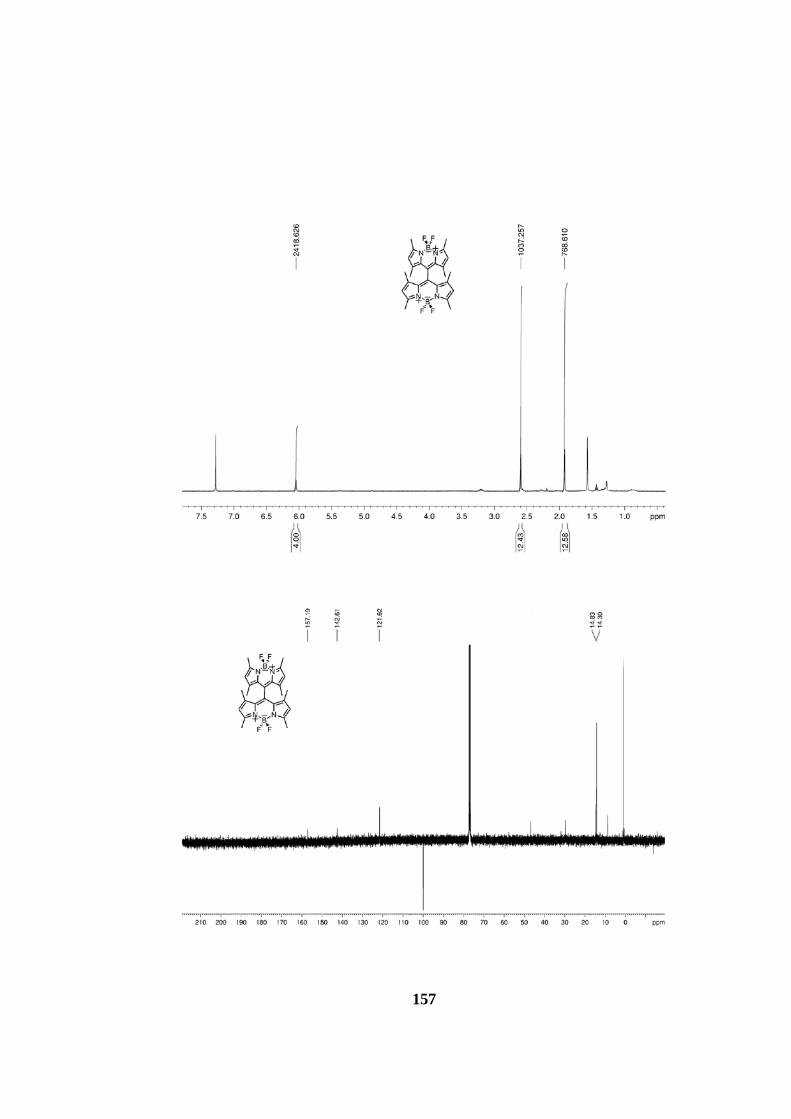
157

158

159

160
7.1.2 Mass Spectra
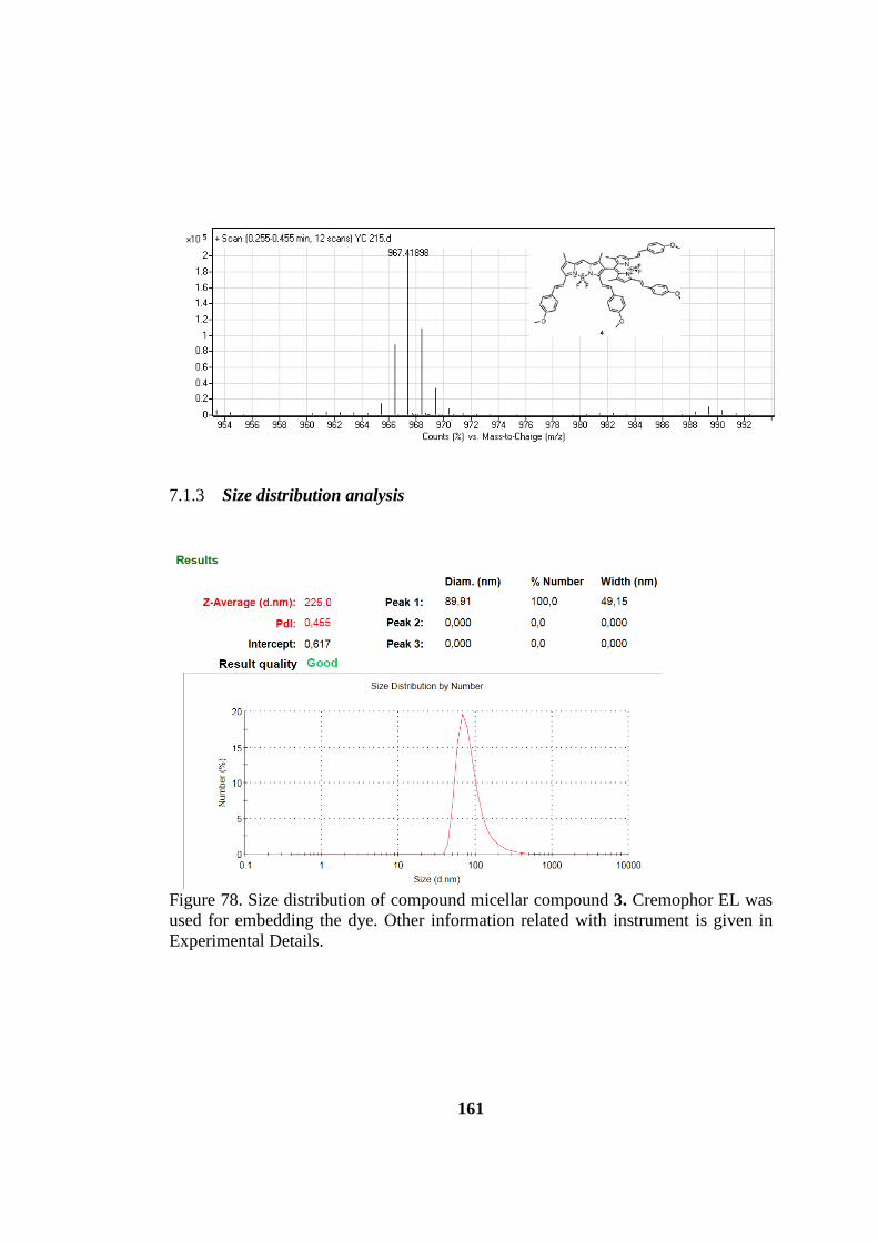
161
7.1.3 Size distribution analysis
Figure 78. Size distribution of compound micellar compound 3. Cremophor EL was used for embedding the dye. Other information related with instrument is given in Experimental Details.
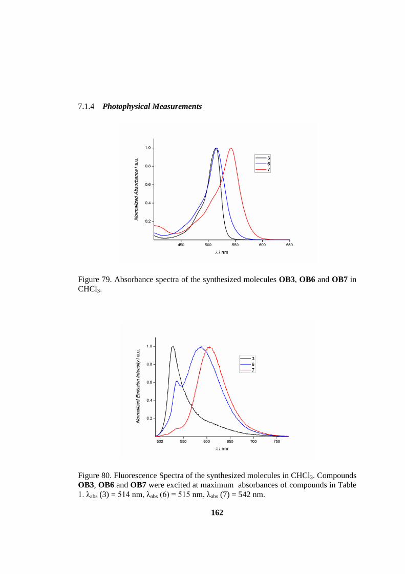
162
7.1.4 Photophysical Measurements
Figure 79. Absorbance spectra of the synthesized molecules OB3, OB6 and OB7 in CHCl3.
Figure 80. Fluorescence Spectra of the synthesized molecules in CHCl3. Compounds OB3, OB6 and OB7 were excited at maximum absorbances of compounds in Table 1. λabs (3) = 514 nm, λabs (6) = 515 nm, λabs (7) = 542 nm.
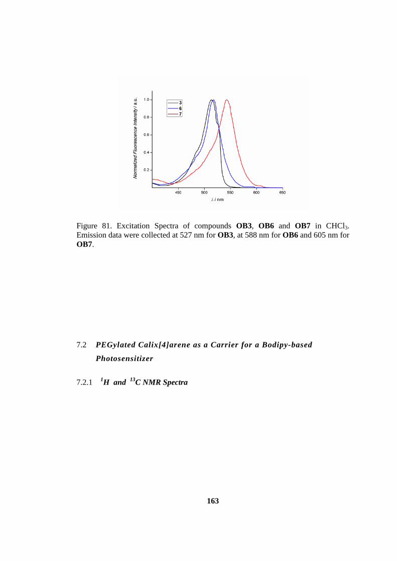
163
Figure 81. Excitation Spectra of compounds OB3, OB6 and OB7 in CHCl3. Emission data were collected at 527 nm for OB3, at 588 nm for OB6 and 605 nm for OB7.
7.2 PEGylated Calix[4]arene as a Carrier for a Bodipy-based
Photosensitizer
7.2.1 1H and 13C NMR Spectra

164

165

166

167
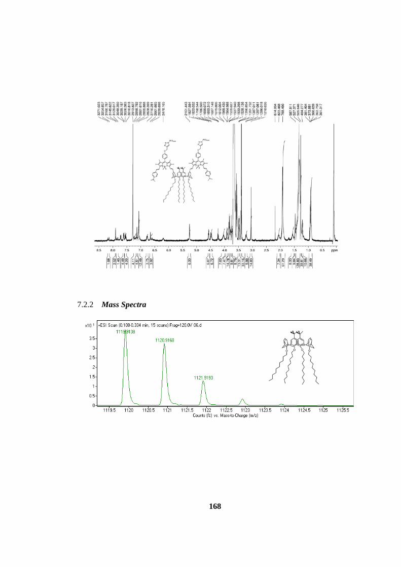
168
7.2.2 Mass Spectra
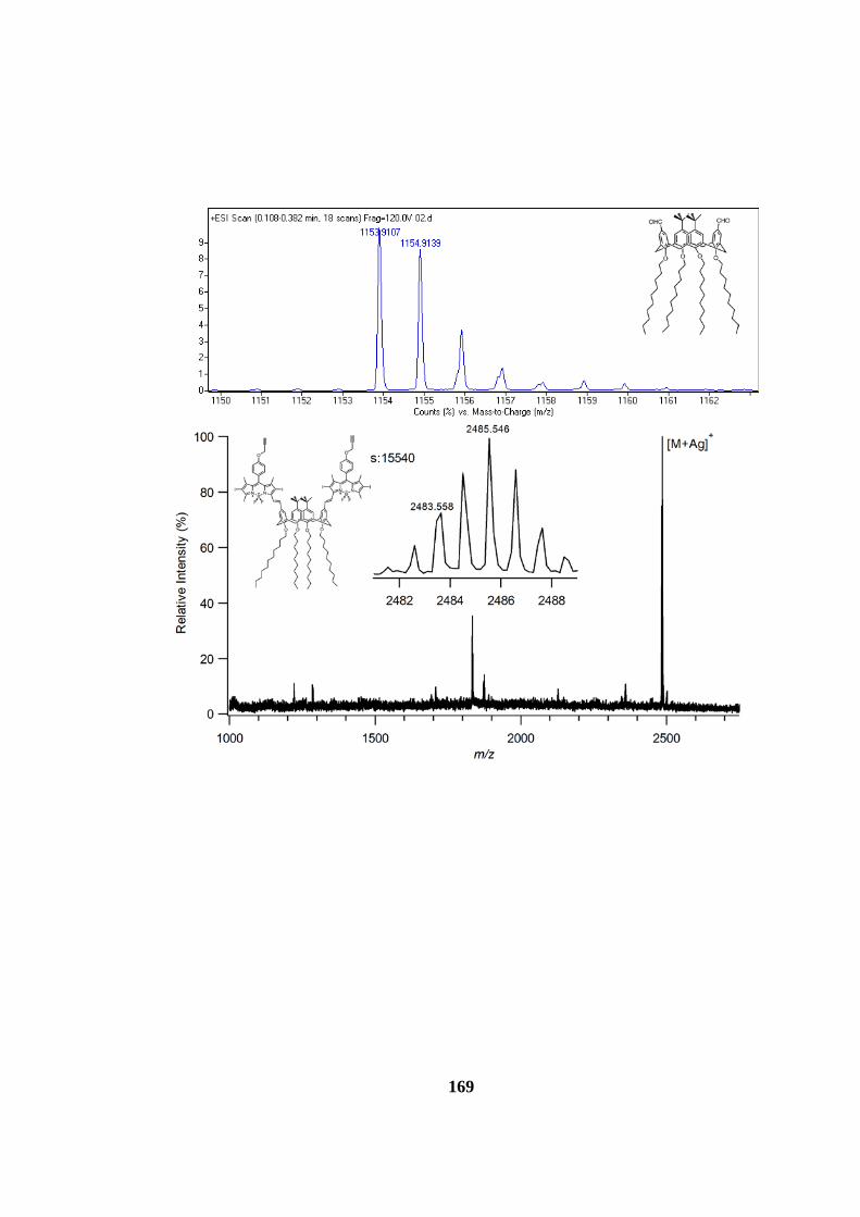
169

170

171
7.3 AXIAL CHIRAL BODIPYS
7.3.1 1H and 13C NMR Spectra
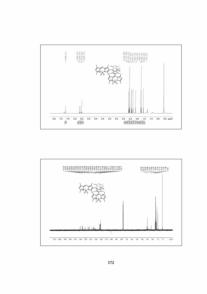
172

173

174

175
7.3.2 Mass Spectra

176

177
7.3.3 Absorbance and Emission Spectra
Absorbance spectrum of AB5 (blue) and AB7 (red)
Fluorescence spectrum of AB5 (blue) and AB7 (red)

178
7.4 THIOPHENE BASED PS’S FOR DSSC
7.4.1 1H and 13C NMR Spectra

179

180
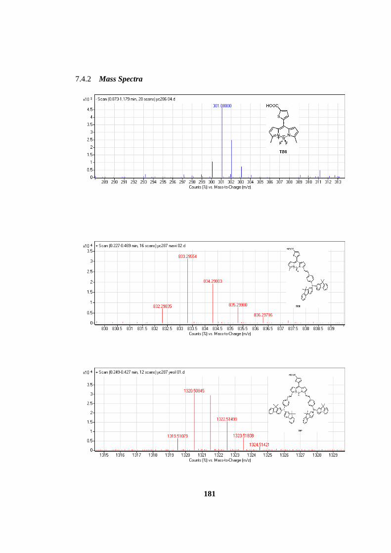
181
7.4.2 Mass Spectra