Embed Size (px)
Citation preview
Korean J Radiol 4(1), March 2003 1
Detection of Small Hypervascular HepatocellularCarcinomas in Cirrhotic Patients: Comparison ofSuperparamagnetic Iron Oxide-Enhanced MRImaging with Dual-Phase Spiral CT
Objective: To compare the performance of superparamagnetic iron oxide(SPIO)-enhanced magnetic resonance (MR) imaging at 1.5T and dual-phase spi-ral computed tomography (CT) for the depiction of small hypervascular hepato-cellular carcinomas (HCCs).
Materials and Methods: Forty-three patients with 70 small nodular HCCs (520 mm; mean, 13.7 mm) were examined. Diagnosis was based on the results ofsurgical biopsy in 22 patients and by the combined assessment of MR imaging,lipiodol CT, alpha feto-protein levels, and angiographic findings in 21. MR imag-ing consisted of respiratory-triggered turbo spin-echo T2-weighted imaging, T1-weighted fast low-angle shot, and T2 -weighted fast imaging with steady-stateprecession imaging before and after SPIO enhancement. CT imaging was per-formed with 5-mm collimation and 1:1.4 pitch, and began 30 and 65 secs after theinjection of 150 mL of contrast medium at a rate of 3 mL/sec. Two blindedobservers reviewed all images independently on a segment-by-segment basis.Diagnostic accuracy was evaluated using receiver operating characteristics(ROC) analysis.
Results: The mean areas (Az) under the ROC curves were 0.85 for SPIO-enhanced MR imaging and 0.79 for dual-phase spiral CT (p < .05). The meansensitivity of SPIO-enhanced MR imaging was significantly higher than that of CT(p < .05), i.e. 70.6% for MR imaging and 58.1% for CT. MR imaging had higherfalse-positive rates than dual-phase spiral CT, but the difference was not statisti-cally significant (3.7% vs 3.3%) (p > .05).
Conclusion: SPIO-enhanced MR imaging is more sensitive than dual-phasespiral CT for the depiction of small hypervascular hepatocellular carcinomas.
epatocellular carcinoma (HCC) is one of the most common globally oc-curring malignant neoplasms. Although hepatic resection is an effectivetreatment for HCC patients, the resectability, operative mortality, and
prognosis for small HCCs differ from those for larger tumors such as those usuallyfound in patients presenting with symptoms (1 5). For large tumors, the operativemortality rate ranges from 6.7% to 36% (3 4), but for small tumors, this rate is 3-12.5% (5). In addition, the five-year survival rate is much better for smaller than forlarger tumors. The need for a preoperative imaging modality that can detect smallHCCs prior to the appearance of clinical symptoms is thus of great importance in clini-cal practice.
Contrast-enhanced spiral CT is the most widely used imaging technique for the de-tection and characterization of focal hepatic lesions. Various studies have shown thatthe use of dual-phase spiral CT imaging during both the arterial and portal venous
Jeong-Min Lee, MD1,2
In-Hwan Kim, MD3
Hyo-Sung Kwak, MD3
Ji-Hyun Youk, MD3
Young-Min Han, MD3
Chong-Soo Kim, MD3
Index terms:Liver, CT Liver, MR imagingLiver, neoplasmsMagnetic resonance (MR),
contrast agents
Korean J Radiol 2003;4:1-8Received June 26, 2002; accepted after revision November 23, 2002.
1Department of Diagnostic Radiology,Seoul National University Hospital, Seoul,Korea; 2Department of Radiation Medicine,Seoul National University MedicalResearch Center, Seoul, Korea; 3Depart-ment of Diagnostic Radiology, ChonbukNational University Hospital, Chonju,Korea
Address reprint requests to:Jeong-Min Lee, MD, Department ofDiagnostic Radiology, Seoul NationalUniversity Hospital, 28 Yongon-dong,Chongno-gu, Seoul 110-744, Korea.Telephone: (822) 760-3154Fax: (822) 743-6385e-mail: [email protected]
H
phases of enhancement leads to further improvement inthe detection rates of hypervascular tumors such as HCC(6 8). However, the detection of hepatocellular carcinomaand hepatocytic nodules such as dysplastic nodules is diffi-cult because cirrhotic liver parenchyma contains fibrosis,regenerative nodules, fatty infiltration, and parenchymalnecrosis (9). Currently, MR imaging is increasingly utilizedfor the detection of hepatic lesions, and the use of super-paramagnetic iron oxide (SPIO), a tissue-specific contrastagent, has the potential to increase the sensitivity andspecificity of hepatic MR imaging (10 12). To our knowl-edge, no study has focused on the comparison of dual-phase contrast-enhanced spiral CT and SPIO-enhanced MRimaging for the detection of small HCCs (less than 2 cm).In the present study, we compare the performance of MRimaging after the administration of SPIO with that of dual-phase contrast-enhanced CT for the detection of small-di-ameter hypervascular HCCs using receiver operating char-acteristics (ROC) analysis.
SUBJECTS AND METHODS
Patients Between October 1998 and January 2002, 120 patients
with suspected focal liver tumors underwent dual-phasespiral CT, unenhanced MR imaging, and SPIO-enhancedMR imaging. We excluded 77 of these, as follows: 49 withHCCs larger than 2 cm in diameter; seven in whom hyper-vascularity of the tumor was not apparent at selective he-patic angiography; seven who underwent radiofrequencythermal ablation; five who underwent surgery more than 4weeks after CT or MRI; and nine who, after transcatheterarterial chemoembolization (TACE), were not available forfollow-up studies of at least 6 months due to a lack of firmevidence of a true-negative segment. The remaining 43 pa-tients, for whom there was acceptable proof of the pres-ence of small hypervascular hepatocellular carcinoma (lessthan 2 cm in diameter), and who had liver cirrhosis, wereincluded in this study.
In all these patients, liver cirrhosis was determined byclinical examination and blood chemistry tests (aspartateaminotransferase, alanine aminotransferase, alkaline phos-phatase, bilirubin, albumin, and globulin). The interval be-tween SPIO-enhanced MR imaging and dual-phase spiralCT was not greater than one month. Direct comparison be-tween MR imaging and CT was made in each imaging sec-tion of the liver.
Lesion ConfirmationA total of 43 patients with 70 small HCCs (5 20 mm,
mean 13.7 mm) were enrolled in the study. Twelve pa-
tients had two HCCs, one patient had three, three patientshad four, and one patient had five, and in each of the re-maining 26 patients, one nodule was present. Proof of hy-pervascular HCC was confirmed using a combination ofpathologic and angiographic findings (n=25) or a combina-tion of angiographic and lipiodol CT findings (n=45).Seventeen patients with 20 lesions underwent hepatic re-section and intraoperative ultrasonography. In five pa-tients, only one attempt was made to perform percuta-neous biopsy of a single lesion per patient, but in three, ad-ditional lesions were detected by both lipiodol and angio-graphic CT. For the other 21 patients with 39 lesions whounderwent TACE, the presence of individual malignant le-sions was confirmed by means of elevated serum FP lev-els, angiographic findings, and lipiodol CT uptake.
Subsegmental TACE was performed by injecting achemotherapeutic drug emulsion, followed by gelatinsponge particles (Gelfoam; Pharmacia-Upjohn, Mich,U.S.A.), as previously described (13). The emulsion con-sisted of lipiodol (Guerbet, Aulnay-sous-Bois, France) ,doxorubicin hydrochloride (Adriamycin; Ildong PharmCo., Seoul, Korea) and non-ionic contrast material(Ultravist 300; Schering, Berlin, Germany ). After perform-ing subsegmental TACE, 2 3 mL of lipiodol was injectedinto the proper hepatic artery in order to detect hiddenHCC at subsequent lipiodol CT, performed using aSomatom Plus-4 scanner (Siemens Medical Systems,Erlangen, Germany) with 5-mm slice thickness two weeksafter TACE. The diagnostic criterion for HCC at lipiodolCT was a round, dense deposit of iodized oil after TACE inwhich this oil was used (14).
Follow-up CT examinations were performed 6 36months later in all patients who had undergone resectivesurgery or TACE. Proof of the absence of HCC nodules inhepatic segments was provided by negative findings at in-traoperative ultrasonography, or a combination of nonodular lipiodol uptake at lipiodol CT and no evidence offurther nodular growth at follow-up CT.
MR Imaging ExaminationsAll MR imaging was performed on a 1.5-T system
(Magnetom Vision; Siemens, Erlangen, Germany) using aphased-array coil for signal reception. Baseline MR imageswere acquired with a respiratory-triggered T2-weightedturbo spin-echo (TSE) sequence, a breath-hold T2*-weight-ed fast imaging with steady-state precession (FISP) se-quence, and a breath-hold T1-weighted fast low-angle shot(FLASH) sequence.
Respiratory-triggered T2-weighted TSE imaging (TRrange/TE, 3300 5500/85) was performed with an echo-train length of 5, a 120 256 matrix, and two signal aver-
Lee et al.
2 Korean J Radiol 4(1), March 2003
ages. Breath-hold T2*-weighted FISP imaging (180/12, 30flip angle, matrix of 96 256, one signal average) was fol-lowed by breath-hold T1-weighted FLASH imaging (120/4,80 flip angle, matrix of 140 256, one signal average).All images were obtained in the transaxial plane, using aphased-array multicoil. For all sequences, a 7-mm slicethickness was used, with a 10% intersection gap and a fieldof view of 35 40 cm, depending on the size of the liver.
SPIO-enhanced MR imaging comprised the respiratory-triggered T2-TSE sequence, the breath-hold T2*-weightedFISP sequence, and the breath-hold T1-weighted FLASHsequence, with the same parameters as those used in base-line MR imaging. The SPIO agent (Feridex; AdvancedMagnetics, Cambridge, Mass., U.S.A.), was administered ata dose of 15 mol of iron per kilogram of body weight,was diluted in 100 mL of 5% dextrose solution and inject-ed intravenously through a specific 5 filter for 30 mins;imaging commenced approximately 70 (range, 50 90)mins after the intravenous infusion of SPIO.
CT ExaminationsFor dual phase contrast-enhanced spiral CT, a Somatom
plus 4 scanner (Siemens Medical Systems, Erlangen,Germany) was used. The scanning parameters were 120kVp, 240 mA, 5-mm collimation, table speed of 7-mm/sec,and a 5-mm reconstruction interval. After unenhanced spi-ral liver scanning, the arterial phase was begun 30 secs af-ter the power injection of 150 mL of nonionic contrast ma-terial (Iopromide [Ultravist 370]; Schering, Berlin,Germany) at a rate of 3 mL/sec. Portal phase scanning wasbegun 65 secs after the start of contrast injection.
Imaging Analysis (Receiver Operating CharacteristicsAnalysis, ROC)
Before performing ROC analysis, a gold standard for thelesions to be investigated was defined by two experiencedabdominal radiologists, who coordinated ROC analysis andreached their decisions by consensus. They analyzed all theimages obtained as well as the operative, laboratory, andhistological findings, evaluating a total of 70 separate focalHCC lesions smaller than 2 cm in diameter.
Two other independent observers, both experienced ab-dominal radiologists, reviewed the contrast-enhanced spi-ral CT and SPIO-enhanced MR images. They were in-formed only that the patients with liver cirrhosis were re-ferred for preoperative assessment of suspected liver ma-lignancy. Images were reviewed on a segment-by-segmentbasis, and to avoid incorrect localization of the lesions, he-patic segmentation according to the Couinaud numberingsystem was drawn directly. A total of 344 segments (61 ofwhich contained 70 HCC nodules, and 283 segments with-
out proof of the presence of HCC lesions) were reviewed.Five of the 61 segments contained more than two nodules,and to ensure correct localization, each observer recordedthe image number and size of each lesion, and added fur-ther comments.
Each observer read two sets of images (set 1: unen-hanced and SPIO-enhanced turbo spin-echo, T1-weightedFLASH, and T2*-weighted FISP images; set 2: pre- andpost-contrast dual-phase spiral CT images), with an inter-val of at least a week between the two readings. Eachrecorded the presence and location of one or more lesions,assigning to each a confidence level on a five-point scale:‘1’ was defined as ‘definitely or almost definitely absent’;‘2’ as ‘probably absent’; ‘3’ as ‘possibly present’; ‘4’ as‘probably present’; and ‘5’ as ‘definitely or almost definite-ly present’.
For each imaging set, a binomial ROC curve was fitted toeach reviewer’s confidence rating data using a maximumlikelihood estimation (ROCKIT 0.9, Charles E. Metz). Thediagnostic accuracy of each imaging modality was deter-mined by calculating the area (A index, Az) under each ob-server-specific binomial ROC curve plotted in the designedsquare. The composite ROC curves used to represent thecombined performance of the two observers were calculat-ed for each set of images using Rockit 0.9 software to ratetheir pooled data. The differences between imaging modal-ities in terms of the mean areas under the ROC curveswere statistically analyzed using the two-tailed Studentt test for paired data. To determine relative sensitivity forHCC, the number of segments assigned level 3 or morefrom among the 70 HCC nodules was noted. The relativesensitivities and specificities for HCC obtained from thetwo radiologists’ and pooled data were calculated for eachmodality, and to ascertain the differences between theimaging modalities and between observers, multiple com-parisons involving Student’s t test were made. A two-tailedp value of less than 0.05 was considered significant.
Agreement between blinded observers is reported belowin terms of kappa values, those greater than 0 indicatingpositive correlation. Values of up to 0.4 indicated positivebut poor correlation; those of 0.41 0.75 indicated goodcorrelation.
RESULTS
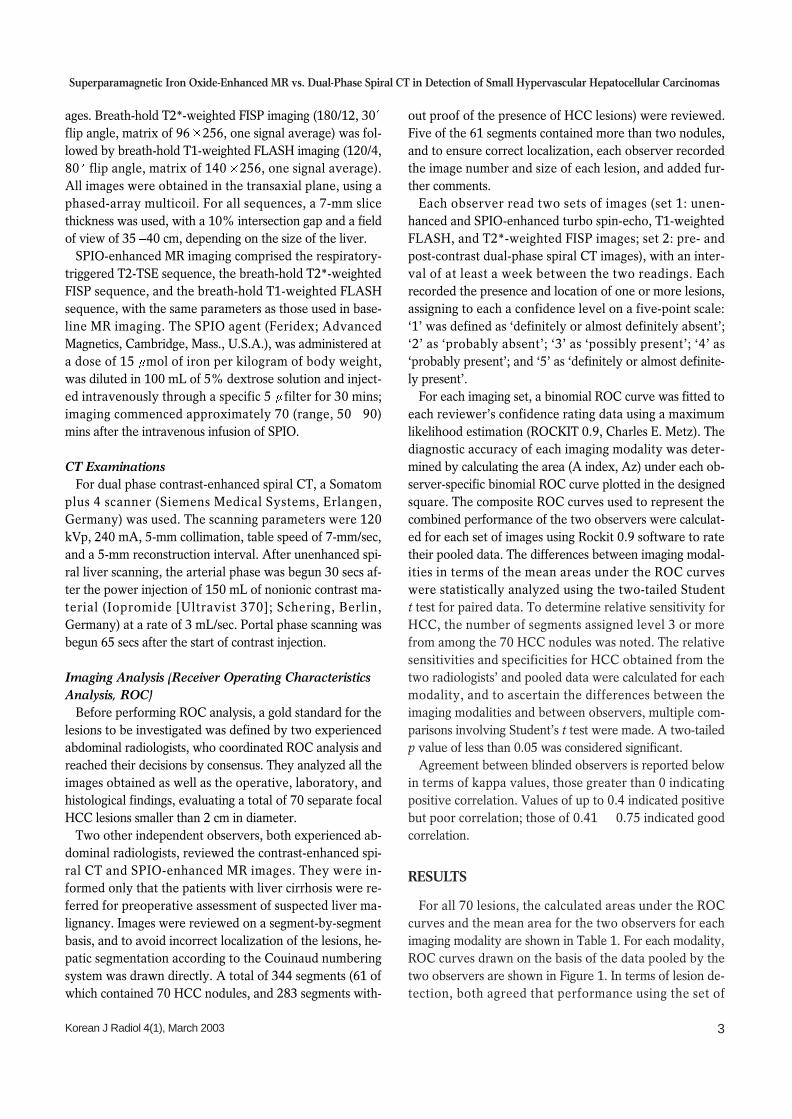
For all 70 lesions, the calculated areas under the ROCcurves and the mean area for the two observers for eachimaging modality are shown in Table 1. For each modality,ROC curves drawn on the basis of the data pooled by thetwo observers are shown in Figure 1. In terms of lesion de-tection, both agreed that performance using the set of
Superparamagnetic Iron Oxide-Enhanced MR vs. Dual-Phase Spiral CT in Detection of Small Hypervascular Hepatocellular Carcinomas
Korean J Radiol 4(1), March 2003 3
SPIO-enhanced MR images was significantly superior tothat obtained at dual-phase spiral CT imaging (p < .05).The mean area under both observers’ ROC curves was0.85 for pre- and post-SPIO-enhanced MR imaging and0.79 for pre- and post-contrast dual-phase spiral CT imag-ing; this difference was statistically significant (p < .05).
Sensitivities and specificities were calculated for each ob-server and for each modality, and mean values were alsodetermined (Table 2). The combination of SPIO-enhanced
MR images was 12.5% more sensitive than that of dual-phase spiral CT images (MR, 70.6%; CT, 58.1%), a statisti-cally significant difference (p < .05). In 18 cases, tumornodules were detected at MR imaging, but not at CT. In 12cases, nodules were smaller than 1cm (Fig. 2). In two cases,additional lesions were detected only at CT; in one ofthese, because the high signal intensity of the gallbladder(GB) interfered with the signal intensity of the true nodule,found at T2-weighted imaging to also be high, the ob-servers missed a 5-mm subcapsular nodule abutting theGB. In the other case, a small nodule was not detected be-cause of the presence of severely injured liver parenchymaof heterogeneous texture. In addition, 11/70 lesions(15.7%) in ten patients were not detected by any modality.
Lee et al.
4 Korean J Radiol 4(1), March 2003
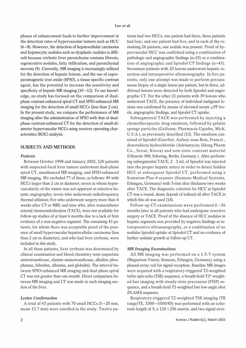
Fig. 2. In this 63-year-old man, a 0.8-cm hepatocellular carcinoma was found in segment I.A. Neither the arterial-phase CT image (left) nor the portal venous-phase CT image (right) revealed the presence of a lesion. B. SPIO-enhanced respiratory-triggered T2-weighted turbo spin-echo image (left) and breath-hold T2*-weighted fast image obtainedwith steady state precession (right) depict the tumor as an area of high signal intensity (arrows). Its presence was surgically confirmed.
A B
Fig. 1. ROC curves describe observer confidence in the detectionof hepatocellular carcinomas depicted at pre- and post-SPIO-en-hanced MR imaging involving T1-weighted FLASH, T2-weightedTSE, and T2 -weighted FISP sequences (diamonds), and anoth-er set obtained at contrast-enhanced dual-phase spiral CT(squares). Note that observers showed more confidence in inter-preting MR images than dual-phase spiral CT images (p < .05).
False-Positive Fraction
ROC CurveT
rue-
Pos
itive
Fra
ctio
n
Table 1. Individual and Mean Areas Under the Curve for Pre-and Post-SPIO-Enhanced MR and Dual-Phase SpiralCT (All Lesions)
Imaging ModalityAz Index
Observer 1 Observer 2 Mean
CT imaging 0.80 0.03 0.78 0.04 0.79 0.02MR imaging 0.86 0.03 0.85 0.03 0.85 0.02
Note. Values are expressed as mean SD.For the comparison of MR imaging with CT, p < .05
Table 2. Sensitivity and Specificity of Pre- and Post-SPIO-Enhanced MR and Dual-Phase Spiral CT (AllLesions)
Imaging ModalityAz Index
Observer 1 Observer 2 Mean
CTSensitivity 58.8 57.4 58.1Specificity 96.5 96.9 96.7
MRSensitivity 72.1 69.1 70.6Specificity 96.2 95.5 95.8
Note. Numbers are percentages.For the comparison of MR imaging with CT, p < .05
Of these false-negative lesions, three were confirmed bypercutaneous biopsy, two by surgical resection and biopsy,and the remaining six by lipiodol CT. Except for one witha diameter of 2 cm, these missed lesions ranged in sizefrom 0.5 to 1.0 cm. The 2.0-cm nodule was located in thesubcapsular dome of segment VIII and was therefore misin-terpreted at both CT and MR imaging as a partial volumeartifact of the cardiac structure.
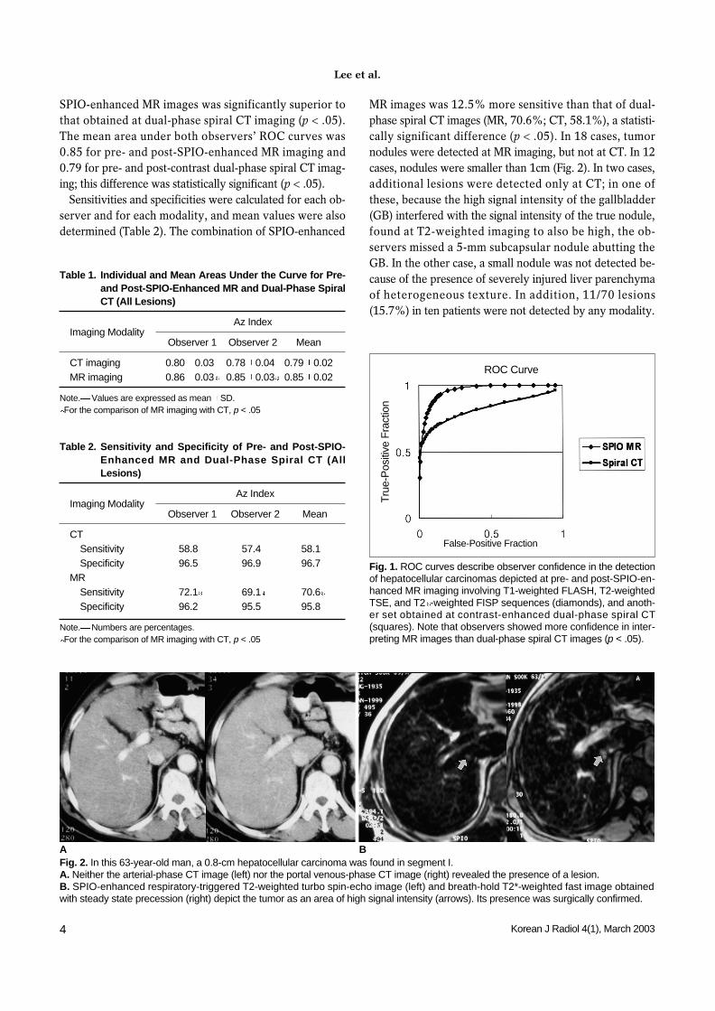
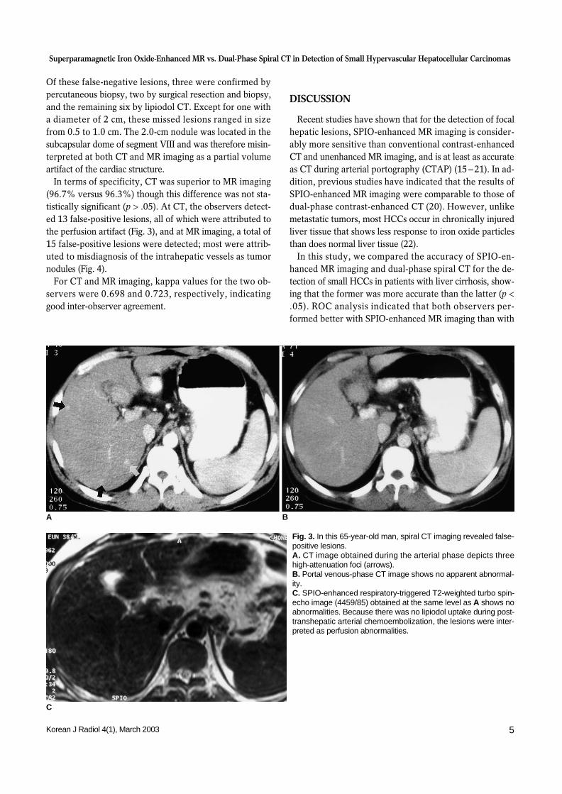
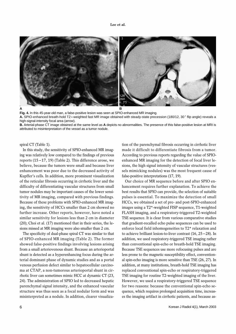
In terms of specificity, CT was superior to MR imaging(96.7% versus 96.3%) though this difference was not sta-tistically significant (p > .05). At CT, the observers detect-ed 13 false-positive lesions, all of which were attributed tothe perfusion artifact (Fig. 3), and at MR imaging, a total of15 false-positive lesions were detected; most were attrib-uted to misdiagnosis of the intrahepatic vessels as tumornodules (Fig. 4).
For CT and MR imaging, kappa values for the two ob-servers were 0.698 and 0.723, respectively, indicatinggood inter-observer agreement.
DISCUSSION
Recent studies have shown that for the detection of focalhepatic lesions, SPIO-enhanced MR imaging is consider-ably more sensitive than conventional contrast-enhancedCT and unenhanced MR imaging, and is at least as accurateas CT during arterial portography (CTAP) (15 21). In ad-dition, previous studies have indicated that the results ofSPIO-enhanced MR imaging were comparable to those ofdual-phase contrast-enhanced CT (20). However, unlikemetastatic tumors, most HCCs occur in chronically injuredliver tissue that shows less response to iron oxide particlesthan does normal liver tissue (22).
In this study, we compared the accuracy of SPIO-en-hanced MR imaging and dual-phase spiral CT for the de-tection of small HCCs in patients with liver cirrhosis, show-ing that the former was more accurate than the latter (p <.05). ROC analysis indicated that both observers per-formed better with SPIO-enhanced MR imaging than with
Superparamagnetic Iron Oxide-Enhanced MR vs. Dual-Phase Spiral CT in Detection of Small Hypervascular Hepatocellular Carcinomas
Korean J Radiol 4(1), March 2003 5
A B
Fig. 3. In this 65-year-old man, spiral CT imaging revealed false-positive lesions.A. CT image obtained during the arterial phase depicts threehigh-attenuation foci (arrows). B. Portal venous-phase CT image shows no apparent abnormal-ity.C. SPIO-enhanced respiratory-triggered T2-weighted turbo spin-echo image (4459/85) obtained at the same level as A shows noabnormalities. Because there was no lipiodol uptake during post-transhepatic arterial chemoembolization, the lesions were inter-preted as perfusion abnormalities.
C
spiral CT (Table 1). In this study, the sensitivity of SPIO-enhanced MR imag-
ing was relatively low compared to the findings of previousreports (15 17, 19) (Table 2). This difference arose, webelieve, because the tumors were small and because liverenhancement was poor due to the decreased activity ofKupffer’s cells. In addition, more prominent visualizationof the reticular fibrosis occurring in cirrhotic liver and thedifficulty of differentiating vascular structures from smalltumor nodules may be important causes of the lower sensi-tivity of MR imaging, compared with previous findings.Because of these problems with SPIO-enhanced MR imag-ing, the sensitivity of HCCs smaller than 2 cm showed nofurther increase. Other reports, however, have noted asimilar sensitivity for lesions less than 2 cm in diameter(20); Choi et al. (17) mentioned that in their series, the le-sions missed at MR imaging were also smaller than 2 cm.
The specificity of dual-phase spiral CT was similar to thatof SPIO-enhanced MR imaging (Table 2). The formershowed false-positive findings involving lesions arisingfrom a small arteriovenous shunt. Because an arterioportalshunt is detected as a hyperenhancing focus during the ar-terial dominant phase of dynamic studies and as a portalvenous perfusion defect similar to hepatocellular carcino-ma at CTAP, a non-tumorous arterioportal shunt in cir-rhotic liver can sometimes mimic HCC at dynamic CT (23,24). The administration of SPIO led to decreased hepaticparenchymal signal intensity, and the enhanced vascularstructure was thus seen as a focal nodular form and wasmisinterpreted as a nodule. In addition, clearer visualiza-
tion of the parenchymal fibrosis occurring in cirrhotic livermade it difficult to differentiate fibrosis from a tumor.According to previous reports regarding the value of SPIO-enhanced MR imaging for the detection of focal liver le-sions, the high signal intensity of vascular structures (ves-sels mimicking nodules) was the most frequent cause offalse-positive interpretations (17, 19).
Our choice of MR sequence before and after SPIO en-hancement requires further explanation. To achieve thebest results that SPIO can provide, the selection of suitablepulses is essential. To maximize the detection of smallHCCs, we obtained a set of pre- and post-SPIO-enhancedimages using a T2*-weighted FISP sequence, T1-weightedFLASH imaging, and a respiratory-triggered T2-weightedTSE sequence. It is clear from various comparative studiesthat gradient-recalled echo pulse sequences can be used toenforce local field inhomogeneities to T2* relaxation andto achieve brilliant lesion-to-liver contrast (16, 25 28). Inaddition, we used respiratory-triggered TSE imaging ratherthan conventional spin-echo or breath-hold TSE imaging.Because TSE sequences use more refocusing pulses and areless prone to the magnetic susceptibility effect, convention-al spin-echo imaging is more sensitive than TSE (26, 27). Inaddition, at many institutions, breath-hold TSE imaging hasreplaced conventional spin-echo or respiratory-triggeredTSE imaging for routine T2-weighted imaging of the liver.However, we used a respiratory-triggered TSE sequencefor two reasons: because the conventional spin-echo se-quence, which requires prolonged acquisition time, increas-es the imaging artifact in cirrhotic patients, and because as-
Lee et al.
6 Korean J Radiol 4(1), March 2003
Fig. 4. In this 45-year-old man, a false-positive lesion was seen at SPIO-enhanced MR imaging.A. SPIO-enhanced breath-hold T2 -weighted fast MR image obtained with steady-state precession (180/12, 30 flip angle) reveals ahigh-signal-intensity focal area (arrow). B. Arterial-phase CT image obtained at the same level as A depicts no abnormalities. The presence of this false-positive lesion at MRI isattributed to misinterpretation of the vessel as a tumor nodule.
A B
cites formation related to poor liver function in patientswith liver cirrhosis makes long breath-holding very diffi-cult. Because a benign cyst shows low signal intensity atSPIO-enhanced MR imaging, despite the high signal inten-sity of other malignant lesions, and an hemangioma showsincreased signal intensity at SPIO-enhanced T1-weightedimaging (29, 30), we used the T1-weighted gradient-echosequence to differentiate HCC from hemangioma and cyst.
This study suffers certain limitations. First, there was in-sufficient histopathologic confirmation of the presence ofliver lesions. Although most lesions not subject to biopsyprobably represent additional sites of HCC or true-positivelesions, some false-positive lesions might have been includ-ed in our study. Second, because only 17 patients under-went surgery and intraoperative ultrasonography, it wasnot absolutely certain that in segments apparently withoutHCC nodules, these were in fact absent (true negative).However, we used relatively strict criteria for the standardof reference regarding true-negative segments, i.e. negativeCT findings after the arterial infusion of iodized oil werecombined with follow-up CT at least six months later.Third, we performed arterial and portal phase imaging af-ter contrast injection. Because some small HCCs may showiso-attenuation to surrounding liver parenchyma at portalphase imaging, the inclusion of equilibrium phase imagingmay increase the likelihood of observing the contrastwashout effect of HCCs and therefore improve the detec-tion rate for small HCCs (31, 32). Last, the spiral CT tech-nology used in this study was not state of the art.Multidetector spiral CT now permits the use of thinnerslice thicknesses and better time resolution than is possiblewith single-detector mode (33), and we believe that its usemay improve the accuracy of CT examinations for theevaluation of focal liver tumors.
In conclusion, MR imaging performed before and afterthe administration of SPIO contrast material and involvingcombined T2- and T2*- weighted sequences and an addi-tional T1-weighted sequence is more sensitive and accuratethan dual-phase spiral CT imaging for the detection ofsmall HCCs. To detect these, a set of SPIO-enhanced MRimaging sequences can thus be used in place of dual-phasespiral CT imaging as a preoperative diagnostic strategy inpatients with cirrhosis or other chronic disease of the liver.
AcknowledgementsThe authors wish to thank Bonnie Hami, M.A., Depart-
ment of Radiology, University Hospitals of Cleveland, forher assistance in preparing and editing this manuscript.
References1. Adson MA, Weiland LH. Resection of primary solid hepatic tu-
mors. Am J Surg 1981;141:18-212. Lim RC, Bongard FS. Hepatocellular carcinoma. Arch Surg
1984;119:637-6423. Fortner JG, Kim DK, Maclean BJ, et al. Major hepatic resection
for neoplasia: personal experience in 108 patients. Ann Surg1978;188:363-369
4. Iwatsuki S, Shaw BW, Starzl TE. Experience with 150 liver re-sections. Ann Surg 1983;197:247-253
5. Kanematsu T, Takenaka L, Matsumata T, Furnita T, SugimachiK, Inokuchi K. Limited hepatic resection effective for selectedcirrhotic patients with primary liver cancer. Ann Surg 1984;199:51-56
6. Lee KH, Choi BI, Han JK, Jang HJ, Kim TK, Han MC. Nodularhepatocellular carcinoma: variation of tumor conspicuity on sin-gle-level dynamic scan and optimization of fixed delay times fortwo-phase helical CT. J Comput Assist Tomogr 2000;24:212-218
7. Kim T, Murakami T, Takahash S, et al. Optimal phases of dy-namic CT for detecting hepatocellular carcinoma: evaluation ofunenhanced and triple-phase images. Abdom Imaging 1999;24:473-480
8. Yamashita Y, Mitsuzaki K, Yi T, et al. Small hepatocellular carci-noma in patients with chronic liver damage: prospective com-parison of detection with dynamic MR imaging and helical CT ofwhole liver. Radiology 1996;200:79-84
9. Miller WJ, Baron RL, Dodd GD III, Federle MP. Malignancies inpatients with cirrhosis: CT sensitivity and specificity in 200 con-secutive transplant patients. Radiology 1994;193:645-650
10. Winter TC III, Freeny PC, Nghiem HV, et al. MR imaging withIV superparamagnetic iron oxide: efficacy in the detection of fo-cal hepatic lesions. AJR Am J Roentgenol 1993;161:1191-1198
11. Ros PR, Freeny PC, Harms SE, et al. Hepatic MR imaging withferumoxides: a multicenter clinical trial of its safety and efficacyin the detection of focal hepatic lesions. Radiology 1995;196:481-488
12. Soyer P. Will ferumoxides-enhanced MR imaging replace CTduring arterial portography in the detection of hepatic metas-tases? Prologue to a promising future. Radiology 1996;200:610-611
13. Matsui O, Kadoya M, Yoshikawa J, et al. Small hepatocellularcarcinoma: treatment with subsegmental transcatheter arterialembolization. Radiology 1993;188:79-83
14. Kubota K, Hisa N, Nashikawa T, et al. Evaluation of hepatocel-lular carcinoma after treatment with transcatheter arterialchemoembolization: comparison of Lipiodol CT, power Dopplersonography, and dynamic MRI. Abdom Imaging 2001;26:184-190
15. Yamamoto H, Yamashita Y, Yoshimatsu S, et al. Hepatocecul-lular carcinoma in cirrhotic livers: detection with unenhancedand iron oxide-enhanced MR imaging. Radiology 1995;195:106-112
16. Tang Y, Yamashita Y, Arakawa A, et al. Detection of hepatocel-lular carcinoma arising in cirrhotic livers: comparison ofGadolinium-and Ferumoxides-enhanced MR imaging. AJR Am JRoentgenol 1999;172:1547-1554
17. Choi D, Kim SH, and Lim JH, et al. Preoperative detection ofhepatocellular carcinoma: ferumoxides-enhanced MR imagingversus combined helical CT during arterial portography and CThepatic arteriography. AJR Am J Roentgenol 2001;176:475-482
18. Hagspiel KD, Neidl KFW, Eichenberger AC, Weder W,Marincek B. Detection of liver metastases: comparison of super-
Superparamagnetic Iron Oxide-Enhanced MR vs. Dual-Phase Spiral CT in Detection of Small Hypervascular Hepatocellular Carcinomas
Korean J Radiol 4(1), March 2003 7
paramagnetic iron oxide-enhanced MR imaging at 1.5-T withdynamic CT, intraoperative US and percutaneous US.Radiology 1995; 196:471-478
19. Ward J, Naik KS, Guthrie JA, Wilson D, Robison PJ. Hepatic le-sion detection: comparison of MR imaging after the administra-tion of superparamagnetic iron oxide with dual-phase CT by us-ing alternative free-response receiver operating characteristicanalysis. Radiology 1999;210:459-466
20. Bluemke DA, Paulson EK, Choti MA, DeSena S, Clavien PA.Detection of hepatic lesions in candidates for surgery: compari-son of ferumoxides-enhanced MR imaging and dual-phase heli-cal CT. AJR Am J Roentgenol 2000;175:1653-1658
21. Seneterre E, Taourel P, Bouvier Y, et al. Detection of hepaticmetastases: ferumoxides-enhanced MR imaging versus unen-hanced MR imaging and CT during arterial portography.Radiology 1996;200:785-792
22. Elizondo G, Weissleder T, Stark DD, et al. Hepatic cirrhosis andhepatitis: MR imaging enhanced with superparamagnetic ironoxide. Radiology 1990;174:797-801
23. Kim TK, Choi BI, Han JK, Chung JW, Park JH, Hand MC.Nontumorous arterioportal shunt mimicking hypervascular tu-mor in cirrhotic liver: two-phase spiral CT findings. Radiology1998;208:597-603
24. Choi BI, Lee KH, Han JK, Lee JM. Hepatic arterioportal shunt:dynamic CT and MR features. Korean J Radiol 2002;3:1-15
25. Josephson L, Lewis J, Jacob P, Hahn PF, Stark DW. The effectsof iron oxides on proton relaxivity. Magn Reson Imaging1988;6:647-653
26. Ward J, Chen F, Guthrie JA, et al. Hepatic lesion detection aftersuperparamagnetic iron oxide enhancement: comparison of five
T2-weighted sequences at 1.0T by using alternative free-re-sponse receiver operating characteristic analysis. Radiology2000;214:159-166
27. Schwartz LH, Seltzer SE, Tempany CM, et al. Superparamag-netic iron oxide hepatic MR imaging: efficacy and safety usingconventional and fast spin-echo pulse sequences. J Magn ResonImaging 1995;5:566-570
28. Kim SH, Choi DI, Lim JH, et al. Optimal pulse sequence for fer-umoxides-enhanced MR imaging used in the detection of hepa-tocellular carcinoma: comparative study using seven pulse se-quences. Korean J Radiol 2002;3:87-97
29. Oudkerk M, van den Heuvel AG, Wielopolski PA, Shmits PIM,Borel Rinkes IHM, Wiggers T. Hepatic lesions: detection withferumoxides-enhanced T1-weighted MR imaging. Radiology1997;203:449-456
30. Grangier C, Tourniaire J, Mentha G, et al. Enhancement of liverhemangioma on T1-weighted MR SE images by superparamag-netic iron oxide particles. J Comput Assist Tomogr 1994;18:888-896
31. Jang HJ, Lim JH, Lee SJ, et al. Hepatocellular carcinoma: arecombined CT duing arterial portography and CT hepatic arteri-ography in addition to triple-phase helical CT all necessary forpreoperative evaluation? Radiology 2000;215:373-380
32. Lim JH, Choi DI, Kim SH, et al. Detection of hepatocellular car-cinoma: value of adding delayed-phase imaging to dual-phasehelical CT. AJR Am J Roentgenol 2002;179:67-73
33. Murakami T, Kim T, Takamura M, et al. Hypervascular hepato-cellular carcinoma: detection with double arterial-phase multi-detector row helical CT. Radiology 2001;218:763-767
Lee et al.
8 Korean J Radiol 4(1), March 2003


![MRI for Detection of Hepatocellular Carcinoma: Comparison ...mriquestions.com/uploads/3/4/5/7/34572113/youk_mn...sions, especially hepatocellular carcinoma [1–3]. However, evaluation](https://img.dokumen.tips/doc/110x75/5f3ced438bc609735d4a5d4b/mri-for-detection-of-hepatocellular-carcinoma-comparison-sions-especially.jpg)