Embed Size (px)
Citation preview
Detection of bovine coronavirus and type Arotavirus in neonatal calf diarrhea and winterdysentery of cattle in Quebec: Evaluation of
three diagnostic methodsRaafat Athanassious, Gregoire Marsolais, Robert Assaf, Serge Dea, Jean-Paul DescOteaux,
Suzanne Dulude, Claude Montpetit
AbstractThe use of direct electron microscopy, enzyme-linkedimmunosorbent assay, and protein A-gold immuno-electron microscopy for the identification of bovinecoronavirus and type A rotavirus were examined.Two hundred and forty-nine samples from diar-rheic calves and winter dysenteric cattle from sevengeographic areas in Quebec were examined for thepresence of viruses by direct electron microscopyof negatively stained preparations. In addition, all thesamples were analyzed by enzyme-linkedimmunosorbent assay, and a random selection of47 samples were also analyzed by protein A-goldimmunoelectron microscopy. Thirty-nine percentof samples examined by direct electron microscopycontained viral particles; bovine coronavirus andtype A rotavirus were the most common virusesinvolved. Overall agreement between any two of themethods used compared favorably with resultsobtained by others using similar methods. The pres-ence of coronavirus and rotavirus in fecal samplesobtained from neonatal calves and the presence ofcoronavirus in samples from winter dysenteric adultcattle suggested their etiological roles in the respec-tive diseases. Furthermore, results from protein A-gold immunoelectron microscopy of coronavirus-like particles implied that a different coronavirus orsome other viruses might be involved in these diseases.Finally, the efficiency of direct electron microscopy,enzyme-linked immunosorbent assay and proteinA-gold immunoelectron microscopy as diagnostictools is discussed.
ResumeDetection de la presence de coronavirus etde rotavirus bovins de type A chez des veauxdiarrheiques et des vaches affecteos de dysen-torie d'hiver dans la province de Quebec : eva-luation de trois methodes de diagnosticCette etude a ete entreprise pour comparer l'efficacitede la microscopie electronique, de l'ELISA et del'immunomarquage proteine A-or colloidal, pour
Centre de recherche en Virologie (Athanassious, Dea, Descoteaux,Dulude), and Ministere de l'Agriculture, des Pecheries et del'Alimentation du Quebec (Marsolais, Assaf, Montpetit),Institut Armand-Frappier, 531, boul. Des Prairies, Laval,Quebec H7N 4Z3.
Reprint requests to Dr. G. Marsolais.
This work was supported by "Le Ministere de l'Agriculture, desPecheries et de l'Alimentation du Quebec".
l'identification des coronavirus et des rotavirusbovins de type A. Deux cent quarante-neuf echan-tillons provenant de veaux diarrheiques et de vachesaffectees de dysenterie d'hiver, issus de sept regionsgeographiques du Quebec, furent examines en micro-scopie electronique, suite a des colorations nega-tives, pour detecter la presence de virus. Tous cesechantillons furent etgalement analyses en ELISAet une selection de 47 specimens fut aussi examineepar la technologie de l'immunomarquage proteineA-or colloidal. Trente-neuf pour cent des echantillonsexamines en microscopie electronique contenaient desparticules virales; les coronavirus et les rotavirusfurent les agents les plus frequemment rencontres.Les resultats obtenus 'a l'aide de ces differentes tech-niques sont comparables a ceux obtenus par d'autreschercheurs ayant utilise les memes techniques. Lamise en evidence de coronavirus et de rotavirus dansles feces et le contenu intestinal de veaux diar-rheiques et la presence de coronavirus dans les fecesd'adultes affectes de dysenterie d'hiver suggerent leurimplication dans l'etiologie de ces deux maladies.De plus, les resultats des examens en microscopie e'lec-tronique suite a l'immunomarquage proteine A-orcolloidal pour l'identification des particules coron-aviriformes, suggerent fortement la presence d'inenouvelle souche de coronavirus ou d'autres virusapparentes au coronavirus dans l'etiologie de cesmaladies. Finalement, 1'efficacite de la microscopieelectronique, de l'ELISA et de l'immunomarquage al'or colloidal est discutee.
Can Vet J 1994; 35: 163-169
Introductionnteric disease in calves is caused by several fac-tors, both infectious and noninfectious (1-4). The
known etiological agents include bacteria (entero-toxigenic Escherichia coli),viruses (rotavirus, coron-avirus) and protozoa (cryptosporidia). Difficulties in theclinical diagnosis of infectious enteritis arise from fre-quent nonspecific clinical signs and lesions, the presenceof asymptomatic infections, the involvement of multi-ple agents, and the interactions of intrinsic and extrin-sic factors that predispose the host to infection.
Coronavirus and rotavirus are the most commonviruses involved in neonatal calf diarrhea (3,5-8).Coronavirus has also been identified in feces of winterdysenteric cattle (9-12). Since calf diarrhea is a majorhealth problem of young animals, it has been inten-sively investigated, and several methods have beenused for the detection of enteropathogenic viruses(6,13-23). However, each method has its limitations.
Ca Ve J Voum 35 Mac 199Can Vet J Volume 35, March 1994 163
Direct electron microscopy (DEM) of negatively stainedparticles is an indispensible tool in viral diagnosis (24),but problems arise when the virus has a pleomorphicmorphology of an uncharacteristic shape and/or size, or
the sample is contaminated with virus-like particles,such as cellular membranes, ribosomes, cellular organelles,and bacteriophages. When this occurs, it is essentialto verify DEM observations by other means. In our
laboratory, DEM of negatively stained preparationsand an enzyme-linked immunosorbent assay (ELISA)(Corota Kit, Institut Armand-Frappier, Laval, Quebec),have been routinely used for the diagnosis of diarrhealinfections of calves; whereas the regional animal pathol-ogy laboratories employ only the ELISA Corota Kitfor diagnosis.The objective of this study was to evaluate the DEM
and ELISA methods, currently employed in our labo-ratory, for the detection of bovine coronavirus and typeA rotavirus in clinical samples from diarrheic calves andwinter dysenteric adult cattle, obtained from seven geo-
graphical areas in Quebec, and to introduce a proteinA-gold immunoelectron microscopic method (PAG-IEM) that would further help in the identification of theseviruses.
Materials and methodsSpecimensSamples used in this survey were submitted from seven
laboratories in Quebec. They consisted of intestinal(73) and fecal (124) samples from diarrheic calves, as
well as fecal (52) samples from winter dysenteric adultcattle. Fecal samples were collected within 5-6 h afterthe onset of clinical signs, and intestinal samples were
taken from calves killed within two days after theappearance of diarrhea. All samples were frozen, for-warded to the laboratory on ice packs, and stored at-70°C until analyzed.
Sample preparationFecal material Equal volumes of fecal samples and0.05 M Tris-HCI buffer, pH 8.0, were mixed and clari-fied by centrifugation at 12,000 X g for 5 min. Clarifiedsupernatants were collected and analyzed by ELISAand electron microscopy (direct and protein A-goldimmunolabeling).
Intestinal epithelium The lumenal surface of intesti-nal segments was scraped to release the maximum num-
ber of epithelial cells. The cells were transferred to an
Eppendorf centrifuge tube, and 0.5 mL water and 0.6 mL0.05 M Tris-HCI buffer were added. The sample was
mixed with a shaker (Vortex, Clay-Adams, New York,USA) and clarified by centrifugation at 12,000 X g for5 min. The supernatant was collected and analyzed byELISA and electron microscopy.
Hyperimmune sera
Bovine rotavirus type A antibody- The Lincoln strainof bovine rotavirus (VR456) was obtained from theAmerican Type Culture Collection (ATCC) (Rockville,Maryland, USA). The virus was propagated in MA-104cell culture (Microbiological Associates, Bethesda,Maryland, USA) in the presence of bovine trypsin andpurified by isopycnic ultracentrifugation in cesium
164
chloride density gradients, as previously described (25).The hyperimmune sera were obtained after immuniza-tion of rabbits and guinea pigs with the purified virus par-
ticles emulsified with Freund's complete or incompleteadjuvant (V/V) (25).
Povine coronavirus antibody The Mebus strain ofbovine coronavirus (VR874) was obtained from theATCC. The virus was propagated on the human rectaltumor cell line (HRT-1 8) (Institut national de la rechercheagronomique, Thiverval-Grignon, France) in the presenceof trypsin, and purified by differential and isopycnic cen-
trifugation in sucrose gradients (25). Fractions of den-sities 1.18 g/mL-120 g/mL, containing the highestconcentration of purified viral particles, were used as
inocula for immunization of rabbits and guinea pigs, as
mentioned above.
Animal care
The rabbits and guinea pigs used for the production ofhyperimmune sera were kept according to establishedstandards (26).
Direct electron microscopyAliquots (25 pL) from clarified fecal specimens or
clarified intestinal epithelial material were placed on
200 mesh, carbon stabilized, formvar-coated grids andnegatively stained for 1 min with 3% phosphotungsticacid solution, pH 6.0. The grids were examined at 75kVin a Hitachi 7000 electron microscope.Enzyme-linked immunosorbent assay
The double antibody form of ELISA (Corota Kit) was
used for the detection of bovine coronavirus and rotavirustype A in fecal and intestinal samples. The microtitrationplates consisted of alternating rows of eight 200 ,iL-wells;the wells in one row were coated with an optimal dilu-tion of guinea pig antibovine corona hyperimmuneserum in 0.05 M sodium carbonate buffer, pH 9.6(1:2000), and wells in the other row were coated withguinea pig antirota type A hyperimmune serum in thesame buffer (1:2000). The plates were incubated at 4°Cfor 14 h. Then, to prevent nonspecific binding, 200 ,uLof casein buffer (0.05 M Tris-HCI buffer, pH 7.4, con-
taining 0.5% casein) were added to each well. Followingan overnight incubation at 4°C, the plates were washedthree times with washing buffer (0.05 M Tris-HCIbuffer, pH 8.0), dried, and stored at -20°C until required.Duplicate fecal and/or intestinal epithelial samples
Can Vet J Volume 35, March 1994
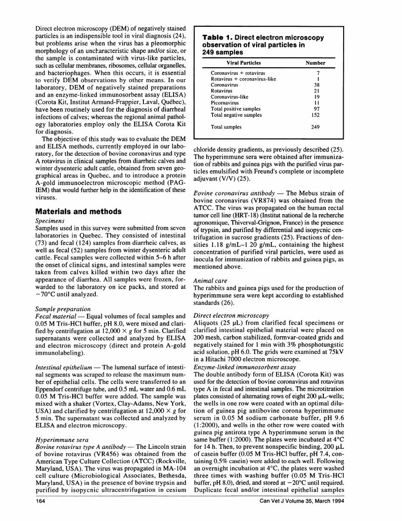
Table 1. Direct electron microscopyobservation of viral particles in249 samples
Viral Particles Number
Coronavirus + rotavirus 7Rotavirus + coronavirus-like ICoronavirus 38Rotavirus 2 1Coronavirus-like 19Picornavirus 11Total positive samples 97Total negative samples 152
Total samples 249
A c .Imum
H
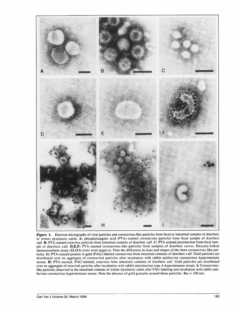
Figure 1. Electron micrographs of viral particles and coronavirus-like particles from fecal or intestinal samples of diarrheicor winter dysenteric cattle. A: phosphotungstic acid (PTA)-stained coronavirus particles from fecal sample of diarrheiccalf. B: PTA-stained rotavirus particles from intestinal contents of diarrheic calf. C: PTA-stained picornavirus from fecal sam-ple of diarrheic calf. D,E,F: PTA-stained coronavirus-like particles from samples of diarrheic calves. Enzyme-linkedimmunosorbent assay (ELISA) tests were negative. Note the difference in sizes and shapes of the three coronavirus-like par-ticles. G: PTA-stained protein A-gold (PAG)-labeled coronavirus from intestinal contents of diarrheic calf. Gold particles aredistributed over an aggregate of coronaviral particles after incubation with rabbit antibovine coronavirus hyperimmuneserum. H: PTA-stained, PAG-labeled, rotavirus from intestinal contents of diarrheic calf. Gold particles are distributedover an aggregate of rotaviral particles after incubation with rabbit antirotavirus type A hyperimmune serum. I: Coronavirus-like particles observed in the intestinal contents of winter dysenteric cattle after PAG-labeling and incubation with rabbit anti-bovine coronavirus hyperimmune serum. Note the absence of gold granules around these particles. Bar = 100 nm.
Can Vet J Volume 35, March 1994
L
165

(100 ,uL) were added to the coated wells and incubatedfor 1 h at room temperature (21±1°C). The wells were
washed three times with 200 ,uL washing buffer, and then100 ,uL of optimal dilution of rabbit hyperimmune anti-bovine corona or antirota type A serum were added. Afteran incubation period of 1 h at room temperature, the wellswere washed with buffer and 100 ,uL of a 1:24,000dilution of peroxidase-labeled antirabbit IgG (ICN,Mississauga, Ontario) were added to each well. Theplates were incubated for 1 h at room temperature,washed, and 100 puL of enzyme substrate solution (thesubstrate solution contained 0.1% hydrogen peroxide and0.02% 3,3'5,5'-tetramethyl benzidine, 1:1; pH 5.0)were added. After further incubation for 10 min, the reac-
tion was terminated by the addition of 50 p.L 2N sulfu-ric acid, and the absorbance was read at 450 nm using a
spectrophotometer (Microplate Autoreader (EL309),Bio-Teck Instruments, Highland Park, Winooski,Vermont, USA). The positive and negative antigencontrols for coronavirus consisted of supernatants frominfected and noninfected HRT-18 cells, respectively;while those for rotavirus consisted of supernatants frominfected and non-infected MA 104 cells. Before beingused as controls, the supernatants were examined by elec-tron microscopy for the presence (positive controls)and absence (negative controls) of viral particles. Resultswere expressed as follows: optical density (OD) .0.200for coronavirus and .0.250 for rotavirus were consideredpositive.
Protein A -gold immunoelectron microscopyA set of aliquots (25pL) from clarified fecal specimens
or clarified intestinal epithelial material was mixedwith the same volume of diluted (10 and 100-fold dilu-
166
tions) rabbit antibovine coronavirus hyperimmuneserum, while another set of aliquots was mixed with thesame volume of diluted (100 and 1000-fold dilutions)rabbit antibovine rotavirus type A hyperimmune serum.
Droplets of these mixtures were deposited on paraffinstrips, and carbon stabilized, Formvar-coated, gridswere floated for 30 min on the droplets. The grids were
then washed for 5 min on several drops of phosphatebuffered saline-Tween (phosphate buffered saline: 0. IMphosphate buffer, pH 7.2, containing 0.85% NaCl,supplemented with 0.05% Tween 20), incubated indiluted protein A-gold 15 nm complex (1:10, v/v) for 30min, rinsed again in 0.1 M phosphate buffer, pH 7.2, fol-lowed by water, and stained with 3% phosphotungsticacid, pH 6.0, or 2% uranyl acetate, for 30 s and thegrids were examined, as mentioned above.
ResultsDirect electron microscopyDirect electron microscopic investigation of negativelystained samples from diarrheic and winter dysenteric cat-tle did not identify virus particles in the majority ofcases. Only 97 of the 249 samples examined (39.0%)showed the presence of various viral particles (Table 1).The two main types of identifiable viral particles
were coronavirus (Figure IA) and rotavirus (Figure IB).Some samples contained aggregates of smaller picorna-viruses (average diameter size 31 nm) (Figure IC).Viral particles were present in single or mixed infection.Rotavirus appeared as stable, spherical structures withdiameters ranging between 65 nm and 70 nm (Figure IB).The structure of coronaviruses was less defined. Most ofthem appeared spherical in shape with diameter sizesranging between 123 nm and 176 nm (Figure IA).
Can Vet J Volume 35, March 1994
Table 2. Comparison of direct electron microscopy(DEM) and enzyme-linked immunosorbent assay(ELISA) from field samples: Coronavirus
DEM/ELISA
Sample +/+ -I- +/- -/+ % Overall agreement
Diarrheic calves (197)a 15 175 5 2 96.4Adult cattle (52) 20 23 5 4 82.7
Total (249) 35 198 10 6 93.6
aNumber of samples
Table 3. Comparison of direct electron microscopy(DEM) and enzyme-linked immunosorbent assay(ELISA) from field samples: Rotavirus
DEM/ELISA
Sample +/+ -1- +1- -1+ % Overall agreement
Diarrheic calves (197)a 29 160 0 8 95.9Adult cattle (52) 0 51 0 1 98.1
Total (249) 29 211 0 9 96.4
aNumber of samples
0la74)
00-CU
Cl)
I
uJ
co0
co0U)
0
1o
0
0
E
0)N
0
(I)-
E
m-Q_
. uJ
o
-111me _00O
0
C_
E00
Oo
oe
o
0.*.EE0
*0OE
iE
@ . C)
~+
Z+
TI
+1
8 +
+l
+
+
Z+
+l
8 ++
XD00
I0
O C)o 000 00
0 0
eN
cli
(71
C1
0060000
0 O 0
0
00600
00N-
cl 00
_ _ON
C)
C C
U. la~c <.c
-
r-
CO
H0
> C)0*.4
C)Q
CO COXz:~rJ~'000066)C C.)C)C)C)o
C) C C) C)0
CO COs: CO .
Can Vet J Volume 35, March 1994 167
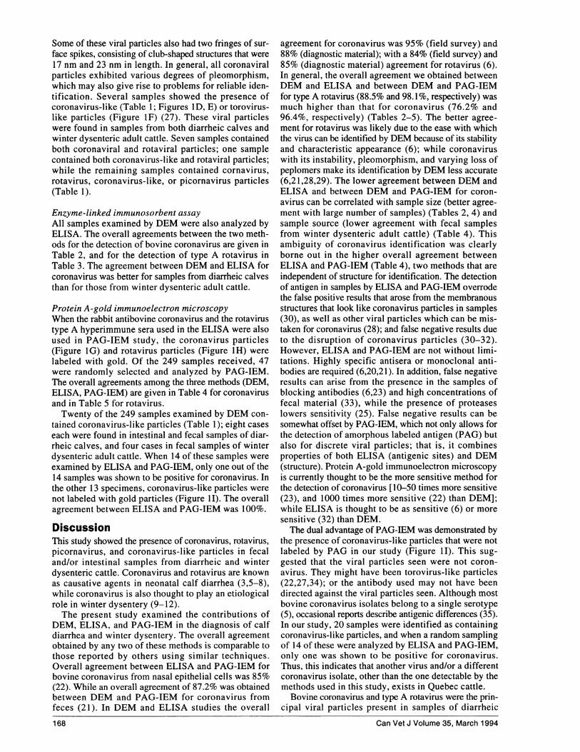
Some of these viral particles also had two fringes of sur-face spikes, consisting of club-shaped structures that were17 nm and 23 nm in length. In general, all coronaviralparticles exhibited various degrees of pleomorphism,which may also give rise to problems for reliable iden-tification. Several samples showed the presence ofcoronavirus-like (Table 1; Figures ID, E) or torovirus-like particles (Figure IF) (27). These viral particleswere found in samples from both diarrheic calves andwinter dysenteric adult cattle. Seven samples containedboth coronaviral and rotaviral particles; one samplecontained both coronavirus-like and rotaviral particles;while the remaining samples -contained cornavirus,rotavirus, coronavirus-like, or picornavirus particles(Table 1).
Enzyme-linked immunosorbent assayAll samples examined by DEM were also analyzed byELISA. The overall agreements between the two meth-ods for the detection of bovine coronavirus are given inTable 2, and for the detection of type A rotavirus inTable 3. The agreement between DEM and ELISA forcoronavirus was better for samples from diarrheic calvesthan for those from winter dysenteric adult cattle.
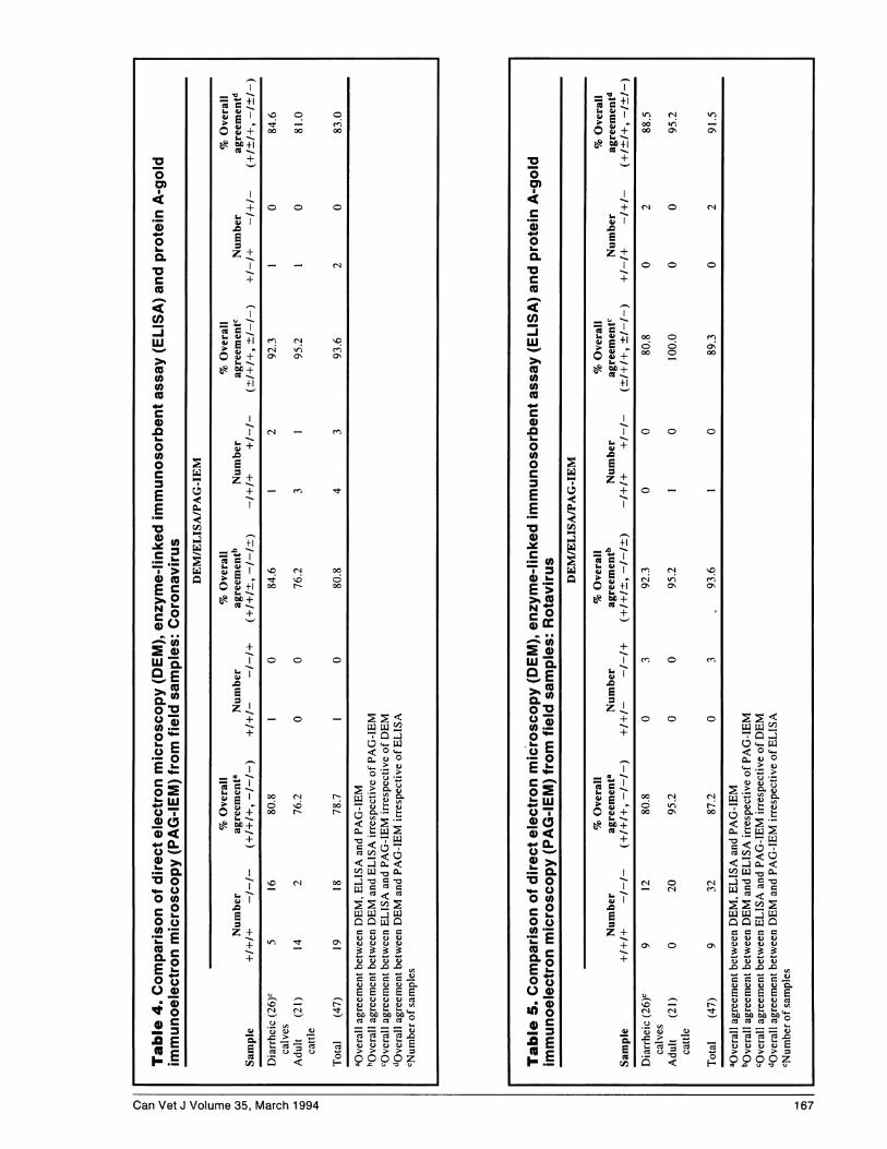
Protein A -gold immunoelectron microscopyWhen the rabbit antibovine coronavirus and the rotavirustype A hyperimmune sera used in the ELISA were alsoused in PAG-IEM study, the coronavirus particles(Figure IG) and rotavirus particles (Figure IH) werelabeled with gold. Of the 249 samples received, 47were randomly selected and analyzed by PAG-IEM.The overall agreements among the three methods (DEM,ELISA, PAG-IEM) are given in Table 4 for coronavirusand in Table 5 for rotavirus.Twenty of the 249 samples examined by DEM con-
tained coronavirus-like particles (Table 1); eight caseseach were found in intestinal and fecal samples of diar-rheic calves, and four cases in fecal samples of winterdysenteric adult cattle. When 14 of these samples wereexamined by ELISA and PAG-IEM, only one out of the14 samples was shown to be positive for coronavirus. Inthe other 13 specimens, coronavirus-like particles werenot labeled with gold particles (Figure 11). The overallagreement between ELISA and PAG-IEM was 100%.
DiscussionThis study showed the presence of coronavirus, rotavirus,picornavirus, and coronavirus-like particles in fecaland/or intestinal samples from diarrheic and winterdysenteric cattle. Coronavirus and rotavirus are knownas causative agents in neonatal calf diarrhea (3,5-8),while coronavirus is also thought to play an etiologicalrole in winter dysentery (9-12).The present study examined the contributions of
DEM, ELISA, and PAG-IEM in the diagnosis of calfdiarrhea and winter dysentery. The overall agreementobtained by any two of these methods is comparable tothose reported by others using similar techniques.Overall agreement between ELISA and PAG-IEM forbovine coronavirus from nasal epithelial cells was 85%(22). While an overall agreement of 87.2% was obtainedbetween DEM and PAG-IEM for coronavirus fromfeces (21). In DEM and ELISA studies the overall
168
agreement for coronavirus was 95% (field survey) and88% (diagnostic material); with a 84% (field survey) and85% (diagnostic material) agreement for rotavirus (6).In general, the overall agreement we obtained betweenDEM and ELISA and between DEM and PAG-IEMfor type A rotavirus (88.5% and 98.1%, respectively) wasmuch higher than that for coronavirus (76.2% and96.4%, respectively) (Tables 2-5). The better agree-ment for rotavirus was likely due to the ease with whichthe virus can be identified by DEM because of its stabilityand characteristic appearance (6); while coronaviruswith its instability, pleomorphism, and varying loss ofpeplomers make its identification by DEM less accurate(6,21,28,29). The lower agreement between DEM andELISA and between DEM and PAG-IEM for coron-avirus can be correlated with sample size (better agree-ment with large number of samples) (Tables 2, 4) andsample source (lower agreement with fecal samplesfrom winter dysenteric adult cattle) (Table 4). Thisambiguity of coronavirus identification was clearlyborne out in the higher overall agreement betweenELISA and PAG-IEM (Table 4), two methods that areindependent of structure for identification. The detectionof antigen in samples by ELISA and PAG-IEM overrodethe false positive results that arose from the membranousstructures that look like coronavirus particles in samples(30), as well as other viral particles which can be mis-taken for coronavirus (28); and false negative results dueto the disruption of coronavirus particles (30-32).However, ELISA and PAG-IEM are not without limi-tations. Highly specific antisera or monoclonal anti-bodies are required (6,20,21). In addition, false negativeresults can arise from the presence in the samples ofblocking antibodies (6,23) and high concentrations offecal material (33), while the presence of proteaseslowers sensitivity (25). False negative results can besomewhat offset by PAG-IEM, which not only allows forthe detection of amorphous labeled antigen (PAG) butalso for discrete viral particles; that is, it combinesproperties of both ELISA (antigenic sites) and DEM(structure). Protein A-gold immunoelectron microscopyis currently thought to be the more sensitive method forthe detection of coronavirus [10-50 times more sensitive(23), and 1000 times more sensitive (22) than DEM];while ELISA is thought to be as sensitive (6) or moresensitive (32) than DEM.The dual advantage of PAG-IEM was demonstrated by
the presence of coronavirus-like particles that were notlabeled by PAG in our study (Figure 1I). This sug-gested that the viral particles seen were not coron-avirus. They might have been torovirus-like particles(22,27,34); or the antibody used may not have beendirected against the viral particles seen. Although mostbovine coronavirus isolates belong to a single serotype(5), occasional reports describe antigenic differences (35).In our study, 20 samples were identified as containingcoronavirus-like particles, and when a random samplingof 14 of these were analyzed by ELISA and PAG-IEM,only one was shown to be positive for coronavirus.Thus, this indicates that another virus and/or a differentcoronavirus isolate, other than the one detectable by themethods used in this study, exists in Quebec cattle.
Bovine coronavirus and type A rotavirus were the prin-cipal viral particles present in samples of diarrheic
Can Vet J Volume 35, March 1994
calves thus confirming the role of these viruses in calfdiarrhea (1,3,5-12,14,21). On the other hand, bovinecoronavirus was the principal viral virus particle presentin samples of winter dysenteric adult cattle, therebysuggesting that bovine coronavirus is one of the highlyprobable causes of winter dysentery (9-12). Of the 52 fecalsamples of winter dysenteric adult cattle, 25 were shownto be positive for coronavirus by DEM and 24 byELISA, with 20 being positive by both DEM and ELISA,while only one sample was positive for type A rotavirusby ELISA. Further confirmation for an etiological roleof bovine coronavirus in winter dysentery was illustratedwhen 21 of these samples were also analyzed by PAG-IEM. Seventeen of the 21 samples were positive forcoronavirus by both ELISA and PAG-IEM, with anadditional positive sample by PAG-IEM but not byELISA. Moreover, only one of these 21 samples was pos-itive for type A rotavirus by both ELISA and PAG-IEM.The results of the present investigation clearly demon-
strated that DEM, ELISA, and PAG-IEM can be effec-tively employed in a centralized diagnostic laboratory.For routine purposes, DEM viral identification, especiallyby an experienced investigator, and/or ELISA analysisis generally sufficient. However, when in doubt, PAG-IEM because of its sensitivity and specificity in thedetection of viral particles or antigen should be used toverify findings (23). Although not suitable for the large-scale screening of specimens, PAG-IEM can proveindispensable for the identification of selected or doubt-ful specimens. cvi
References1. Saif LJ, Theil KW, eds. Viral Diarrheas in Man and Animals. Boca
Raton, Florida: CRC Press, 1990.2. Acres SD, Saunders JR, Radostits OM. Acute undifferentiated
neonatal diarrhea of beef calves: The prevalence of enterotoxigenicE. coli, reo-like (rota) virus and other enteropathogens in cow-calfherds. Can Vet J 1977; 18: 113-121.
3. Almeida JD, Craig CR, Hall TE. Multiple viruses present in thefaeces of a scouring calf. Vet Rec 1978; 102: 170-171.
4. Moon HW, McClurkin AW, Isaacson RE, Pohlenz J, Skartvedt SM,Gillette KG, Baetz AL. Pathogenic relationships of rotavirus,Escherichlia coli, and other agents in mixed infections in calves.J Am Vet Med Assoc 1978; 173: 577-583.
5. Dea S, Roy RS, Begin ME. Physiochemical and biological prop-erties of neonatal calf diarrhea coronaviruses isolated in Quebecand comparison with the Nebraska calf coronavirus. Am J Vet Res1980; 41: 23-29.
6. Reynolds DJ, Chasey D, Scott AC, Bridger JC. Evaluation ofELISA and electron microscopy for the detection of coronavirusand rotavirus in bovine faeces. Vet Rec 1984; 114: 397-401.
7. Durham PJK, Hassard LE, Norman GR, Yemen RL. Viruses andvirus-like particles detected during examination of feces from calvesand piglets with diarrhea. Can Vet J 1989; 30: 876-881.
8. Carman PS, Hazlett MJ. Bovine coronavirus infection in Ontario,1990-1991. Can Vet J 1992; 33: 812-814.
9. Van Kruiningen HJ, Hiestand L, Hill DL, Tilton RC, Ryan RW.Winter dysentery in dairy cattle: recent findings. Compend ContinEduc Pract Vet 1985; 7: S591-S598.
10. Van Kruiningen HJ, Khairallah LH, Sasseville VG. Wyand MS,Post JE. Calfhood coronavirus enterocolitis: a clue to the etiologyof winter dysentery. Vet Pathol 1987; 24: 564-567.
11. Saif LI, Redman DR, Brock KV, KohlerEM, Heckert RA. Winterdysentery in adult dairy cattle: detection of coronavirus in the fae-ces. Vet Rec 1988; 123: 300-301.
12. Saif LI, Brock KV, Redman DR, Kohler EM. Winter dysentery indairy herds: electron microscopic and serological evidence for anassociation with coronavirus infection. Vet Rec 1991; 128:447-449.
13. England JJ, Frye CS, Enright EA. Negative contrast electronmicroscopic diagnosis of viruses of neonatal calf diarrhea. CornellVet 1976; 66: 172-181.
14. Marsolais G, Assaf R, Montpetit C, Marois P. Diagnosis of viralagents associated with neonatal calf diarrhea. Can J Comp Med1978;42: 168-171.
15. Edwards S, Chasey D, Naphtine P, Banks J, Hewitt-Taylor C,Cranage MP. A comparison of three rapid diagnostic methods forthe detection of rotavirus infection in calves. Vet Microbiol1987; 13: 19-25.
16. Czerny CP, Eichhorn W. Characterization of monoclonal andpolyclonal antibodies to bovine enteric coronavirus: establishmentof an efficient ELISA for antigen detection in feces. Vet Microbiol1989; 20: 111-122.
17. Thorns CJ, Bell MM, Chasey D, Chesham J, Roeder PL.Development of monoclonal antibody ELISA for simultaneousdetection of bovine coronavirus, rotavirus serogroup A andEscherichia coli K 99 antigen in feces of calves. Am J Vet Res1992; 53: 36-43.
18. Rodgers SJ, Baldwin CA. Comparison of a commercial visible reac-tion disc enzyme immunoassay to a commercial spectrophotometricbead immunoassay and electron microscopy of the diagnosis ofrotavirus. J Vet Diagn Invest 1991; 3: 342-343.
19. Hammami S, Sawyer MM, Castro AE, Holmberg CA, Osburn Bl.Detection of rotavirus in fecal samples from calves by a cell cul-ture indirect immunofluorescence, an Ag-capture ELISA, a tissueculture ELISA and a commercial Ag-capture ELISA. J Vet DiagnInvest 1989; 1: 72-73.
20. Crouch C, Raybould T, Acre S. Monoclonal antibody captureenzyme linked immunosorbent assay for detection of bovineenteric coronavirus. J Clin Microbiol 1984; 19: 388-393.
21. El-Ghorr AA, Snodgrass DR, Scott FMM. Evaluation of animmunogold electron microscopy technique for detecting bovinecoronavirus. J Virol Meth 1988; 19: 215-224.
22. Heckert RA, Saif LI, Myers GW. Development of protein A-goldimmunoelectron microscopy for detection of bovine coronavirusin calves: comparison with ELISA and direct immunofluores-cence of nasal epithelial cells. Vet Microbiol 1989;19: 217-231.
23. Dea S, Garzon S. Identification of coronaviruses by the use of indi-rect protein A-gold immunoelectron microscopy. J Vet DiagnInvest 1991; 3: 297-305.
24. Hyatt AD. The application of electron microscopy to veterinaryvirus diagnosis. Aust Vet J 1989; 66: 445-449.
25. Dea S, Tijssen P. Detection of turkey enteric coronavirus byenzyme-linked immunosorbent assay and differentiation fromother coronaviruses. Am J Vet Res 1989; 50: 226-231.
26. Canadian Council on Animal Care. Guide to the Care and Use ofExperimental Animals. Vol I and 2. Ottawa: Canadian Council onAnimal Care, 1980.
27. Koopmans M, van Wuijckhuise-Sjouke L, Schukken YH,Cremers H, Horzinek MC. Association of diarrhea in cattle withtorovirus infections on farms. Am J Vet Res 1991; 52: 1769-1773.
28. Dea S, Roy RS, Elazhary MASY. La diarrhee neonatale due aucoronavirus du veau. Une revue de la litterature. Can Vet J 198 1;22: 51-58.
29. Crouch C, Acres S. Prevalence of rotavirus and coronavirus anti-gens in the feces of normal cows. Can J Comp Med 1984; 48:340-342.
30. Eugster AK, Sneed L. Viral intestinal infections of animals andman. Comp Immunol Microbiol Infect Dis 1980; 2: 417-435.
31. Gibbs EPJ, Smale CJ, Voyle CA. Electron microscopy as an aid tothe rapid diagnosis of virus diseases of veterinary importance. VetRec 1980; 106: 451-458.
32. Flewett TH. Electron microscopy in the diagnosis of infectiousdiarrhea. J Am Vet Med Assoc 1978; 173: 538-543.
33. Crouch CF, Bielefeldt OH, Watts TC, Babiuk LA. Chronic shed-ding of bovine enteric coronavirus antigen-antibody complexesby clinically normal cows. J Gen Virol 1985; 66: 1489-1500.
34. Woode GN, Reed DE, Runnels PL, Herrig MA, Hill HT. Studieswith a classified isolate from diarrheic calves. Vet Microbiol1982; 7: 221-240.
35. Dea 5, Roy RS, Elazhary MASY. Antigenic variations among calfdiarrhea coronaviruses by immunodiffusion and counterimmuno-electrophoresis. Ann Rech Vet 1982; 13: 35 1-356.
Can Vet J Volume 35, March 1994 169