Embed Size (px)
DESCRIPTION
DHF
Citation preview
DENGUE
Practice Essentials
Dengue is the most common arthropod-borne viral (arboviral) illness in humans. It is transmitted by mosquitoes of the genus Aedes, which are widely distributed in subtropical and tropical areas of the world.
A small percentage of persons who have previously been infected by one dengue serotype develop bleeding and endothelial leak upon infection with another dengue serotype. This syndrome is termed dengue hemorrhagic fever.
Dengue fever is typically a self-limiting disease with a mortality rate of less than 1%. When treated, dengue hemorrhagic fever has a mortality rate of 2-5%, but when left untreated, the mortality rate is as high as 50%.
Essential update: Dengue may be underrecognized in the United States
As suggested by a recently reported case of a woman aged 63 years who died from complications of dengue acquired in New Mexico or Texas in 2012, the disease may not be adequately recognized in the United States as a source of potentially fatal acute febrile illness. The patient had initially been diagnosed with West Nile virus, but a postmortem bone marrow biopsy revealed the presence of dengue virus.[1, 2]
In addition, the patient’s records revealed that she met the clinical case definition for hemophagocytic lymphohistiocytosis, a hyperinflammatory syndrome that is sometimes associated with dengue and that in this instance was the cause of death.
Signs and symptoms
Many patients with dengue experience a prodrome of chills, erythematous mottling of the skin, and facial flushing, which may last for 2-3 days. Children younger than 15 years usually have a nonspecific febrile syndrome, which may be accompanied by a maculopapular rash.
Accompanying symptoms in patients with dengue may include any of the following:
• Headache• Retro-orbital pain• Severe myalgias: Especially of the lower back, arms, and legs• Arthralgias: Usually of the knees and shoulders• Nausea and vomiting (diarrhea is rare)• Rash: A maculopapular or macular confluent rash over the face, thorax,
and flexor surfaces, with islands of skin sparing• Weakness• Altered taste sensation• Anorexia• Sore throat• Mild hemorrhagic manifestations (eg, petechiae, bleeding gums,
epistaxis, menorrhagia, hematuria)• Lymphadenopathy

Dengue hemorrhagic fever
The initial phase of dengue hemorrhagic fever is similar to that of dengue fever and other febrile viral illnesses. Shortly after the fever breaks (or sometimes within 24 hours before), signs of plasma leakage appear, along with the development of hemorrhagic symptoms such as bleeding from sites of trauma, gastrointestinal bleeding, and hematuria. Patients may also present with abdominal pain, vomiting, febrile seizures (in children), and a decreased level of consciousness.
If left untreated, dengue hemorrhagic fever most likely progresses to dengue shock syndrome. Common symptoms in impending shock include abdominal pain, vomiting, and restlessness. Patients also may have symptoms related to circulatory failure.
See Clinical Presentation for more detail.
Diagnosis
Laboratory criteria for the diagnosis of dengue include 1 or more of the following:
• Isolation of the dengue virus from serum, plasma, leukocytes, or autopsy samples
• Demonstration of a fourfold or greater change in reciprocal immunoglobulin G (IgG) or IgM antibody titers to 1 or more dengue virus antigens in paired serum samples
• Demonstration of dengue virus antigen in autopsy tissue via immunohistochemistry or immunofluorescence or in serum samples via enzyme immunoassay (EIA)
• Detection of viral genomic sequences in autopsy tissue, serum, or cerebral spinal fluid (CSF) samples via polymerase chain reaction (PCR) assay
The following laboratory tests should also be performed in the workup of patients with possible dengue:
• Complete blood count (CBC)• Metabolic panel• Serum protein and albumin levels• Liver panel• Disseminated intravascular coagulation (DIC) panelCharacteristic findings in dengue fever are as follows:
• Thrombocytopenia (platelet count < 100 x 109/L)• Leukopenia• Mild to moderate elevation of aspartate aminotransferase and alanine
aminotransferase values
In patients with dengue hemorrhagic fever, the following may be present:
• Increased hematocrit level secondary to plasma extravasation and/or third-space fluid loss
• Hypoproteinemia• Prolonged prothrombin time
• Prolonged activated partial thromboplastin time• Decreased fibrinogen• Increased amount of fibrin split productsGuaiac testing for occult blood in the stool should be performed on all patients in whom dengue virus infection is suspected. Urinalysis identifies hematuria.
Imaging studies
• Chest radiography• Head computed tomography (CT) scanning without contrast: To detect
intracranial bleeding or cerebral edema from dengue hemorrhagic fever
• Ultrasonography: To detect fluid in the chest and abdominal cavities, pericardial effusion, and a thickened gallbladder wall, in dengue hemorrhagic fever
See Workup for more detail.
Management
Oral rehydration therapy is recommended for patients with moderate dehydration caused by high fever and vomiting.
Patients who develop signs of dengue hemorrhagic fever warrant closer observation. Admission for intravenous fluid administration is indicated for patients who develop signs of dehydration, such as the following:
• Tachycardia• Prolonged capillary refill time• Cool or mottled skin• Diminished pulse amplitude• Altered mental status• Decreased urine output• Rising hematocrit• Narrowed pulse pressure• HypotensionPatients with internal or gastrointestinal bleeding may require transfusion, and patients with coagulopathy may require fresh frozen plasma.
See Treatment and Medication for more detail.
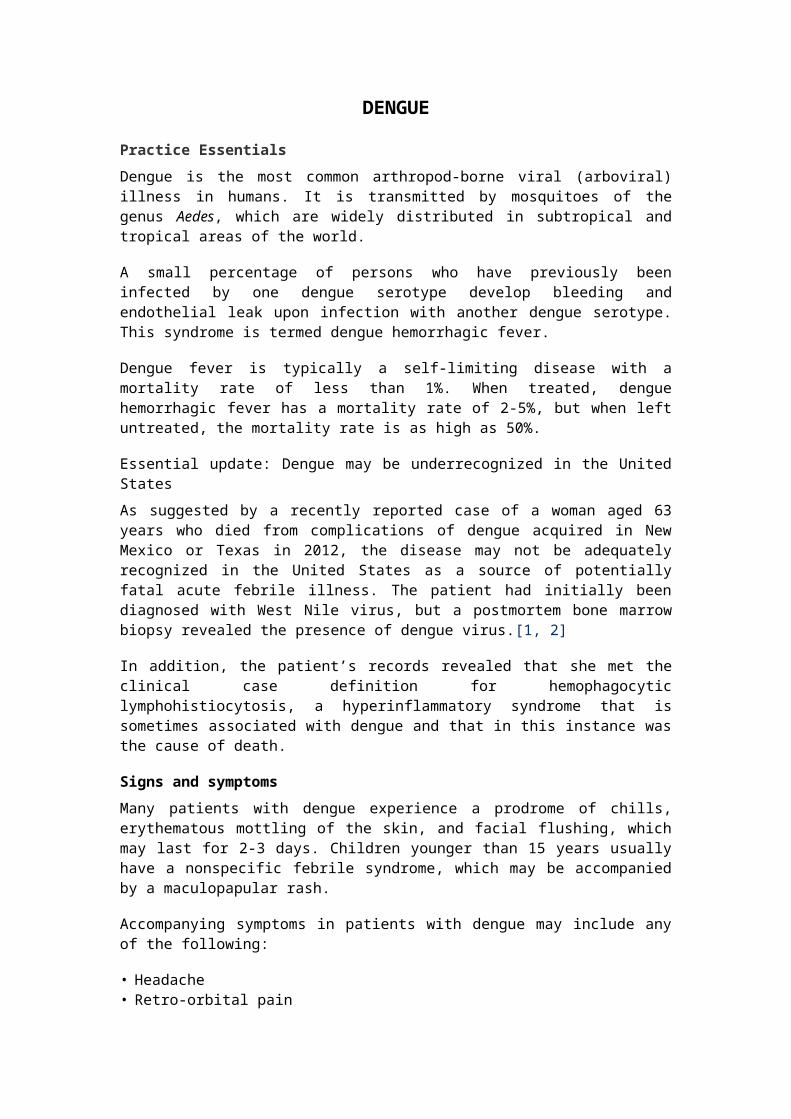
Image library
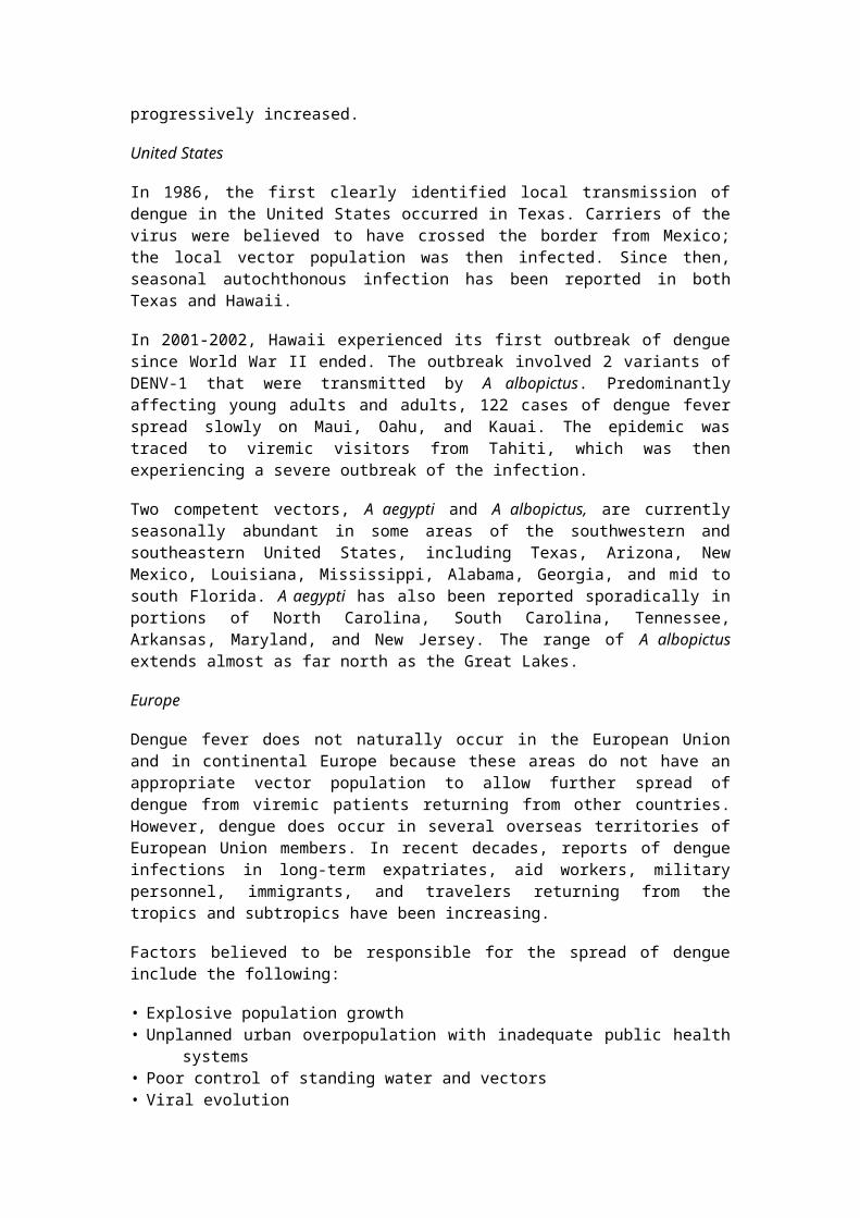
Drawing of Aedes aegypti mosquito.
Background
Dengue is the most common arthropod-borne viral (arboviral) illness in humans. Globally, 2.5-3 billion individuals live in approximately 112 countries that experience dengue transmission. Annually, approximately 50-100 million individuals are infected. It is caused by infection with 1 of the 4 serotypes of dengue virus, which is a Flavivirus (a genus of single-stranded nonsegmented RNA viruses). Infection with one dengue serotype confers lifelong homotypic immunity to that serotype and a very brief period of partial heterotypic immunity to other serotypes, but a person can eventually be infected by all 4 serotypes. Several serotypes can be in circulation during an epidemic.
Dengue is transmitted by mosquitoes of the genus Aedes, which are widely distributed in subtropical and tropical areas of the world (see the image below).
Initial dengue infection may be asymptomatic (50-90%),[3] may result in a nonspecific febrile illness, or may produce the symptom complex of classic dengue fever (DF). Classic dengue fever is marked by rapid onset of high fever, headache, retro-orbital pain, diffuse body pain (both muscle and bone), weakness, vomiting, sore throat, altered taste sensation, and a centrifugal maculopapular rash, among other manifestations. The severity of the pain led to the term breakbone fever to describe dengue.
A small percentage of persons who have previously been infected by one dengue serotype develop bleeding and endothelial leak upon infection with another dengue serotype. This syndrome is termed dengue hemorrhagic fever (DHF).
Dengue hemorrhagic fever has also been termed dengue vasculopathy. Vascular leakage in these patients results in hemoconcentration and serous effusions and can lead to circulatory collapse. This, in conjunction with severe hemorrhagic complications, can lead to dengue shock syndrome, which poses a greater fatality risk than bleeding per se.[4]
Dengue virus transmission follows 2 general patterns: epidemic dengue and hyperendemic dengue. Epidemic dengue transmission occurs when dengue virus is introduced into a region as an isolated event that involves a single viral strain. If the number of vectors and susceptible pediatric and adult hosts is sufficient, explosive transmission can occur, with an infection incidence of 25-50%. Mosquito-control efforts, changes in weather, and herd immunity contribute to the control of these epidemics. Transmission appears to begin in urban centers and then spreads to the rest of the country.[5] This is the current pattern of transmission in parts of Africa and South America, areas of Asia where the virus has reemerged, and small island nations. Travelers to these areas are at increased risk of acquiring dengue during these periods of epidemic transmission.
Hyperendemic dengue transmission is characterized by the continuous circulation of multiple viral serotypes in an area where a large pool of susceptible hosts and a competent vector (with or without seasonal variation) are constantly present. This is the predominant pattern of global transmission. In areas of hyperendemic dengue, antibody prevalence increases with age, and most adults are immune. Hyperendemic transmission appears to be a major risk for dengue hemorrhagic fever.
Travelers to these areas are more likely to be infected than are travelers to areas that experience only epidemic transmission.[6]
Because the signs and symptoms of dengue fever are nonspecific, attempting laboratory confirmation of dengue infection by serodiagnosis, polymerase chain reaction (PCR), or culture is important. Serodiagnosis is made on the basis of a rise in antibody titer in paired IgG or IgM specimens. Results vary depending on whether the infection is primary or secondary (see Presentation and Workup). Dengue is a reportable disease in the United States; known or suspected cases should be reported to public health authorities.
Dengue fever is usually a self-limited illness. Supportive care with analgesics, judicious fluid replacement, and bed rest is usually sufficient. Successful management of severe dengue requires intravascular volume replacement, with careful attention to fluid management and proactive treatment of hemorrhage. Admission to an intensive care unit is indicated for patients with dengue shock syndrome (see Treatment).
DENV-1 and DENV-2
Serotype 1 dengue (DENV-1) was introduced into a largely susceptible population in Cuba in 1977. Serosurveys indicated that more than 44% of the population was infected, with only mild disease reported. The first dengue hemorrhagic fever epidemic in the Americas occurred in Cuba in 1981 and involved serotype 2 dengue (DENV-2), with hundreds of thousands of cases of dengue in both children and adults, 24,000 cases of dengue hemorrhagic fever, 10,000 cases of dengue shock syndrome, and 158 reported deaths.
In 1997, Asian genotype DENV-2 was reintroduced, and dengue shock syndrome and dengue hemorrhagic fever were seen only in adults who had previously been infected with DENV-1 in 1977. Disease and case-fatality rates were higher in those who had been infected with DENV-2 20 years after their initial DENV-1 infection than those who were infected 4 years apart.
Data from other countries supports the finding that the severity of secondary dengue infections appears to intensify with longer intervals between infections.[7, 8] Since then, dengue fever and dengue hemorrhagic fever cases have progressively increased.
United States
In 1986, the first clearly identified local transmission of dengue in the United States occurred in Texas. Carriers of the virus were believed to have crossed the border from Mexico; the local vector population was then infected. Since then, seasonal autochthonous infection has been reported in both Texas and Hawaii.
In 2001-2002, Hawaii experienced its first outbreak of dengue since World War II ended. The outbreak involved 2 variants of DENV-1 that were transmitted by A albopictus. Predominantly affecting young adults and adults, 122 cases of dengue fever spread slowly on Maui, Oahu, and Kauai. The epidemic was traced to viremic visitors from Tahiti, which was then

experiencing a severe outbreak of the infection.
Two competent vectors, A aegypti and A albopictus, are currently seasonally abundant in some areas of the southwestern and southeastern United States, including Texas, Arizona, New Mexico, Louisiana, Mississippi, Alabama, Georgia, and mid to south Florida. A aegypti has also been reported sporadically in portions of North Carolina, South Carolina, Tennessee, Arkansas, Maryland, and New Jersey. The range of A albopictus extends almost as far north as the Great Lakes.
Europe
Dengue fever does not naturally occur in the European Union and in continental Europe because these areas do not have an appropriate vector population to allow further spread of dengue from viremic patients returning from other countries. However, dengue does occur in several overseas territories of European Union members. In recent decades, reports of dengue infections in long-term expatriates, aid workers, military personnel, immigrants, and travelers returning from the tropics and subtropics have been increasing.
Factors believed to be responsible for the spread of dengue include the following:
• Explosive population growth• Unplanned urban overpopulation with inadequate public health systems• Poor control of standing water and vectors• Viral evolution• Increased international recreational, business, and military travel to
endemic areasAll of these factors must be addressed to control the spread of dengue and other mosquito-borne infections. Unplanned urbanization is believed to have had the largest impact on disease amplification in individual countries, whereas travel is believed to have had the largest impact on global spread.[3, 5, 6, 8, 9]
Travel surveillance
Over the past decade, the GeoSentinel Network of Travel Medicine providers has demonstrated that dengue has become more frequently diagnosed than malaria in travelers returning from tropical areas other than Africa. Such sentinel travel surveillance can augment global and national public health surveillance. More recent studies have not supported an earlier suggestion that climate change is also directly responsible for increased transmission.[7, 6, 8]
Pathophysiology
Dengue fever is a mosquito-borne viral disease caused by 1 of 4 closely related but antigenically distinct serotypes of dengue virus, serotypes DENV-1 through DEN-4.[10] Infection with one dengue serotype confers lifelong homotypic immunity and a very brief period of partial heterotypic immunity, but each individual can eventually be infected by all 4 serotypes. Several serotypes can be in circulation during an epidemic.
The Aedes mosquito
Dengue viruses are transmitted by the bite of an infected Aedes (subgenus Stegomyia) mosquito.[11] Globally, Aedes aegypti is the predominant highly efficient mosquito vector for dengue infection, but the Asian tiger mosquito, Aedes albopictus, and other Aedes species can also transmit dengue with varying degrees of efficiency (see the images below).
Drawing of Aedes aegypti mosquito.
Aedes albopictus. From CDC Public Domain.Aedes mosquito species have adapted well to human habitation, often breeding around dwellings in small amounts of stagnant water found in old tires or other small containers discarded by humans. Humans are their preferred hosts.
Female Aedes mosquitoes are daytime feeders. They inflict an innocuous bite, usually on the back of the neck and the ankles, and are easily disturbed during a blood meal, causing them to move on to finish a meal on another individual, making them efficient vectors. Not uncommonly, entire families develop infection within a 24- to 36-hour period, presumably from the bites of a single infected mosquito.
Hosts for transmission
Humans serve as the primary reservoir for dengue. Certain nonhuman primates in Africa and Asia also serve as hosts but do not develop dengue hemorrhagic fever. Mosquitoes acquire the virus when they feed on a carrier of the virus. Persons with dengue viruses in their blood can transmit the viruses to the mosquito 1 day before the onset of the febrile period. The patient can remain infectious for the next 6-7 days.
The mosquito can transmit dengue if it immediately bites another host. In addition, transmission occurs after 8-12 days of viral replication in the mosquito's salivary glands (extrinsic incubation period). The virus does not adversely affect the mosquito. The mosquito remains infected for the remainder of its life. The life span of A aegypti is usually 21 days but ranges from 15 to 65 days. Vertical transmission of dengue virus in mosquitoes has been documented.[12] The eggs of Aedes mosquitoes withstand long periods of desiccation, reportedly as long as 1 year, but are killed by temperatures of less than 10°C. Rare cases of vertical dengue transmission have been reported. In addition, rare reports of human-to-human transmission via needle-stick injuries have been published.[13]
Once inoculated into a human host, dengue has an incubation period of 3-14 days (average 4-7 days) while viral replication takes place in target dendritic cells. Infection of target cells, primarily those of the reticuloendothelial system, such as dendritic cells, hepatocytes, and endothelial cells,[14, 15, 16, 17] result in the production of immune mediators that serve to shape the quantity, type, and duration of cellular and humoral immune response to both the initial and subsequent virus infections.[14, 18, 19, 20, 21, 22, 23]
Dengue viral infections frequently are not apparent. In most cases, especially in children younger than 15 years, the patient is asymptomatic or has a mild undifferentiated febrile illness lasting 5-7 days. Classic dengue fever primarily occurs in nonimmune, nonindigenous adults and children and is typically self-limiting. Recovery is usually complete by 7-10 days. Dengue hemorrhagic fever and dengue shock syndrome usually occur around the third to seventh day of illness during a second dengue infection in persons with preexisting actively or passively (maternally) acquired immunity to a heterologous dengue virus serotype.
Dengue fever
Dengue presents in a nonspecific manner similarly to that of many other viral and bacterial illnesses. Fever typically begins on the third day of illness and persists 5-7 days, abating with the cessation of viremia. Fever may reach 41C°. Occasionally, and more frequently in children, the fever abates for a day and recurs, a pattern that is termed a saddleback fever; however, this pattern is more commonly seen in dengue hemorrhagic fever.
Leukopenia, lymphopenia near the end of the febrile phase, and thrombocytopenia are common findings in dengue fever and are believed to be caused by direct destructive actions of the virus on bone marrow precursor cells. The resulting active viral replication and cellular destruction in the bone marrow are believed to cause the bone pain. Approximately one third of patients with dengue fever may have mild hemorrhagic symptoms, including petechiae, gingival bleeding, and a positive tourniquet test (>20 petechiae in an area of 2.5 X 2.5 cm). Dengue fever is rarely fatal.
Dengue hemorrhagic fever
Dengue hemorrhagic fever occurs less frequently than dengue fever but has a more dramatic clinical presentation. In most of Asia, where it first was described, dengue hemorrhagic fever is primarily a disease of children. However, in the Americas, and more recently reported in Taiwan, dengue hemorrhagic fever has an equal distribution in all ages.
Dengue hemorrhagic fever typically begins with the initial manifestations of dengue fever. The acute febrile illness (temperatures ≤40°C), like that of dengue fever, lasts approximately 2-7 days. However, in persons with dengue hemorrhagic fever, the fever reappears, giving a biphasic or saddleback fever curve.
Along with biphasic fever, patients with dengue hemorrhagic fever have progressive thrombocytopenia, increasing hematocrit (20% absolute rise

from baseline) and low albumin (signs of hemoconcentration preceding shock), more obvious hemorrhagic manifestations (>50% of patients have a positive tourniquet test), and progressive effusions (pleural or peritoneal). Lymphocytosis, often with atypical lymphocytes, commonly develops before defervescence or the onset of shock. Transaminase levels may be mildly elevated or present in the several thousands associated with hepatomegaly in those patients with acute hepatitis. Low fibrinogen and elevated fibrin split products are signs of disseminated intravascular coagulation. Severe metabolic acidosis and circulatory failure can occur.
The critical feature of dengue hemorrhagic fever is plasma leakage. Plasma leakage is caused by increased capillary permeability and may manifest as hemoconcentration, as well as pleural effusion and ascites. Bleeding is caused by capillary fragility and thrombocytopenia and may manifest in various forms, ranging from petechial skin hemorrhages to life-threatening gastrointestinal bleeding.
Liver damage manifests as increases in levels of alanine aminotransferase and aspartate aminotransferase, low albumin levels, and deranged coagulation parameters (prothrombin time, partial thromboplastin time).[24, 25] In persons with fatal dengue hepatitis, infection was demonstrated in more than 90% of hepatocytes and Kupffer cells with minimal cytokine response (tumor necrosis factor [TNF]–alpha, interleukin [IL]–2). This is similar to that seen with fatal yellow fever and Ebola infections.[24]
As the term implies, dengue shock syndrome is essentially dengue hemorrhagic fever with progression into circulatory failure, with ensuing hypotension, narrow pulse pressure (< 20 mm Hg), and, ultimately, shock and death if left untreated. Death may occur 8-24 hours after onset of signs of circulatory failure. The most common clinical findings in impending shock include hypothermia, abdominal pain, vomiting, and restlessness.
Secondary infection
The immunopathology of dengue hemorrhagic fever/dengue shock syndrome remains incompletely understood. Most patients who develop dengue hemorrhagic fever or dengue shock syndrome have had prior infection with one or more dengue serotypes. When an individual is infected with another serotype (ie, secondary infection) and produces low levels of nonneutralizing antibodies, these antibodies, directed against 1 of 2 surface proteins (precursor membrane protein and envelope protein), when bound by macrophage and monocyte Fc receptors, have been proposed to fail to neutralize virus and instead form an antigen-antibody complex.
This results in increased viral entry into macrophages bearing IgG receptors, allowing unchecked viral replication with higher viral titers and increased cytokine production and complement activation, a phenomenon called antibody-dependent enhancement.[26, 27]
The affected macrophages release vasoactive mediators that increase vascular permeability, leading to vascular leakage, hypovolemia, and shock. This mechanism, along with individual host and viral genome

variations, plays an active role in pathogenesis. Infants born to mothers who have had dengue, as maternally derived dengue neutralizing IgGs wane, are also thought to be at risk for enhanced disease.[26, 27]
Some researchers suggest that T-cell immunopathology may play a role, with increased T-cell activation and apoptosis. Increased concentrations of interferon have been recorded 1-2 days following fever onset during symptomatic secondary dengue infections.[28] The activation of cytokines, including TNF-alpha, TNF receptors, soluble CD8, and soluble IL-2 receptors, has been correlated with disease severity.[14]
Cuban studies have shown that stored serum sample analysis demonstrated progressive loss of cross-reactive neutralizing antibodies to DENV-2 as the interval since DENV-1 infection increased.[21] In addition, certain dengue strains, particularly those of DENV-2, have been proposed to be more virulent, in part because more epidemics of dengue hemorrhagic fever have been associated with DENV-2 than with the other serotypes.
Etiology
Dengue infection is caused by dengue virus (DENV), which is a single-stranded RNA virus (approximately 11 kilobases long) with an icosahedral nucleocapsid and covered by a lipid envelope. The virus is in the family Flaviviridae, genus Flavivirus, and the type-specific virus is yellow fever.
The dengue virus has 4 related but antigenically distinct serotypes: DENV-1, DENV-2, DENV-3, and DENV-4. Genetic studies of sylvatic strains suggest that the 4 serotypes evolved from a common ancestor in primate populations approximately 1000 years ago and that all 4 separately emerged into a human urban transmission cycle 500 years ago in either Asia or Africa.[3, 29] Albert Sabin speciated these viruses in 1944. Each serotype is known to have several different genotypes. Viral genotype and serotype, and the sequence of infection with different serotypes, appear to affect disease severity.
Living in endemic areas of the tropics (or warm, moist climates such as the southern United States) where the vector mosquito thrives is an important risk factor for infection.[10, 30, 31, 32, 33] Poorly planned urbanization combined with explosive global population growth brings the mosquito and the human host into close proximity. Increased air travel easily transports infectious diseases between populations.
Prognosis
Dengue fever is typically a self-limiting disease with a mortality rate of less than 1%. When treated, dengue hemorrhagic fever has a mortality rate of 2-5%. When left untreated, dengue hemorrhagic fever has a mortality rate as high as 50%. Survivors usually recover without sequelae and develop immunity to the infecting serotype.
The fatality rate associated with dengue shock syndrome varies by country, from 12-44%. In a 1997 Cuban epidemic, the fatality rate in patients who met criteria for dengue hemorrhagic fever or dengue shock

syndrome was approximately 6%. The mortality rate associated with dengue fever is less than 1%. Data from the 1997 Cuban epidemic suggest that, for every clinically apparent case of dengue fever, 13.9 cases of dengue infection went unrecognized because of absent or minimal symptoms.
A 2005 review from Singapore of 14,209 patients found that useful predictors of death included the following[41] :
• Atypical presentations• Significant comorbid illness• Abnormal serum markers (including albumin and coagulation studies)• Secondary bacterial infectionsFactors that affect disease severity include the following:
• Patient age• Pregnancy• Nutritional status• Ethnicity• Sequence of infection with different dengue serotypes• Virus genotype• Quality and extent of available medical careComplications and sequelae of dengue virus infections are rare but may include the following:
• Cardiomyopathy• Seizures, encephalopathy, and viral encephalitis• Hepatic injury• Depression• Pneumonia• Iritis• Orchitis• OophoritisIn 20-30% of dengue hemorrhagic fever cases, the patient develops shock, known as the dengue shock syndrome. Worldwide, children younger than 15 years constitute 90% of dengue hemorrhagic fever patients[36] ; however, in the Americas, dengue hemorrhagic fever occurs in both adults and children.
Although dengue is an extremely important arboviral illness globally, literature evaluating the economic impact is fairly sparse, with some conflicting findings. A recent expert panel assessment and 2 studies in the Americas recommended additional research to fill important information gaps, including disease outcomes and accurate statistics regarding disease burden, that could better inform future decision making regarding control and prevention.[42, 43, 44]
A 5-year prospective study in Thai children examined the relative economic burden of dengue infection in children on the local population. Most disability-adjusted life years (DALYs) lost to dengue resulted from long-term illness in children who had not been hospitalized. The infecting serotype appeared to be the major determinant of DALYs lost, with DEN-2 and DEN-3 responsible for 59%. The mean cost of illness from dengue was significantly higher than that from other febrile illnesses studied.[40]

A prospective study examined the direct and indirect costs of dengue infection in 1695 pediatric and adult patients in 8 countries. The average illness lasted 11.9 days for ambulatory patients and 11 days for hospitalized patients. Hospitalized students lost 5.6 days of school. Those at work lost 9.9 work days. Overall mean costs were more than double (1394 international dollars [I$]) for hospitalized cases. With an annual average of 594,000 cases the aggregate economic cost was estimated to be at least I$587 million, without factoring in underreporting of disease and dengue surveillance and vector control costs. This represents a significant global economic burden in low-income countries.[44]
History
Patients with dengue will have a history of living in, or recent travel to, a region where the disease is endemic. The incubation period is 3-14 days (average, 4-7 days); symptoms that begin more than 2 weeks after a person departs from an endemic area are probably not due to dengue.
Many patients experience a prodrome of chills, erythematous mottling of the skin, and facial flushing (a sensitive and specific indicator of dengue fever). The prodrome may last for 2-3 days. Children younger than 15 years usually have a nonspecific febrile syndrome, which may be accompanied by a maculopapular rash. Classic dengue fever begins with sudden onset of fever, chills, and severe (termed breakbone) aching of the head, back, and extremities, as well as other symptoms. The fever lasts 2-7 days and may reach 41°C. Fever that lasts longer than 10 days is probably not due to dengue.
Pain and other accompanying symptoms may include any of the following:
• Headache• Retro-orbital pain• General body pain (arthralgias, myalgias)• Nausea and vomiting (however, diarrhea is rare)• Rash• Weakness• Altered taste sensation• Anorexia• Sore throat• Mild hemorrhagic manifestations (eg, petechiae, bleeding gums,
epistaxis, menorrhagia, hematuria)• LymphadenopathyRash in dengue fever is a maculopapular or macular confluent rash over the face, thorax, and flexor surfaces, with islands of skin sparing. The rash typically begins on day 3 and persists 2-3 days.
Fever typically abates with the cessation of viremia. Occasionally, and more commonly in children, the fever abates for a day and then returns, a pattern that has been called saddleback fever. A second rash may occur within 1-2 days of defervescence, lasting 1-5 days; it is morbilliform, is maculopapular, spares the palms and soles, and occasionally desquamates.
Recovery is complete but slow, with fatigue and exhaustion often persisting after the fever has subsided. The convalescent phase may last
for 2 weeks.
Patients are at risk for development of dengue hemorrhagic fever or dengue shock syndrome at approximately the time of defervescence. Abdominal pain in conjunction with restlessness, change in mental status, hypothermia, and a drop in the platelet count presages the development of dengue hemorrhagic fever.
Of patients with dengue hemorrhagic fever, 90% are younger than 15 years. The initial phase of dengue hemorrhagic fever is similar to that of dengue fever and other febrile viral illnesses. Shortly after the fever breaks (or sometimes within 24 hours before), signs of plasma leakage appear, along with the development of hemorrhagic symptoms such as bleeding from sites of trauma, gastrointestinal bleeding, and hematuria. Patients may also present with abdominal pain, vomiting, febrile seizures (in children), and a decreased level of consciousness.
If left untreated, dengue hemorrhagic fever most likely progresses to dengue shock syndrome. Common symptoms in impending shock include abdominal pain, vomiting, and restlessness. Patients also may have symptoms related to circulatory failure.
Physical Examination
Dengue fever presents in a nonspecific manner and may not be distinguishable from other viral or bacterial illness. According to the Pan American Health Organization (PAHO), the clinical description of dengue fever is an acute febrile illness of 2-7 days duration associated with 2 or more of the following:
• Severe and generalized headache• Retro-orbital pain• Severe myalgias, especially of the lower back, arms, and legs• Arthralgias, usually of the knees and shoulders• Characteristic rash• Hemorrhagic manifestations• LeukopeniaAdditional findings may include the following:
• Injected conjunctivae• Facial flushing, a sensitive and specific predictor of dengue infection• Inflamed pharynx• Lymphadenopathy• Nausea and vomiting• Nonproductive cough• Tachycardia, bradycardia, and conduction defectsUp to half of patients with dengue fever develop a characteristic rash. The rash is variable and may be maculopapular or macular. Petechiae and purpura may develop as hemorrhagic manifestations. Hemorrhagic manifestations most commonly include petechiae and bleeding at venipuncture sites.
A tourniquet test is often positive. This test is performed by inflating a blood pressure cuff on the upper arm to midway between diastolic and
systolic blood pressures for 5 minutes. The results are considered to be positive if more than 20 petechiae per square inch are observed on the skin in the area that was under pressure. Other hemorrhagic manifestations include nasal or gingival bleeding, melena, hematemesis, and menorrhagia.
Neurologic manifestations such as seizures and encephalitis/encephalopathy have been reported in rare cases of dengue infection. Some of these cases did not display other typical features of dengue infection. Other neurologic complications associated with dengue infection include neuropathies, Guillain-Barré syndrome, and transverse myelitis.
Dengue hemorrhagic fever
Findings for dengue hemorrhagic fever are similar to those for dengue fever and include the following:
• Biphasic fever curve• Hemorrhagic findings more pronounced than in dengue fever• Signs of peritoneal effusion, pleural effusion, or bothMinimal criteria for the diagnosis of dengue hemorrhagic fever, according to the World Health Organization (WHO), are as follows[45] :
• Fever• Hemorrhagic manifestations (eg, hemoconcentration, thrombocytopenia,
positive tourniquet test)• Circulatory failure, such as signs of vascular permeability (eg,
hypoproteinemia, effusions)• HepatomegalyIn addition, conjunctival injection develops in approximately one third of patients with dengue hemorrhagic fever. Optic neuropathy has been reported and occasionally results in permanent and significant visual impairment.[46] Pharyngeal injection develops in almost 97% of patients with dengue hemorrhagic fever. Generalized lymphadenopathy is observed.
Hepatomegaly is present more often in dengue shock syndrome than in milder cases. Hepatic transaminase levels may be mildly to moderately elevated. Encephalopathy is a rare complication that may result from a combination of cerebral edema, intracranial hemorrhage, anoxia, hyponatremia, and hepatic injury.
Dengue shock syndrome
Findings of dengue shock syndrome include the following:
• Hypotension• Bradycardia (paradoxical) or tachycardia associated with hypovolemic
shock• Hepatomegaly• Hypothermia• Narrow pulse pressure (< 20 mm Hg)• Signs of decreased peripheral perfusion
Diagnostic Considerations
Studies indicate that as many as 50% of dengue cases may be misdiagnosed, as a result of inaccurate assessment of the signs and symptoms of disease presentation. This inaccuracy can lead to increased cost of treatment, such as unneeded hospitalizations, as well as possibly increased morbidity and mortality due to volume overload from overzealous use of intravenous fluids.[47]
A Belgian study examined predictors of diagnosis in 1962 febrile travelers and expatriates returning from the tropics. After malaria was ruled out, the main predictors of dengue infection included skin rash, thrombocytopenia, and leukopenia.[48]
Dengue must be carefully differentiated from preeclampsia during pregnancy. An overlap of symptoms and signs, including thrombocytopenia, impaired liver function, capillary leak, ascites, and decreased urine output may make this clinically challenging. Definitive diagnosis is confirmed via serology.
Rare cases of vertical dengue transmission have been reported. If the mother acquires infection in the peripartum period, newborns should be evaluated for dengue with platelet counts and serologic studies.[49, 50]
Other problems to be considered in the differential diagnosis of dengue include the following:
• Chikungunya virus• Mayaro fever• Ross River fever• Sindbis virus• Ebola virus• Hemorrhagic fever viruses• River Virus• Chikungunya• Orbivirus• West Nile encephalitis• Roseola infantum• Scarlet fever• Idiopathic thrombocytopenic purpura
Differential Diagnoses
• Arenaviruses• Hepatitis, Viral• Influenza• Leptospirosis• Malaria• Meningitis• Rickettsial Infection• Rocky Mountain Spotted Fever• Typhus• Yellow Fever
Approach Considerations
Because the signs and symptoms of dengue fever are nonspecific, attempting laboratory confirmation of dengue infection is important.

Laboratory criteria for diagnosis include one or more of the following:
• Isolation of the dengue virus from serum, plasma, leukocytes, or autopsy samples
• Demonstration of a fourfold or greater change in reciprocal immunoglobulin G (IgG) or immunoglobulin M (IgM) antibody titers to one or more dengue virus antigens in paired serum samples
• Demonstration of dengue virus antigen in autopsy tissue via immunohistochemistry or immunofluorescence or in serum samples via enzyme immunoassay (EIA)
• Detection of viral genomic sequences in autopsy tissue, serum, or cerebral spinal fluid (CSF) samples via polymerase chain reaction (PCR)
A reverse-transcriptase PCR test has demonstrated promise, yielding a serotype-specific diagnosis very rapidly.[51, 52] However, this test is currently available only in research laboratories.
The following laboratory tests should also be performed:
• Complete blood count (CBC)• Metabolic panel• Serum protein and albumin levels• Liver panel• Disseminated intravascular coagulation (DIC) panelCharacteristic findings in dengue fever are thrombocytopenia (platelet count < 100 x 109/L), leukopenia, and mild-to-moderate elevation of aspartate aminotransferase and alanine aminotransferase values. Jaundice and acute liver failure are uncommon. Peak liver enzyme levels occur later than other complications in adults studied prospectively in Vietnam. Enzyme levels begin to rise during the early stage and peak during the second week. Clinically severe involvement was found to be idiosyncratic and infrequent but did contribute to severe bleeding.[53]
A hematocrit level increase greater than 20% is a sign of hemoconcentration and precedes shock. The hematocrit level should be monitored at least every 24 hours to facilitate early recognition of dengue hemorrhagic fever and every 3-4 hours in severe cases of dengue hemorrhagic fever or dengue shock syndrome.
In patients with dengue hemorrhagic fever, the following may be present:
• Increased hematocrit level secondary to plasma extravasation and/or third-space fluid loss
• Hypoproteinemia• Prolonged prothrombin time• Prolonged activated partial thromboplastin time• Decreased fibrinogen• Increased amount of fibrin split productsSigns of early coagulopathy may be as subtle as a guaiac test that is positive for occult blood in the stool. Guaiac testing should be performed on all patients in whom dengue virus infection is suspected.
Typing and crossmatching of blood should be performed in cases of severe dengue hemorrhagic fever or dengue shock syndrome because blood

products may be required.
Urinalysis identifies hematuria. Cultures of blood, urine, CSF, and other body fluids should be performed as necessary to exclude or confirm other potential causes of the patient's condition.
Arterial blood gas should be assessed in patients with severe cases to assess pH, oxygenation, and ventilation.
Electrocardiography may demonstrate nonspecific changes as a result of fever, electrolyte disturbances, tachycardia, or medications. The usefulness of these changes as a marker of cardiac involvement is unclear.
Biopsy of the skin lesions in patients with nonfatal, uncomplicated dengue fever reveals an abnormality of the small blood vessels. Endothelial swelling, perivascular edema, and mononuclear cell infiltration are the primary histologic findings.
Perform chest radiography to look for pleural effusions and bronchopneumonia. Right-sided pleural effusion is typical. Bilateral pleural effusions are common in patients with dengue shock syndrome. Head computed tomography without contrast may be indicated in patients with altered level of consciousness, to detect intracranial bleeding or cerebral edema from dengue hemorrhagic fever.
Since January 2010, dengue has been a reportable illness in the United States. Report known or suspected cases of dengue fever, dengue hemorrhagic fever, or dengue shock syndrome to public health authorities. Such reports should include the following:
• Patient demographics and recent travel history• Case classification• Date of onset of illness• Whether hospitalization was necessary• OutcomeWhen multiple patients are involved, reports should include the number of cases of dengue fever and dengue hemorrhagic fever/dengue shock syndrome stratified by age, number of confirmed cases and serotypes, and number of hospitalizations and deaths.
Complete Blood Cell Count
Leukopenia, often with lymphopenia, is observed near the end of the febrile phase of illness. Lymphocytosis, with atypical lymphocytes, commonly develops before defervescence or shock. A systematic review found that patients with dengue had significantly lower total WBC, neutrophil, and platelet counts than patients with other febrile illnesses in dengue-endemic populations.[54]
A hematocrit level increase greater than 20% is a sign of hemoconcentration and precedes shock. The hematocrit level should be monitored at least every 24 hours to facilitate early recognition of dengue hemorrhagic fever and every 3-4 hours in severe cases of dengue

hemorrhagic fever or dengue shock syndrome.
Thrombocytopenia has been demonstrated in up to 50% of dengue fever cases. Platelet counts less than 100,000 cells/μL are seen in dengue hemorrhagic fever or dengue shock syndrome and occur before defervescence and the onset of shock. The platelet count should be monitored at least every 24 hours to facilitate early recognition of dengue hemorrhagic fever.
Metabolic Panel and Liver Enzymes
Hyponatremia is the most common electrolyte abnormality in patients with dengue hemorrhagic fever or dengue shock syndrome. Metabolic acidosis is observed in those with shock and must be corrected rapidly. Elevated blood urea nitrogen (BUN) levels are observed in those with shock. Acute kidney injury is uncommon.[55, 56]
Transaminase levels may be mildly elevated into the several thousands in patients with dengue hemorrhagic fever who have acute hepatitis. Low albumin levels are a sign of hemoconcentration.
Coagulation Studies
Coagulation studies may help to guide therapy in patients with severe hemorrhagic manifestations. Findings are as follows:
• Prothrombin time is prolonged• Activated partial thromboplastin time is prolonged• Low fibrinogen and elevated fibrin degradation product levels are signs
of disseminated intravascular coagulation
Serum Studies
Serum specimens should be sent to the laboratory for serodiagnosis, PCR, and viral isolation. Because the signs and symptoms of dengue fever are nonspecific, attempting laboratory confirmation of dengue infection is important. Serodiagnosis is made based on a rise in antibody titer in paired specimens obtained during the acute stage and during convalescence. Results vary depending on whether the infection is primary or secondary.
The IgM capture enzyme-linked immunosorbent assay (MAC-ELISA) has become the most widely used serologic assay for dengue. Other tests are also used, however, including the following:
• Complement fixation (CF)• Neutralization test (NT)• Hemagglutination inhibition (HI)• IgG ELISA• NS1 strip test[57] Draw serum specimens for diagnosis as soon as possible after the onset of illness or hospitalization and at the time of death or discharge from the hospital. Immediately place specimens on wet ice and send to the laboratory. Obtain a second (ie, convalescent) blood sample for
convalescent-phase serologic testing 7-21 days after the acute-phase serum specimen was drawn. Ideally, draw the convalescent-phase serum specimen 10 days after the acute-phase specimen.
A European study found that if only a single serum sample is available, a single positive result on enzyme-linked ELISA (PanBio IgM or IgG) has a high rate of false positivity and should be confirmed using a second, more specific diagnostic technique. In the absence of further testing, platelet and white blood cell counts can be diagnostically helpful, because the combination of thrombocytopenia and leukopenia is present in 40.4% of confirmed cases but in only 6.1% of false-positive cases.[58, 59]
Ultrasonography
Ultrasonography is a potentially timely, cost-effective, and easily used modality in the evaluation of potential dengue hemorrhagic fever. Positive and reliable ultrasonographic findings include fluid in the chest and abdominal cavities, pericardial effusion, and a thickened gallbladder wall. Thickening of the gallbladder wall may presage clinically significant vascular permeability.[4, 60]
The utility of previous studies was limited because patients underwent only a single scan. However, in a study by Srikiatkhachorn et al, daily serial ultrasonographic examinations of the thorax and abdomen proved useful in the evaluation of patients with suspected dengue hemorrhagic fever.[60]
Plasma leakage was detected in some patients within 3 days of fever onset. Pleural effusion was the most common sign. Based on ultrasonographic findings, dengue hemorrhagic fever was predicted in 12 patients before hemoconcentration criteria had been met.
Case Definitions
Cases are classified as suspected dengue if they are compatible with the clinical description. They are classified as probable dengue if they are compatible with the clinical definition and satisfy one or more of the following criteria:
• Supportive serology (reciprocal hemagglutination-inhibition antibody titer greater than 1280, comparable IgG EIA titers, or positive IgM antibody test in late acute or convalescent-phase serum specimen)
• Occurrence at the same location and time as other confirmed cases of dengue fever
A confirmed case of dengue is one that is compatible with the clinical definition and is confirmed by the laboratory.
Criteria for the diagnosis of dengue hemorrhagic fever include a probable or confirmed case of dengue infection and hemorrhagic tendencies as evidenced by one or more of the following:
• A positive result from the tourniquet test• Petechiae, ecchymoses, or purpura• Bleeding from the mucosa, gastrointestinal tract, injection sites, or other
sites• Hematemesis or melena and thrombocytopenia (< 100,000 cells/μL)• Evidence of plasma leakage due to increased vascular permeabilityPlasma leakage may manifest as one or more of the following:
• Greater than 20% rise in average hematocrit level for age and sex• Greater than 20% drop in hematocrit level following volume
replacement compared with baseline• Signs of plasma leakage (eg, pleural effusion, ascites, hypoproteinemia)Dengue shock syndrome is diagnosed in cases meeting all of the above criteria plus evidence of circulatory failure, such as the following:
• Rapid, weak pulse• Narrow pulse pressure (< 20 mm Hg), with increased peripheral vascular
resistance (PVR) and elevated diastolic pressure• Hypotension• Cool, clammy skin• Altered mental status, although the patient may initially remain alertThe onset of shock may be subtle, indicated by raised diastolic pressure and increased PVR in an alert patient.
WHO classification
The accuracy of the World Health Organization (WHO) classification system for dengue has been called into question.[61] A study in Indonesian children found that the WHO classification system was in only modest agreement with the intuitive classification by treating physicians, whereas several modified classification systems were in good agreement.[62]
The WHO classification system was found to have a sensitivity of 86% for the detection of dengue shock syndrome.[18] Modified systems that added the above early predictors of compensated shock and considered models using varying combinations of evidence of hemorrhagic tendencies, thrombocytopenia, and hemoconcentration were found to yield higher sensitivities (88-99%).
Approach Considerations
Dengue fever is usually a self-limited illness. There is no specific antiviral treatment currently available for dengue fever. The World Health Organization (WHO) has provided a number of free publications about dengue.
Supportive care with analgesics, fluid replacement, and bed rest is usually sufficient. Acetaminophen may be used to treat fever and relieve other symptoms. Aspirin, nonsteroidal anti-inflammatory drugs (NSAIDs), and corticosteroids should be avoided. Management of severe dengue requires careful attention to fluid management and proactive treatment of hemorrhage.
Single-dose methylprednisolone showed no mortality benefit in the treatment of dengue shock syndrome in a prospective, randomized, double-blind, placebo-controlled trial.[63] The Novartis Institute for Tropical Diseases (NITD) in Singapore is carrying out research to find

inhibitors of dengue viral target proteins to reduce the viral load during active infection.[64]
Suspected Dengue
Oral rehydration therapy is recommended for patients with moderate dehydration caused by high fever and vomiting. Patients with known or suspected dengue fever should have their platelet count and hematocrit measured daily from the third day of illness until 1-2 days after defervescence. Patients with clinical signs of dehydration and patients with a rising hematocrit level or falling platelet count should have intravascular volume deficits replaced under close observation. Those who improve can continue to be monitored in an outpatient setting, and those who do not improve should be admitted to the hospital for continued hydration.
Patients who develop signs of dengue hemorrhagic fever warrant closer observation. Admission for intravenous fluid administration is indicated for patients who develop signs of dehydration, such as the following:
• Tachycardia• Prolonged capillary refill time• Cool or mottled skin• Diminished pulse amplitude• Altered mental status• Decreased urine output• Rising hematocrit• Narrowed pulse pressure• Hypotension
Severe Dengue
Successful management of severe dengue requires careful attention to fluid management and proactive treatment of hemorrhage. Admission to an intensive care unit is indicated for patients with dengue shock syndrome.
Patients may need a central intravenous line for volume replacement and an arterial line for accurate blood pressure monitoring and frequent blood tests. Exercise caution when placing intravascular catheters because of the increased bleeding complications of dengue hemorrhagic fever. Urethral catheterization may be useful to strictly monitor urine output.
Intravascular volume deficits should be corrected with isotonic fluids such as Ringer lactate solution. Boluses of 10-20 mL/kg should be given over 20 minutes and may be repeated. If this fails to correct the deficit, the hematocrit value should be determined. If it is rising, limited clinical information suggests that a plasma expander may be administered. Starch, dextran 40, or albumin 5% at a dose of 10-20 mL/kg may be used. One study has suggested that starch may be preferable because of hypersensitivity reactions to dextran.[65]
If the patient does not improve after infusion of a plasma expander, blood loss should be considered. Patients with internal or gastrointestinal bleeding may require transfusion, and patients with coagulopathy may
require fresh frozen plasma.
After patients with dehydration are stabilized, they usually require intravenous fluids for no more than 24-48 hours. Intravenous fluids should be stopped when the hematocrit falls below 40% and adequate intravascular volume is present. At this time, patients reabsorb extravasated fluid and are at risk for volume overload if intravenous fluids are continued. Do not interpret a falling hematocrit value in a clinically improving patient as a sign of internal bleeding.
Platelet and fresh frozen plasma transfusions may be required to control severe bleeding. A case report demonstrated good improvement following intravenous anti-D globulin administration in 2 patients. The authors proposed that, as in immune thrombocytopenic purpura from disorders other than dengue, intravenous anti-D produces Fcγ receptor blockade to raise platelet counts.[66]
Patients who are resuscitated from shock rapidly recover. Patients with dengue hemorrhagic fever or dengue shock syndrome may be discharged from the hospital when they meet the following criteria:
• Afebrile for 24 hours without antipyretics• Good appetite, clinically improved condition• Adequate urine output• Stable hematocrit level• At least 48 hours since recovery from shock• No respiratory distress• Platelet count greater than 50,000 cells/μLPregnant patients
Dengue in pregnancy must be carefully differentiated from preeclampsia. An overlap of signs and symptoms, including thrombocytopenia, capillary leak, impaired liver function, ascites, and decreased urine output may make this clinically challenging. Pregnant women with dengue fever respond well to the usual therapy of fluids, rest, and antipyretics. However, 3 cases of maternal death due to dengue fever in the third trimester have been reported. An awareness of the clinical and laboratory manifestations of dengue in pregnancy should allow its early recognition and the institution of appropriate treatment. If the mother acquires infection in the peripartum period, newborns should be evaluated for dengue with serial platelet counts and serological studies.[67, 68]
Diet and Activity
No specific diet is necessary for patients with dengue fever. Patients who are able to tolerate oral fluids should be encouraged to drink oral rehydration solution, fruit juice, or water to prevent dehydration from fever, lack of oral intake, or vomiting. Return of appetite after dengue hemorrhagic fever or dengue shock syndrome is a sign of recovery.
Bed rest is recommended for patients with symptomatic dengue fever, dengue hemorrhagic fever, or dengue shock syndrome. Permit the patient to gradually resume their previous activities, especially during the long period of convalescence.

Prevention
The only way to prevent dengue virus acquisition is to avoid being bitten by a vector mosquito. Although this can be accomplished by avoiding travel to areas where dengue is endemic, that is not an ideal strategy because it would require a person to avoid most tropical and subtropical regions of the world, many of which are popular travel and work destinations. Other measures are as follows:
• Wear N,N-diethyl-3-methylbenzamide (DEET)–containing mosquito repellant
• Wear protective clothing, preferably impregnated with permethrin insecticide
• Remain in well-screened or air-conditioned places• The use of mosquito netting is of limited benefit, as Aedes are day-biting
mosquitoes• Eliminate the mosquito vector using indoor spraysThe most widely used mosquito-control technique, spraying cities to kill adult mosquitoes, is not effective. Efforts should target the larval phase with larvicides and cleaning up larvae habitats. Poor sanitation and poor refuse control provide excellent conditions for mosquito larvae to grow. Hurricanes and other natural disasters increase the habitat for mosquito growth in urban areas by increasing rubble and garbage, which act as water reservoirs.
Breeding of vector mosquitoes can be reduced by eliminating small accumulations of stagnant water around human habitats (eg, disposing of old tires, covering water receptacles, and changing water in birdbaths daily. Support community-based vector control programs (including source reduction) and the use of vectoricidal agents, including predatory copepods as biological control agents.[69, 70, 71, 72]
Outbreaks of dengue will increasingly cross common borders of endemic and disease-free countries unless the following measures are undertaken:
• Increased health surveillance• Prompt reporting of new cases• Heightened professional awareness• Public education
Vaccine Development
No vaccine is currently approved for the prevention of dengue infection. Because immunity to a single dengue strain is the major risk factor for dengue hemorrhagic fever and dengue shock syndrome, a vaccine must provide high levels of immunity to all 4 dengue strains to be clinically useful.[73]
Immunogenic, safe tetravalent vaccines have been developed and are undergoing clinical trials.[74] Candidate vaccines include a live-attenuated virus, recombinant envelope proteins, and an inactivated virus.[75, 76, 77] The estimates of the time needed for further testing of candidate vaccines range from 5-10 years. Sanofi Pasteur has reported successful results of phase II trials of its tetravalent recombinant live attenuated vaccine.[78, 79] Registration is anticipated in 2012.
Medication Summary
No specific antiviral medication is currently available to treat dengue. The treatment of dengue fever is symptomatic and supportive in nature. Bed rest and mild analgesic-antipyretic therapy are often helpful in relieving lethargy, malaise, and fever associated with the disease. Acetaminophen (paracetamol) is recommended for treatment of pain and fever. Aspirin, other salicylates, and nonsteroidal anti-inflammatory drugs (NSAIDs) should be avoided.
Patients with dengue hemorrhagic fever or dengue shock syndrome may require intravenous volume replacement. Plasma volume expanders can be used in patients who do not respond to isotonic fluids.
Analgesics
Class Summary
These agents are used to reduce fever. They inhibit central synthesis and the release of prostaglandins that mediate the effect of endogenous pyrogens in the hypothalamus and, thus, promote the return of the set-point temperature to normal.
View full drug informationAcetaminophen (Tylenol, Feverall, Acephen, Mapap)
Acetaminophen (paracetamol) reduces fever by acting directly on hypothalamic heat-regulating centers, which increases dissipation of body heat via vasodilation and sweating. It is used in dengue infections to relieve pain and lower temperature when fever is thought to contribute to patient discomfort.
Crystalloids for Fluid Therapy
Class Summary
Isotonic (0.9%) sodium chloride solution or lactated Ringer solution is administered intravenously to maintain intravascular volume, blood pressure, and urine output.
Lactated Ringer solution/isotonic sodium chloride solution
These fluids are used to expand intravascular volume. Both fluids are essentially isotonic and have equivalent volume restorative properties. Although administration of large quantities of either fluid may lead to some differences in metabolic changes, for practical purposes and in most situations, these differences are clinically irrelevant. Importantly, no demonstrable difference in hemodynamic effect, morbidity, or mortality exists with either product.
Volume Expanders
Class Summary

Plasma volume expanders are used in the treatment of intravascular volume deficits or shock to restore intravascular volume, blood pressure, and tissue perfusion.
View full drug informationDextran 40 (LMD)
Dextran 40 is a polymer of glucose. When infused, it increases intravascular volume, blood pressure, and capillary perfusion. It is used to restore intravascular volume when isotonic crystalloid administration is inadequate for that purpose.
View full drug informationAlbumin (Albuminar-5, Buminate, Plasbumin 5)
Human albumin is a sterile solution of albumin, which is the major plasma protein responsible for the colloid oncotic pressure of blood. It is pooled from blood, serum, plasma, or placenta from healthy donors. Infusion of albumin results in a shift of fluid from the extracellular space into the bloodstream, thereby decreasing hemoconcentration and blood viscosity.
Albumin may be administered wide open when treating shock. Patient response must be assessed before repeating the dose.
View full drug informationHetastarch (Hespan, Hextend)
Hydroxyethyl starch is a sterile solution of the starch responsible for the colloid oncotic pressure of blood. Hetastarch produces volume expansion through its highly colloidal starch structure.


![Developmental Dysplasia of the Hip: [Print] - eMedicine … · 2017-07-13 · emedicine.medscape.com eMedicine Specialties > Orthopedic Surgery > Hip Developmental Dysplasia of the](https://img.dokumen.tips/doc/110x75/5e8dfff173e63d53604f5cb5/developmental-dysplasia-of-the-hip-print-emedicine-2017-07-13-emedicinemedscapecom.jpg)

![Pemphigus Vulgaris [Print] - eMedicine Dermatology Vulgaris .pdf · emedicine.medscape.com eMedicine Specialties > Dermatology > Bullous Diseases Pemphigus Vulgaris Bassam Zeina,](https://img.dokumen.tips/doc/110x75/5c984ab609d3f21c3a8b874e/pemphigus-vulgaris-print-emedicine-vulgaris-pdf-emedicinemedscapecom.jpg)


![Nosocomial Pneumonia: [Print] - eMedicine Infectious Diseases](https://img.dokumen.tips/doc/110x75/613cffa50c37c14a830ceb8a/nosocomial-pneumonia-print-emedicine-infectious-diseases.jpg)

![ptosis [emedicine]](https://img.dokumen.tips/doc/110x75/577cdd4a1a28ab9e78acb3ee/ptosis-emedicine.jpg)
![Brian Hill - Head and Neck Carcinoma in the Young Patient [Print] - EMedicine Otola](https://img.dokumen.tips/doc/110x75/577cb15f1a28aba7118ba7ac/brian-hill-head-and-neck-carcinoma-in-the-young-patient-print-emedicine.jpg)
