Embed Size (px)
Citation preview
Deletions of Retinoblastoma 1 (Rb1) and Its RepressingTarget S Phase Kinase-associated protein 2 (Skp2) AreSynthetic Lethal in Mouse Embryogenesis*
Received for publication, January 26, 2016, and in revised form, March 8, 2016 Published, JBC Papers in Press, March 10, 2016, DOI 10.1074/jbc.M116.718049
Hongling Zhao1, Hongbo Wang2, Frederick Bauzon, Zhonglei Lu, Hao Fu, Jinhua Cui, and Liang Zhu3
From the Department of Developmental and Molecular Biology, and Ophthalmology & Visual Sciences, and Medicine, The AlbertEinstein Comprehensive Cancer Center and Liver Research Center, Albert Einstein College of Medicine, Bronx, New York 10461
Tumor suppressor pRb represses Skp2, a substrate-recruitingsubunit of the SCFSkp2 ubiquitin ligase. Rb1�/� mice incur “two-hit” pituitary tumorigenesis; Skp2�/�;Rb1�/� mice do not.Rb1�/� embryos die on embryonic day (E) 14.5–15.5. Here, wereport that Skp2�/�;Rb1�/� embryos died on E11.5, establish-ing an organismal level synthetic lethal relationship betweenRb1 and Skp2. On E10.5, Rb1�/� placentas showed similarlyactive proliferation and similarly inactive apoptosis as WT pla-centa, whereas Rb1�/� embryos showed ectopic proliferationwithout increased apoptosis in the brain. Combining Skp2�/�
did not reduce proliferation or increase apoptosis in the placen-tas but induced extensive apoptosis in the brain. We condition-ally deleted Rb1 in neuronal lineage with Nes-Cre and repro-duced the brain apoptosis in E13.5 Nes-Cre;Rb1lox/lox;Skp2�/�
embryos, demonstrating their synthetic lethal relationship at acell autonomous level. Nes-Cre-mediated Rb1 deletion in-creased expression of proliferative E2F target genes in the brainsof Skp2�/� embryos; the increases rose higher with activation ofexpression of apoptotic E2F target genes in Skp2�/� embryos.The brain apoptosis was independent of p53 but coincident withproliferation. The highly activated expression of proliferativeand apoptotic E2F target genes subsided with gradually reducedroles of Skp2 in preventing p27 protein accumulation in thebrain in late gestation, allowing the embryos to reach full termwith normally sized brains. These findings establish that Rb1and Skp2 deletions are synthetic lethal and suggest how thislethal relationship might be circumvented, which could helpdesign better therapies for pRb-deficient cancer.
Children who inherit one null allele of the retinoblastoma 1gene (RB1) are RB1�/� in all cells and develop retinoblastomawith full penetrance, and the retinoblastoma cells are invariably
RB1�/�. These cases represent the classic “two-hit” RB1-defi-cient tumorigenesis. Mice that inherited one knock-out allele ofRb1 (the mouse homolog of RB1) are Rb1�/� in all cells anddevelop pituitary melanotroph tumors with full penetrance,and the tumor cells are invariably Rb1�/�. Rb1�/� mice there-fore model the two-hit RB1-deficient tumorigenesis. Biochem-ical studies have identified many repressing targets of pRb(encoded by RB1), with E2F1, E2F2, and E2F3 being the beststudied (1). It is hypothesized that RB1-deficient tumorigenesisis caused by abnormal activation of pRb repressing targets fol-lowing the inactivation of pRB. When Rb1�/� mice were usedto test this hypothesis, the combined deletion of E2f1 (2) or E2f3(3) inhibited pituitary tumorigenesis in Rb1�/� mice to variousdegrees.
pRb represses Skp2 by inhibiting its binding to p27 (4),reducing its mRNA expression via E2F (5, 6), and promoting itsdegradation via APC/C (7). Combined deletion of Skp2 inhib-ited pituitary melanotroph tumorigenesis in Rb1�/� mice,establishing Skp2 as a functionally significant pRb repressingtarget in two-hit Rb1-deficient tumorigenesis. Remarkably,whereas combined deletion of pRb repressing targets E2f1 (2),E2f3 (3), and Id2 (8) slowed pituitary tumorigenesis andextended the survival of Rb1�/� mice by a few months, pitui-tary of Skp2�/�;Rb1�/� mice were free of oncogenic lesions at17 months of age when littermate Skp2�/�;Rb1�/� mice alldied of pituitary tumors before 13 months of age. Artificial dele-tion of Rb1 in melanotrophs by Pomc-Cre revealed apoptosis inpituitary intermediate lobe in 10-day-old Skp2�/�;Pomc-Cre;Rb1lox/lox mice and reduction of the intermediate lobe to a layerone cell thick by 7 weeks of age. These observations suggest asurvival role of Skp2 in Rb1-deficient melanotroph tumorigen-esis, shedding light on how Skp2�/� prevents pituitary tumor-igenesis in Skp2�/�;Rb1�/� mice.
Rb1 is an essential gene in mouse embryogenesis, becauseRb1�/� embryogenesis becomes lethal on embryonic day 14.5–15.5 (E14.5–15.4)4 (9 –11). Rb1�/� embryogenesis providesanother experimental system to test the functional significanceof biochemically identified pRb repressing targets. In previousstudies, when deletion of a pRb repressing target inhibited pitu-itary tumorigenesis in Rb1�/� mice, the same deletionextended survival of Rb1�/� embryogenesis (12–14). A
* This work was supported by National Institutes of Health GrantsRO1CA127901and RO1CA131421 (to L. Z.), Albert Einstein ComprehensiveCancer Research Center Grant 5P30CA13330, and Liver Research CenterGrant 5P30DK061153. The authors declare that they have no conflicts ofinterest with the contents of this article.
1 Recipient of Department of Defense Prostate Cancer Research Program(PCRP) Postdoctoral Fellowship PC121837.
2 Present address: Dept. of Pathology, University of Texas Health Science Cen-ter, San Antonio, TX 78229.
3 Irma T. Hirschl Career Scientist Award recipient. To whom correspondenceshould be addressed: Dept. of Developmental and Molecular Biology,Albert Einstein College of Medicine, 1300 Morris Park Ave., Rm. U-521,Bronx, NY 10461. Tel.: 718-430-3320; Fax: 718-430-8975; E-mail: [email protected].
4 The abbreviations used are: En, embryonic day n; IHC, immunohistochemis-try; IF, immunofluorescence; PCNA, proliferating cell nuclear antigen;qPCR, quantitative PCR; H&E, hematoxylin and eosin.
crossmarkTHE JOURNAL OF BIOLOGICAL CHEMISTRY VOL. 291, NO. 19, pp. 10201–10209, May 6, 2016
© 2016 by The American Society for Biochemistry and Molecular Biology, Inc. Published in the U.S.A.
MAY 6, 2016 • VOLUME 291 • NUMBER 19 JOURNAL OF BIOLOGICAL CHEMISTRY 10201
by guest on June 15, 2019http://w
ww
.jbc.org/D
ownloaded from
straightforward paradigm therefore emerged: if a biochemicallyidentified pRb repressing target is functionally significant, itsdeletion would lessen the effects of Rb1 inactivation, mani-fested as inhibition of pituitary tumorigenesis to extend sur-vival of Rb1�/� mice and correction of defects in Rb1�/�
embryogenesis to extend survival of Rb1�/� embryos. In thecurrent study, we determined how Skp2, whose deletion moreeffectively inhibited pituitary tumorigenesis in Rb1�/� micethan all other pRb repressing targets tested so far, fits into thisparadigm.
Experimental Procedures
Mice—Nes-Cre mice were obtained from The Jackson Labo-ratory. Rb1�/� (9), Rb1lox/lox (15), Skp2�/� (16), and Trp53lox/lox
mice (17) have been described. Nes-Cre was genotyped usingprimers Cre F (5�-ATGCCCAAGAAGAAGAGGAAGGT-3�)and Cre R (5�-GAAATCAGTGCGTTCGAACG CTAGA-3�).Other genotyping details were previously described (18, 19).The mice used in this study were on mixed C57BL/6J�129Sv�FVB strain background. The mice were maintainedunder pathogen-free conditions in the Albert Einstein Collegeof Medicine animal facility. All procedures were reviewed andapproved by Einstein Animal Care Committee, conforming toaccepted standards of humane animal care.
Embryos Gestation Determination and Analyses—Gestationand embryo age were determined by timed mating. Male andfemale mice were put in the same cage in late evening on day 1and were separated in early morning on day 2. The embryo agewas counted as E0.5 on day 2 if the female mice were foundpregnant later. Pregnant females were intraperitoneallyinjected with BrdU (0.1 mg/g) 2 h before harvest. Embryos atvarious ages were harvested and were fixed in 10% formalin(Fisher Scientific, SF 100-4), embedded in paraffin wax, andsectioned. For embryos older than E15.5, decalcification wasperformed before routine processing to paraffin embedding.Embryo histology studies were conducted together with AlbertEinstein Cancer Center mouse pathology core. Embryo brainsused for RT-PCR and Western blots were dissected, snap fro-zen, and kept in �80 °C freezer.
Immunohistochemistry (IHC), Immunofluorescence (IF)Staining, and Microscopy—For IHC and IF staining, embryoswere sectioned at 5-�m thickness. The slides were deparaf-finized, hydrated, and incubated in a steamer for 20 min insodium citrate buffer (Vector Labs, H3301) for antigenretrieval. Sections were first treated with 3% H2O2 to quenchendogenous peroxidase, washed several times, blocked with10% normal goat serum, and then incubated in primary anti-bodies at 4 °C overnight. The following antibodies were used:BrdU (CalBiochem, NA61, 1:100 dilution), PCNA (Santa CruzBiotechnology, sc-56, 1:100 dilution), and aCasp3 (Cell Signal-ing Technology, 9664S, 1:100 dilution). SuperPictureTM kit(Invitrogen, 879263 and 879163) was used to detect signals inIHC staining. IF detection of PCNA was done with rhodamine-labeled goat anti-mouse IgG (Thermo Scientific, 31660, 1:100dilution), aCasp3 was detected by TSATMPLUS fluorescencekit (NEL741001KT, PerkinElmer Life Science), and TUNELstaining was performed with an apoptosis detection kit (Milli-pore, S7100 and S7110). IHC staining was counterstained with
Harris Hematoxylin (Poly Scientific R&D Corp, S212), andIF staining was counterstained with DAPI (Sigma-Aldrich,D-9564). The images were visualized with a Nikon Eclipse Ti-Umicroscope, captured with Olympus DP71 camera and DPController software (3.2.1.276), and saved with DP managersoftware (3.1.1.208). The images were further processed byAdobe Photoshop. For BrdU and TUNEL staining quantifica-tion, pictures were taken under 400� magnificence. Approxi-mately 400 –500 total cells were counted in each sample.
RT-qPCR and Western Blots—For RT-qPCR analyses, totalRNA was isolated from indicated embryo brains with RNeasykit (Qiagen), and 2.5 �g of RNA was reverse transcribed tocDNA in a 20-�l reaction with Superscript first strand syn-thesis system (Invitrogen). After 20 times dilution, �2 �l ofcDNA of each sample was used as template for real time PCRsusing SYBR Green dye (Applied Biosystems). All RT-qPCRresults were normalized with GAPDH in the same samples andpresented as the average of results from at least three embryobrains for each genotype. The primers for RT-qPCR weredescribed before (20). For Western blot analyses, embryo brainswere homogenized with Dounce glass homogenizer in radio-immune precipitation assay buffer. Protein concentrationswere determined by Bio-Rad protein assay kit (catalog no.500-0006) using a SmartSpecTM 3000 spectrophotometer forequal loading by protein content. The same amount of proteinextract was loaded on SDS gel, blotted, and probed with thefollowing antibodies: pRb (BD Pharmingen, 554136, 1:1000dilution), PCNA (Santa Cruz Biotechnology, sc-56, 1:500 dilu-tion), E2F1 (Santa Cruz Biotechnology, sc-193, 1:200 dilu-tion), Cyclin A (Santa Cruz Biotechnology, sc-751, 1:200dilution), Cyclin E (Santa Cruz Biotechnology, sc-481, 1:200dilution), Skp2 (Santa Cruz Biotechnology, sc-7164, 1:200 dilu-tion), p73 (Abcam, ab40658, 1:1000 dilution), phosphorylated-Histon H3 (Cell Signaling, 9701L, 1:1000 dilution), aCasp3 (CellSignaling Technology, 9664S, 1:1000 dilution), p27 (BD Trans-duction Lab, 610242, 1:1000 dilution), and Tubulin (Sigma-Al-drich, T6074, 1:5000 dilution). 8% SDS-PAGE gel was used forpRb protein analysis, 12% gels were used for pHH3 and aCasp3analyses, and 10% gels were used for other protein analyses. Theprotein marker for Western blot was from Crystalgen Inc. (cat-alog no. 65-0671). At least three embryo brains of indicatedgenotypes at different embryonic stages were analyzed withhighly reproducible results.
Statistical Analysis—Differences in BrdU, TUNEL labeling,and RT-qPCR between indicated samples were analyzed bytwo-sided Student’s t test. p � 0.05 is considered as statisticallysignificant. Embryo survival analyses were analyzed by Fisher’sexact test (*, p � 0.05; **, p � 0.01; ***, p � 0.005).
Results
Skp2�/�;Rb1�/� Embryogenesis Is Lethal on E11.5—Todefine functional relationships between Skp2�/� and Rb1�/�
in embryogenesis, we mated Skp2�/�;Rb1�/� mice to combineSkp2�/� and Rb1�/� in embryogenesis. Skp2�/� mice are via-ble with normal life span (16), whereas Rb1�/� embryos die onE14.5-E15.5 (9 –11). Table 1 shows the survival and death (pres-ence or absence of a beating heart) of three key genotypes (WT,Rb1�/�, and Skp2�/�;Rb1�/�) between E10.5 and E13.5. We
Skp2 and Rb1 Deletions Are Synthetic Lethal
10202 JOURNAL OF BIOLOGICAL CHEMISTRY VOLUME 291 • NUMBER 19 • MAY 6, 2016
by guest on June 15, 2019http://w
ww
.jbc.org/D
ownloaded from
found dead Rb1�/� embryos at low frequencies on E10.5 (1 of19), E11.5 (2 of 19), E12.5 (0 of 9), and E13.5 (1 of 7). These lowfrequencies increased to 1 of 8, 9 of 16, 3 of 4, and 5 of 5,respectively, in Skp2�/�;Rb1�/� embryos. The increases arestatistically significant on E11.5 (p � 0.0088), E12.5 (p � 0.014),and E13.5 (p � 0.015). Thus, Skp2�/� and Rb1�/� are syntheticlethal at the organismal level in mouse embryogenesis.
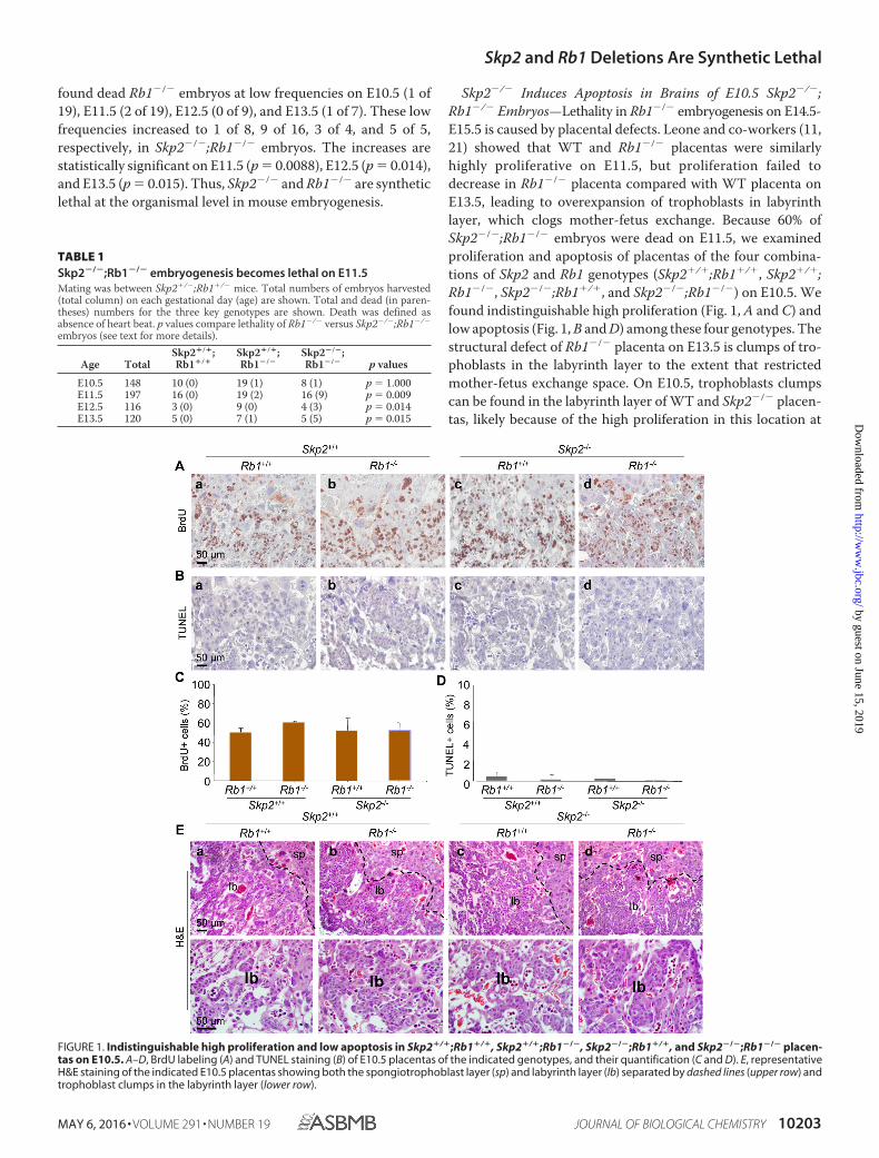
Skp2�/� Induces Apoptosis in Brains of E10.5 Skp2�/�;Rb1�/� Embryos—Lethality in Rb1�/� embryogenesis on E14.5-E15.5 is caused by placental defects. Leone and co-workers (11,21) showed that WT and Rb1�/� placentas were similarlyhighly proliferative on E11.5, but proliferation failed todecrease in Rb1�/� placenta compared with WT placenta onE13.5, leading to overexpansion of trophoblasts in labyrinthlayer, which clogs mother-fetus exchange. Because 60% ofSkp2�/�;Rb1�/� embryos were dead on E11.5, we examinedproliferation and apoptosis of placentas of the four combina-tions of Skp2 and Rb1 genotypes (Skp2�/�;Rb1�/�, Skp2�/�;Rb1�/�, Skp2�/�;Rb1�/�, and Skp2�/�;Rb1�/�) on E10.5. Wefound indistinguishable high proliferation (Fig. 1, A and C) andlow apoptosis (Fig. 1, B and D) among these four genotypes. Thestructural defect of Rb1�/� placenta on E13.5 is clumps of tro-phoblasts in the labyrinth layer to the extent that restrictedmother-fetus exchange space. On E10.5, trophoblasts clumpscan be found in the labyrinth layer of WT and Skp2�/� placen-tas, likely because of the high proliferation in this location at
FIGURE 1. Indistinguishable high proliferation and low apoptosis in Skp2�/�;Rb1�/�, Skp2�/�;Rb1�/�, Skp2�/�;Rb1�/�, and Skp2�/�;Rb1�/� placen-tas on E10.5. A–D, BrdU labeling (A) and TUNEL staining (B) of E10.5 placentas of the indicated genotypes, and their quantification (C and D). E, representativeH&E staining of the indicated E10.5 placentas showing both the spongiotrophoblast layer (sp) and labyrinth layer (lb) separated by dashed lines (upper row) andtrophoblast clumps in the labyrinth layer (lower row).
TABLE 1Skp2�/�;Rb1�/� embryogenesis becomes lethal on E11.5Mating was between Skp2�/�;Rb1�/� mice. Total numbers of embryos harvested(total column) on each gestational day (age) are shown. Total and dead (in paren-theses) numbers for the three key genotypes are shown. Death was defined asabsence of heart beat. p values compare lethality of Rb1�/� versus Skp2�/�;Rb1�/�
embryos (see text for more details).
Age TotalSkp2�/�;Rb1�/�
Skp2�/�;Rb1�/�
Skp2�/�;Rb1�/� p values
E10.5 148 10 (0) 19 (1) 8 (1) p � 1.000E11.5 197 16 (0) 19 (2) 16 (9) p � 0.009E12.5 116 3 (0) 9 (0) 4 (3) p � 0.014E13.5 120 5 (0) 7 (1) 5 (5) p � 0.015
Skp2 and Rb1 Deletions Are Synthetic Lethal
MAY 6, 2016 • VOLUME 291 • NUMBER 19 JOURNAL OF BIOLOGICAL CHEMISTRY 10203
by guest on June 15, 2019http://w
ww
.jbc.org/D
ownloaded from
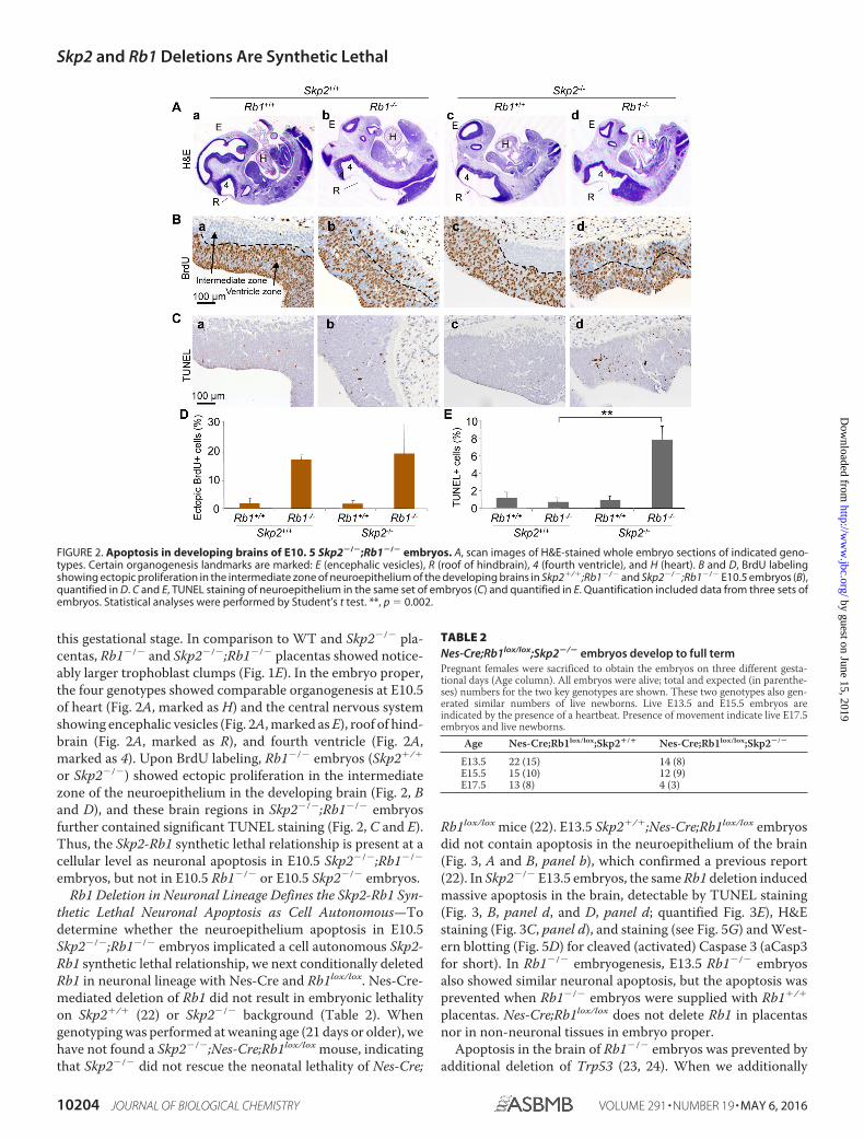
this gestational stage. In comparison to WT and Skp2�/� pla-centas, Rb1�/� and Skp2�/�;Rb1�/� placentas showed notice-ably larger trophoblast clumps (Fig. 1E). In the embryo proper,the four genotypes showed comparable organogenesis at E10.5of heart (Fig. 2A, marked as H) and the central nervous systemshowing encephalic vesicles (Fig. 2A, marked as E), roof of hind-brain (Fig. 2A, marked as R), and fourth ventricle (Fig. 2A,marked as 4). Upon BrdU labeling, Rb1�/� embryos (Skp2�/�
or Skp2�/�) showed ectopic proliferation in the intermediatezone of the neuroepithelium in the developing brain (Fig. 2, Band D), and these brain regions in Skp2�/�;Rb1�/� embryosfurther contained significant TUNEL staining (Fig. 2, C and E).Thus, the Skp2-Rb1 synthetic lethal relationship is present at acellular level as neuronal apoptosis in E10.5 Skp2�/�;Rb1�/�
embryos, but not in E10.5 Rb1�/� or E10.5 Skp2�/� embryos.Rb1 Deletion in Neuronal Lineage Defines the Skp2-Rb1 Syn-
thetic Lethal Neuronal Apoptosis as Cell Autonomous—Todetermine whether the neuroepithelium apoptosis in E10.5Skp2�/�;Rb1�/� embryos implicated a cell autonomous Skp2-Rb1 synthetic lethal relationship, we next conditionally deletedRb1 in neuronal lineage with Nes-Cre and Rb1lox/lox. Nes-Cre-mediated deletion of Rb1 did not result in embryonic lethalityon Skp2�/� (22) or Skp2�/� background (Table 2). Whengenotyping was performed at weaning age (21 days or older), wehave not found a Skp2�/�;Nes-Cre;Rb1lox/lox mouse, indicatingthat Skp2�/� did not rescue the neonatal lethality of Nes-Cre;
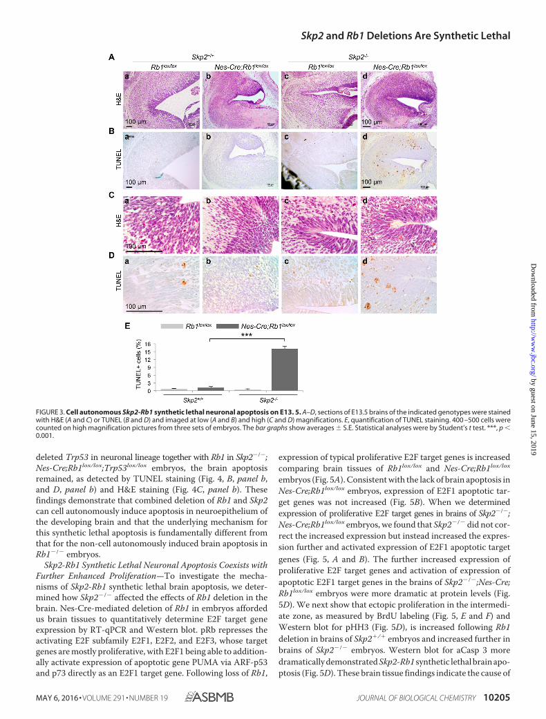
Rb1lox/lox mice (22). E13.5 Skp2�/�;Nes-Cre;Rb1lox/lox embryosdid not contain apoptosis in the neuroepithelium of the brain(Fig. 3, A and B, panel b), which confirmed a previous report(22). In Skp2�/� E13.5 embryos, the same Rb1 deletion inducedmassive apoptosis in the brain, detectable by TUNEL staining(Fig. 3, B, panel d, and D, panel d; quantified Fig. 3E), H&Estaining (Fig. 3C, panel d), and staining (see Fig. 5G) and West-ern blotting (Fig. 5D) for cleaved (activated) Caspase 3 (aCasp3for short). In Rb1�/� embryogenesis, E13.5 Rb1�/� embryosalso showed similar neuronal apoptosis, but the apoptosis wasprevented when Rb1�/� embryos were supplied with Rb1�/�
placentas. Nes-Cre;Rb1lox/lox does not delete Rb1 in placentasnor in non-neuronal tissues in embryo proper.
Apoptosis in the brain of Rb1�/� embryos was prevented byadditional deletion of Trp53 (23, 24). When we additionally
FIGURE 2. Apoptosis in developing brains of E10. 5 Skp2�/�;Rb1�/� embryos. A, scan images of H&E-stained whole embryo sections of indicated geno-types. Certain organogenesis landmarks are marked: E (encephalic vesicles), R (roof of hindbrain), 4 (fourth ventricle), and H (heart). B and D, BrdU labelingshowing ectopic proliferation in the intermediate zone of neuroepithelium of the developing brains in Skp2�/�;Rb1�/� and Skp2�/�;Rb1�/� E10.5 embryos (B),quantified in D. C and E, TUNEL staining of neuroepithelium in the same set of embryos (C) and quantified in E. Quantification included data from three sets ofembryos. Statistical analyses were performed by Student’s t test. **, p � 0.002.
TABLE 2Nes-Cre;Rb1lox/lox;Skp2�/� embryos develop to full termPregnant females were sacrificed to obtain the embryos on three different gesta-tional days (Age column). All embryos were alive; total and expected (in parenthe-ses) numbers for the two key genotypes are shown. These two genotypes also gen-erated similar numbers of live newborns. Live E13.5 and E15.5 embryos areindicated by the presence of a heartbeat. Presence of movement indicate live E17.5embryos and live newborns.
Age Nes-Cre;Rb1lox/lox;Skp2�/� Nes-Cre;Rb1lox/lox;Skp2�/�
E13.5 22 (15) 14 (8)E15.5 15 (10) 12 (9)E17.5 13 (8) 4 (3)
Skp2 and Rb1 Deletions Are Synthetic Lethal
10204 JOURNAL OF BIOLOGICAL CHEMISTRY VOLUME 291 • NUMBER 19 • MAY 6, 2016
by guest on June 15, 2019http://w
ww
.jbc.org/D
ownloaded from
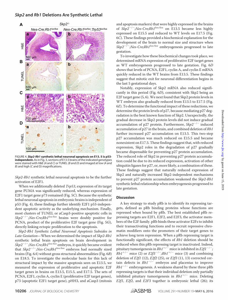
deleted Trp53 in neuronal lineage together with Rb1 in Skp2�/�;Nes-Cre;Rb1lox/lox;Trp53lox/lox embryos, the brain apoptosisremained, as detected by TUNEL staining (Fig. 4, B, panel b,and D, panel b) and H&E staining (Fig. 4C, panel b). Thesefindings demonstrate that combined deletion of Rb1 and Skp2can cell autonomously induce apoptosis in neuroepithelium ofthe developing brain and that the underlying mechanism forthis synthetic lethal apoptosis is fundamentally different fromthat for the non-cell autonomously induced brain apoptosis inRb1�/� embryos.
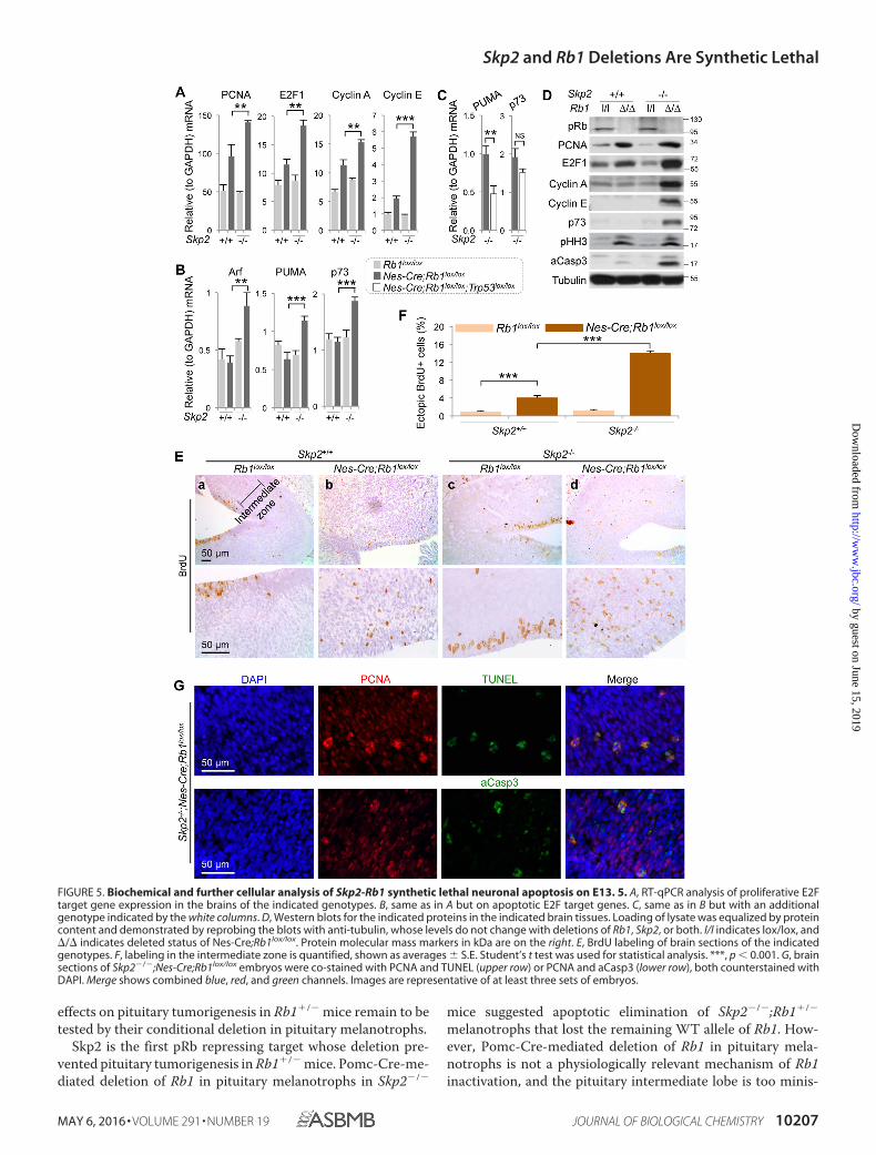
Skp2-Rb1 Synthetic Lethal Neuronal Apoptosis Coexists withFurther Enhanced Proliferation—To investigate the mecha-nisms of Skp2-Rb1 synthetic lethal brain apoptosis, we deter-mined how Skp2�/� affected the effects of Rb1 deletion in thebrain. Nes-Cre-mediated deletion of Rb1 in embryos affordedus brain tissues to quantitatively determine E2F target geneexpression by RT-qPCR and Western blot. pRb represses theactivating E2F subfamily E2F1, E2F2, and E2F3, whose targetgenes are mostly proliferative, with E2F1 being able to addition-ally activate expression of apoptotic gene PUMA via ARF-p53and p73 directly as an E2F1 target gene. Following loss of Rb1,
expression of typical proliferative E2F target genes is increasedcomparing brain tissues of Rb1lox/lox and Nes-Cre;Rb1lox/lox
embryos (Fig. 5A). Consistent with the lack of brain apoptosis inNes-Cre;Rb1lox/lox embryos, expression of E2F1 apoptotic tar-get genes was not increased (Fig. 5B). When we determinedexpression of proliferative E2F target genes in brains of Skp2�/�;Nes-Cre;Rb1lox/lox embryos, we found that Skp2�/� did not cor-rect the increased expression but instead increased the expres-sion further and activated expression of E2F1 apoptotic targetgenes (Fig. 5, A and B). The further increased expression ofproliferative E2F target genes and activation of expression ofapoptotic E2F1 target genes in the brains of Skp2�/�;Nes-Cre;Rb1lox/lox embryos were more dramatic at protein levels (Fig.5D). We next show that ectopic proliferation in the intermedi-ate zone, as measured by BrdU labeling (Fig. 5, E and F) andWestern blot for pHH3 (Fig. 5D), is increased following Rb1deletion in brains of Skp2�/� embryos and increased further inbrains of Skp2�/� embryos. Western blot for aCasp 3 moredramatically demonstrated Skp2-Rb1 synthetic lethal brain apo-ptosis (Fig. 5D). These brain tissue findings indicate the cause of
FIGURE 3. Cell autonomous Skp2-Rb1 synthetic lethal neuronal apoptosis on E13. 5. A–D, sections of E13.5 brains of the indicated genotypes were stainedwith H&E (A and C) or TUNEL (B and D) and imaged at low (A and B) and high (C and D) magnifications. E, quantification of TUNEL staining. 400 –500 cells werecounted on high magnification pictures from three sets of embryos. The bar graphs show averages S.E. Statistical analyses were by Student’s t test. ***, p �0.001.
Skp2 and Rb1 Deletions Are Synthetic Lethal
MAY 6, 2016 • VOLUME 291 • NUMBER 19 JOURNAL OF BIOLOGICAL CHEMISTRY 10205
by guest on June 15, 2019http://w
ww
.jbc.org/D
ownloaded from
Skp2-Rb1 synthetic lethal neuronal apoptosis to be the furtheractivation of E2F1.
When we additionally deleted Trp53, expression of its targetgene PUMA was significantly reduced, whereas expression ofE2F1 target gene p73 remained (Fig. 5C). Because the syntheticlethal neuronal apoptosis in embryonic brains is independent ofp53 (Fig. 4), these findings further identify E2F1 p53-indepen-dent apoptotic activity as the underlying mechanism. Finally,most clusters of TUNEL or aCasp3-positive apoptotic cells inSkp2�/�;Nes-Cre;Rb1lox/lox brains were doubly positive forPCNA, product of the proliferative E2F target gene (Fig. 5G),directly linking ectopic proliferation to the apoptosis.
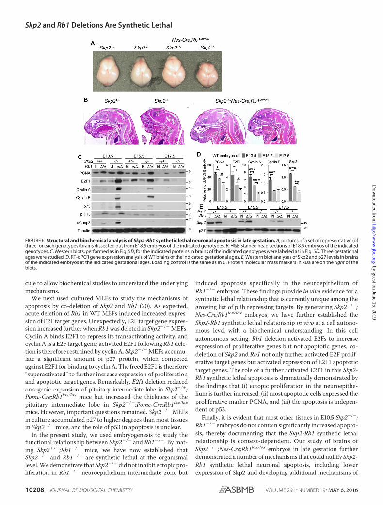
Skp2-Rb1 Synthetic Lethal Neuronal Apoptosis Subsides inLate Gestation—When we determined the impact of Skp2-Rb1synthetic lethal brain apoptosis on brain development inSkp2�/�;Nes-Cre;Rb1lox/lox embryos, it quickly became evidentthat Skp2�/�;Nes-Cre;Rb1lox/lox embryos had normally sizedbrains (Fig. 6A) without gross structural abnormalities (Fig. 6B)on E18.5. To investigate the molecular basis for this lack ofstructural impact by the massive apoptosis seen on E13.5, wemeasured the expression of proliferative and apoptotic E2Ftarget genes in brains on E13.5, E15.5, and E17.5. The sets ofPCNA, E2F1, cyclin A, cyclin E (proliferative E2F target genes),p73 (apoptotic E2F1 target gene), pHH3, and aCasp3 (mitosis
and apoptosis markers) that were highly expressed in the brainsof Skp2�/�;Nes-Cre;Rb1lox/lox on E13.5 became less highlyexpressed on E15.5 and reduced to WT levels on E17.5 (Fig.6C). These findings provided a biochemical explanation for thedevelopment of the brain to normal size and structure whenSkp2�/�;Nes-Cre;Rb1lox/lox embryogenesis progressed to lategestation.
To investigate how these biochemical changes took place, wedetermined mRNA expression of proliferative E2F target genesas WT embryogenesis progressed to late gestation. Fig. 6Dshows that levels of PCNA, E2F1, cyclin A, and cyclin E mRNAquickly reduced in the WT brains from E13.5. These findingssuggest that mitotic exit for neuronal differentiation begins inthe last 5 gestational days.
Notably, expression of Skp2 mRNA also reduced signifi-cantly in this period (Fig. 6D), consistent with Skp2 being anE2F target gene (5, 6). We next found that Skp2 protein levels inWT embryos also gradually reduced from E13.5 to E17.5 (Fig.6E). To determine the functional impact of these reductions, wedetermine the protein levels of p27, because mediating p27 deg-radation is the best known function of Skp2. Unexpectedly, thegradual decrease in Skp2 protein levels did not induce gradualaccumulation of p27 protein. Furthermore, Skp2�/� inducedaccumulation of p27 in the brain, and combined deletion of Rb1further increased p27 accumulation on E13.5. This two-stepp27 accumulation was much reduced on E15.5 and becamenonexistent on E17.5. These findings suggest that, with reducedexpression, Skp2 roles in the degradation of p27 graduallybecame dispensable for preventing p27 protein accumulation.The reduced role of Skp2 in preventing p27 protein accumula-tion could be due to its reduced expression, activation of otherubiquitin ligases for p27, or, more likely, a combination of these.These findings suggest that naturally reduced expression ofSkp2 and naturally increased Skp2-independent mechanismsto prevent p27 protein accumulation weakened the Skp2-Rb1synthetic lethal relationship when embryogenesis progressed tolate gestation.
Discussion
A key strategy to study pRb is to identify its repressing tar-gets, defined as pRb binding proteins whose functions arerepressed when bound by pRb. The best established pRb re-pressing targets are E2F1, E2F2, and E2F3, the activator mem-bers of the E2F family. pRb binds these activator E2Fs to inhibittheir transactivating functions and to recruit repressive chro-matin modifiers onto the promoters of their target genes toachieve long term repression. When a pRb repressing target isfunctionally significant, the effects of Rb1 deletion should bereduced when this pRb repressing target is inactivated. Indeed,pituitary tumorigenesis in Rb1�/� mice is inhibited in E2f1�/�;Rb1�/� mice (2) or E2f3�/�;Rb1�/� mice (3) and combiningdeletion of E2f1 (12), E2f2 (25), or E2f3 (11, 13) corrected cer-tain defects in Rb1�/� embryos and placentas to improveRb1�/� embryogenesis. A weakness shared by these three pRbrepressing targets is that their individual deletion only partiallyinhibited pituitary tumorigenesis in Rb1�/� mice. DeletingE2f1, E2f2, and E2F3 together is embryonic lethal (26); its
FIGURE 4. Skp2-Rb1 synthetic lethal neuronal apoptosis on E13. 5 is p53-independent. As in Fig. 3, sections of E13.5 brains of the indicated genotypeswere stained with H&E (A and C) or TUNEL (B and D) and imaged at low (A andB) and high (C and D) magnifications.
Skp2 and Rb1 Deletions Are Synthetic Lethal
10206 JOURNAL OF BIOLOGICAL CHEMISTRY VOLUME 291 • NUMBER 19 • MAY 6, 2016
by guest on June 15, 2019http://w
ww
.jbc.org/D
ownloaded from
effects on pituitary tumorigenesis in Rb1�/� mice remain to betested by their conditional deletion in pituitary melanotrophs.
Skp2 is the first pRb repressing target whose deletion pre-vented pituitary tumorigenesis in Rb1�/� mice. Pomc-Cre-me-diated deletion of Rb1 in pituitary melanotrophs in Skp2�/�
mice suggested apoptotic elimination of Skp2�/�;Rb1�/�
melanotrophs that lost the remaining WT allele of Rb1. How-ever, Pomc-Cre-mediated deletion of Rb1 in pituitary mela-notrophs is not a physiologically relevant mechanism of Rb1inactivation, and the pituitary intermediate lobe is too minis-
FIGURE 5. Biochemical and further cellular analysis of Skp2-Rb1 synthetic lethal neuronal apoptosis on E13. 5. A, RT-qPCR analysis of proliferative E2Ftarget gene expression in the brains of the indicated genotypes. B, same as in A but on apoptotic E2F target genes. C, same as in B but with an additionalgenotype indicated by the white columns. D, Western blots for the indicated proteins in the indicated brain tissues. Loading of lysate was equalized by proteincontent and demonstrated by reprobing the blots with anti-tubulin, whose levels do not change with deletions of Rb1, Skp2, or both. l/l indicates lox/lox, and/ indicates deleted status of Nes-Cre;Rb1lox/lox. Protein molecular mass markers in kDa are on the right. E, BrdU labeling of brain sections of the indicatedgenotypes. F, labeling in the intermediate zone is quantified, shown as averages S.E. Student’s t test was used for statistical analysis. ***, p � 0.001. G, brainsections of Skp2�/�;Nes-Cre;Rb1lox/lox embryos were co-stained with PCNA and TUNEL (upper row) or PCNA and aCasp3 (lower row), both counterstained withDAPI. Merge shows combined blue, red, and green channels. Images are representative of at least three sets of embryos.
Skp2 and Rb1 Deletions Are Synthetic Lethal
MAY 6, 2016 • VOLUME 291 • NUMBER 19 JOURNAL OF BIOLOGICAL CHEMISTRY 10207
by guest on June 15, 2019http://w
ww
.jbc.org/D
ownloaded from
cule to allow biochemical studies to understand the underlyingmechanisms.
We next used cultured MEFs to study the mechanisms ofapoptosis by co-deletion of Skp2 and Rb1 (20). As expected,acute deletion of Rb1 in WT MEFs induced increased expres-sion of E2F target genes. Unexpectedly, E2F target gene expres-sion increased further when Rb1 was deleted in Skp2�/� MEFs.Cyclin A binds E2F1 to repress its transactivating activity, andcyclin A is a E2F target gene; activated E2F1 following Rb1 dele-tion is therefore restrained by cyclin A. Skp2�/� MEFs accumu-late a significant amount of p27 protein, which competedagainst E2F1 for binding to cyclin A. The freed E2F1 is therefore“superactivated” to further increase expression of proliferationand apoptotic target genes. Remarkably, E2f1 deletion reducedoncogenic expansion of pituitary intermediate lobe in Skp2�/�;Pomc-Cre;Rb1lox/lox mice but increased the thickness of thepituitary intermediate lobe in Skp2�/�;Pomc-Cre;Rb1lox/lox
mice. However, important questions remained. Skp2�/� MEFsin culture accumulated p27 to higher degrees than most tissuesin Skp2�/� mice, and the role of p53 in apoptosis is unclear.
In the present study, we used embryogenesis to study thefunctional relationship between Skp2�/� and Rb1�/�. By mat-ing Skp2�/�;Rb1�/� mice, we have now established thatSkp2�/� and Rb1�/� are synthetic lethal at the organismallevel. We demonstrate that Skp2�/� did not inhibit ectopic pro-liferation in Rb1�/� neuroepithelium intermediate zone but
induced apoptosis specifically in the neuroepithelium ofRb1�/� embryos. These findings provide in vivo evidence for asynthetic lethal relationship that is currently unique among thegrowing list of pRb repressing targets. By generating Skp2�/�;Nes-Cre;Rb1lox/lox embryos, we have further established theSkp2-Rb1 synthetic lethal relationship in vivo at a cell autono-mous level with a biochemical understanding. In this cellautonomous setting, Rb1 deletion activated E2Fs to increaseexpression of proliferative genes but not apoptotic genes; co-deletion of Skp2 and Rb1 not only further activated E2F prolif-erative target genes but activated expression of E2F1 apoptotictarget genes. The role of a further activated E2F1 in this Skp2-Rb1 synthetic lethal apoptosis is dramatically demonstrated bythe findings that (i) ectopic proliferation in the neuroepithe-lium is further increased, (ii) most apoptotic cells expressed theproliferative marker PCNA, and (iii) the apoptosis is indepen-dent of p53.
Finally, it is evident that most other tissues in E10.5 Skp2�/�;Rb1�/� embryos do not contain significantly increased apopto-sis, thereby documenting that the Skp2-Rb1 synthetic lethalrelationship is context-dependent. Our study of brains ofSkp2�/�;Nes-Cre;Rb1lox/lox embryos in late gestation furtherdemonstrated a number of mechanisms that could nullify Skp2-Rb1 synthetic lethal neuronal apoptosis, including lowerexpression of Skp2 and developing additional mechanisms of
FIGURE 6. Structural and biochemical analysis of Skp2-Rb1 synthetic lethal neuronal apoptosis in late gestation. A, pictures of a set of representative (ofthree for each genotypes) brains dissected out from E18.5 embryos of the indicated genotypes. B, H&E-stained head sections of E18.5 embryos of the indicatedgenotypes. C, Western blots, performed as in Fig. 5D, for the indicated proteins in brains of the indicated genotypes were labeled as in Fig. 5D. Three gestationalages were studied. D, RT-qPCR gene expression analysis of WT brains of the indicated gestational ages. E, Western blot analyses of Skp2 and p27 levels in brainsof the indicated embryos at the indicated gestational ages. Loading control is the same as in C. Protein molecular mass markers in kDa are on the right of theblots.
Skp2 and Rb1 Deletions Are Synthetic Lethal
10208 JOURNAL OF BIOLOGICAL CHEMISTRY VOLUME 291 • NUMBER 19 • MAY 6, 2016
by guest on June 15, 2019http://w
ww
.jbc.org/D
ownloaded from
p27 degradation to render Skp2 dispensable in preventing p27accumulation.
Inactivation of pRb is a highly recurrent feature of cancercells, and inhibiting pRb repressing targets is a logical andintensively pursued therapeutic strategy for pRb-deficient can-cer. The identification of Skp2-Rb1 synthetic lethal relationshipilluminated a highly attractive new strategy for pRb-deficientcancer. It is increasingly clear that cancer as an heterogeneousdisease cannot be cured by a single treatment, no matter howeffective it might be for certain cases. It is further evident thatno matter how effective a treatment is at the beginning, canceralways evolves to resist it. The Skp2-Rb1 synthetic lethal apo-ptosis in embryogenesis show these two features, and our find-ings suggest the cancer characteristics that are best suited forthe Skp2-Rb1 synthetic lethal treatment strategy (genetic inac-tivation of pRb with high expression of Skp2), how these can-cers might develop resistance to this treatment (select for addi-tional abilities to degrade p27), and how the resistant cancermight be treated (combining targeting the other p27 ubiquitinligases).
Author Contributions—H. Z., H. W., and L. Z. conceived and coor-dinated the study. H. Z., H. W., F. B., Z. L., H. F., and J. C. performedthe experiments. H. Z. and L. Z. wrote the paper.
Acknowledgments—We thank Dr. Keiko Nakayama and Dr. KeiichNakayama for providing the Skp2 KO mice.
References1. Trimarchi, J. M., and Lees, J. A. (2002) Sibling rivalry in the E2F family.
Nat. Rev. Mol. Cell Biol. 3, 11–202. Yamasaki, L., Bronson, R., Williams, B. O., Dyson, N. J., Harlow, E., and
Jacks, T. (1998) Loss of E2F-1 reduces tumorigenesis and extends thelifespan of Rb1�/� mice. Nat. Genet. 18, 360 –364
3. Ziebold, U., Lee, E. Y., Bronson, R. T., and Lees, J. A. (2003) E2F3 loss hasopposing effects on different pRB-deficient tumors, resulting in suppres-sion of pituitary tumors but metastasis of medullary thyroid carcinomas.Mol. Cell Biol. 23, 6542– 6552
4. Ji, P., Jiang, H., Rekhtman, K., Bloom, J., Ichetovkin, M., Pagano, M., andZhu, L. (2004) An Rb-Skp2-p27 pathway mediates acute cell cycle inhibi-tion by Rb and is retained in a partial-penetrance Rb mutant. Mol. Cell 16,47–58
5. Zhang, L., and Wang, C. (2006) F-box protein Skp2: a novel transcriptionaltarget of E2F. Oncogene 25, 2615–2627
6. Yung, Y., Walker, J. L., Roberts, J. M., and Assoian, R. K. (2007) A Skp2autoinduction loop and restriction point control. J. Cell Biol. 178,741–747
7. Binné, U. K., Classon, M. K., Dick, F. A., Wei, W., Rape, M., Kaelin, W. G.,Jr., Näär, A. M., and Dyson, N. J. (2007) Retinoblastoma protein and ana-phase-promoting complex physically interact and functionally cooperateduring cell-cycle exit. Nat. Cell Biol. 9, 225–232
8. Lasorella, A., Rothschild, G., Yokota, Y., Russell, R. G., and Iavarone, A.(2005) Id2 mediates tumor initiation, proliferation, and angiogenesis in Rbmutant mice. Mol. Cell Biol. 25, 3563–3574
9. Jacks, T., Fazeli, A., Schmitt, E. M., Bronson, R. T., Goodell, M. A., andWeinberg, R. A. (1992) Effects of an Rb mutation in the mouse. Nature359, 295–300
10. Lee, E. Y., Chang, C.-Y., Hu, N., Wang, Y.-C., Lai, C.-C., Herrup, K., Lee,W.-H., and Bradley, A. (1992) Mice deficient for Rb are nonviable and
show defects in neurogenesis and haematopoiesis. Nature 359, 288 –29411. Wenzel, P. L., Wu, L., de Bruin, A., Chong, J. L., Chen, W. Y., Dureska, G.,
Sites, E., Pan, T., Sharma, A., Huang, K., Ridgway, R., Mosaliganti, K.,Sharp, R., Machiraju, R., Saltz, J., Yamamoto, H., Cross, J. C., Robinson,M. L., and Leone, G. (2007) Rb is critical in a mammalian tissue stem cellpopulation. Genes Dev. 21, 85–97
12. Tsai, K. Y., Hu, Y., Macleod, K. F., Crowley, D., Yamasaki, L., and Jacks, T.(1998) Mutation of E2f-1 suppresses apoptosis and inappropriate S phaseentry and extends survival of Rb-deficient mouse embryos. Mol. Cell 2,293–304
13. Ziebold, U., Reza, T., Caron, A., and Lees, J. A. (2001) E2F3 contributesboth to the inappropriate proliferation and to the apoptosis arising in Rbmutant embryos. Genes Dev. 15, 386 –391
14. Lasorella, A., Noseda, M., Beyna, M., Yokota, Y., and Iavarone, A. (2000)Id2 is a retinoblastoma protein target and mediates signalling by Myconcoproteins. Nature 407, 592–598
15. Sage, J., Miller, A. L., Pérez-Mancera, P. A., Wysocki, J. M., and Jacks, T.(2003) Acute mutation of retinoblastoma gene function is sufficient forcell cycle re-entry. Nature 424, 223–228
16. Nakayama, K., Nagahama, H., Minamishima, Y. A., Matsumoto, M., Na-kamichi, I., Kitagawa, K., Shirane, M., Tsunematsu, R., Tsukiyama, T.,Ishida, N., Kitagawa, M., Nakayama, K., and Hatakeyama, S. (2000) Tar-geted disruption of Skp2 results in accumulation of cyclin E andp27(Kip1), polyploidy and centrosome overduplication. EMBO J. 19,2069 –2081
17. Marino, S., Vooijs, M., van Der Gulden, H., Jonkers, J., and Berns, A. (2000)Induction of medulloblastomas in p53-null mutant mice by somatic inac-tivation of Rb in the external granular layer cells of the cerebellum. GenesDev. 14, 994 –1004
18. Wang, H., Bauzon, F., Ji, P., Xu, X., Sun, D., Locker, J., Sellers, R. S., Na-kayama, K., Nakayama, K. I., Cobrinik, D., and Zhu, L. (2010) Skp2 isrequired for survival of aberrantly proliferating Rb1-deficient cells and fortumorigenesis in Rb1�/� mice. Nat. Genet. 42, 83– 88
19. Zhao, H., Bauzon, F., Fu, H., Lu, Z., Cui, J., Nakayama, K., Nakayama, K. I.,Locker, J., and Zhu, L. (2013) Skp2 deletion unmasks a p27 safeguard thatblocks tumorigenesis in the absence of pRb and p53 tumor suppressors.Cancer Cell 24, 645– 659
20. Lu, Z., Bauzon, F., Fu, H., Cui, J., Zhao, H., Nakayama, K., Nakayama, K. I.,and Zhu, L. (2014) Skp2 suppresses apoptosis in Rb1-deficient tumours bylimiting E2F1 activity. Nat. Commun. 5, 3463
21. Wu, L., de Bruin, A., Saavedra, H. I., Starovic, M., Trimboli, A., Yang, Y.,Opavska, J., Wilson, P., Thompson, J. C., Ostrowski, M. C., Rosol, T. J.,Woollett, L. A., Weinstein, M., Cross, J. C., Robinson, M. L., and Leone, G.(2003) Extra-embryonic function of Rb is essential for embryonic devel-opment and viability. Nature 421, 942–947
22. MacPherson, D., Sage, J., Crowley, D., Trumpp, A., Bronson, R. T., andJacks, T. (2003) Conditional mutation of Rb causes cell cycle defects with-out apoptosis in the central nervous system. Mol. Cell Biol. 23, 1044 –1053
23. Morgenbesser, S. D., Williams, B. O., Jacks, T., and DePinho, R. A. (1994)p53-dependent apoptosis produced by Rb-deficiency in the developingmouse lens. Nature 371, 72–74
24. Macleod, K. F., Hu, Y., and Jacks, T. (1996) Loss of Rb activates bothp53-dependent and independent cell death pathways in the developingmouse nervous system. EMBO J. 15, 6178 – 6188
25. Dirlam, A., Spike, B. T., and Macleod, K. F. (2007) Deregulated E2f-2underlies cell cycle and maturation defects in retinoblastoma null eryth-roblasts. Mol. Cell Biol. 27, 8713– 8728
26. Chong, J. L., Wenzel, P. L., Sáenz-Robles, M. T., Nair, V., Ferrey, A., Hagan,J. P., Gomez, Y. M., Sharma, N., Chen, H. Z., Ouseph, M., Wang, S. H.,Trikha, P., Culp, B., Mezache, L., Winton, D. J., Sansom, O. J., Chen, D.,Bremner, R., Cantalupo, P. G., Robinson, M. L., Pipas, J. M., and Leone, G.(2009) E2f1–3 switch from activators in progenitor cells to repressors indifferentiating cells. Nature 462, 930 –934
Skp2 and Rb1 Deletions Are Synthetic Lethal
MAY 6, 2016 • VOLUME 291 • NUMBER 19 JOURNAL OF BIOLOGICAL CHEMISTRY 10209
by guest on June 15, 2019http://w
ww
.jbc.org/D
ownloaded from
and Liang ZhuHongling Zhao, Hongbo Wang, Frederick Bauzon, Zhonglei Lu, Hao Fu, Jinhua Cui
) Are Synthetic Lethal in Mouse EmbryogenesisSkp2Kinase-associated protein 2 () and Its Repressing Target S PhaseRb1Deletions of Retinoblastoma 1 (
doi: 10.1074/jbc.M116.718049 originally published online March 10, 20162016, 291:10201-10209.J. Biol. Chem.
10.1074/jbc.M116.718049Access the most updated version of this article at doi:
Alerts:
When a correction for this article is posted•
When this article is cited•
to choose from all of JBC's e-mail alertsClick here
http://www.jbc.org/content/291/19/10201.full.html#ref-list-1
This article cites 26 references, 9 of which can be accessed free at
by guest on June 15, 2019http://w
ww
.jbc.org/D
ownloaded from