Embed Size (px)
Citation preview

SKP2 Oncogene Is a Direct MYC Target Gene and MYCDown-regulates p27KIP1 through SKP2 in Human LeukemiaCells*□S
Received for publication, July 29, 2010, and in revised form, January 13, 2011 Published, JBC Papers in Press, January 18, 2011, DOI 10.1074/jbc.M110.165977
Gabriel Bretones‡1, Juan C. Acosta‡1,2, Juan M. Caraballo‡1, Nuria Ferrandiz‡1,2, M. Teresa Gomez-Casares§,Marta Albajar‡¶, Rosa Blanco‡, Paula Ruiz‡, Wen-Chun Hung�, M. Pilar Albero**, Ignacio Perez-Roger**,and Javier Leon‡3
From the ‡Departamento de Biología Molecular, Instituto de Biomedicina y Biotecnología de Cantabria, Universidad de Cantabria,Consejo Superior de Investigaciones Cientıficas, SODERCAN (Sociedad para el Desarrollo de Cantabria), 39011 Santander, Spain,the §Servicio de Hematología, Hospital Dr. Negrín, 35020 Las Palmas de Gran Canaria, Spain, the ¶Servicio de Hematología,Hospital Universitario Marques de Valdecilla-Instituto de Investigacion y Formacion Marques de Valdecilla, 39009 Santander,Spain, the �Institute of Biomedical Sciences, National Sun Yat-Sen University, Kaohsiung 804, Republic of China, and the**Department of Chemistry, Biochemistry and Molecular Biology, Cardenal Herrera-CEU University, 46113 Moncada, Spain
SKP2 is the ubiquitin ligase subunit that targets p27KIP1
(p27) for degradation. SKP2 is induced in the G1-S transit ofthe cell cycle, is frequently overexpressed in human cancer,and displays transformation activity in experimental models.Here we show that MYC induces SKP2 expression at themRNA and protein levels in human myeloid leukemia K562cells with conditional MYC expression. Importantly, in thesesystems, induction of MYC did not activate cell proliferation,ruling out SKP2 up-regulation as a consequence of cell cycleentry. MYC-dependent SKP2 expression was also detected inother cell types such as lymphoid, fibroblastic, and epithelialcell lines. MYC induced SKP2 mRNA expression in theabsence of protein synthesis and activated the SKP2 pro-moter in luciferase reporter assays. With chromatin immu-noprecipitation assays, MYC was detected bound to a regionof human SKP2 gene promoter that includes E-boxes. TheK562 cell line derives from human chronic myeloid leukemia.In a cohort of chronic myeloid leukemia bone marrow sam-ples, we found a correlation between MYC and SKP2 mRNAlevels. Analysis of cancer expression databases also indicateda correlation between MYC and SKP2 expression in lym-phoma. Finally, MYC-induced SKP2 expression resulted in adecrease in p27 protein in K562 cells. Moreover, silencing ofSKP2 abrogated the MYC-mediated down-regulation of p27.Our data show that SKP2 is a direct MYC target gene and thatMYC-mediated SKP2 induction leads to reduced p27 levels.The results suggest the induction of SKP2 oncogene as a newmechanism for MYC-dependent transformation.
c-MYC (hereafterMYC) is an oncogenic transcription factorof the helix-loop-helix/leucine zipper protein family. MYCexerts a wide array of biological functions in different cellularmodels related to cell cycle control, genomic instability, ener-getic metabolism, protein synthesis, intercellular communica-tion, and control of cell differentiation (for reviews see Refs.1–3). MYC forms heterodimers with the protein MAX thatbind to E-boxes in the regulatory regions of target genes. MYCregulates �1000 genes and binds to 15% of genomic loci (4–7).Most target genes are activated byMYC in an E-box-dependentmanner, although an important fraction of MYC target genesare repressed by MYC (reviewed in Refs. 8 and 9). Consistentwith the biological activities of MYC, its expression is deregu-lated in a wide array of human solid tumors and in leukemia,often associated to tumor progression (10–12).SKP2 is an oncogenic protein frequently overexpressed in
human cancers. Moreover, SKP2 behaves as an oncogene intransformation assays (13, 14). SKP2 encodes the F-box proteinof the ubiquitin ligase SCFSKP2 complex; this complex is com-prised by three core subunits RBX1, CUL1, and SKP1, whereasthe substrate is bound by SKP2 along with the small proteinCKS1 (14, 15). More than 20 SKP2 substrates have been found,including several proteins involved in cell cycle control (e.g.cyclin E, p21, p57, and E2F1) (14), and SKP2 can suppress p53-mediated apoptosis (16). Nonetheless, the main oncogenicmechanism of SKP2 is attributed to degradation of the cyclin-dependent kinase (CDK)4 inhibitor p27KIP1 (hereafter p27)(13).p27 was originally described as a CDK inhibitor, with cyclin
E-CDK2 complexes as its primary targets (17, 18). Low p27levels are associated with a poor prognosis in most tumors (19,20). p27 is regulated mainly at the protein stability level. Thedegradation of p27 by proteosomes is preceded by ubiquityla-tion (21), which is mediated by the SCFSKP2 complex, in whichSKP2 is the p27-recognizing subunit (22–24). To be recognized
* This study was supported by Grant PN-SAF08-01581 from the Spanish Min-isterio de Ciencia e Innovacion, Grant ISCIII-RETIC RD06/0020/0017 fromthe Spanish Ministerio de Sanidad y Consumo, a grant from University ofCantabria-IFIMAV (to J. L.), and Grants PI06-0285 and Proyecto UniversidadCardenal Herrera-Santander (to I. P.-R.).
□S The on-line version of this article (available at http://www.jbc.org) containssupplemental Table S1 and Figs. S1–S3.
1 Supported by fellowships from Spanish Ministerio de Ciencia e Innovacion.2 Present address: MRC Clinical Sciences Centre, Imperial College Faculty of
Medicine, Hammersmith Hospital Campus, London, United Kingdom.3 To whom correspondence should be addressed: Departamento de Biología
Molecular, Facultad de Medicina, IBBTEC, Cardenal Herrera Oria s/n, 39011Santander, Spain. Tel.: 34-942-201952; E-mail: [email protected].
4 The abbreviations used are: CDK, cyclin-dependent kinase; 4HT, 4-hydroxy-tamoxifen; CHX, cycloheximide; CML, chronic myeloid leukemia; TPA,phorbol-12-myristate-13-acetate; qPCR, quantitative PCR; sh, shorthairpin.
THE JOURNAL OF BIOLOGICAL CHEMISTRY VOL. 286, NO. 11, pp. 9815–9825, March 18, 2011© 2011 by The American Society for Biochemistry and Molecular Biology, Inc. Printed in the U.S.A.
MARCH 18, 2011 • VOLUME 286 • NUMBER 11 JOURNAL OF BIOLOGICAL CHEMISTRY 9815
by guest on May 25, 2018
http://ww
w.jbc.org/
Dow
nloaded from
by the SCFSKP2 complex, p27 must be bound to cyclin-CDKcomplexes and phosphorylated at Thr-187. p27 bound to cyclinE-CDK2 is a target for phosphorylation by the bound CDK2 ora second active cyclin E-CDK2 complex at Thr-187 (reviewedin Refs. 25 and 26). The oncogenic activity of SKP2 is attrib-uted mainly to low p27 levels, and consistently, the levels ofSKP2 and p27 inversely correlate in many tumors and cellmodels (13, 14).MYC and p27 show functional antagonism in proliferation:
MYC and the loss of p27 cooperate in animal carcinogenesismodels (27), and MYC abrogates p27 function in proliferation.The antagonistic effect of MYC is mediated through severalmechanisms (reviewed in Refs. 11). MYC down-regulatesmurine p27 at the transcriptional level (28, 29); in addition, itinduces cyclin D2 and CDK4, which sequester p27 in CDK-cyclin complexes (30, 31); finally, MYC induces expression ofCUL1 (32) and CKS1 (33), both components of the SCFSKP2complex. A correlation between MYC and SKP2 expressionwas recently described in amurine lymphomamodel, with onlymodest effects on p27 levels (34). The previous studies arenonetheless hampered by the fact that SKP2 is induced in cellcycle stimulation, which is also a well known MYC activity.This has made it difficult to distinguish whether SKP2 induc-tion is a direct effect of MYC. Here we studied the MYC-SKP2-p27 axis in different cell models with conditionalMYCexpression in which MYC is induced in cell cycle-arrestedcells.We show for here that SKP2 is a directMYC target geneand that MYC-mediated SKP2 up-regulation contributes top27 degradation.
EXPERIMENTAL PROCEDURES
Cell Lines and Cell Proliferation—K562 and K562-derivedcell lines were cultured in RPMI 1640mediumwith 8% fetal calfserum and antibiotics. Unless otherwise stated, the cells (2.5 �105 cells/ml) were treated with 75 �M ZnSO4, 200 nM 4-hy-droxytamoxifen (4HT) (Sigma), 10 nM phorbol-12-myristate-13-acetate (TPA), and 1 �M imatinib. Transient transfectionswere performed in a nucleofector (Amaxa) following the man-ufacturer’s instructions. Short hairpin expression vectors usedwere shMyc (35) and pSR-shSKP2 (36). KmycJ are K562 cellswith a MYC gene inducible by ZnSO4 (37). KMER4 are K562cells stably transfected with a vector expressing the MycERconstruct.5 MycER is activated by 4HT (38). Kp27-5 cells areK562 cells stably transfected with a p27 gene inducible by Zn2�
(39). Kp27MER are Kp27-5 cells transduced with the MycERgene (40). KmycBp53 are KmycB cells expressing a p53 mutantthat is activated at 32 °C (41). KMERshSKP2 are KMER4 cellsinfected with retrovirus expressing pSR-shSKP2 and selectedwith 1 �g/ml puromycin. pSR-shSKP2 was constructed byinserting the siRNA sequence AAGGGAGTGACAAA-GACTTTGTTCAAGAGACAAAGTCTTTGTCACTCCCTT(36) into theBglII andHindIII sites of pSUPER-retro vector (thesequence of the hairpin loop is underlined). KMERpSR areKMER4 cells infected with the empty retroviral vectorpSUPER-retro. TM1 are derived from the Mv1Lu lung epithe-lial cell line with a tetracyclin-repressible MYC gene (42).
HO15.19Myc-null rat fibroblasts were generated fromparentalTGR-1 by homologous recombination (43). TM1, HO15.19,and TGR1 cells were cultured in DMEM supplemented with10% fetal calf serum and antibiotics. P493-6 cells are immortal-ized human B cells expressing a tetracyclin-repressible MYCgene (44) and were cultured in RPMI 1640 with 10% fetal calfserum and antibiotics. Cell proliferationwas determined by cellcounting in a hemocytometer.Nude Mice Xenografts—KMERpSR or KMERshSKP2 cells
(107) were resuspended in 0.2 ml of RPMI/Geltrex (Invitrogen)(1:1). The cell suspension was injected subcutaneously into theright and left flanks of 6-week-old female athymic nude mice(Hsd:Athymic Nude-Foxn1 nu/nu; Harlan Laboratories mod-els); after 21–24 days, the mice were euthanized, and thetumors were weighed.mRNA Analysis—Total RNA from cell lines and bone mar-
row cells was isolated using the RNeasy kit (Qiagen). RT wasperformed with i-Script reverse transcriptase (Bio-Rad). Quan-titative PCR (qPCR) was performed with the SYBRGreen PCRkit (Bio-Rad). The sequences of primers used and ampliconsizes are shown in supplemental Table S1. The data were nor-malized to ribosomal protein S14 mRNA levels.Immunoblots—Total cell lysates and immunoblots were car-
ried out as described (39). The blots were developed with sec-ondary antibodies conjugated to IRDye680 and IRDye800 (Li-Cor Biosciences) and visualized in an Odyssey scanner. Theantibodies used were anti-actin (goat polyclonal, sc-1616),MYC (sc-764), SKP2 (sc-7164), p27 (sc-528), �-tubulin (sc-5546) (all rabbit polyclonals from Santa Cruz Biotechnology),p27 monoclonal antibody (K-25020; Transduction Labs), andThr(P)-187-p27 (rabbit polyclonal, 71-7700; Invitrogen).Chromatin Immunoprecipitation—The cells (5 � 107) were
fixed in 1% formaldehyde, lysedwith SDS, and sonicated, essen-tially as described (45). ChIP was performed using Dynabeads-Protein G (Dynal Biotech) coupled to anti-MYC antibody andrabbit IgG as specificity control. Quantitative PCR of elutedDNA was performed with the primers of SKP2 and lactatedehydrogenase genes indicated in supplemental Table S1.Immunofluorescence Analysis—Kp27MER cells were treated
withZnSO4 and/or 4HT for 24 h and immunostainedwith anti-SKP2 and -p27 antibodies as described and revealed with fluo-rescein- or rhodamine-conjugated secondary antibodies asdescribed (40). The cells were mounted with Vectashieldmounting medium (Vector Laboratories) with DAPI to visual-ize nuclei.Luciferase Assays—The cells (3 � 106) were electroporated
with an Amaxa electroporator and Mirus Ingenio (Mirus BioLLC) transfection reagent. KmycJ cells were transfected with 3�g of the �1148-SKP2 promoter (46) or pGL3basic vector(Promega) as control. K562 or KmycJ cells were nucleofectedwith 1.5 �g of 4�Ebox-Luc or the MYC-unresponsive4�EboxMut-Luc reporter constructs (47). K562 cells werenucleofected with 1.5 �g of pSR-shSKP2 or pSUPER-retro vec-tor. The cells were lysed, and luciferase activitywasmeasured induplicate in a dual luciferase reporter gene assay system (Pro-mega). The data were normalized to Renilla luciferase values(0.5 �g of pRL-TK vector in each transfection).5 M. Albajar and J. Leon, submitted for publication.
MYC-SKP2-p27 Axis in Human Leukemia Cells
9816 JOURNAL OF BIOLOGICAL CHEMISTRY VOLUME 286 • NUMBER 11 • MARCH 18, 2011
by guest on May 25, 2018
http://ww
w.jbc.org/
Dow
nloaded from

Chronic Myeloid Leukemia (CML) Samples—Bone marrowmononuclear cells (prepared by Ficoll-Hypaque) from 4healthy controls, 31 CML patients at diagnosis, and 8 patientsthat achieved the remission of the leukemia after treatment(Complete Molecular Response) (48) were studied. Thepatients were from the Hospital Universitario Marques de Val-decilla (Santander) and Hospital Universitario Dr. Negrín (LasPalmas). The study was approved by the ethics committees ofboth hospitals.
RESULTS
Induction of MYC Is Associated to Induction of SKP2—Westudied the MYC-SKP2 relationship in cell models in which (i)ectopic MYC expression is activated or induced and (ii) MYCinduction does not stimulate cell proliferation, a critical condi-tion to avoid SKP2 up-regulation subsequent to cell cycle pro-gression. In the Kp27MER cell line, p27 can be induced withZnSO4, and MYC can be activated with 4HT (Fig. 1A, toppanel). Large scale gene expression profiling in these cellsshowed that MYC activation provoked up-regulation of SKP2mRNA (40).We set out to validate this microarray data. Induc-tion of p27 resulted in rapid growth arrest (Fig. 1A) and SKP2expression was down-regulated (Fig. 1C), as predicted. Impor-tantly, MYC activation did not modify p27-mediated growtharrest in this model (Fig. 1A), confirming previous results (40).MYC activation (i.e. 4HT treatment) nonetheless resulted in a2-fold increase in SKP2 mRNA in proliferation-arrestedKp27MERcells (Fig. 1B, black bars).Moreover,MYCactivationinduced robust up-regulation of SKP2 protein, as detected byimmunoblot (Fig. 1C, compare lanes 1 and 2 and lanes 3 and 4).The immunoblot also showed down-regulation of endogenous
MYC following activation of ectopic MYC. This well knowneffect of ectopic MYC (49, 50) confirmed MycER activation by4HT. In contrast toMycER-expressing cells, 4HT did notmod-ify SKP2 expression in Kp27-5 cells, in which p27 is induced byZn2� but which do not carry the MycER allele (Fig. 1B).We used KMER4 cells treated with 1 �M imatinib, a BCR-
ABL inhibitor that causes rapid down-regulation of endoge-nous MYC in K562 cells (51). MYC activation with 4HT inKMER4 cells did not rescue the proliferation arrestmediated byimatinib (Fig. 1D). Treatment of KMER4 with imatinib led todown-regulation of SKP2mRNA, as detected by RT-qPCR (Fig.1E), and protein, as detected by immunoblot (Fig. 1F). Activationof MYC in the presence of imatinib resulted in a 2-fold inductionof SKP2 mRNA after 24 h (Fig. 1E) and protein (Fig. 1F, comparelanes 1 and 2 and lanes 3 and 4). Although BCR-ABL was previ-ously reported to induceSKP2(36,52)andto induceMYC(51,53),here we show that ectopic MYC up-regulates SKP2 in prolifera-tion-arrested cells with inhibited BCR-ABL.The KmycJ cell line is a K562 derivative that bears a ZnSO4-
inducible MYC allele (37). Treatment of K562 and KmycJ cellswith 10 nM TPA resulted in growth arrest (Fig. 2A). TPA alsoinduced amarked down-regulation ofMYC, and as anticipated,SKP2 mRNA and protein expression were also repressed byTPA (supplemental Fig. S1). MYC induction did not rescuecells from TPA-induced growth arrest (Fig. 2A), confirmingprevious reports (54). We induced MYC in KmycJ cells pre-treated (12 h) with 10 nM TPA, i.e. when the cells had very lowendogenous MYC levels. In these TPA-arrested cells, MYCinduction with Zn2� resulted in a notable increase in SKP2mRNA, as assessed by RT-qPCR (Fig. 2B), and in protein, as
FIGURE 1. SKP2 induction by MYC in Kp27MER and KMER cell lines. A, the Kp27MER cell model (top panel). Proliferation rates of Kp27MER cells treated with200 nM 4HT and 75 �M ZnSO4 were measured by counting viable cells in a hemocytometer (bottom panel). B, SKP2 mRNA expression in Kp27MER cells andparental Kp27-5 cells. The cells were treated for 12 h with 200 nM 4HT and 50 �M ZnSO4, and mRNA expression was determined by RT-qPCR. The values are themeans � S.E. of three experiments. C, immunoblot showing SKP2 up-regulation in response to MYC in Kp27MER cells treated with 4HT and ZnSO4 as indicated.D, the KMER4 cell model (top panel). Proliferation rates of KMER cells treated with 1 �M imatinib and 200 nM 4HT (bottom panel). E, KMER4 cells were treated with1 �M imatinib and 200 nM 4HT (24 h), and SKP2 mRNA levels were determined by RT-qPCR. The values are the means � S.E. of three experiments. F, KMER4 cellswere treated with imatinib and 4HT as indicated, and the levels of SKP2, MYC, MycER, and actin (loading control) in total cell lysates were assayed byimmunoblot.
MYC-SKP2-p27 Axis in Human Leukemia Cells
MARCH 18, 2011 • VOLUME 286 • NUMBER 11 JOURNAL OF BIOLOGICAL CHEMISTRY 9817
by guest on May 25, 2018
http://ww
w.jbc.org/
Dow
nloaded from

determined in immunoblot (Fig. 2C). The results were similarwhen we used a different MYC-inducible K562 cell line(KmycB) (not shown).Finally, we assayed SKP2 induction by MYC in the
KmycBp53 cell line. This is a K562-derived cell line bearing ap53 mutant that is activated at 32 °C and a Zn2�-inducibleMYC transgene (41). MYC induction did not antagonize thep53-mediated proliferation arrest at 32 °C (Fig. 2D). We foundthatMYC inductionwith Zn2� resulted in the increase of SKP2mRNA (Fig. 2E) and SKP2 protein (Fig. 2F), despite the fact thatthe cells were growth-arrested by p53 at 32 °C. We concludethat MYC induction provokes SKP2 induction independentlyof cell cycle activation.Silencing of MYC Is Associated with Repression of SKP2 in
Several Cell Types—To confirm the effects of MYC on SKP2expression, we silenced MYC in K562 cells using siRNA. K562cells were nucleofected with a short hairpin MYC vector (pRS-shMYC) (35). MYC silencing was confirmed by immunoblotandwas accompanied by down-regulation of SKP2 protein (Fig.3A) andmRNA (Fig. 3B). MYC silencing also resulted in down-regulation of B23/nucleophosmin mRNA (a MYC target geneused as positive control) but not of SKP1 mRNA, whichencodes another component of the SCFSKP2 complex used hereas negative control (Fig. 2B). To confirm the effect of MYC onSKP2 expression in cell models other than K562, we studiedSKP2 expression in P493-6 cells, a human lymphoblastoid cellline carrying a tetracycline-repressible MYC transgene (55). Inthese cells, doxycycline treatment led to rapidMYC down-reg-ulation accompanied by down-regulation of SKP2 protein (Fig.3C) andmRNA (Fig. 3D).We also testedTM1 cells, amink lungepithelial cell line also carrying a tet-off humanMYCallele (42).
In TM1 cells, doxycycline addition provoked MYC and SKP2down-regulation (Fig. 3E), although the cells continued to grow(not shown).Finally, we used the Myc-null rat cell line HO15.19, which
does not express MYC (43). We analyzed MYC and SKP2expression after serum deprivation in Myc-null and parentalcells (TGR1). SKP2 levels were much lower but detectable inMyc-null cells. In contrast to parental cells, however, there waslittle decrease in SKP2 levels in response to serum deprivation(Fig. 3F). We conclude that MYC is a major regulator of SKP2expression in cells from different tissues and species.MYCInducesSKP2mRNAin theAbsenceofProteinSynthesis—
To assess whether MYC directly activated transcription of theSKP2 gene, we performed the induction experiments in thepresence of the protein synthesis inhibitor cycloheximide(CHX) using Kp27MER cells, in which MYC can be activatedwith 4HT and p27 can be induced by ZnSO4. The cells werepretreated with 10 nM TPA (12 h) to down-regulate endoge-nous MYC and then treated with 10 �g/ml CHX for differentperiods. CHX effectiveness was confirmed by lack of inductionof p27 protein in response to Zn2� (Fig. 4A, compare p27 inlanes 1 and 2 versus lanes 3 and 4 in the absence of 4HT andlanes 5 and 6 versus lanes 7 and 8 in its presence). As predicted,endogenous MYC showed a high degradation rate. MycER wasmore stable in presence of 4HT (Fig. 4A, compareMycER levelsin lanes 9 and 10 versus lanes 11 and 12), although the under-lying molecular mechanism is unclear. In sharp contrast, wefound that SKP2 was a long-lived protein (�6 h). SKP2 mRNAlevels were determined by RT-qPCR. The results showed that4HT activation of MycER led to increased SKP2 mRNA levels,even in the absence of synthesis of new proteins; this was prob-
FIGURE 2. SKP2 induction by MYC in KmycJ and KmycBp53 cell lines. A, the KmycJ cell model (top panel). Proliferation rates of KMycJ cells treated with 10nM TPA and 75 �M ZnSO4 were measured by counting viable cells in a hemocytometer (bottom panel). B, KmycJ cells were treated with 10 nM TPA for 12 h andthen induced with 75 �M ZnSO4 as indicated. SKP2 mRNA levels were measured by RT-qPCR. C, KmycJ cells were treated as in B, and expression of SKP2, MYC,and actin (loading control) was assayed by immunoblot. D, the KmycBp53 cell model (top panel). Proliferation rates of KMycBp53 cells were treated with 75 �M
ZnSO4 and incubated at 37 or 32 °C (bottom panel). E, KmycBp53 cells were incubated (37 or 32 °C) alone or with ZnSO4 for 24 h. SKP2 mRNA levels weredetermined by RT-qPCR. The values are the means � S.E. of three experiments. F, KmycBp53 cells were treated as in E, and the levels of SKP2, MYC, and actin(loading control) in total cell lysates were assayed by immunoblot.
MYC-SKP2-p27 Axis in Human Leukemia Cells
9818 JOURNAL OF BIOLOGICAL CHEMISTRY VOLUME 286 • NUMBER 11 • MARCH 18, 2011
by guest on May 25, 2018
http://ww
w.jbc.org/
Dow
nloaded from

ably dependent on theMycER activated by 4HT (Fig. 4B). BothMYC andMycER protein levels were very low after 6 h of CHXtreatment, precluding analysis of SKP2 mRNA expression atlonger treatment times.MYC Activates and Binds to the Human SKP2 Promoter—
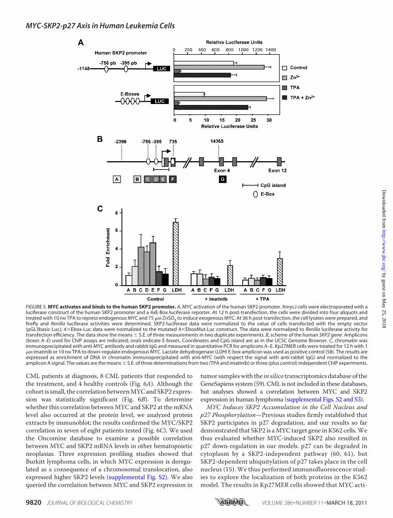
We tested the ability of MYC to activate the human SKP2 pro-moter through promoter-luciferase assays. We transfectedKmycJ cells with a luciferase reporter containing 1148 bp ofSKP2 promoter (46). The results showed that the SKP2 pro-moter was activated by MYC (Fig. 5A, top panel). Consistentwith MYC repression by TPA, MYC transactivation activitywas reduced in TPA-treated cells after transfection and waselevated when MYC was induced by Zn2�. As a positive con-trol, we used a luciferase reporter bearing four E-boxes(4�Ebox-Luc), which was also activated by MYC (Fig. 5A, bot-
tom panel). The empty luciferase construct and the reporterwith four mutated E-boxes (4�EboxMut-Luc) were used fornormalization. These results suggested that SKP2 could be adirect MYC target gene.Sequence analysis of the human SKP2 gene revealed several
E-boxes in the promoter and in the second exon.We thus usedChIP to analyze promoter occupancy byMYC. The localizationof E-boxes in the human SKP2 gene and the amplicons analyzedin the ChIP experiments are shown in Fig. 5B. ChIP demon-strated MYC binding to a region with two E-boxes (CATGCGand CACGCG), mapping at �756 and �389 upstream of thetranscription start site (Fig. 5C); both sequences are describedas high affinity E-boxes for MYC in vivo (56, 57). As negativecontrols, we used K562 cells treated with imatinib and TPA,which inhibitMYC expression (Fig. 1, E and F, and supplemen-tal Fig. S1). MYC binding was dramatically reduced after bothtreatments (Fig. 5C). This MYC-binding region was alsodetected in K562 and HeLa cells in the ENCODE genome-wideChIP sequencing project, published by the UCSC genomebrowser (assembly NCBI36/hg18, Yale/UC Davis/Harvardstudy). MYC binding to the E-box of the lactate dehydrogenaseA (58) was used as positive control (Fig. 5C). Taken collectively,our results indicate that human SKP2 is a direct MYC targetgene, at least in human cells.MYC and SKP2 Expression Correlated in Chronic Myeloid
Leukemia Cells—The K562 cell line is derived from a humanCMLpatient in blast crisis. To confirm the correlation betweenSKP2 andMYC expression in vivo, we used RT-qPCR to deter-minemRNA levels of SKP2 andMYC in the bonemarrow of 31
FIGURE 3. SKP2 is down-regulated after MYC repression. A, K562 cells werenucleofected with a short hairpin RNA vector for MYC (shMYC) or the emptyvector (Vect.), and MYC, SKP2, and actin expression were assayed by immu-noblot 24 and 36 h after nucleofection. B, K562 cells were transfected as inA; at 24 h after nucleofection, the levels of SKP2, B23/nucleophosmin (positivecontrol), and SKP1 (negative control) mRNA were determined by RT-qPCR.The values are the means � S.E. of three independent transfections. C, humanlymphoid P493-6 cells were treated with 1 �g/ml doxycyclin (Dox), and SKP2,MYC, and actin expression were assayed by immunoblot. D, P493-6 cells weretreated as in C, and SKP2 mRNA expression was determined by RT-qPCR.E, TM1 cells were treated with 2 �g/ml doxycyclin to repress MYC for theperiods indicated, and SKP2, MYC, and actin expression were determined byimmunoblot. F, Myc-null HO15.19 rat fibroblasts and parental TGR1 wereserum-deprived for the indicated times, and SKP2, MYC, and actin expressionanalyzed by immunoblot.
FIGURE 4. MYC induces SKP2 in the absence of protein synthesis.A, Kp27MER cells were pretreated for 12 h with 10 nM TPA and then treatedwith 10 �g/ml CHX, 4HT, and ZnSO4 for 1– 6 h. Expression of p27, SKP2, MYC,MycER, and actin were determined by immunoblot. B, SKP2 mRNA expressionin Kp27MER was determined by RT-qPCR after 6 h of exposure to cyclohexi-mide, ZnSO4, and 4HT as indicated. The data show the means � S.E. of threeindependent experiments. Ctrol, control.
MYC-SKP2-p27 Axis in Human Leukemia Cells
MARCH 18, 2011 • VOLUME 286 • NUMBER 11 JOURNAL OF BIOLOGICAL CHEMISTRY 9819
by guest on May 25, 2018
http://ww
w.jbc.org/
Dow
nloaded from
CML patients at diagnosis, 8 CML patients that responded tothe treatment, and 4 healthy controls (Fig. 6A). Although thecohort is small, the correlation betweenMYCand SKP2 expres-sion was statistically significant (Fig. 6B). To determinewhether this correlation betweenMYC and SKP2 at themRNAlevel also occurred at the protein level, we analyzed proteinextracts by immunoblot; the results confirmed the MYC/SKP2correlation in seven of eight patients tested (Fig. 6C). We usedthe Oncomine database to examine a possible correlationbetween MYC and SKP2 mRNA levels in other hematopoieticneoplasias. Three expression profiling studies showed thatBurkitt lymphoma cells, in which MYC expression is deregu-lated as a consequence of a chromosomal translocation, alsoexpressed higher SKP2 levels (supplemental Fig. S2). We alsoqueried the correlation between MYC and SKP2 expression in
tumor sampleswith the in silico transcriptomics database of theGeneSapiens system (59). CML is not included in these databases,but analyses showed a correlation between MYC and SKP2expression in human lymphoma (supplemental Figs. S2 and S3).MYC Induces SKP2 Accumulation in the Cell Nucleus and
p27 Phosphorylation—Previous studies firmly established thatSKP2 participates in p27 degradation, and our results so fardemonstrated that SKP2 is aMYC target gene inK562 cells.Wethus evaluated whether MYC-induced SKP2 also resulted inp27 down-regulation in our models. p27 can be degraded incytoplasm by a SKP2-independent pathway (60, 61), butSKP2-dependent ubiquitylation of p27 takes place in the cellnucleus (15). We thus performed immunofluorescence stud-ies to explore the localization of both proteins in the K562model. The results in Kp27MER cells showed that MYC acti-
FIGURE 5. MYC activates and binds to the human SKP2 promoter. A, MYC activation of the human SKP2 promoter. KmycJ cells were electroporated with aluciferase construct of the human SKP2 promoter and a 4xE-Box luciferase reporter. At 12 h post-transfection, the cells were divided into four aliquots andtreated with 10 nM TPA to repress endogenous MYC and 75 �M ZnSO4 to induce exogenous MYC. At 36 h post-transfection, the cell lysates were prepared, andfirefly and Renilla luciferase activities were determined. SKP2-luciferase data were normalized to the value of cells transfected with the empty vector(pGL3basic-Luc); 4�Ebox-Luc data were normalized to the mutated 4�EboxMut-Luc construct. The data were normalized to Renilla luciferase activity fortransfection efficiency. The data show the means � S.E. of three measurements in two duplicate experiments. B, scheme of the human SKP2 gene. Amplicons(boxes A–E) used for ChIP assays are indicated; ovals indicate E-boxes. Coordinates and CpG island are as in the UCSC Genome Browser. C, chromatin wasimmunoprecipitated with anti-MYC antibody and rabbit IgG and measured in quantitative PCR for amplicons A–E. Kp27MER cells were treated for 12 h with 1�M imatinib or 10 nM TPA to down-regulate endogenous MYC. Lactate dehydrogenase (LDH) E-box amplicon was used as positive control (58). The results areexpressed as enrichment of DNA in chromatin immunoprecipitated with anti-MYC (with respect the signal with anti-rabbit IgG) and normalized to theamplicon A signal. The values are the means � S.E. of three determinations from two (TPA and imatinib) or three (plus control) independent ChIP experiments.
MYC-SKP2-p27 Axis in Human Leukemia Cells
9820 JOURNAL OF BIOLOGICAL CHEMISTRY VOLUME 286 • NUMBER 11 • MARCH 18, 2011
by guest on May 25, 2018
http://ww
w.jbc.org/
Dow
nloaded from

vation resulted in increased nuclear SKP2 and decreasednuclear p27 levels in most cells. It is of note that some cellsshowing higher nuclear SKP2 levels also showed lower p27(Fig. 7A).Theactual substrateof theSCFSKP2complex isp27phosphor-
ylated in threonine 187 (26). ForMYC-mediated degradation ofp27 via SKP2 up-regulation, it is thus necessary that a substan-tial fraction of p27 be in the formofThr(P)-187-p27. In a kineticstudy to determine total p27 and Thr(P)-187-p27 in Kp27MERcells treated with Zn2� and 4HT (4–24 h), MYC initially pro-voked an increase in the phosphorylated p27 fraction (10-foldafter 8 h compared with control cells; Fig. 7B). At longer treat-ment times, the phospho-p27 fraction and the total p27 levelswere reduced, as anticipated. We performed a dose-responsestudy to analyze total p27 and Thr(P)-187-p27 levels in cellstreated with 50 or 75 �M Zn2� and 4HT (12 h). The resultsagain showed that MYC activation resulted in a significantincrease in the Thr(P)-187-p27 fraction (Fig. 7C). Thisincreased p27 phosphorylation can be explained in part by theMYC-induced up-regulation of CDK2 and cyclins in these cells(40).MYC Reduces p27 Protein Levels in K562—The previous
results indicated that SKP2 and p27 colocalize in the nucleusand that a significant fraction of p27 is phosphorylated in Thr-187, a prerequisite to acting as a SKP2 substrate.We assayed the
MYC effect on ectopic p27 levels after p27 induction with 50�M ZnSO4. At this ZnSO4 concentration, p27 levels were nothigh enough to titrate out the SCFSKP2 system, as occurs withhigher ZnSO4 doses (Fig. 7B and data not shown). Immunoblotanalysis showed that MYC activation with 4HT resulted in asignificant decrease in p27 levels when p27 was induced withZn2� (Fig. 8A, compare lanes 1 and 2 and lanes 3 and 4). Con-sistent with the reduction in p27, the proliferation rate wasaugmented by MYC in Kp27MER cells treated with 50 �M
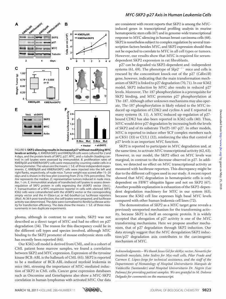
ZnSO4 (data not shown and Ref. 40). In contrast, 4HT did notreduce p27 levels in parental Kp27-5 cells, which bear a Zn2�-inducible p27 but not the MycER construct (Fig. 8B). We alsotested the MYC effect on the endogenous p27 that is up-regu-lated when the KMER4 cells reach quiescence by culture satu-ration (�1.5 � 106 cells/ml after 3 days of culture). Immuno-blot results demonstrated that SKP2 levels were higher and p27was lower in cells with activated MYC (Fig. 8C).As a second approach to testing the MYC-SKP-p27 axis in
ourmodel, we infected KMER4 cells with retrovirus expressinga short hairpin SKP2 (shSKP2) vector to generate the cell lineKMERshSKP2. Immunoblot results confirmed that SKP2 levelswere reduced by shSKP2 and that p27 levels were concomi-tantly increased (Fig. 9A) compared with the KMERpSR cellline expressing the empty vector. In agreement with theincreased p27 abundance, shSKP2-expressing cells grew at a
FIGURE 6. MYC and SKP2 expression in CML patients. A, RT-qPCR analysis of MYC and SKP2 mRNA expression in 31 samples from CML patients at diagnosisin chronic phase (D), 8 patients in complete molecular response under imatinib treatment (T), and 4 healthy donors (H). The data are the means of threedeterminations, arranged from low to high MYC expression. mRNA values were normalized to the levels of ribosomal protein small 14 in each case. B, corre-lation between MYC and SKP2 levels in CML patients. The solid line indicates the linear prediction. The Pearson’s r, and p values are indicated. C, immunoblotshowing SKP2 and MYC expression in samples of CML patients at diagnosis. A picture of the gel stained with Coomassie (Coom.) Blue is shown to assess proteinloading.
MYC-SKP2-p27 Axis in Human Leukemia Cells
MARCH 18, 2011 • VOLUME 286 • NUMBER 11 JOURNAL OF BIOLOGICAL CHEMISTRY 9821
by guest on May 25, 2018
http://ww
w.jbc.org/
Dow
nloaded from

reduced rate (Fig. 9B) and showed cell accumulation in the G1phase (not shown). This difference in growth rates was repro-duced in vivo; whereas KMERpSR xenografts readily formedtumors in nude mice, KMERshSKP2 cells did not form tumors,or theyweremuch smaller (Fig. 9,C andD). Consistentwith thelack of SKP2, p27 levels were not reduced by MYC in cellsexpressing shSKP2 (not shown). Previous reports describe apositive effect of SKP2 on MYC stability and transcriptionalactivity (62, 63). Nonetheless, we found no significant changesin MYC levels after SKP2 silencing in K562 cells (Fig. 9A). We
also testedMYC transcriptional activity after SKP2 silencing bythe shSKP2 expression vector. SKP2 silencing was confirmedby immunoblot (Fig. 9E), but luciferase assays failed to show asignificant SKP2 effect on MYC-dependent transcriptionalactivity (Fig. 9F). Collectively, our results show that MYCinduces SKP2 and that this induction is responsible, at least inpart, for p27 down-regulation.
DISCUSSION
MYC stimulates proliferation, and SKP2 expression is asso-ciated with cell cycle progression (actually, SKP2 stands for Sphase kinase-associated protein 2). This has made it difficult todate to distinguish whether SKP2 induction is a direct or anindirect effect of MYC. Here we used four K562 cell-basedmodels in whichMYC can be induced or activated in cell cycle-arrested cells. Proliferation arrest was achieved by severalapproaches: ectopic p27 overexpression, p53 activation, TPA,and imatinib treatments. In all cases, MYC induction resultedin SKP2 up-regulation. In TM1 epithelial cells,MYC repressionsimilarly provoked SKP2 down-regulation although cells con-tinued to proliferate. SKP2 regulation by MYC was thereforenot an indirect effect of MYC-induced cell cycle progression.Moreover, our data strongly suggest that SKP2 is a direct MYCtarget gene based on three additional observations: (i) MYCup-regulates SKP2mRNA expressionwhen protein synthesis isinhibited, (ii) MYC activates the SKP2 promoter in luciferasereporter assays, and (iii) MYC binds to an E-box in the 5� reg-ulatory region of human SKP2 in ChIP assays. Up-regulation ofSKP2 was also recently reported in MYC-induced murine lym-
FIGURE 7. SKP2 colocalizes with p27 in the cell nucleus and Myc inducesp27 phosphorylation. A, immunofluorescence of p27 and SKP2 proteins inKp27MER cells exposed for 24 h to ZnSO4 and 4HT. Most 4HT-treated cellsexpressed nuclear SKP2, but two fields were selected to show cells with lowSKP2 and high p27 levels (arrows). Most cells exposed to ZnSO4 showed highp27 levels and low SKP2 in the nuclei, but a field was selected showing a cellwith high SKP2 and low p27 (arrow). The nuclei were DAPI-stained.B, Kp27MER cells were treated for 4 –24 h with 75 �M ZnSO4. Total p27, Thr(P)-187-p27 (pT187-p27), and actin (as loading control) were assayed by immu-noblotting. The third panel shows the signal of total p27 (red) and phospho-p27 (green) to show the coexpression of both forms (yellow). Total p27 andThr(P)-187-p27 ratios as determined by densitometry are shown (bottompanel). C, Kp27MER cells were treated for 12 h with 200 nM 4HT and 50 or 75�M ZnSO4, as indicated. Total p27, Thr(P)-187-p27 (pT187-p27), and actin(loading control) were assayed by immunoblot. Total p27:Thr(P)-187-p27 sig-nal ratios, as determined by densitometry, are shown (bottom panel).
FIGURE 8. MYC-induced SKP2 correlates with p27 down-regulation.A, expression of p27 in Kp27MER after MYC activation. The cells were treatedwith 200 nM 4HT and 50 �M ZnSO4 for 24 and 48 h. p27, SKP2, and MYC proteinlevels were determined by immunoblot. B, Kp27-5 cells were treated for 48 hwith ZnSO4 and 4HT, and protein levels of SKP2, MYC, p27 and actin (loadingcontrol) were determined by immunoblot. C, KMER4 cells were cultured for76 h to reach quiescence caused by cell density, alone or with 200 nM 4HT toactivate MYC. SKP2, p27, and MycER protein levels were determined byimmunoblot.
MYC-SKP2-p27 Axis in Human Leukemia Cells
9822 JOURNAL OF BIOLOGICAL CHEMISTRY VOLUME 286 • NUMBER 11 • MARCH 18, 2011
by guest on May 25, 2018
http://ww
w.jbc.org/
Dow
nloaded from
phoma, although in contrast to our results, SKP2 was notdescribed as a direct target of MYC and had no effect on p27degradation (34). The reason for this discrepancy could lie inthe different cell types and species involved, although MYCbinding to the SKP2 promoter of mouse embryonic stem cellshas recently been reported (64).Our K562 cell model is derived fromCML, and in a cohort of
CML patient bone marrow samples, we found a correlationbetween SKP2 andMYC expression. Expression of the tyrosinekinase BCR-ABL is the hallmark of CML (65). SKP2 is reportedto be a mediator of BCR-ABL-induced myeloid leukemia inmice (66), stressing the importance of MYC-mediated induc-tion of SKP2 in CML cells. Cancer gene expression databasessuch as Oncomine and GeneSapiens also show a MYC-SKP2correlation in human lymphomas with activated MYC. Our data
are consistent with recent reports that SKP2 is among the MYC-induced genes in transcriptional profiling analysis in murinehematopoietic stem cells (67) and in genome-wide transcriptionalresponse toMYC silencing in human breast carcinoma cells (68).SKP2 is nonetheless subject to complex regulation by several tran-scription factors besides MYC, and SKP2 expression should thusnot be expected to correlate toMYC in all cell types or tumors.However, our results show that MYC is required for serum-dependent SKP2 expression in rat fibroblasts.p27 can be degraded via SKP2-dependent and -independent
systems (61, 69). The phenotype of Skp2�/� mice and cells isrescued by the concomitant knock-out of the p27 (Cdkn1B)gene, however, indicating that the main transformation mech-anismof SKP2 is linked to p27 degradation (70, 71). In ourK562model, SKP2 induction by MYC also results in reduced p27levels. Moreover, Thr-187 phosphorylation is a prerequisite forSKP2 binding, and MYC promotes p27 phosphorylation atThr-187. Although other unknownmechanismsmay also oper-ate, Thr-187 phosphorylation is likely related to the MYC-in-duced up-regulation of CDK2 and cyclins A and E reported inmany systems (8, 11). A MYC-induced up-regulation of p27-bound CDK2 has also been reported in K562 cells (40). Thus,MYCwould drive p27 degradation by increasing both the levelsof SKP2 and of its substrate Thr(P)-187-p27. In other models,MYC is reported to induce other SCF complex members suchas CKS1 (33) or CUL1 (32), reinforcing the idea that control ofp27 levels is an important MYC function.SKP2 is reported to participate in MYC degradation and, at
the same time, to activateMYC transcriptional activity (62, 63).However, in our model, the SKP2 effect on MYC levels wasmarginal, in contrast to the decrease observed in p27. In addi-tion, we detected no effect on MYC transcriptional activity asmeasured with luciferase reporters. This discrepancy might bedue to the different cell types used in our study. A recent reportshowed that MYC degradation in hematopoietic cells is onlydependent on FBW7 ubiquitin ligase and not on SKP2 (67).Another possible explanation is exhaustion of the SKP2-depen-dent degradation machinery for MYC in our system (63),because the K562 cell line expresses high basal MYC levelscompared with other human leukemia cell lines (72).The demonstration of SKP2 as a MYC target gene reveals a
previously unreported mechanism for the transforming activ-ity, because SKP2 is itself an oncogenic protein. It is widelyaccepted that abrogation of p27 activity is one of the MYCtransforming mechanisms. Here we propose another mecha-nism, that of p27 degradation through SKP2 induction. Ourdata strongly suggest that the MYC deregulation/SKP2 induc-tion/p27 degradation axis contributes to the carcinogenicmechanism of MYC.
Acknowledgments—We thank Jesus Gil for shMyc vector, Novartis forimatinib mesylate, John Sedivy for Myc-null cells, Pilar Frade andCarmen E. Lopez-Jorge for technical assistance, and the staff of theDepartments of Hematology of Hospital Universitario Marques deValdecilla (Santander) and Hospital Universitario Dr. Negrín (LasPalmas) for providing patient samples. We are grateful to M. DoloresDelgado for comments on the manuscript.
FIGURE 9. SKP2 silencing results in increased p27 without modifying MYClevels or activity. A, KMERshSKP2 and KMERpSR cells were cultured for 2 and4 days, and the protein levels of SKP2, p27, MYC, and �-tubulin (loading con-trol) in cell lysates were assessed by immunoblot. B, proliferation rates ofKMERpSR and KMERshSKP2 cells were measured by counting viable cells in ahemocytometer. The values are the means � S.E. of three independent exper-iments. C, KMERpSR and KMERshSKP2 cells were injected into the left andright flanks, respectively, of nude mice. Tumor weight was scored after 15–20days and is shown in the box plot (covering from 25 to 75% percentiles). Theline represents the median. D, representative tumors induced in nude mice.Bar, 1 cm. E, immunoblot analysis of transfected cell lysates to assess down-regulation of SKP2 protein in cells expressing the shSKP2 vector (Vect.).F, transactivation of a MYC-responsive reporter in cells with silenced SKP2.K562 cells were cotransfected with the shSKP2 vector or the correspondingempty vector and the 4�Ebox-Luc or 4xE-boxMut-Luc luciferase reporters(Mut). At 36 h post-transfection, the cell lysates were prepared, and luciferaseactivity was determined. The data were normalized to Renilla luciferase activ-ity for transfection efficiency. The data show the means � S.E. of three mea-surements in two duplicate experiments.
MYC-SKP2-p27 Axis in Human Leukemia Cells
MARCH 18, 2011 • VOLUME 286 • NUMBER 11 JOURNAL OF BIOLOGICAL CHEMISTRY 9823
by guest on May 25, 2018
http://ww
w.jbc.org/
Dow
nloaded from

REFERENCES1. Eilers, M., and Eisenman, R. N. (2008) Genes Dev. 22, 2755–27662. Meyer, N., and Penn, L. Z. (2008) Nat. Rev. Cancer 8, 976–9903. Leon, J., Ferrandiz, N., Acosta, J. C., and Delgado, M. D. (2009) Cell Cycle
8, 1148–11574. Zeller, K. I., Zhao, X., Lee, C. W., Chiu, K. P., Yao, F., Yustein, J. T., Ooi,
H. S., Orlov, Y. L., Shahab, A., Yong, H. C., Fu, Y., Weng, Z., Kuznetsov,V. A., Sung, W. K., Ruan, Y., Dang, C. V., andWei, C. L. (2006) Proc. Natl.Acad. Sci. U.S.A. 103, 17834–17839
5. Orian, A., van Steensel, B., Delrow, J., Bussemaker, H. J., Li, L., Sawado, T.,Williams, E., Loo, L. W., Cowley, S. M., Yost, C., Pierce, S., Edgar, B. A.,Parkhurst, S. M., and Eisenman, R. N. (2003) Genes Dev. 17, 1101–1114
6. Li, Z., Van Calcar, S., Qu, C., Cavenee, W. K., Zhang, M. Q., and Ren, B.(2003) Proc. Natl. Acad. Sci. U.S.A. 100, 8164–8169
7. Fernandez, P. C., Frank, S. R., Wang, L., Schroeder, M., Liu, S., Greene, J.,Cocito, A., and Amati, B. (2003) Genes Dev. 17, 1115–1129
8. Dang, C. V., O’Donnell, K. A., Zeller, K. I., Nguyen, T., Osthus, R. C., andLi, F. (2006) Semin. Cancer Biol 16, 253–264
9. Patel, J. H., Loboda, A. P., Showe,M. K., Showe, L. C., andMcMahon, S. B.(2004) Nat. Rev. Cancer 4, 562–568
10. Nesbit, C. E., Tersak, J. M., and Prochownik, E. V. (1999) Oncogene 18,3004–3016
11. Lutz, W., Leon, J., and Eilers, M. (2002) Biochim. Biophys. Acta 1602,61–71
12. Delgado, M. D., and Leon, J. (2010) Genes Cancer 1, 605–61613. Hershko, D. D. (2008) Cancer 112, 1415–142414. Frescas, D., and Pagano, M. (2008) Nat. Rev. Cancer 8, 438–44915. Nakayama, K. I., and Nakayama, K. (2006) Nat. Rev. Cancer 6, 369–38116. Kitagawa, K., Kotake, Y., and Kitagawa, M. (2009) Cancer Sci. 100,
1374–138117. Polyak, K., Lee, M. H., Erdjument-Bromage, H., Koff, A., Roberts, J. M.,
Tempst, P., and Massague, J. (1994) Cell 78, 59–6618. Coats, S., Flanagan, W. M., Nourse, J., and Roberts, J. M. (1996) Science
272, 877–88019. Chu, I. M., Hengst, L., and Slingerland, J. M. (2008) Nat. Rev. Cancer 8,
253–26720. Slingerland, J., and Pagano, M. (2000) J. Cell. Physiol. 183, 10–1721. Pagano, M., Tam, S. W., Theodoras, A. M., Beer-Romero, P., Del Sal, G.,
Chau, V., Yew, P. R., Draetta, G. F., and Rolfe, M. (1995) Science 269,682–685
22. Carrano, A. C., Eytan, E., Hershko, A., and Pagano, M. (1999) Nat. CellBiol. 1, 193–199
23. Sutterluty, H., Chatelain, E.,Marti, A.,Wirbelauer, C., Senften,M.,Muller,U., and Krek, W. (1999) Nat. Cell Biol. 1, 207–214
24. Tsvetkov, L. M., Yeh, K. H., Lee, S. J., Sun, H., and Zhang, H. (1999) Curr.Biol. 9, 661–664
25. Galea, C. A., Wang, Y., Sivakolundu, S. G., and Kriwacki, R. W. (2008)Biochemistry 47, 7598–7609
26. Lu, Z., and Hunter, T. (2010) Cell Cycle 9, 2342–235227. Martins, C. P., and Berns, A. (2002) EMBO J. 21, 3739–374828. Yang, W., Shen, J., Wu, M., Arsura, M., FitzGerald, M., Suldan, Z., Kim,
D. W., Hofmann, C. S., Pianetti, S., Romieu-Mourez, R., Freedman, L. P.,and Sonenshein, G. E. (2001) Oncogene 20, 1688–1702
29. Chandramohan, V., Jeay, S., Pianetti, S., and Sonenshein, G. E. (2004)J. Immunol. 172, 5522–5527
30. Perez-Roger, I., Kim, S. H., Griffiths, B., Sewing, A., and Land, H. (1999)EMBO J. 18, 5310–5320
31. Bouchard, C., Thieke, K., Maier, A., Saffrich, R., Hanley-Hyde, J., Ansorge,W., Reed, S., Sicinski, P., Bartek, J., and Eilers, M. (1999) EMBO J. 18,5321–5333
32. O’Hagan, R. C., Ohh, M., David, G., de Alboran, I. M., Alt, F. W., Kaelin,W. G., Jr., and DePinho, R. A. (2000) Genes Dev. 14, 2185–2191
33. Keller, U. B., Old, J. B., Dorsey, F. C., Nilsson, J. A., Nilsson, L., MacLean,K. H., Chung, L., Yang, C., Spruck, C., Boyd, K., Reed, S. I., and Cleveland,J. L. (2007) EMBO J. 26, 2562–2574
34. Old, J. B., Kratzat, S., Hoellein, A., Graf, S., Nilsson, J. A., Nilsson, L.,Nakayama, K. I., Peschel, C., Cleveland, J. L., and Keller, U. B. (2010)Mol
Cancer Res. 8, 353–36235. Bernard, D., Pourtier-Manzanedo, A., Gil, J., and Beach, D. H. (2003)
J. Clin. Invest. 112, 1724–173136. Andreu, E. J., Lledo, E., Poch, E., Ivorra, C., Albero, M. P., Martínez-Cli-
ment, J. A., Montiel-Duarte, C., Rifon, J., Perez-Calvo, J., Arbona, C.,Prosper, F., and Perez-Roger, I. (2005) Cancer Res. 65, 3264–3272
37. Delgado, M. D., Lerga, A., Canelles, M., Gomez-Casares, M. T., and Leon,J. (1995) Oncogene 10, 1659–1665
38. Littlewood, T. D., Hancock, D. C., Danielian, P. S., Parker,M.G., and Evan,G. I. (1995) Nucleic Acid Res. 23, 1686–1690
39. Munoz-Alonso, M. J., Acosta, J. C., Richard, C., Delgado, M. D., Sedivy, J.,and Leon, J. (2005) J. Biol. Chem. 280, 18120–18129
40. Acosta, J. C., Ferrandiz, N., Bretones, G., Torrano, V., Blanco, R., Richard,C., O’Connell, B., Sedivy, J., Delgado, M. D., and Leon, J. (2008)Mol. Cell.Biol. 28, 7286–7295
41. Ceballos, E., Delgado, M. D., Gutierrez, P., Richard, C., Muller, D.,Eilers, M., Ehinger, M., Gullberg, U., and Leon, J. (2000) Oncogene 19,2194–2204
42. Warner, B. J., Blain, S. W., Seoane, J., and Massague, J. (1999) Mol. Cell.Biol. 19, 5913–5922
43. Mateyak, M. K., Obaya, A. J., Adachi, S., and Sedivy, J. M. (1997) CellGrowth Differ. 8, 1039–1048
44. Schuhmacher, M., Staege, M. S., Pajic, A., Polack, A., Weidle, U. H.,Bornkamm, G. W., Eick, D., and Kohlhuber, F. (1999) Curr. Biol. 9,1255–1258
45. Shang, Y., Hu, X., DiRenzo, J., Lazar,M.A., andBrown,M. (2000)Cell 103,843–852
46. Huang, Y. C., and Hung, W. C. (2006) J. Cell. Physiol. 209, 363–36947. Kiessling, A., Sperl, B., Hollis, A., Eick, D., and Berg, T. (2006) Chem. Biol.
13, 745–75148. Baccarani,M., Cortes, J., Pane, F., Niederwieser, D., Saglio, G., Apperley, J.,
Cervantes, F., Deininger, M., Gratwohl, A., Guilhot, F., Hochhaus, A.,Horowitz, M., Hughes, T., Kantarjian, H., Larson, R., Radich, J., Simons-son, B., Silver, R. T., Goldman, J., and Hehlmann, R. (2009) J. Clin. Oncol.27, 6041–6051
49. Penn, L. J., Brooks, M. W., Laufer, E. M., and Land, H. (1990) EMBO J. 9,1113–1121
50. Grignani, F., Lombardi, L., Inghirami, G., Sternas, L., Cechova, K., andDalla-Favera, R. (1990) EMBO J. 9, 3913–3922
51. Gomez-Casares,M. T., Vaque, J. P., Lemes, A.,Molero, T., Delgado,M.D.,and Leon, J. (2004) Haematologica 89, 241–243
52. Chen, J. Y., Wang, M. C., and Hung, W. C. (2009) Leuk. Res. 33,1520–1524
53. Xie, S., Lin, H., Sun, T., and Arlinghaus, R. B. (2002) Oncogene 21,7137–7146
54. Lerga, A., Crespo, P., Berciano,M., Delgado,M. D., Canelles,M., Cales, C.,Richard, C., Ceballos, E., Gutierrez, P., Ajenjo, N., Gutkind, S., and Leon, J.(1999) Cell Growth Differ. 10, 639–654
55. Pajic, A., Spitkovsky, D., Christoph, B., Kempkes, B., Schuhmacher, M.,Staege, M. S., Brielmeier, M., Ellwart, J., Kohlhuber, F., Bornkamm, G.W.,Polack, A., and Eick, D. (2000) Int. J. Cancer 87, 787–793
56. Grandori, C., Mac, J., Siebelt, F., Ayer, D. E., and Eisenman, R. N. (1996)EMBO J. 15, 4344–4357
57. Kim, J., Lee, J. H., and Iyer, V. R. (2008) Plos One 3, e179858. Kim, J. W., Zeller, K. I., Wang, Y., Jegga, A. G., Aronow, B. J., O’Donnell,
K. A., and Dang, C. V. (2004)Mol. Cell. Biol. 24, 5923–593659. Kilpinen, S., Autio, R., Ojala, K., Iljin, K., Bucher, E., Sara, H., Pisto, T.,
Saarela,M., Skotheim, R. I., Bjorkman,M.,Mpindi, J. P., Haapa-Paananen,S., Vainio, P., Edgren,H.,Wolf,M., Astola, J., Nees,M.,Hautaniemi, S., andKallioniemi, O. (2008) Genome Biol. 9, R139
60. Hara, T., Kamura, T., Nakayama, K., Oshikawa, K., Hatakeyama, S., andNakayama, K. (2001) J. Biol. Chem. 276, 48937–48943
61. Kamura, T., Hara, T., Matsumoto, M., Ishida, N., Okumura, F., Hat-akeyama, S., Yoshida, M., Nakayama, K., and Nakayama, K. I. (2004) Nat.Cell Biol. 6, 1229–1235
62. Kim, S. Y., Herbst, A., Tworkowski, K. A., Salghetti, S. E., andTansey,W. P.(2003)Mol. Cell 11, 1177–1188
63. von der Lehr, N., Johansson, S., Wu, S., Bahram, F., Castell, A., Cetinkaya,
MYC-SKP2-p27 Axis in Human Leukemia Cells
9824 JOURNAL OF BIOLOGICAL CHEMISTRY VOLUME 286 • NUMBER 11 • MARCH 18, 2011
by guest on May 25, 2018
http://ww
w.jbc.org/
Dow
nloaded from

C., Hydbring, P., Weidung, I., Nakayama, K., Nakayama, K. I., Soderberg,O., Kerppola, T. K., and Larsson, L. G. (2003)Mol. Cell 11, 1189–1200
64. Kim, J., Woo, A. J., Chu, J., Snow, J. W., Fujiwara, Y., Kim, C. G., Cantor,A. B., and Orkin, S. H. (2010) Cell 143, 313–324
65. Melo, J. V., and Barnes, D. J. (2007) Nat. Rev Cancer 7, 441–45366. Agarwal, A., Bumm, T. G., Corbin, A. S., O’Hare, T., Loriaux, M., Van-
Dyke, J., Willis, S. G., Deininger, J., Nakayama, K. I., Druker, B. J., andDeininger, M. W. (2008) Blood 112, 1960–1970
67. Reavie, L., Della Gatta, G., Crusio, K., Aranda-Orgilles, B., Buckley, S. M.,Thompson, B., Lee, E., Gao, J., Bredemeyer, A. L., Helmink, B. A., Zavadil,J., Sleckman, B. P., Palomero, T., Ferrando, A., and Aifantis, I. (2010) Nat.Immunol. 11, 207–215
68. Cappellen, D., Schlange, T., Bauer,M.,Maurer, F., andHynes, N. E. (2007)EMBO Rep. 8, 70–76
69. Miranda-Carboni, G. A., Krum, S. A., Yee, K., Nava, M., Deng, Q. E.,Pervin, S., Collado-Hidalgo, A., Galic, Z., Zack, J. A., Nakayama, K., Na-kayama, K. I., and Lane, T. F. (2008) Genes Dev. 22, 3121–3134
70. Nakayama, K., Nagahama, H., Minamishima, Y. A., Miyake, S., Ishida, N.,Hatakeyama, S., Kitagawa, M., Iemura, S., Natsume, T., and Nakayama,K. I. (2004) Dev. Cell 6, 661–672
71. Kossatz, U., Dietrich, N., Zender, L., Buer, J., Manns, M. P., and Malek,N. P. (2004) Genes Dev. 18, 2602–2607
72. Delgado, M. D., Chernukhin, I. V., Bigas, A., Klenova, E. M., and Leon, J.(1999) FEBS Lett. 444, 5–10
MYC-SKP2-p27 Axis in Human Leukemia Cells
MARCH 18, 2011 • VOLUME 286 • NUMBER 11 JOURNAL OF BIOLOGICAL CHEMISTRY 9825
by guest on May 25, 2018
http://ww
w.jbc.org/
Dow
nloaded from
Albero, Ignacio Perez-Roger and Javier LeónGómez-Casares, Marta Albajar, Rosa Blanco, Paula Ruiz, Wen-Chun Hung, M. Pilar
Gabriel Bretones, Juan C. Acosta, Juan M. Caraballo, Nuria Ferrándiz, M. Teresathrough SKP2 in Human Leukemia Cells
KIP1SKP2 Oncogene Is a Direct MYC Target Gene and MYC Down-regulates p27
doi: 10.1074/jbc.M110.165977 originally published online January 18, 20112011, 286:9815-9825.J. Biol. Chem.
10.1074/jbc.M110.165977Access the most updated version of this article at doi:
Alerts:
When a correction for this article is posted•
When this article is cited•
to choose from all of JBC's e-mail alertsClick here
Supplemental material:
http://www.jbc.org/content/suppl/2011/01/18/M110.165977.DC1
http://www.jbc.org/content/286/11/9815.full.html#ref-list-1
This article cites 72 references, 25 of which can be accessed free at
by guest on May 25, 2018
http://ww
w.jbc.org/
Dow
nloaded from