Embed Size (px)
Citation preview

David Hart
Dec 12, 2006

Heme

Porphyrins
• Cyclic compounds that bind metal ions• Chlorphyll (Mg2+)
– Central to solar energy utilization
• Heme (Fe2+) – Most prevalent metalloporphyrin in
humans– Central to oxygen sensing and utilization
• Cobalamin (Cobalt)•


The Heme Pocket in Hemoglobin

Heme
• One ferrous (Fe2+) atom in the center of the tetrapyrrole ring of Protoporphyrin IX
• Prosthetic group for– Hemoglobin and Myoglobin– The Cytochromes– Catalase and Tryptophan pyrrolase– Nitric Oxide Synthase
• Turnover of Hemeproteins (Hemoglobin, etc) is coordinated with synthesis and degradation of porphyrins
• Bound iron is recycled

Lecture Outline
• Heme function
• Heme synthesis and regulation
• Iron metabolism
• Porphyrias
• Heme degradation

Heme Function• Oxygen sensing (heme and hemoproteins)• Oxygen transport (hemoglobin)• Oxygen storage (myoglobin)• Electron transport (cytochromes)• Oxidation (cyrochrome p450, tryptophan
pyrrolase, guanylate cyclase …)• Decomposition and activation of H2O2
(catalase and peroxidase)• Nitric Oxide Synthesis• Regulation of cellular processes• Effector of apoptosis

N
NH N
HN
B
CD
Porphyrin: Cyclic molecule formed by linkage of four pyrrole rings through methenyl bridges
A

Porphyrin Side Chains
• M = Methyl (-CH3)
• V = Vinyl (-CH=CH2)
• P = Propionyl (-CH2-CH2-COO-)
• A = Acetyl (-CH2-COO-)
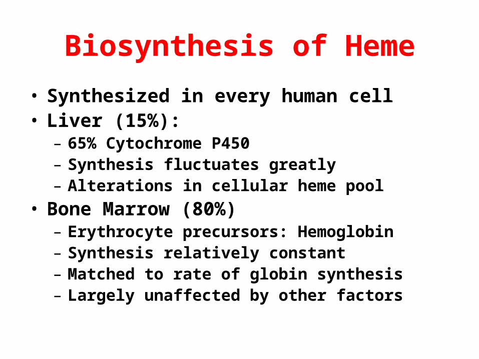
Biosynthesis of Heme
• Synthesized in every human cell• Liver (15%):
– 65% Cytochrome P450– Synthesis fluctuates greatly– Alterations in cellular heme pool
• Bone Marrow (80%)– Erythrocyte precursors: Hemoglobin– Synthesis relatively constant– Matched to rate of globin synthesis– Largely unaffected by other factors

COOH
CH2
CH2
COSCoACH2 NH2
COOH
SUCCINYL CoA
GLYCINE
All Carbon and Nitrogen atoms provided by 2 building blocks:

COOH
CH2
CH2
COSCoACH2 NH2
COOH
SUCCINYL CoA
GLYCINE isDecarboxylated
IN MITOCHONDRIA
AMINOLEVULINIC ACID SYNTHASE
- CO2

COOH
CH2
CH2
C=OCH2
NH2
Condense to form: AMINOLEVULINIC ACID (ALA)
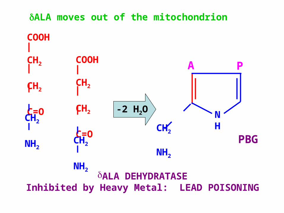
MOVES OUT OF THE MITOCHONDRION

COOH
CH2
CH2
C=OCH2
NH2
COOH
CH2
CH2
C=OCH2
NH2
2 Molecules dehydrated byALA DEHYDRATASE
-2 H2O

COOH
CH2
CH2
CC
NH
COOH
CH2
C
CCH2
NH2
To form Porphobilinogen (PBG)

COOH
CH2
CH2
NH
COOH
CH2
CH2
NH2
Porphobilinogen (PBG)
AcetateCH2COO-
PropionateCH2CH2COO-

Porphobilinogen (PBG)
NH
CH2
NH2
A P

NH
CH 2
NH 2
A
PN
H
CH
2
NH2
A
P
NH
CH2
NH2
A
P
NH
CH2
NH2
A
P

Hydroxymethylbilane synthase& Uroporphyrinogen III synthase
• Four PBG molecules condense• Ring closure • Isomerization

NH
NH HN
HN
A B
CD
A
A
P
A
P
P
P
A
Uroporphyrinogen III

NH
NH HN
HNHOOC-H2C-
HOOC-H2C-
-CH2-CH2-COOH
-CH2-COOH
CH2
CH2
COOH
CH2
CH2
COOH
COOHCH2
CH2
COOHCH2
Uroporphyrinogen III

Series of decarboxylations & oxidations• Porphyrinogens:
– Chemically reduced– Colorless intermediates
• Porphyrins:– Intensely colored– Fluorescent
• Uroporphyrinogen III• Coproporphyrinogen IIIMoves back into Mitochondrion• Protoporphyrinogen IX• Protoporphyrin IX

NH
N HN
NH3C-
H3C-
-CH=CH2
-CH3
CH2
CH2
COOH
CH2
CH2
COOH
CH3
Protoporphyrin IX
CH=CH2

HEMEFe2+ chelated by Protoporphyrin IX
Assisted by Ferrochelatase
CH3-
Regulation of Heme Synthesis

AMINOLEVULINIC ACID SYNTHASE
• Two tissue-specific isozymes• Coded on separate genes• In Liver, heme represses synthesis and
activity of ALAS – Heme can be used for treatment of acute
porphyric attack
• In RBC heme synthesis regulation is more complex– Coordinated with globin synthesis

COOH
CH2
CH2
COSCoACH2 NH2
COOH
SUCCINYL CoA
GLYCINE
IN MITOCHONDRIA
AMINOLEVULINIC ACID SYNTHASERATE-CONTROLLING STEP IN
HEPATIC HEME SYNTHESIS
COOH
CH2
CH2
C=OCH2
NH2
ALA

BonkovskyASH Education BookDecember 2005

Disorders of Heme Synthesis
• X-linked Sideroblastic Anemia
• Lead Poisoning
• Iron Deficiency Anemia
• The Porphyrias

X-linked Sideroblastic Anemia
X-linked Sideroblastic Anemia
ALAS Requires Pyridoxal Phosphate as Coenzyme
Some Sideroblastic Anemiasimprove with Pyridoxine (B6)
COOH
CH2
CH2
C=OCH2
NH2
COOH
CH2
CH2
C=OCH2
NH2
-2 H2O
ALA moves out of the mitochondrion
ALA DEHYDRATASEInhibited by Heavy Metal: LEAD POISONING
PBG
NH
CH2
NH2
A P

Lead Poisoning

Lead PoisoningALAD and FerrochelataseAre particularly sensitive
to Lead inhibition
Lead Poisoning
Fe + PPIX
Ferrochelatase
Heme

Iron Metabolism
• Reactive Transition Metal (Fe2+ Fe3+)• Normally present complexed with proteins
that limit its reactivity• Both iron deficiency and iron overload cause
cellular defects and disease• Most available iron generated by
macrophages that recycle red cell iron• Dietary Fe3+ in duodenum converted to Fe2+
and absorbed by duodenal enterocyte

Iron35% of Earth’s mass
nasa

Fe3+
Heme
Fe2+
Fe2+
BloodApicalDuodenal Enterocyte
MitochondrialHemeSynthesis
HepatocyteMacrophageErythroid Cell
diFe3+
Transferrin
GUTContents

NEJM June 2004

Fe2+
Fe2+
Blood
Macrophage
RBC
HemoglobinHaptoglobin
HemeHemopexin
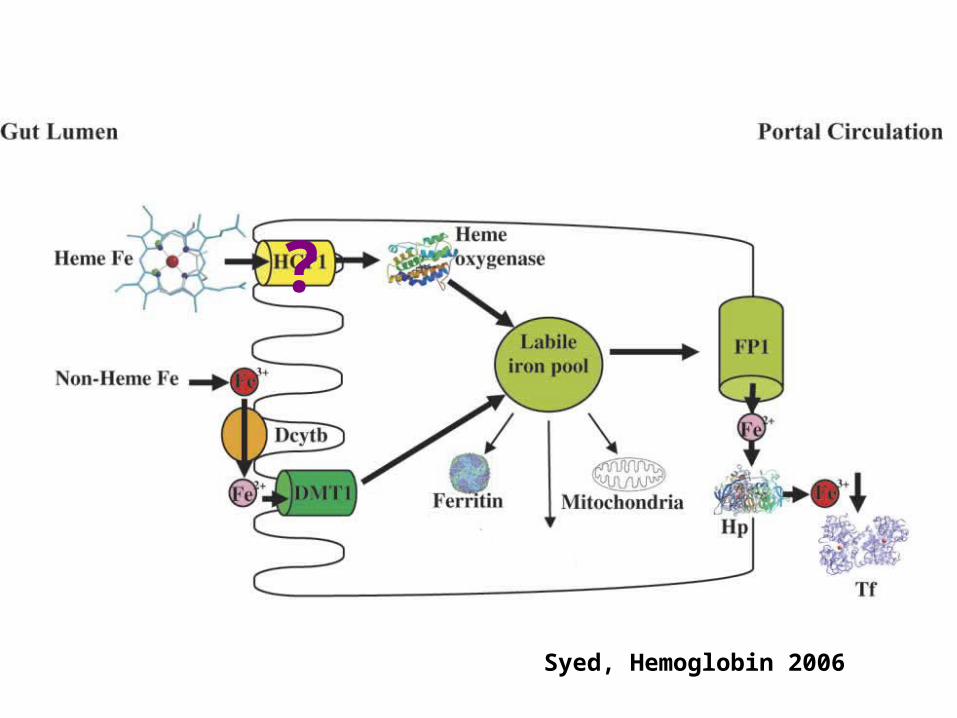
Syed, Hemoglobin 2006
?

http://walz.med.harvard.edu

Hentze, Muckenthaler & AndrewsCell, Vol 117, 285-297, April 30, 2004
Hepcidin

Hepcidin: 25 Amino AcidsJ Med Genet 2004

Nem
eth
et
al,
Sci
ence
, D
ec 2
004

Beutler,ScienceDec 2004

Hentze, Muckenthaler & AndrewsCell, Vol 117, 285-297, April 30, 2004
Ferroportin

Genetic Hemochromatosis Disruption of Hepcidin / Ferroportin
• Autosomal Recessive– HFE C282Y/C282Y– TfR2– Hemojuvelin– Hepcidin
• Autosomal Dominant– Ferroportin
medlib.med.utah.edu
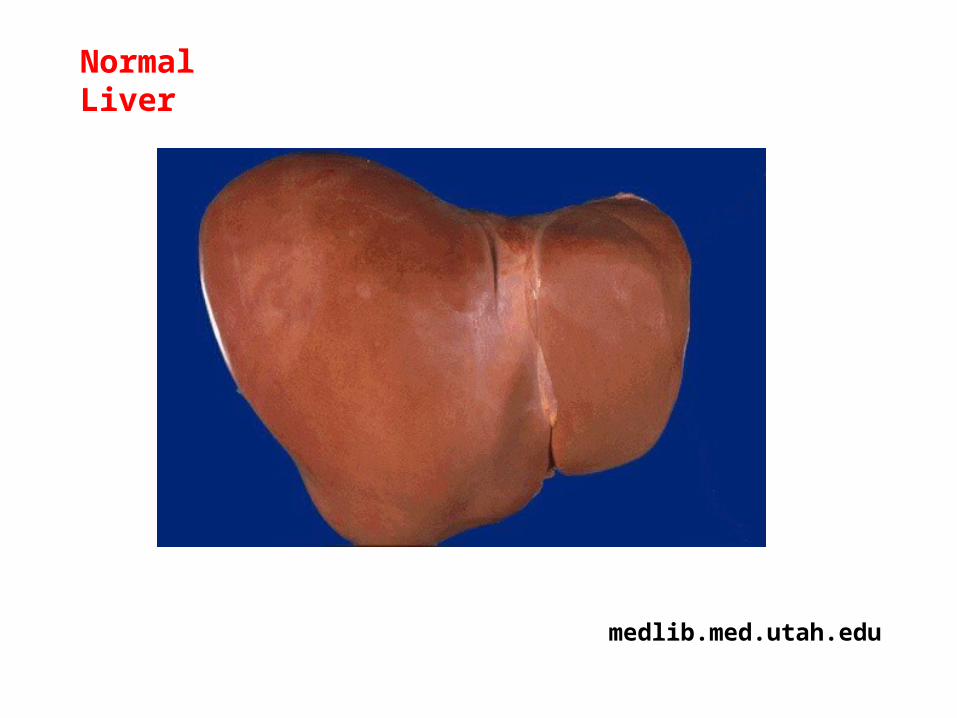
NormalLiver

www.med.niigata-u.ac.j
Granular, Dark Reddish BrownSurface of Liver in Hemochromatosis

http://eduserv.hscer.washington.edu
Iron Accumulation in Chronic Disease

Ring SideroblastPrussian Blue stains Iron
In Mitochondria
www.uchsc.edu

Iron Deficiency Anemia
Hypochromic,Microcytic

http://eduserv.hscer.washington.edu
Normal Red Blood Cells


www.lsuagcenter.co
Spinach:Non-HemeIron LessReadilyAbsorbed
OxalatesPhytatesTanninsFiberCalcium

www.mcgil.com/food/pics
Heme Iron is More Readily Absorbed

www.agnet.org/library
Iron Deficient Spinach“Chlorosis”

www.geoimagery.com
Harvesting Latex

www.sentientkinetics.com
Geophagia

www.awesomedrinks.com
Pagophagia

Solemnity Scale:0 = No smiles/hour
5 = “wreathed”In smiles

www.drmhijazy.com
Spoon Nails

Blue Sclera

Disorders of Heme Synthesis
• X-linked Sideroblastic Anemia
• Lead Poisoning
• Iron Deficiency Anemia
• The Porphyrias

Heme
porphuros
(purple)

Heme Synthesis: Porphyrias
• 8 Enzymatic Reactions
• 7 Deficiencies: “Porphyrias”
• Most are Autosomal Dominant
• Hepatic or Erythroid depending on main site of synthesis / accumulation

Porphyrias
• Accumulation and excretion of porphyrins– Pattern depends on which enzyme affected
• Multiple alleles• Acute and Chronic
– Acute: Neurovisceral attacks
• Porphyrin accumulation: Photosensitivity– Formation of reactive oxygen species– Damage tissues, Release lysosomal enzymes

Very Rare Recessive Porphyria
ALA-D Porphyria
Lead Poisoning

AcuteHepatic
PBG and ALA Accumulate in Urine
PBG in Urine: Diagnostic ScreenUrine darkens with exposure
NOT photosensitiveNeuro-visceral attacks
Precipitated by Drugs, EtOHwhich induce cytochrome P450
Hydroxymethylbilane Synthase
Lead Poisoning
ALA-D Porphyria