Embed Size (px)
Citation preview
Amino Acids Metabolism Part II
Conversion of amino acids to specialized products
Shyamal D. Desai Ph.D.Department of Biochemistry & Molecular BiologyMEB. Room # 7107Phone- [email protected]
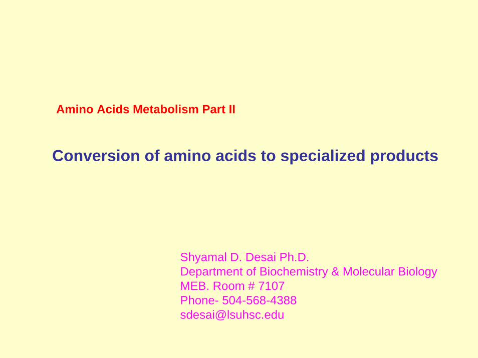
Nitrogen metabolismN2 Atmospheric nitrogen N2 is most abundant but is too
inert for use in most biochemical processes.
Dietary proteinsAtmospheric nitrogen is acted upon by bacteria (nitrogen fixation) and plants to nitrogen containing compounds. We assimilate these compounds as proteins (amino acids) in our diets.
Amino acids
Body proteinsOther nitrogen containing compounds
Carbon skeletons
NH4+Urea
excreted
Lecture I
Lecture III
Lecture II
α-aminogroups
Conversion of nitrogeninto specialized products
Enters various metabolic pathways
Disposal of Nitrogen
Amino acids synthesis& degradation
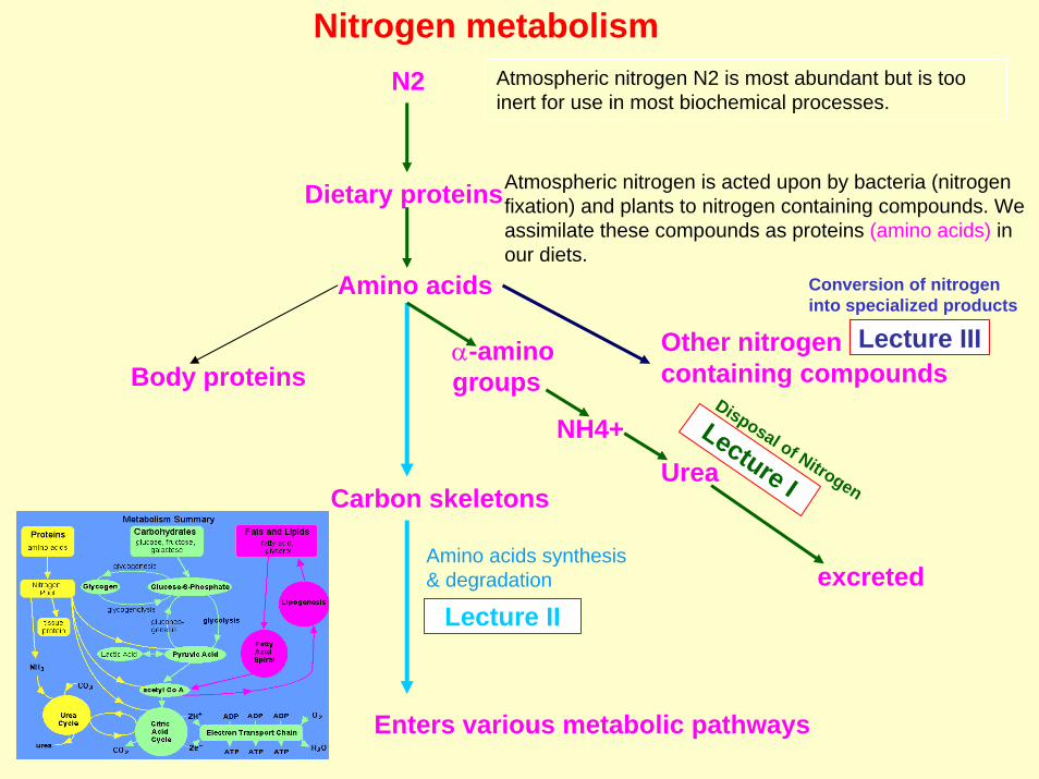
Amino Acids as precursors of nitrogen-containing compounds
Porphyrin metabolism
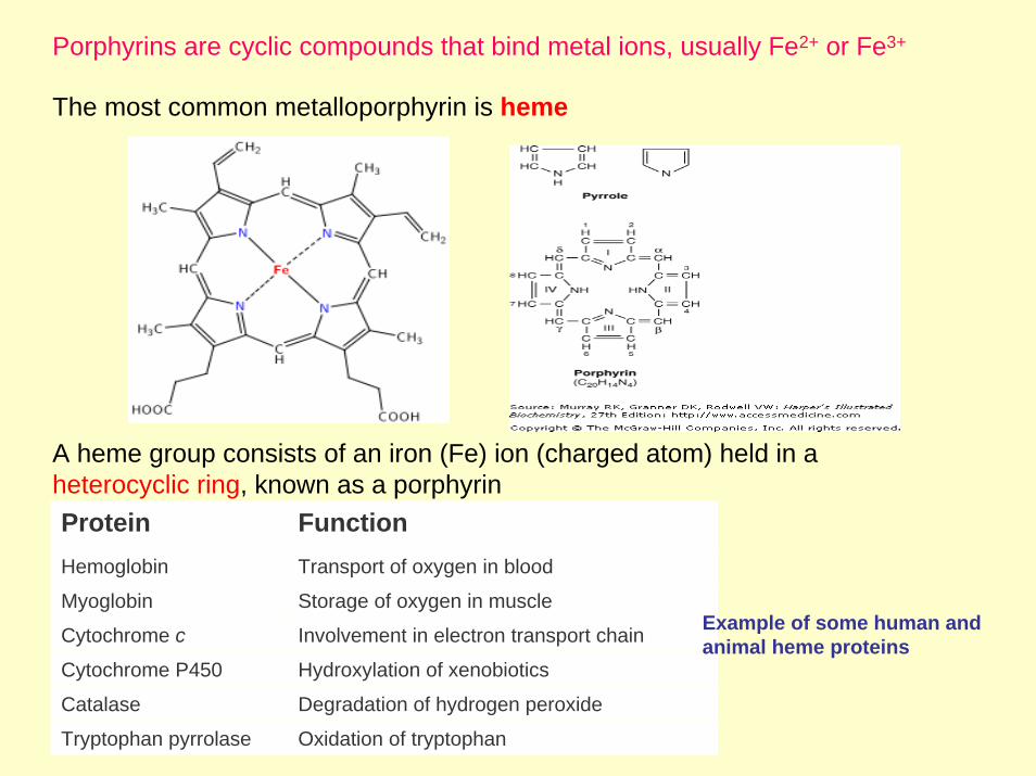
Porphyrins are cyclic compounds that bind metal ions, usually Fe2+ or Fe3+
The most common metalloporphyrin is heme
A heme group consists of an iron (Fe) ion (charged atom) held in a heterocyclic ring, known as a porphyrinProtein FunctionHemoglobin Transport of oxygen in blood
Myoglobin Storage of oxygen in muscle
Cytochrome c Involvement in electron transport chain Example of some human and animal heme proteins
Cytochrome P450 Hydroxylation of xenobiotics
Catalase Degradation of hydrogen peroxide
Tryptophan pyrrolase Oxidation of tryptophan
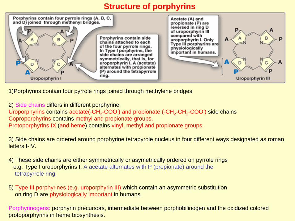
Structure of porphyrins
1)Porphyrins contain four pyrrole rings joined through methylene bridges
2) Side chains differs in different porphyrine.Uroporphyrins contains acetate(-CH2-COO-) and propionate (-CH2-CH2-COO-) side chainsCoproporphyrins contains methyl and propionate groups.Protoporphyrins IX (and heme) contains vinyl, methyl and propionate groups.
3) Side chains are ordered around porphyrine tetrapyrole nucleus in four different ways designated as roman letters I-IV.
4) These side chains are either symmetrically or asymetrically ordered on pyrrole ringse.g. Type I uroporphyrins I, A acetate alternates with P (propionate) around thetetrapyrrole ring.
5) Type III porphyrines (e.g. uroporphyrin III) which contain an asymmetric substitution on ring D are physiologically important in humans.
Porphyrinogens: porphyrin precursors, intermediate between porphobilinogen and the oxidized colored protoporphyrins in heme biosyhthesis.
Boisynthesis of heme
Heme synthesis occurs in all cells due to the requirement for heme as a prosthetic group on enzymes and electron transport chain proteins. By weight, the major locations of heme synthesis are the liver (cytochrome p450) and the erythroidprogenitor cells (Hemoglobin) of the bone marrow.
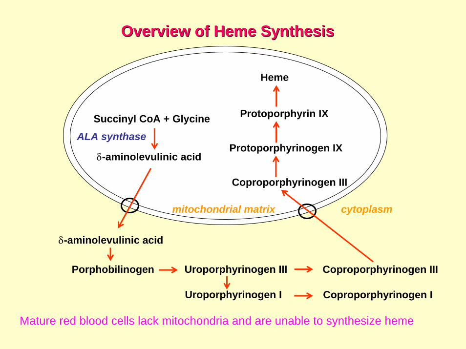
Overview of Overview of HemeHeme SynthesisSynthesis
Uroporphyrinogen I Coproporphyrinogen I
Succinyl CoA + Glycine
δ-aminolevulinic acid
δ-aminolevulinic acid
Porphobilinogen Uroporphyrinogen III Coproporphyrinogen III
Coproporphyrinogen III
Protoporphyrinogen IX
Protoporphyrin IX
Heme
ALA synthase
cytoplasmmitochondrial matrix
Mature red blood cells lack mitochondria and are unable to synthesize heme
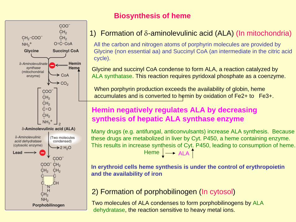
1) Formation of δ-aminolevulinic acid (ALA) (In mitochondria) All the carbon and nitrogen atoms of porphyrin molecules are provided by Glycine (non essential aa) and Succinyl CoA (an intermediate in the citric acid cycle).
Glycine and succinyl CoA condense to form ALA, a reaction catalyzed by ALA synthatase. This reaction requires pyridoxal phosphate as a coenzyme.
When porphyrin production exceeds the availability of globin, hemeaccumulates and is converted to hemin by oxidation of Fe2+ to Fe3+.
2) Formation of porphobilinogen (In cytosol)Two molecules of ALA condenses to form porphobilinogens by ALAdehydratase, the reaction sensitive to heavy metal ions.
Biosynthesis of heme
Hemin negatively regulates ALA by decreasing synthesis of hepatic ALA synthase enzyme Many drugs (e.g. antifungal, anticonvulsants) increase ALA synthesis. Because these drugs are metabolized in liver by Cyt. P450, a heme containing enzyme. This results in increase synthesis of Cyt. P450, leading to consumption of heme.
Heme ALA
In erythroid cells heme synthesis is under the control of erythropoietin and the availability of iron

Biosynthesis of heme
3) Formation of uroporphyrinogen (In cytosol)The condensation of four molecules of porphobillinogens results in theformation of tetrapyrrrole, hydroxymethylbilane, a reaction catalyzed by
hydroxymethylbilane synthase
**
* *
A
P P
A
P
AIsomerization and cyclization by uroporphyinogen III synthase leads tothe formation of Uroporphyrinogen III
Uropprphyrinogen III undergoes decarboxylation at its acetate groups, generating coproporphyrinogen III, a reaction carried out by uroporphyrinogen decarboxylase
Two propionate side chains are decarboxylated to vinyl groups generatingprotoporphyrinogen IX, which is then oxidized to protoporphyrin IX.
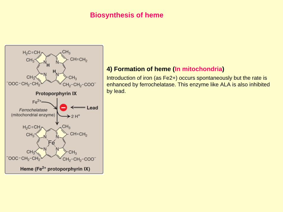
Biosynthesis of heme
4) Formation of heme (In mitochondria)Introduction of iron (as Fe2+) occurs spontaneously but the rate is enhanced by ferrochelatase. This enzyme like ALA is also inhibitedby lead.
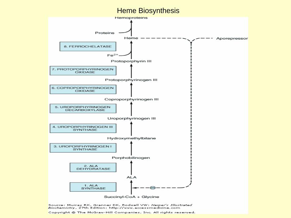
Heme Biosynthesis

Overview of Overview of HemeHeme SynthesisSynthesis
Uroporphyrinogen I Coproporphyrinogen I
Succinyl CoA + Glycine
δ-aminolevulinic acid
δ-aminolevulinic acid
Porphobilinogen Uroporphyrinogen III Coproporphyrinogen III
Coproporphyrinogen III
Protoporphyrinogen IX
Protoporphyrin IX
Heme
ALA synthase
cytoplasmmitochondrial matrix
Mature red blood cells lack mitochondria and are unable to synthesize heme
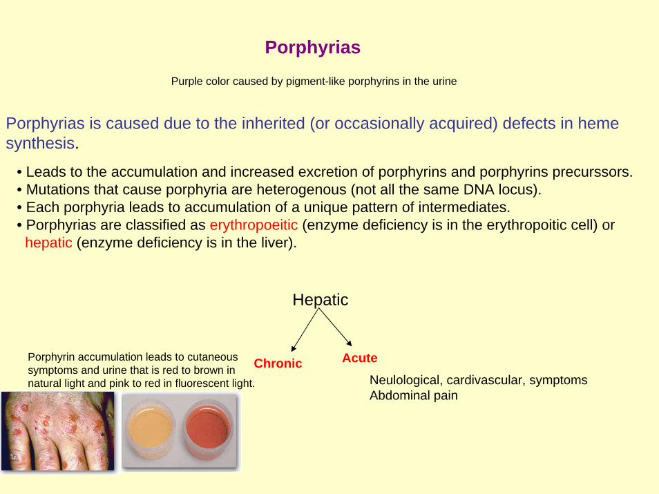
PorphyriasPurple color caused by pigment-like porphyrins in the urine
Porphyrias is caused due to the inherited (or occasionally acquired) defects in hemesynthesis.
• Leads to the accumulation and increased excretion of porphyrins and porphyrins precurssors. • Mutations that cause porphyria are heterogenous (not all the same DNA locus).• Each porphyria leads to accumulation of a unique pattern of intermediates.• Porphyrias are classified as erythropoeitic (enzyme deficiency is in the erythropoitic cell) or
hepatic (enzyme deficiency is in the liver).
AcuteChronic
Hepatic
Porphyrin accumulation leads to cutaneoussymptoms and urine that is red to brown innatural light and pink to red in fluorescent light. Neulological, cardivascular, symptoms
Abdominal pain

PorphyriasHepatic PorphyriasAccumulatedIntermediatesName Deficient enzyme Photosensitivity
Acute intermittentporphria (Acute)
Hydroxymethylbiilanesynthtase
Protoporphyrin and ALA in the urine
-
Protoporphyrinogenoxidase
Protoporphyrinogen IX and other intermeditesprior to the block in the urine
Variegate porphyria(Acute) +
HeriditaryCoproporphyria (Acute)
Coproporphyrinogenoxidase
Coproporphyrinogen III otherintermedites prior to the block in the urine
+
Erythropoietic porphyria
Erythropoieticprotoporphyria
Protoporphyrins accumulate in theBone marrow, erythrocytes andplasma
+Ferrochelatase
Congenital Erythropoieticporphyria
Uroporphyrinogen IIIsynthatase
+Uroporphyrinogen I and coporphyrinogen I urine
Hepatic and ErythropoieticporphyriaPorphyria CutaneaTarda (Chronic)
Uroporphyrinogendecarboxylase
Uroporphyrinogen I and coporphyrinogen Iin urine
+
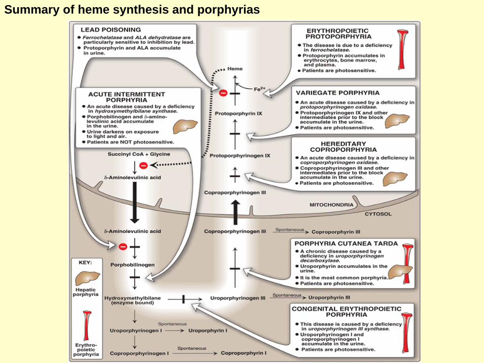
Summary of heme synthesis and porphyrias

Summary of Major Findings in the Porphyrias
Table 31–2. Summary of Major Findings in the Porphyrias.1
Enzyme Involved2
Type, Class, and MIM Number
Major Signs and Symptoms
Results of Laboratory Tests
1. ALA synthase(erythroid form)
X-linked sideroblastic anemia3
(erythropoietic) (MIM 301300)Anemia Red cell counts and hemoglobin
decreased
2. ALA dehydratase
ALA dehydratase deficiency (hepatic) (MIM 125270)
Abdominal pain, neuropsychiatric symptoms
Urinary ALA and coproporphyrin III increased
3. UroporphyrinogenI synthase4
Acute intermittent porphyria(hepatic) (MIM 176000)
Abdominal pain, neuropsychiatric symptoms
Urinary ALA and PBG increased
4. UroporphyrinogenIII synthase
Congenital erythropoietic(erythropoietic) (MIM 263700)
No photosensitivity Urinary, fecal, and red cell uroporphyrin I increased
5. Uroporphyrinogendecarboxylase
Porphyria cutanea tarda(hepatic) (MIM 176100)
Photosensitivity Urinary uroporphyrin I increased
6. Coproporphyrinogen oxidase
Hereditary coproporphyria(hepatic) (MIM 121300)
Photosensitivity, abdominal pain, neuropsychiatricsymptoms
Urinary ALA, PBG, and coproporphyrin III and fecal coproporphyrin III increased
7. Protoporphyrinogen oxidase
Variegate porphyria (hepatic) (MIM 176200)
Photosensitivity, abdominal pain, neuropsychiatricsymptoms
Urinary ALA, PBG, and coproporphyrin III and fecal protoporphyrin IX increased
8. Ferrochelatase Protoporphyria (erythropoietic) (MIM 177000)
Photosensitivity Fecal and red cell protoporphyrin IX increased
Porphyrias Contd-----
Lead poisoning
*Ferrochelatase and ALA synthase are inhibited
*Protoporphyrin and ALA accumulate in urine
Photosensitivity
It is due to the porphyrin-mediated formation of superoxide radicals from oxygen. These reactive species can oxidatively damage membranes, and cause the release of lysosomal enzymes. Destruction of cellular components cause photosensitivity.
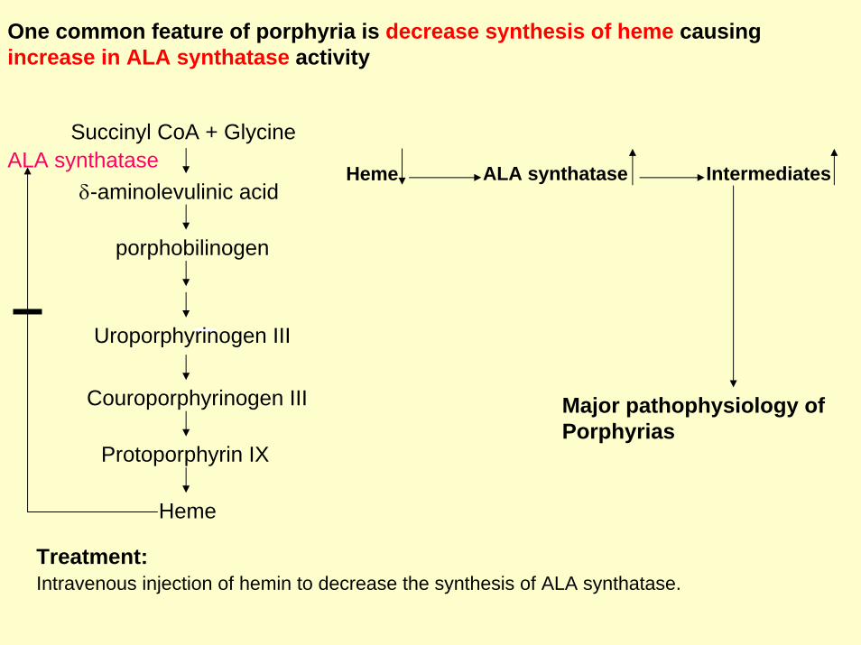
One common feature of porphyria is decrease synthesis of heme causingincrease in ALA synthatase activity
Succinyl CoA + Glycine
δ-aminolevulinic acidALA synthatase
porphobilinogen
Uroporphyrinogen III
Couroporphyrinogen III
Protoporphyrin IX
Heme
Heme ALA synthatase Intermediates
Major pathophysiology of Porphyrias
Treatment:Intravenous injection of hemin to decrease the synthesis of ALA synthatase.

Degradation of hemeRBCs last for 120 days and are degraded by reticuloendothelial (RE) system [liver and spleen].
About 85% of heme destined for degradation comes from RBCs and 15% from cytochromes, and immature RBCs. 1) Formation of bilirubin
a) Microsomal heme oxygenase hydroxylates methenyl bridge betweentwo pyrrole rings with concomitant oxidation of Fe2+ to Fe3+.
b) A second oxidation by the same enzyme results in the cleavage of the porphyrin ring resulting in biliverdin (green color).
c) Biliverdin is then reduced by biliverdin reductase, forming the bilirubin (red-orange).
2) Uptake of bilirubin by liverBilirubin then binds to serum albumin and is transported to the liver.
3) Formation of bilirubin diglucuronideBilirubin is then conjugated to two molecules of glucuronic acid by the enzyme bilirubin glucuronyl-transferase using UDP-glucuronic acid as a glucuronate donor (to increase the solubility of bilirubin)
4) Secretion of bilirubin into bile Conjugated form of bilirubin is the secreted into the bile. 5) Formation of urobilinsBilirubin diglucuronide is hydrolyzed and reduced by bacteria in the gut to yield Urobilinogen------oxidized to stercobilin.
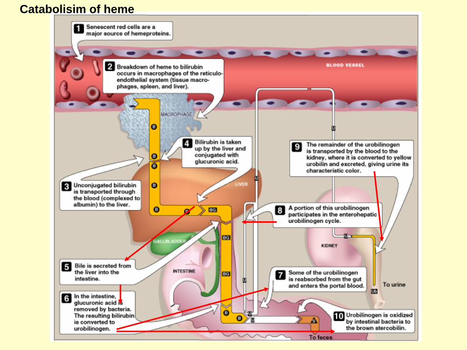
Catabolisim of heme

Yellow color of the skin, nailbeds, and sclerae(whites of the eyes) caused due to deposition of Bilirubin.
Jaundice
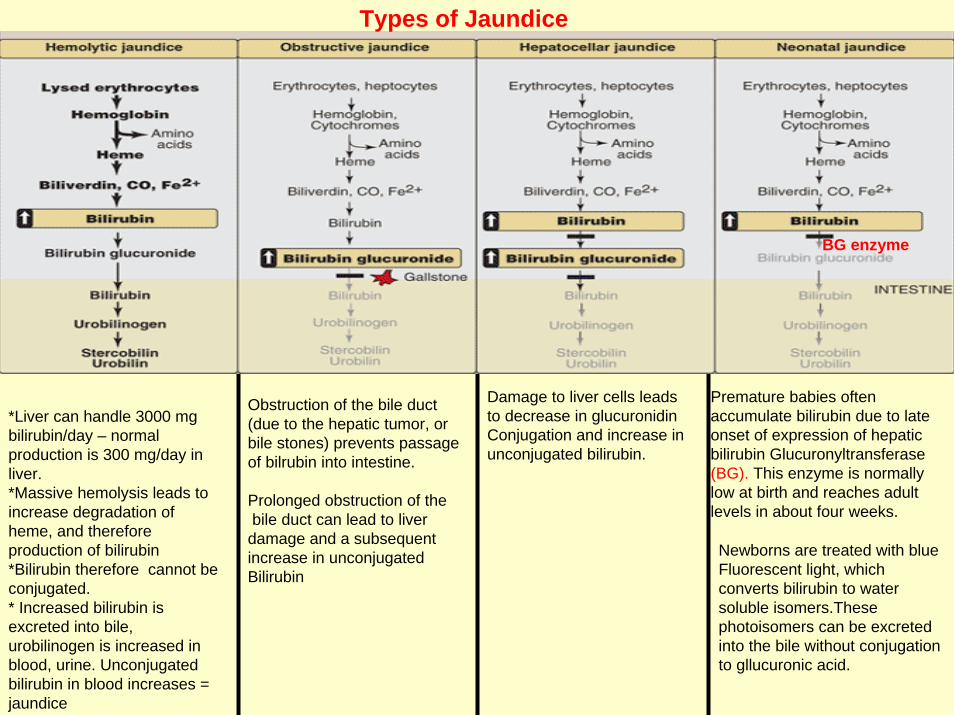
*Liver can handle 3000 mg bilirubin/day – normalproduction is 300 mg/day in liver. *Massive hemolysis leads to increase degradation of heme, and therefore production of bilirubin*Bilirubin therefore cannot be conjugated. * Increased bilirubin is excreted into bile, urobilinogen is increased in blood, urine. Unconjugatedbilirubin in blood increases = jaundice
Damage to liver cells leadsto decrease in glucuronidinConjugation and increase in unconjugated bilirubin.
Types of Jaundice
Obstruction of the bile duct(due to the hepatic tumor, orbile stones) prevents passage of bilrubin into intestine.
Prolonged obstruction of thebile duct can lead to liver
damage and a subsequent increase in unconjugatedBilirubin
Premature babies often accumulate bilirubin due to late onset of expression of hepatic bilirubin Glucuronyltransferase(BG). This enzyme is normally low at birth and reaches adult levels in about four weeks.
BG enzyme
Newborns are treated with blue Fluorescent light, which converts bilirubin to water soluble isomers.Thesephotoisomers can be excreted into the bile without conjugation to gllucuronic acid.

Determination of Bilirubin concentration
Van der Bargh reaction
Diazopyrroles (red color)Diazotized sulfanilic acid + Bilirubin
Measured Calorimetrically
Other nitrogen containing compounds
CatecholaminesDopamine, norepinephrine (noradrenaline)and epinephrin (adrenaline) are biologically active amines and are collectively called as Catecholeamines.
* Dopamine and norepinephrine functions as a neurotransmitters.
Outside the nervous system, norepinephrine and its methylated derivative, epinephrine regulates carbohydrate and lipid metabolism.
They are released from storage vehicles in the adrenal medulla in response to stress(fright, exercise, cold, and low levels of blood glucose).
They increase the degradation of glycogen,and triglycerides, as well as increase blood pressure and the output of heart.

Synthesis of catecholamine
Catecholamines are synthesized from Tyrosine
•Tyrosine is hydroxylated by tyrosine hydroxylase (rate limiting step in the pathway) to form DOPA.
•DOPA is decarboxylated by DOPA decarboxylase (pyridoxal phosphate requiring enzyme)to form dopamine.
•Dopamine is then hydroxylated by Dopamine β-hydroxylase to give norepinephrine.•Epinephrine is formed by N-methylation reaction using S-adenosylmethionine as
a methyl donor.
Parkinson’s disease is caused due to the production of insufficient dopamine synthesis in brain
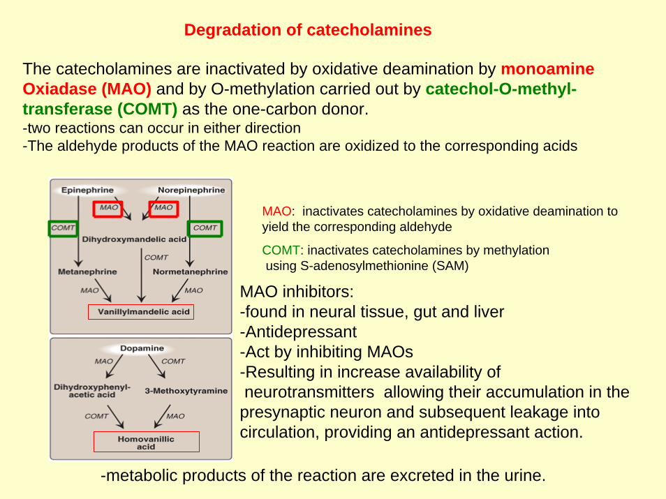
Degradation of catecholamines
The catecholamines are inactivated by oxidative deamination by monoamineOxiadase (MAO) and by O-methylation carried out by catechol-O-methyl-transferase (COMT) as the one-carbon donor.-two reactions can occur in either direction-The aldehyde products of the MAO reaction are oxidized to the corresponding acids
MAO inhibitors: -found in neural tissue, gut and liver-Antidepressant-Act by inhibiting MAOs-Resulting in increase availability ofneurotransmitters allowing their accumulation in the presynaptic neuron and subsequent leakage into circulation, providing an antidepressant action.
MAO: inactivates catecholamines by oxidative deamination to yield the corresponding aldehyde
COMT: inactivates catecholamines by methylationusing S-adenosylmethionine (SAM)
-metabolic products of the reaction are excreted in the urine.

Histamines-A chemical messenger that mediates a wide range of cellular responses, includingallergic and inflammatory reactions, gastric acid secretion, and possibly neurotransmission in the brain.
Pyridoxalphosphate
They are secreted by mast cells as a result of allergic reactions or trauma
Antihistamines are used to block histamine production during allergic reactions

Serotonin (5-hydroxytrptamine)
-mostly found in the cells of intestinal mucosa-smaller amounts occurs in CNS were it functions as a neurotransmitter-also found in platelet-has roles in pain perception, affective disorders, regulation of sleep, temperature,and blood pressure.
Also degraded by MAO.
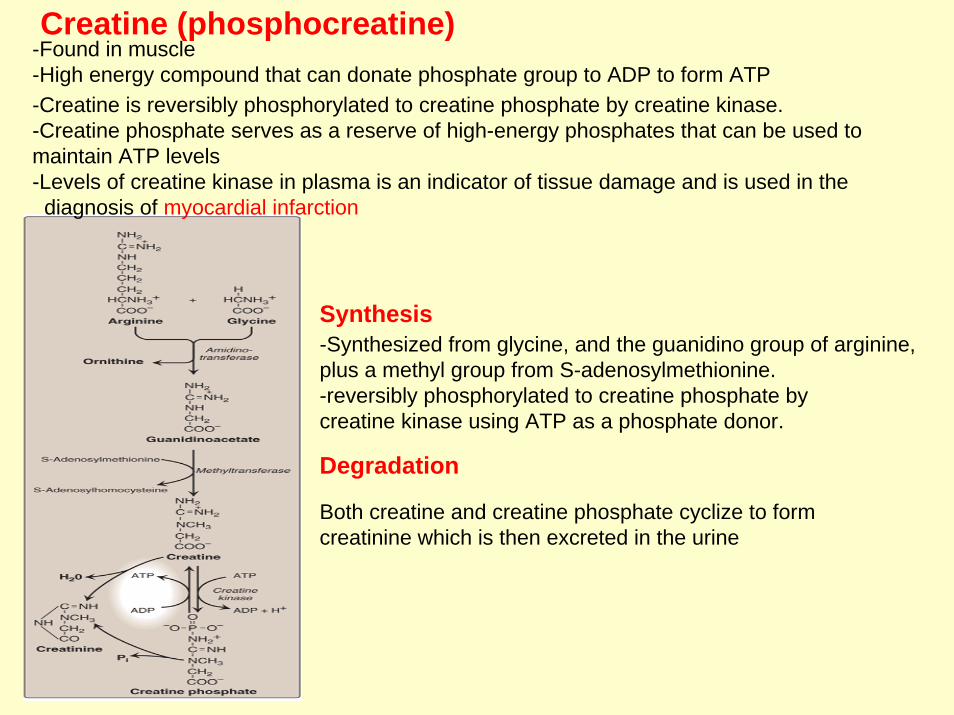
Creatine (phosphocreatine)-Found in muscle-High energy compound that can donate phosphate group to ADP to form ATP
-Synthesized from glycine, and the guanidino group of arginine, plus a methyl group from S-adenosylmethionine. -reversibly phosphorylated to creatine phosphate by creatine kinase using ATP as a phosphate donor.
-Creatine is reversibly phosphorylated to creatine phosphate by creatine kinase. -Creatine phosphate serves as a reserve of high-energy phosphates that can be used to maintain ATP levels-Levels of creatine kinase in plasma is an indicator of tissue damage and is used in the
diagnosis of myocardial infarction
Synthesis
Degradation
Both creatine and creatine phosphate cyclize to form creatinine which is then excreted in the urine

Melanin
-Pigment that occurs in several tissues, e.g. in eye, skin, and hair.-Synthesized from tyrosine in the epidermis by melaocytes-Function is to protect tissues from sun-light-Defect in melanin formation occurs in albinism due to the defective copper-containing enzyme tyrosinase.


Lecture I
II Digestion of dietary proteinsa) Gastric enzymesb) Pancreatic enzymesc) Small Intestinal enzymes (proteases cascade)d) Amino acid specificity for proteolytic enzymes
Amino Acids pool
Supplied Depleteda) Degradation
(Lysosomal and proteasome)b) Dietary proteinc) Do novo synthesis
a) Synthesis of body proteinsb) Precurssors for essential
N-containing molecules
I
III) How amino acids are transported in to cellsTransport systems
IV) Removal of nitrogen from amino acidsa) Transamination (aminotransferases)b) Oxidative deamination (Glutamine dehyrogenase)
V) Urea cycleReactions of urea cycle: a) locations b) sequence b) enzymes for each reaction c) end products for each reactionsd) ATP requirements e) sources of nitrogens in urea
VI) Metabolism of ammoniaa) Sources of ammonia, b) transport of ammonia, c) Urea cycle defects in humans

Lecture III) Essential and non essential amino acidsNames of the essential and non essential aa
II) Glucogenic and ketogenic amino acidsa) Why amino acids are classified as glucogenic and ketogenic or both?b) Seven intermediates of carbon skeletonc) Amino acids that form those intermediates
III) Catabolism of the branched-chain amino acids
IV) Biosynthesis of nonessential amino acidsa) Syhthesis from α-keto acidsb) Synthesis by amidationc) Synthesis of proline, serine, glycine, cysteine, tyrosine
V) Metabolic defects in amino acid metabolisma) Phenylketoureab) Maple Syrup urine diseasec) Albinismd) Homocystinuriae) Alkaptouria
Defective enzymeAmino acid involvedAccumulated intermediateCharacteristics
Lecture III
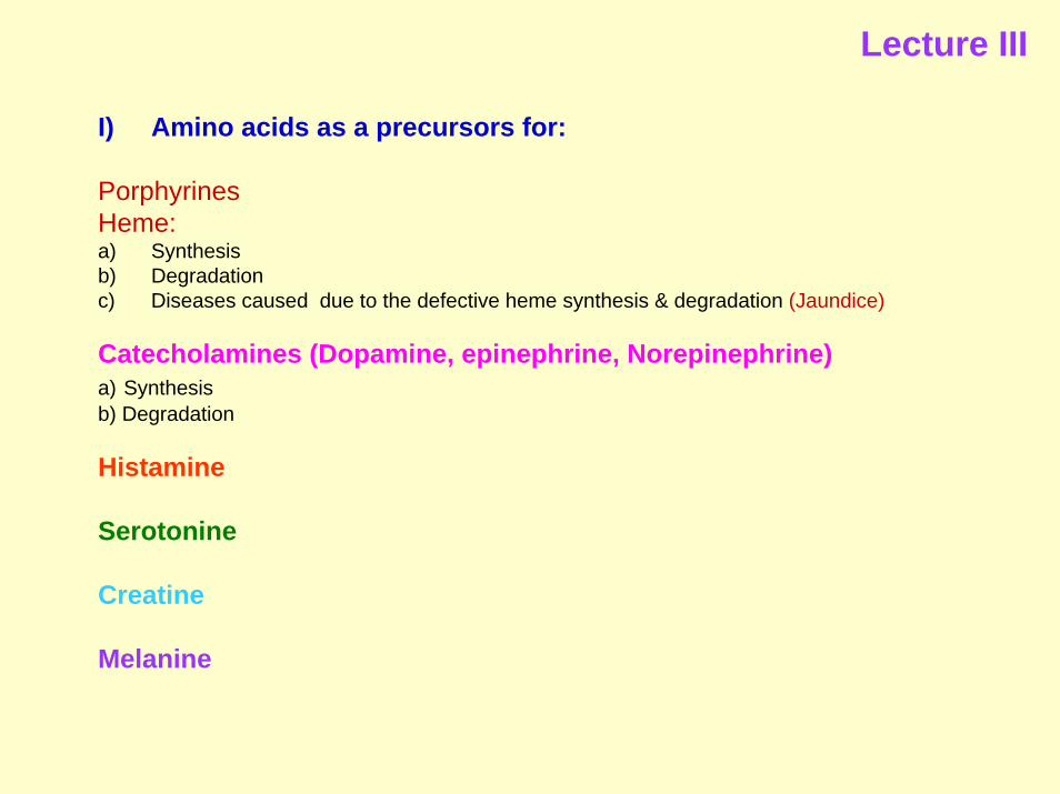
I) Amino acids as a precursors for:
PorphyrinesHeme:a) Synthesis b) Degradation c) Diseases caused due to the defective heme synthesis & degradation (Jaundice)
Catecholamines (Dopamine, epinephrine, Norepinephrine)a) Synthesis b) Degradation
Histamine
Serotonine
Creatine
Melanine