Embed Size (px)
Citation preview

MCraniofacial Surgery
InformationforPatientsandFamilies
The Melbourne Craniofacial CentreThe Royal Children’s Hospital
Melbourne Craniofacial Unit

The Melbourne Craniofacial
Unit provides complete care and
treatment for children with all
types of craniofacial disorders.
This includes children with birth
defects, deformity following
accident, or craniofacial growth
disorders. Since 1979, our group
of highly trained, coordinated
specialists has cared for over
two thousand patients and their
families. This centre of excellence
has one of the best safety records
for this type of complex surgery.
Melbourne Craniofacial Unit

Craniofacial SurgeryInformation
forPatientsandFamilies 2
T he craniofacial team brings together health
professionals from many different fields to
ensure that patients and their families receive
the best care. The team consists of plastic surgeons,
neurosurgeons, oral and maxillofacial surgeons,
dentists, ophthalmologists, anaesthetists, geneticists,
orthodontists, photographers, orthotists (helmet
remodelling), psychiatrists, social workers and specially
trained nursing staff, both in theatre and on the wards.
The craniofacial team

3
Consultation
On initial referral to the Craniofacial Unit, you and
your child will usually be seen in the craniofacial clinic
where many of the team members will be present and
all aspects of care can be addressed. There is usually
a general discussion and some information will be given
concerning the likely course of action and possible
treatment.
After this initial consultation in the Craniofacial Clinic,
you will be referred for specific one-on-one consultation
with individual team members as required for ongoing
treatment and care. It is during this one-to-one
consultation that your specific questions will be
discussed in detail. Every team member is committed to
getting to know you and your child well during this
cooperative experience. Importantly, you will also get
to know members of the team and the role each will
play in your child’s care.
If surgery is recommended, x-rays, scans, and other
investigations will be planned as needed. After one of
these consultations, it is often advisable to visit the
ward where your child will stay. One of the nurses can
show you photographs of other similar patients, so that
you can be best prepared for the early post operative
changes that occur.

CraniofacialSurgeryInformation
forPatientsandFamilies 4
What will happen duringyour child’s hospital stay? If your child is having an extensive procedure
admission may be the day prior to surgery.
This will allow you and your child to familiarise
with the ward and staff. Anaesthetic checks and
pre-operative blood checks can be finalised For less
extensive procedures your child may be admitted
on the day of surgery.
Usually there is a single fold-out bed where one parent
can sleep beside their child. For patients who come a
long distance, further accommodation may be arranged.
On the day of the operation, your child can not have
solid food for six hours prior to the procedure. This
helps to ensure a safe anaesthetic which is very impor-
tant. Clear fluids may be given closer to the time of
surgery; you will be advised of this by the anaesthetist.
At the time of surgery, you will accompany your child
to the pre-operative area. It is important to inform the
staff how to contact you during and after the surgery.
We will try to give you an accurate estimate of how long
the surgery will take. However, this time is hard to
predict and can vary.
When finished, the surgeon will let you know what
was accomplished during the procedure and how your
child is doing. When your child is awake and settled you
will be allowed into the recovery room.
5
After most craniofacial procedures, your child may have
various drips, catheters, arm splints, and bandages in
place to help with fluid requirements, pain relief, and
other supportive measures.
For a major procedure, your child is usually in hospital
for approximately four or five days, however this may
vary. After a few days the bandages will be removed
and your child’s hair can be washed. Significant post-
operative swelling can make it difficult for the eyelids
to open. This is normal and the children tolerate this
well. They can still communicate with their family
members and it often only takes two or three days
before they can open their eyes. Post-operative pain is
easily controlled on the ward with medication.Usually
patients can go home when their eyes are open and
they are eating and drinking normally.

Craniofacial SurgeryInformation
forPatientsandFamilies 6
Before going home, you will be given appropriate
instructions, and sometimes a moulding or
protective helmet will be ordered. Usually you
and your child will be seen approximately one week
after discharge. Any staples or stitches are easily
removed during this visit. It is important during the
first six weeks after craniofacial surgery to protect your
child from falls and injuries. After six weeks they are
usually at no greater risk than any other child. Long
term, protective head gear should be worn for contact
sports such as football, cycling, skiing, and surfing, just
like any normal child.
Follow up is often required after surgery. Once things
have stabilized, most children are reviewed annually.
For some conditions a series of operations may be
required. This would be determined and explained early
in the planning stages. Many patients only require one
operation but there is always a chance
that further minor adjustments will be needed.
After discharge

7
T he human head contains numerous bones
surrounding the brain; together these bones
are called the skull (or cranium). The bones
of the face are suspended from the anterior
portion of the cranium. This bony skeleton
is responsible for the shape and form of
the face and head. Abnormalities of
this underlying bony skeleton lead to
craniofacial problems and correction
of these deformities is the object of
craniofacial surgery.
The bones of the cranium are separated by sutures.
These sutures allow early growth to occur. Normal
sutures in a developing baby are quite wide with a
large separation between the bones. This allows the
rapidly growing brain to expand without restriction,
displacing the cranial bones outwards. Most of the
growth of the brain and the skull has occurred by
the age of two, with periods of rapid growth during
the first nine months of life.
As the growth of the brain approaches completion
the bony plates fuse together.
The facial bones develop in a similar fashion, although
the time of rapid growth is in the teenage years. It is
for this reason that many operations on the facial bones
are delayed until later in puberty when most of the
growth is complete, whereas cranial deformities are
often corrected in infancy.
Craniofacial anatomy and growth
Frontal bone
Parietal bone
Face
Occipital bone
Zygomatic bone
Metopic suture
Sagittal suture
Sagittal suture
Coronal suture
Craniofacial SurgeryInformation
forPatientsandFamilies 8
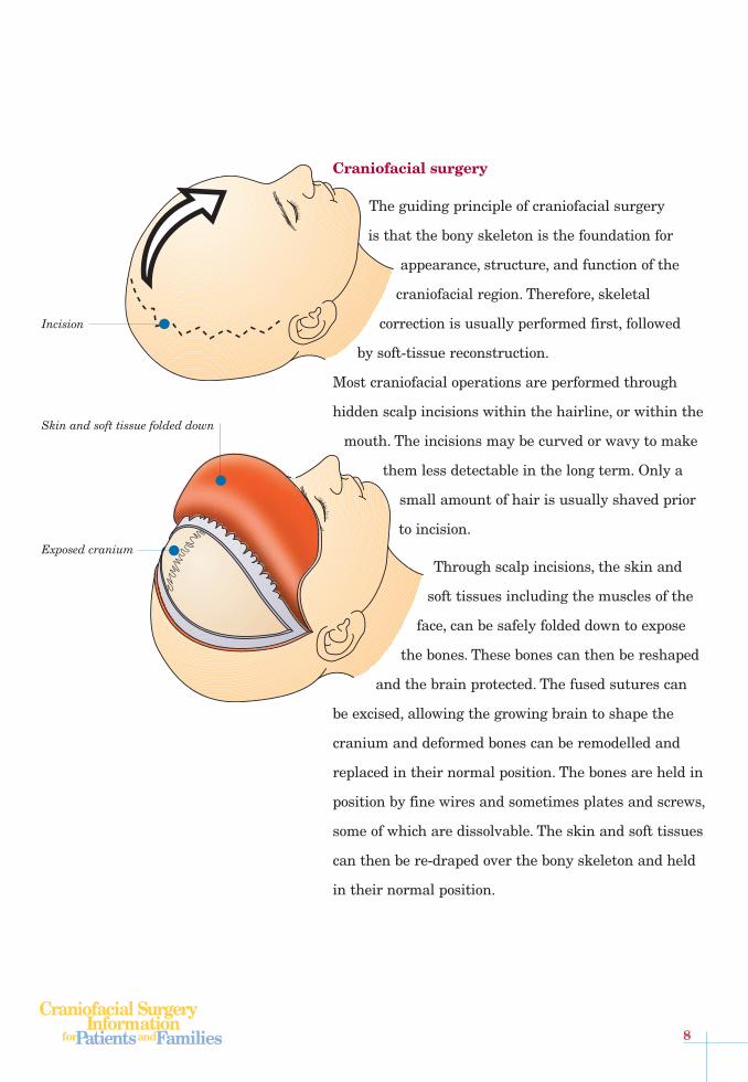
Craniofacial surgery
The guiding principle of craniofacial surgery
is that the bony skeleton is the foundation for
appearance, structure, and function of the
craniofacial region. Therefore, skeletal
correction is usually performed first, followed
by soft-tissue reconstruction.
Most craniofacial operations are performed through
hidden scalp incisions within the hairline, or within the
mouth. The incisions may be curved or wavy to make
them less detectable in the long term. Only a
small amount of hair is usually shaved prior
to incision.
Through scalp incisions, the skin and
soft tissues including the muscles of the
face, can be safely folded down to expose
the bones. These bones can then be reshaped
and the brain protected. The fused sutures can
be excised, allowing the growing brain to shape the
cranium and deformed bones can be remodelled and
replaced in their normal position. The bones are held in
position by fine wires and sometimes plates and screws,
some of which are dissolvable. The skin and soft tissues
can then be re-draped over the bony skeleton and held
in their normal position.
Incision
Skin and soft tissue folded down
Exposed cranium
9
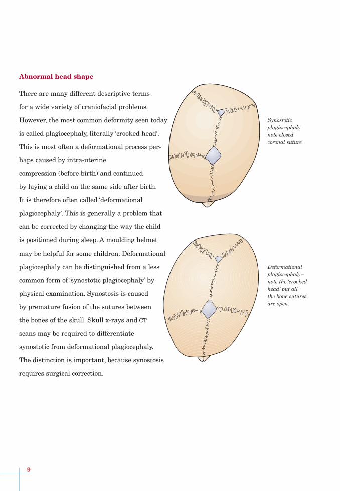
Abnormal head shape
There are many different descriptive terms
for a wide variety of craniofacial problems.
However, the most common deformity seen today
is called plagiocephaly, literally ‘crooked head’.
This is most often a deformational process per-
haps caused by intra-uterine
compression (before birth) and continued
by laying a child on the same side after birth.
It is therefore often called ‘deformational
plagiocephaly’. This is generally a problem that
can be corrected by changing the way the child
is positioned during sleep. A moulding helmet
may be helpful for some children. Deformational
plagiocephaly can be distinguished from a less
common form of ‘synostotic plagiocephaly’ by
physical examination. Synostosis is caused
by premature fusion of the sutures between
the bones of the skull. Skull x-rays and CT
scans may be required to differentiate
synostotic from deformational plagiocephaly.
The distinction is important, because synostosis
requires surgical correction.
Deformational plagiocephaly–note the ‘crooked head’ but all the bone sutures are open.
Synostoticplagiocephaly–note closed coronal suture.

Craniofacial SurgeryInformation
forPatientsandFamilies 10
T he sutures between the bones of the skull
sometimes close or fuse prematurely. When
this occurs it results in a deformity in the shape
of the skull, which may get worse as the skull grows
during the first two years of life. This is termed
synostosis (or craniosynostosis). Normally the two
frontal bones are separated by the metopic suture,
the two parietal bones by the sagittal suture, the
coronal suture lies between the frontal and parietal
bones, and the lambdoid suture between the occipital
and parietal bones.
The particular deformity and resultant shape of the
head is determined by which suture has prematurely
fused. If the metopic suture fuses between the frontal
bones, the forehead develops a triangular shape with a
prominent ‘keel’. There is a recession of the region
lateral to the eyes (temples) with a decreased distance
between the eyes (hypotelorism). The resultant head
shape is termed trigonocephaly.
Nonsyndromic craniosynostosis
Patient age 3 and 21 after infant correcion
Metopic synostosis CT of metopic synostosis
Plagiocephaly CT of plagiocephaly
2 yearsafter surgery 12 years after surgery

11
If one of the coronal sutures between the frontal and
parietal bones fuse then the resultant head shape is
called plagiocephaly and consists of a flattening of the
forehead on that side, a bulge posteriorly, a widening
of the bony orbit, and malposition to the nose and chin.
As mentioned above this needs to be carefully
distinguished from deformational plagiocephaly.
If the sagittal suture between the two parietal bones
becomes fused, growth is restricted from side to side
and the growing brain pushes forward and backwards,
producing an elongated head shape known as
scaphocephaly.
If both coronal sutures are fused, the length of the
skull from front to back is reduced and there is a
compensatory widening to give a short, squarish head
which is termed brachycephaly.
If one of the lambdoid sutures is fused, the changes
are similar to plagiocephaly but more pronounced at
the back of the skull.
More than one suture may be fused, although
this usually occurs within the setting of one of the
craniofacial syndromes described later.
When multiple sutures are fused, the shape of the head
often resembles a cloverleaf and this has been termed
cloverleaf skull or Kleeblattschadel deformity. This is
very serious because brain growth will be restricted.
Sagittal synostosis CT of sagittal synostosis
Sagittal synostosis 6 months after surgery
Cloverleaf deformity
Cloverleaf deformity 8 weeks after surgery

Craniofacial SurgeryInformation
forPatientsandFamilies 12
T here are over 200 craniofacial syndromes. These
syndromes feature premature fusion of the skull
associated with an abnormal gene. Therefore,
syndromes often run in families. Affected children may
have other features of abnormal head and facial
growth, as well as peripheral orthopaedic disorders. The
common craniofacial syndromes are Crouzon, Apert,
Pfeiffer, and Saethre Chotzen syndrome.
Recent laboratory studies have revealed that there
are alterations in the molecular structure of particular
genes which are responsible for producing protein
molecules called fibroblast growth factor receptors.
The role of these genes and proteins is currently being
investigated in many centres throughout the world,
including ours, and may lead to a better understanding
of these conditions as well as potential for prenatal
diagnoses in selected patients.
These genetic changes can lead to a wide range
of anomalies in tissues and organs apart from the
craniofacial skeleton. Genetic counselling forms an
important part in the management of families
affected by these conditions.
Syndromic Synostoses

13
Crouzon Syndrome
Crouzon syndrome consists of coronal suture fusion
on both sides, producing a short, wide head (brachy-
cephaly) in combination with under development of the
middle of the face, including the upper jaw. This can
result in small eye sockets, leading to prominent eyes
and the infant can have difficulty closing its eyelids.
Apert Syndrome
In Apert syndrome there is also coronal suture fusion
leading to brachycephaly as well as poor development of
the middle of the face and upper jaw. The skull is a lit-
tle different than with Crouzon, tending to be of a larg-
er size, with a very prominent forehead which may
grow upward in a tower-like fashion (turricephaly).
In addition, patients with Apert syndrome have severe
complex webbing of the fingers and toes, which is called
syndactyly. They may have anomalies of the brain as
well as an abnormal drainage of fluid around the brain
(hydrocephalus).
Pfeiffer Syndrome
Pfeiffer’s syndrome consists of brachycephaly due to
coronal synostosis in association with broad thumbs
and great toes.
Saethre Chotzen syndrome
These patients have coronal suture fusion with
brachycephaly as well as a prominent forehead, low
hairline and bilateral drooping (ptosis) of the upper
eyelids. They may have slightly short fingers and toes
with mild webbing between their fingers.
Crouzon syndrome
Apert syndrome
Saethre Chotzen syndrome

Craniofacial SurgeryInformation
forPatientsandFamilies 14
T here are many disorders of growth that can
result in over or underdevelopment of the facial
bones and jaws. These can all be improved
with craniofacial surgery. Binder syndrome and fibrous
dysplasia are typical examples of these disorders.
Binder Syndrome
Binder Syndrome is characterised by a deficiency in
growth of the mid-face between the mouth and brow.
This results in children who have a poorly developed
nose and poor jaw alignment. Treatment for this
condition usually includes orthodontics as well
orthognathic surgery to reposition the jaw to allow
the upper and lower teeth to meet together.
Once the midface has been placed in the proper
position with this surgery, attention can be directed
towards the nose, which can be supported and enlarged
with a bone graft.
In very severe cases, the nasal skin can be stretched
from an early age with artificial implants. In this
manner, an excellent early facial appearance can be
achieved which can prevent teasing at school. These
procedures usually begin after the eruption of the
secondary teeth, at approximately 7–11 years of age,
but final surgery is often not completed until growth
is finished.
Disorders of growth
Binder syndrome
Binder syndrome with temporary nasal enlargement
Binder syndrome – 20 year follow up after nasal reconstruction and advancement of the mid face

15
Fibrous dysplasia
Fibrous dysplasia is one of several over-growth
syndromes. It is a generally a benign disease of bone
that is characterized by the replacement of normal
bone with tissue similar to scar (fibrous tissue).
It can involve one or more sites and is typically noted
at around 10 years of age. It can present as simple facial
swelling, and can progress to gross asymmetry with
over-growth of the craniofacial skeleton. Surgery can
be performed to remodel this overgrowth or even to
prevent blindness.

CraniofacialSurgeryInformation
forPatientsandFamilies 16
Tessier Clefts
Rarely facial clefting may occur which is more
extensive than the more common cleft lip and palate.
These clefts are of varying severity from minor areas
of hair loss or notching to complete clefts in the skin
and bone. They are classified according to the ‘Tessier
Clock’ as illustrated. Usually excellent results can be
achieved with craniofacial surgical correction.
Craniofacial microsomia
Craniofacial microsomia entails a lack of development
of one or both sides of the face.
It has many names including hemifacial microsomia
and Goldenhar syndrome. This condition has a wide
range of manifestations. In mild cases patients have
one sided facial underdevelopement with skin tags in
front of their ears. Severely effected patients sometimes
have a profoundly affected, small jaw and are missing
an ear.
As it is a variable condition, some patients are very
mild and require no active treatment, while others
require orthodontics and jaw surgery to improve the
way the teeth meet (occlusion) and facial symmetry.
Craniofacial surgery and tissue transfers to improve
the facial appearance are needed for severe cases and
often construction of an entire ear is needed.
Clefting disorders14
5
6
7
8
9
10111213
1
3
42
0
30
7
Rare Tessier Clefts involving upperjaw, nose and skull
Tessier Cleft patient after reconstruction
Craniofacial microsomia

17
Treacher Collins
Treacher Collins syndrome is a genetic disorder of
facial growth on both sides. It consists of abnormal jaw
growth, with clefts in cheek bones and lower eyelids.
Ear malformations are seen, and patients are often
deaf. Good results can be achieved with craniofacial
surgery and hearing aids can be fashioned around
artificial or constructed ears.
TraumaMajor injuries to the face and skull can now be
successfully treated with craniofacial surgery
techniques. Trauma can include damage to the soft
tissues of the eyelids, lips, nose and cheeks as well
as to the bony structures. Craniofacial principles are
applied in order to rebuild the facial skeleton, limit
scars and improve function.
TumoursOccasionally young children with tumours are brought
to the attention of the craniofacial team. If cancerous,
these tumours are managed in combination with the
Oncology Unit. Benign tumours can often be treated
with simple surgery alone with excellent cosmetic
results. Some of the aggressive tumours can be cured
with a combination of pre-operative chemotherapy,
surgical excision of the area, and accurate reconstruc-
tion of the craniofacial skeleton and soft tissues to
produce a healthy, normal-looking child.
Tumour resection and reconstruction
Treacher Collins syndrome

Craniofacial SurgeryInformation
forPatientsandFamilies 18
Encephalocele
An encephalocele results from failure in formation of
the structures covering the brain in the embryo leading
to a hernia or outpouching of brain tissue through a
hole in the skull. Encephaloceles commonly present
between the eyes at the top of the nose. They can also
be found in other positions on the skull. There are
often associated changes in the shape of the skull and
eye sockets. The nose is often severely deformed.
Craniofacial principles apply to the correction of these
deformities with a wide exposure through the incision
across the top of the head. A direct incision over the
protruding encephalocele is frequently needed. The
lining around the brain is dissected and the contents
reduced back into the skull and the lining repaired.
The cranial bones surrounding the defect may then
need to be removed, reshaped or replaced with bone
grafts. Excess skin may then need to be excised or
rearranged.
Encephalocele Encephalocele after surgery
Encephalocele Encephalocele after surgery

19
Conclusion
The Melbourne Craniofacial Unit
based at the Royal Children’s Hospital
in Melbourne draws together the
expertise of world-class specialists.
• The aim is to help families and children
with craniofacial disorders due to
congenital problems (present at birth),
damage from trauma, or tumours.
• Our aim is to provide integrated complete
care for patients and their families,
enabling the children to achieve their
full potential in life.
• If you have any questions please feel free
to contact our craniofacial co-ordinator
telephone 03 9345 6582
facsimile 03 9345 4649
email [email protected]
www.rch.org.au/plastic