Embed Size (px)
Citation preview
Original Article
The CR-CO discrepancy and its effect on cephalometric measurements
Madelaine Shildkraut, DDS, MCID; David P. Wood, DDS, MCID; W. Stuart Hunter, DDS, MS, PhD
he importance of centric relation has been debated for many years. Dawson' described centric relation as a maxillo-
mandibular relationship in which the properly aligned condyle and disc are in the most supe- rior position in contact with the posterior surface of the articular eminence, irrespective of vertical dimension or tooth position. Lucia2 believes that ine correct centric relation is essential for coordi- nation of the occluding tooth surfaces and the temporomandibular joint.
The dental literature contains as few as two studies directly addressing the matter of centric relation and cephalometrics. Wood3 studied
"centrically related cephalometrics" with a sample of 30 patients whose casts were mounted on a Whip-mix articulator (using face-bow and centric bite). His "shadowgraph technique" per- mitted the comparison between centric occlusion and centric relation. Limitations due to the ra- diographic enlargement factor allowed the mea- surement of only ' a small number of cephalometric angles. Wood stated that "al- though the statistical analysis suggests the accu- racy of the shadowgraph, it by no means renders the technique clinically applicable". He did, how- ever, conclude that mounted casts and "cent$- cally related cephalometrics" offer more accurate
Abstract The purpose of the present study was to compare cephalometric measurements derived from a centric occlusion (CO)
tracing with those of a converted centric relation (CR) tracing. The sample consisted of 68 consecutively treated patients, with a CR-CO discrepancy of 2 mm or greater in either the horizontal and/or vertical planes, measured at the condyles from mounted models. Comparisons were also made within the sample between the 39 females and 29 males; and the 35 skeletal Class I and 33 Class I I patients.
In analyzing the CR-CO discrepancy, the vertical component was greater than the horizontal in 96% of the sample. Every patient had a vertical component, although 10% had no horizontal component. Correlations between the horizontal discrepancy and the two tracings showed high values for approximately 50% of the measures, whereas little correlation was found with the vertical discrepancy.
Paired t tests used to compare the CO and CR cephalometric values demonstrated significant differences (p<0.05) for the majority of the values studied. However, there generally were no differences between the groups of males and females, or between the skeletal Class I and Class I I individuals. The results of this study suggest that to make a correct orthodontic diagnosis the mandible should be placed in centric relation rather than in the more traditional centric occlusion.
Key Words CR-CO discrepancy Converted ceph Centric relation
Submitted: May 1991 Revised and accepted: January 1994 Angle Orthod 1994;64(5):333-342.
The Angle Orthodontist Vo1.64 No. 5 1994 333
Shildkraut; Wood; Hunter
Table I Description of CR-CO conversion (originated by Slavicek and
modified by Corbett and Williams)
On a CO tracing, draw Frankfort Horizontal (FH) and functional occlusal plane (FOP).
Draw the axis-orbital plane (AOP) as a line 6.5 degrees from FH through orbitale extending through the neck of the condyle.
Record the vertical overbite by drawing a short line parallel to the FOP extending through the incisal edge of the lower incisor.
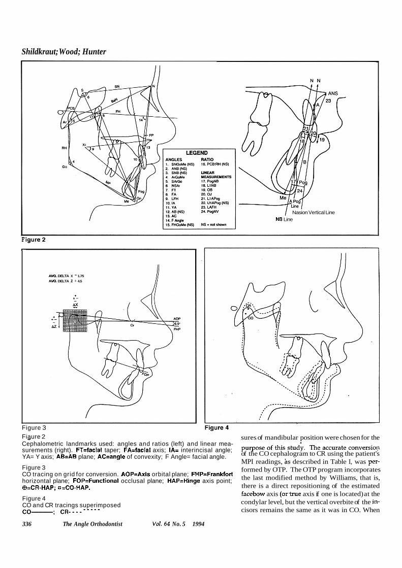
Locate and mark the estimated hinge axis point (HAP) on the CO tracing as 213 the width of the condyle forward from its posterior aspect on the AOP.
Plot the Ax and Az MPI readings on a graph sheet, as shown in Figure 3.
Overlay the CO tracing on the graph sheet registering at the estimated HAP at the crosshair and superimposed on the AOP (large horizontal line on graph sheet).
Mark a new CR HAP on the CO tracing from the graph sheet.
Using a new piece of tracing paper placed on the CO tracing draw the mandible, lower teeth, vertical overbite line, lower lip and chin soft tissue, and HAP.
Superimpose the HAP of the new tracing on the CR HAP of the first tracing and .rotate on this point to the best superimposition of vertical overbite line.
10. Trace the maxilla, upper teeth and remaining landmarks to make a centric relation tracing (Figure 4 shows the difference in mandibu- lar position between a CO [solid line] and CR [broken line] tracing as one is superimposed over the other).
information than hand-articulated casts. Wood used the term "dynamic occlusion"
which differentiates tooth-directed centric occlu- sion from centric relation dictated by the joints and ligaments.
Williamson et aL4 used the "centric-ceph" tech- nique on a sample of 46 patients divided into groups of Angle Class I and Angle Class I1 cases. They concluded that there were differences in cephalometric measurements with respect to the mandibular position, though most differences were slight. They found that Class I1 patients ex- hibited the largest discrepancies.
The role of occlusion in orthodontics has been studied by many authors. Aubrey5 observed that to finish a case in centric relation it is essential to remove functional interferences. Therefore he stated that one must "adapt teeth to joints and not ask the joints to adapt to the teeth". Roth has been advocating the need to treat patients in cen- tric relation since the 1 9 7 0 ~ . ~ ~ He believes that
The Angle Orthodontist VoL 64 No. 5 1994
there is a relationship between temporomandibu- lar pain-dysfunction and occlusal interferences6 and has described the criteria necessary to achieve functional occl~sion.~
Timm et al? observed that ". . . a common error is to accept a tooth arrangement that is Class I in centric occlusion but is actually Class I1 when the mandible is in centric relation". Therefore it is important to make a diagnosis from mounted casts in centric relation to correct the "true" and not the apparent malocclusion.
However, it is seldom possible to have a head- film in CR when planning treatment. To circum- vent this problem, Slavi~ek '~ ,~~ developed a tech- nique permitting the transfer of information obtained from mounted casts, which allows the transformation of a CO tracing into a CR tracing. Slavicek's approach was later modified by Maurice C. Corbett (Carmel Valley, Calif.) and modified again by Robert E. Williams (Mountain View, Calif.). The modified Slavicek approach has made possible an accurate evaluation of a malocclusion in centric relation.
The purpose of the study was to determine if there was a significant difference between 24 cephalometric measurements of mandibular po- sition derived from a centric occlusion (CO) trac- ing compared to those of a converted centric relation (CR) tracing. If statistically significant differences exist between CR and CO, this could affect the diagnosis and treatment planning nec- essary to correct the malocclusion.
Materials and methods Sample
Diagnostic records from a sample of 131 con- secutively treated patients (none of whom had TMD symptoms pretreatment) were collected from the private practice of one orthodontist in London, Ontario. The records included an ini- tial lateral cephalogram and all diagnostic casts mounted by the same clinician on the same SAM2 (Great Lakes Orthodontics, Buffalo, New York) articulator using an estimated face-bowl2 and cen- tric relation bite registration. The methodology 'for taking the centric relation bite registration was the same as outlined in Appendix I by Wood et al.I3 The amount of condylar distraction present was recorded using a mandibular position indi- cator (MPI)11,12 and a centric occlusion wax bite registration. The MPI is an instrument that al- lows the clinician or researcher to evaluate the magnitude and directional displacement that oc- curs in the condylar axis from CR to CO. The na- ture of the slide at the level of the occlusion most often does not necessarily reflect the condylar
CR-CO discrepancy movement. The MPI is a modified upper mem- ber of the SAM2 articulator in which the condy- lar housings have been replaced with laterally sliding cubes that contact the medial poles of the condylar elements (balls) when related to the lower member of the articulator. A right and left
paper that has a 1 nun square grid is p:aced on the outside of each sliding cube of the kiPI. In the center of each cube is an internal pin that perforates the grid paper and thereby marks CR the condylar axis. Once the grid papers have been fixed to the sliding cubes and perforated, the maxillary cast is attached to the MPI. The CO wax bite registration as described by Wood and KomeI2 is then placed on the upper cast. The 1 )wer cast that is fixed to the lower member of tile articulator is then placed firmly into the CO wax. The CO wax allows the approximation of the models into maximum intercuspation (plas- ter to plaster contact) and yet prevents rocking. The incisal guide pin of the MPI is then dropped onto the incisal table of the lower member of the articulator to further stabilize things. To record ! ~ e condylar axis position in CO, articulating pa-
per is held against the sliding cubes (with the graph paper affixed). The cubes are then slid out laterally t~ contact the condylar elements (balls) of the lower member of the articulator thereby marking the paper. The difference between the perforation by the pin on the graph paper (CR po- 5ition) and the mark of the articulating paper (CO ,)osition) is recorded in units of half millimeters in the horizontal (X) and vertical (Z) planes. For the horizontal (X) plane, when the CO mark is posterior to the CR perforation, the value is re- corded as negative and when anterior the value is positive. For the vertical (Z) plane, when the CO mark is inferior the value is recorded as posi- five. By definition condylar position is always ..uperior in CR. Therefore the articulator mark (CO) should always be inferior to the perforation (CR) if the patient has no TMJ symptoms, the casts have been mounted correctly and the CR bite has been taken correctly.
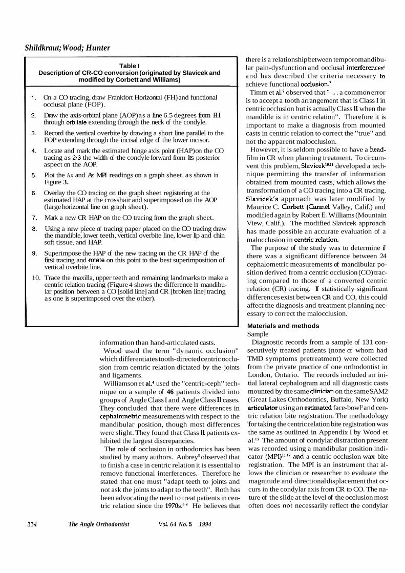
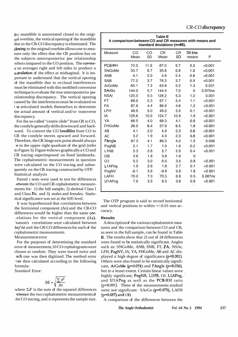
Any patients who did not have a CR-CO dis- crepancy (averaged between right and left sides) o f 2 mm or greater in either the horizontal (Ax) and/or vertical (Az) sagittal planes were elimi- nated. The resulting sample size for study was 68 patients (or 52% of the consecutively treated sample). The adolescents (64 patients) ranged from 9 to 17 years with a mean age of 12.8 years. The adults (4 patients) ranged from 19 to 35 years with a mean age of 27 years. An analysis of the ix or Az values showed that 55% of the study
sample had a value between 2 mm and 3 mm,
4
Superior
3
- 2
, 1
AZ 0
1 1
2 + 3
Inferior
4
Posterior
@ = Centric relation position
* = 2 or more recordings ( ) superimposed
Anterior
38% had a value between 3 mm and 4 mm, and Figure 1
7% had a value of 4 mm or greater. A graphic' Graphic representa- tion of averaged MPI representation of the averaged MPI recordings is recordings (n=68)
shown in Figure 1. For comparison, the sample of 68 was divided
into groups of skeletal Class I and skeletal Class I1 cases. Any case with an ANB angle of 24 de- grees was considered Class 11. This resulted in 35 Class I cases and 33 Class I1 cases. For the de- termination of sex differences, comparisons were made between the 39 females and 29 Gales.
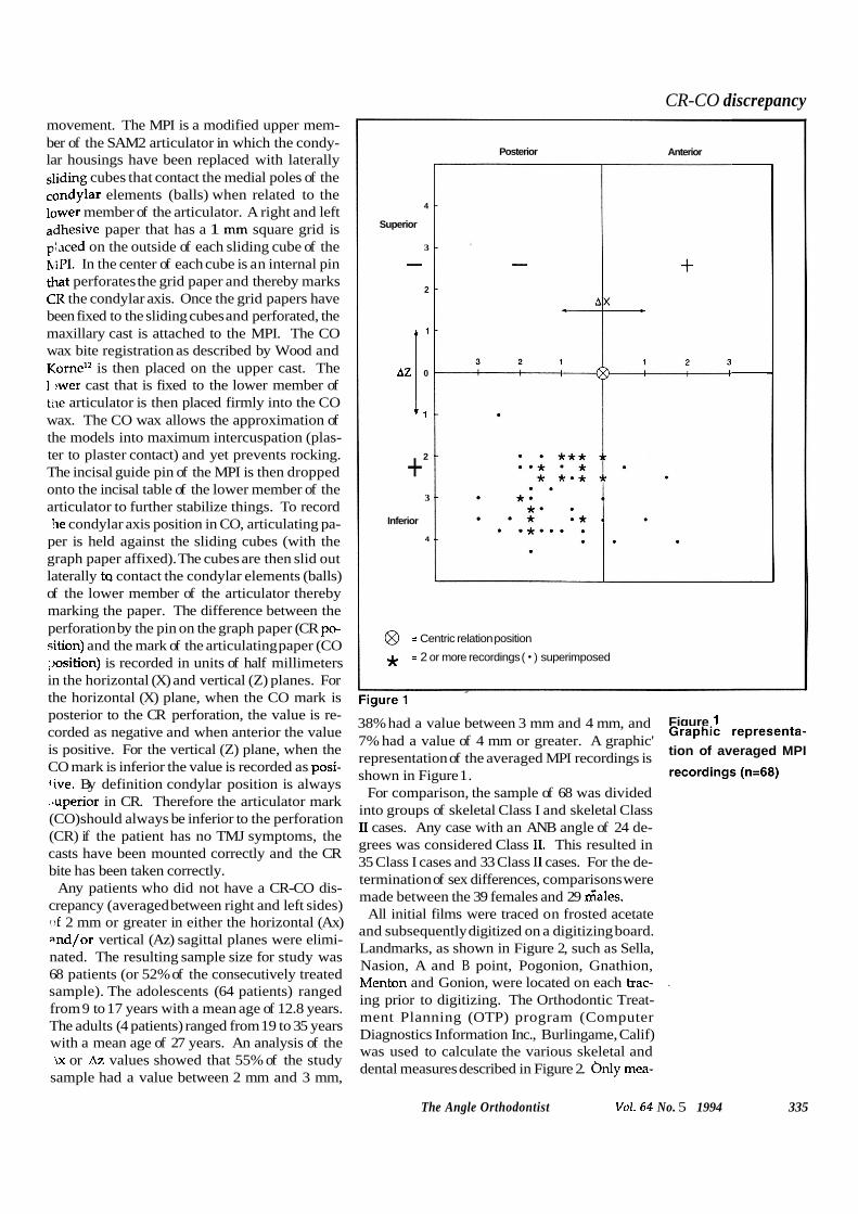
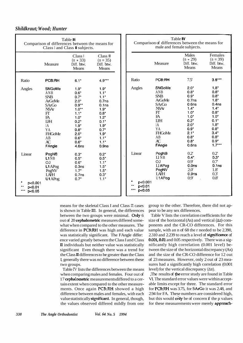
All initial films were traced on frosted acetate and subsequently digitized on a digitizing board. Landmarks, as shown in Figure 2, such as Sella, Nasion, A and B point, Pogonion, Gnathion, Menton and Gonion, were located on each trac- .
ing prior to digitizing. The Orthodontic Treat- ment Planning (OTP) program (Computer Diagnostics Information Inc., Burlingame, Calif) was used to calculate the various skeletal and dental measures described in Figure 2. m y mea-
The Angle Orthodontist Vo1.64 No. 5 1994 335
Shildkraut; Wood; Hunter
/ Nasion Vertical Line NB Line
F r
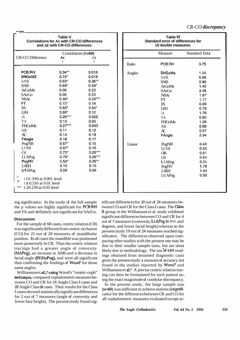
Figure 3 Fiaure 2
AVO. DELTA X - 1.75
AVO. DELTA Z + 4.5
sures of mandibular position were chosen for the " Cephalometric landmarks used: angles and ratios (left) and linear mea- purpose of *is stud;. -l-he accurate conversiol? surements (right). FT=facial taper; FA=facial axis; IA= interincisal angle; YA= Y axis; AB=AB plane; AC=angle of convexity; F Angle= facial angle. of the CO cephalogram to CR using the patient's
MPI readings, i s described in Table I, was per- Figure 3 formed by OTP. The OTP program incorporates CO tracing on grid for conversion. AOP=Axis orbital plane; FHP=Frankfort horizontal plane; FOP=Functional occlusal plane; HAP=Hinge axis point; the last modified method by Williams, that is,
&CR-HAP; ::=CO-HAP. there is a direct repositioning of the estimated facebow axis (ortrue axis if one is located) at the
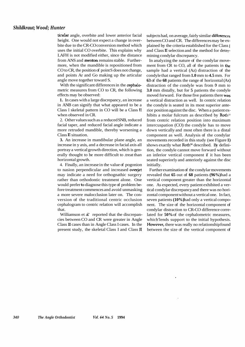
Figure 4 CO and CR tracings superimposed condylar level, but the vertical overbite of the in- cop; CR-- - - - - - - - cisors remains the same as it was in CO. When
336 The Angle Orthodontist Vo1.64 No. 5 1994
CR-CO discrepancy the mandible is autorotated closed to the origi- nal overbite, the vertical opening of the mandible due to the CR-CO discrepancy is eliminated. The closing to the original overbite allows one to mea- sure only the effect that the CR position has on the subjects anteroposterior jaw relationship when compared to the CO position. The conver- s on averages right and left sides to produce a simulation of the effect at midsagittal. It is im- portant to understand that the vertical opening of the mandible due to occlusal interferences must be eliminated with this modified conversion technique to evaluate the true anteroposterior jaw relationship discrepancy. The vertical opening caused by the interferences must be evaluated on t le articulated models themselves to determine tne actual amount of vertical and/or transverse discrepancy.
For the so-called "centric slide" from CR to CO, the condyle generally shifts downward and back- ward. To convert the CO headfilm from CO to CR the condyle moves upward and forward. Therefore, the CR hinge axis point should always )e in the upper right quadrant of the grid (refer
to Figure 3). Figure 4 shows graphically a CO and CR tracing superimposed on fixed landmarks. The cephalometric measurements in question were calculated on the CO tracing and subse- quently on the CR tracing constructed by OTP. Statistical analysis
Paired t tests were used to test for differences >etween the CO and CR cephalometric measure-
ments for: 1) the full sample; 2) skeletal Class I and Class 11s; and 3) males and females. Statis- tical significance was set at the 0.05 level.
It was hypothesized that correlations between the horizontal component (Ax) and the CR-CO differences would be higher than the same cor- .elations for the vertical component (Az). 'earson's correlations were calculated between Ax/Az and the CR-CO differences for each of the cephalometric measurements. Measurement error
For the purposes of determining the standard error of measurement, 10 CO cephalograms were chosen at random. They were traced twice and aach one was then digitized. The method error *./as then calculated according to the following formula: Standard Error:
where Id2 is the sum of the squared differences letween the two cephalometric measurements of
:he CO tracing, and n represents the sample size.
Table II A comparison between CO and CR measures with means and
standard deviations (n=68).
Measure CO CO CR CR Dif.btw. Mean SD Mean SD means P
PCB:RH SNGoMe ANB SNB ArGoMe SArGo NSAr FT FA LFH I A YA FHGoMe AB AC FAngle PogNB L1 NB OB OJ L1 APog PogNV LAFH U1 APog
The OTP program is said to record horizontal and vertical positions to within +/-0.01 mm ac- curacy.
Results A description of the various cephalometric mea-
sures and the comparison between CO and CR, as seen in the full sample, can be found in Table 11. The results show that 21 out of 24 differences were found to be statistically significant. Angles such as: SNGoMe, ANB, SNB, FT, PA, NSAr, LFH, PogNV, IA, YA, FHGoMe, AB and AC dis- played a high degree of significance (p<0.001). Others were also found to be statistically signifi- cant, ArGoMe (p=0.031) and FAngle (p=0.036), but to a lesser extent. Certain linear values were highly significant; PogNB, LINB, OJ, LlAPog, and UlAPog as well as the PCB:RH ratio (p<0.001). Three of the measurements studied were not significant: SArGo (p=0.975), LAFH (p=0.087) a d OB.
A comparison of the differences between the
The Angle Orthodontist Vo1.64 No. 5 1994 337
Shildkrau t; Wood; Hunter
Table Ill Comparison of differences between the means for
Class I and Class II subjects.
Class 1 Class I I (n = 33) (n = 35)
Measure Diff. btw. Diff. btw. Means Means
Ratio PCB:RH
Angles SNGoMe ANB SNB ArGoMe SArGo NSAr FT FA LFH I A YA FHGoMe AB AC FAngle
Linear PogNB L1 NB OJ L1 APog PogNV LAFH U1 APog
peo.001 *' peO.01 "' pe0.05
- Table IV
Comparison of differences between the means for male and female subjects.
Males Females (n = 29) (n = 39)
Measure Diff. btw. Diff. btw. Means Means
Ratio PCB:RH 7.5' 3.8'"
Angles SNGoMe ANB SNB ArGoMe SArGo NSAr FT FA LFH I A YA FHGoMe AB AC FAngle
Linear PogNB 0.2' 0.2' Ll NB 0.4'. 0.3* OJ 0.9' 0.7' Ll APog O.Ons O.lns PogNV 2.0' 1.8' LAFH 0.9ns 0.3' U 1 APog 0.9' . 0.8'
peo.001 " peO.01 *** pe0.05
means for the skeletal Class I and Class II cases is shown in Table 111. In general, the differences between the two groups were minimal. Only 6 out of 20 cephalometric measures differed some- what when compared to the other measures. The difference in PCB:RH was high and each value was statistically significant. The FAngle differ: ence varied greatly between the Class I and Class I1 individuals but neither value was statistically significant Even though there was a trend for the Class I1 differences to be greater than the Class I, generally there was no difference between these two groups.
Table IV lists the differences between the means when comparing males and females. Four out of 17 cephalometric measurements differed to a cer- tain extent when compared to the other measure- ments. Once again PCB:RH showed a high difference between males and females, with each value statistically significant. In general, though, the values observed differed mildly from one
group to the other. Therefore, there did not ap- pear to be any sex differences. Table V lists the correlation coefficients for the
size of the horizontal (Ax) and vertical (Az) com- ponents and the CR-CO differences. For this sample, with an n of 68 the r needed to be 2.390, 2.310 and 2.239 to reach a level of sigqificance of 0.001,0.01 and 0.05 respectively. There was a sig- nificantly high correlation (0.001 level) be- tween the size of the horizontal discrepancy (Ax) and the size of the CR-CO difference for 12 out of 23 measures. However, only 2 out of 23 mea- sures had a significantly high correlation (0.001 level) for the vertical discrepancy (Az). ,The results of @e error study are found in Table
VI. The standard error values were within accept- able limits except for three. The standard error for PCB:RH was 3.75, for SAGO it was 2.48, and 2.94 for FA. These numbers are considered high, but this would ody be of concern if the p values for these measurements were merely approach-
The Angle Orthodontist Vo1.64 No. 5 1994
CR-CO discrepancy --
Table V Correlations for Ax with CR-CO differences
and Az with CR-CO differences. -
Correlations (n=68) CR-CO Difference Ax Az
r r
PCB:RH SNGoME ANB SNB ArGoMe SArGo NSAr FT FA LFH I A YA FHGoMe AB AC FAngle PogNB L1 NB OJ Ll APog PogNV LAFH U1 APog
* r 10.390 at 0.001 level . ** r 1 0.310 at 0.01 level 1 *** r 20.239 at 0.05 level
Table VI Standard error of differences for
10 double measures.
Measure Standard Error
Ratio
Angles
Linear
SNGoMe ANB SNB ArGoMe SArGo NSAr FT FA LFH I A YA FHGoMe AB AC FAngle
PogNB Ll NB OB OJ L1 APog PogNV LAFH U 1 APog
ing significance. In the study of the full sample the p values are highly significant for PCB:RH and FA and definitely not significant for SArGo.
Discussion For the sample of 68 cases, centric relation (CR)
was sigruficantly different from centric occlusion (CO) for 21 out of 24 measures of mandibular position. In all cases the mandible was positioned more posteriorly in CR. Thus the centric relation tracings had a greater angle of convexity (NAPog), an increase in ANB and a decrease in facial angle (FHNaPog), and were all significant thus confirming the findings of Wood3 for those same angles.
Williamson et a1.,4 using Wood's "centric-ceph" technique, compared cephalometric measures be- tween CO and CR for 18 Angle Class I cases and 28 Angle Class I1 cases. Their results for the Class I cases showed statistically significant differences for 2 out of 7 measures (angle of convexity and lower face height). The present study found sig-
nificant differences for 20 out of 24 measures be- tween CO and CR for the Class I cases. The Clsiss I1 group in the Williamson et al. study exhibited significant differences between CO and CR for 4 out of 7 measures (convexity, LlAPog in mm and degrees, and lower facial height) whereas in the present study 19 out of 24 measures reached sig- nificance. The differences observed upon com- paring other studies with the present one may be due to their smaller sample sizes, but are most likely due to methodology. The use Gf MPI read- ings obtained from mounted diagnostic casts gives the present study a measure of accuracy not found in the studies reported by Wood3 and Williamson et aL4 A precise centric relation trac- ing can then be formulated for each patient us-. ing the exact magnitude of condylar discrepancy.
In the present study, the large sample size (n=68) was sufficient to achieve statistical signifi- cance for the differences between CR and CO for all cephalometric measures evaluated except ar-
The Angle Orthodon tist Vo1.64 No. 5 1994 339
Shildkraut; Wood; Hunter ticular angle, overbite and lower anterior facial height. One would not expect a change in over- bite due to the CR-CO conversion method which uses the initial CO overbite. This explains why LAFH is not modified either, since the distance from ANS and menton remains stable. Further- more, when the mandible is repositioned from CO to CR, the position of point S does not change, and points Ar and Go making up the articular angle move together toward S.
With the significant differences in the cephalo- metric measures from CO to CR, the following effects may be observed:
1. In cases with a large discrepancy, an increase in ANB can signify that what appeared to be a Class I skeletal pattern in CO will be a Class I1 when observed in CR.
2. Other values such as a reduced SNB, reduced facial taper, and reduced facial angle indicate a more retruded mandible, thereby worsening a Class I1 situation.
3. An increase in mandibular plane angle, an increase in y axis, and a decrease in facial axis all portray a vertical growth direction, which is gen- erally thought to be more difficult to .treat than horizontal growth.
4. Finally, an increase in the value of pogonion to nasion perpendicular and increased overjet may indicate a need for orthognathic surgery rather than orthodontic treatment alone. One would prefer to diagnose this type of problem be- fore treatment commences and avoid unmasking a more severe malocclusion later on. The con- version of the traditional centric occlusion cephalogram to centric relation will accomplish that.
Williamson et al.' reported that the discrepan- cies between CO and CR were greater in Angle Class I1 cases than in Angle Class I cases. In the present study, the skeletal Class I and Class I1
subjects had, on average, fairly similar differences between CO and CR. The differences may be ex- plained by the criteria established for the Class I and Class I1 selection and the method for deter- mining condylar discrepancy.
In analyzing the nature of the condylar move- ment from CR to CO, all of the patients in the sample had a vertical (Az) distraction of the condyle that ranged from 1.0 mm to 4.5 mm. For 63 of the 68 patients the range of horizontal (Ax) distraction of the condyle was from 0 mm to 3.0 mm distally, but for 5 patients the condyle moved forward. For those five patients there was a vertical distraction as well. In centric relation the condyle is seated in its most superior ante- rior position against the disc. When a patient ex- hibits a molar fulcrum as described by RothiJ from centric relation position into maximum intercuspation (CO) the condyle has to move down vertically and most often there is a distal component as well. Analysis of the condylar movements recorded in this study (see Figure 1) shows exactly what RoW4 described. By defini- tion, the condyle cannot move forward without an inferior vertical component if it has been seated superiorly and anteriorly against the disc initially.
Further examination of the condylar movements revealed that 65 out of 68 patients (96%) had a vertical component greater than the horizontal one. As expected, every patient exhibited a ver- tical condylar discrepancy and there was no hori- zontal component without a vertical one. In fact, seven patients (10%) had only a vertical compo- nent. The size of the horizontal component of condylar distraction to CR-CO difference corre- lated for 50% of the cephalometric measures, which'lends support to the initial hypothesis. ow ever, there was really no relationship found between the size of the vertical component of
The Angle Orthodontist Vo1.64 No. 5 1994
CR-CO discrepancy co;tdylar distraction to CR-CO difference for the sample studied.
In the present study Ax and/or Az were found to be greater than 2 mm in 50% of an orthodontic population (68 out of 131 patients). Twenty-five percent of the 131 cases had a condylar distrac- tion 23 mm. It is not possible to determine cl airside which patients will have a large discrep- a: ~ y . The significant difference in cephalometric measures of the mandible positioned in CR and in CO reinforces the need to mount every case and diagnose the malocclusion from centric rela- tion. The other added benefit of mounted diag- nostic casts is 'a documentation of condylar position at the start of orthodontic treatment.
One goal in orthodontic treatment is to protect t .le temporomandibular joint. Therefore a treat- ment goal approaching centric relation in every patient would be desirable. In a society where the incidence of litigation has increased, orth- odontists interested in functional occlusion may have the ability to defend themselves with records of condylar position before and after orth- :dontic t~eatment?~ Aubrey5 reinforces the need to finish cases in
centric relation occlusion and to remove func- tional interferences. He also views the impor- tance o i CR as a stable, reproducible, and comfortable position for the temporomandibular joint. The presence of occlusal interferences cre- ates problems such as subluxation of the joint and ventual occlusal wear, especially when there is
a lack of anterior guidance. There is a need to study in more depth the CR-
CO discrepancy as it relates to cephalometrics. This would demand a longitudinal CR-CO dis- crepancy study (MPI readings) with pretreat- ment, posttreatment, and postretention values for the same sample. The data have shown that cen- ric relation should be considered as the starting
point for proper diagnosis and treatment planning.
Conclusions 1. Measures of mandibular position were sig-
nificantly different between a centric occlusion. tracing and the same tracing converted to centric relation.
2. The differences appeared to apply equally between the skeletal Class I and Class I1 groups, or between males and females.
3. The condyle was always vertically distracted and most often positioned distally when the teeth were in centric occlusion.
4. The vertical component (Az) was almost al- ways greater than the horizontal (Ax) component. 5. To avoid errors in diagnosis, treatment plans
should be formulated from lateral cephalograms that have been converted to centric relation.
Acknowledgments The authors wish to acknowledge the Alpha
Omega Foundation for their support in the writ- ing of this paper.
Author Address Dr. David P. Wood 1270 Princess Royal Avenue Nanaimo, British Columbia Canada V9S 327 M . Shildkraut is in private practice in Montreal,
Quebec, Canada. D.P. Wood was chairman of the Division of Gradu-
ate Orthodontics, University of Western Ontario, Lon- don, Ontario, Canada.
W . Stuart Hunter is Professor Emeritus in the Di- vision of Graduate Orthodontics, Faculty of Dentisty, University of Western Ontario, London, Ontario, Canada.
The Angle Ortl~odontist Vo1. 64 No. 5 1994 341