Embed Size (px)
Citation preview

8/11/2019 Cornelia de Lange Syndrome - A Recognizable Fetal Phenotype
http://slidepdf.com/reader/full/cornelia-de-lange-syndrome-a-recognizable-fetal-phenotype 1/5
Fax +41 61 306 12 34
E-Mail [email protected]
Case Report
Fetal Diagn Ther 2009;26:50–53
DOI: 10.1159/000236361
Cornelia de Lange Syndrome:A Recognizable Fetal Phenotype
F.A. Wilminka D.N.M. Papatsonisa E.W.M. Grijseelsb M.W. Wesselsc
a Department of Obstetrics and Gynecology, Amphia Hospital, Breda, Departments of b Obstetrics and Gynecology,
and c Clinical Genetics, Erasmus Medical Center, Rotterdam, The Netherlands
in 2003 because of stagnation at the first stage of labor. At a ges-tational age of 40 weeks a healthy daughter of 3,975 g was born.The parents were non-consanguineous and without dysmorphicfeatures or congenital anomalies. The family history did not men-tion any congenital anomalies or relevant diseases. In this preg-nancy there was no drug abuse or infection, and serologic screen-ing for HIV, hepatitis B and syphilis was negative.
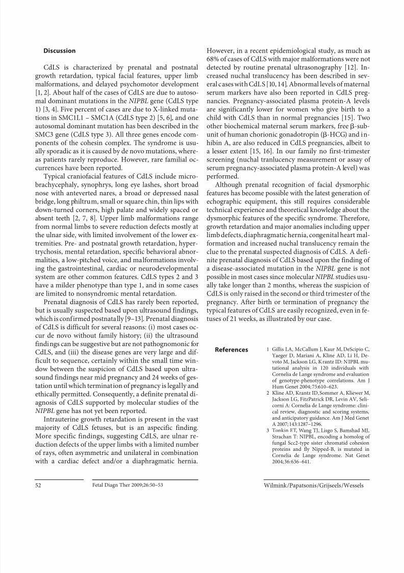
At routine sonography there was suspicion of a diaphragmat-ic hernia (fig. 1) and intrauterine growth retardation. Structural
Key Words
Cornelia de Lange syndrome Phenotype, fetal Hernia,
diaphragmatic Anomalies, limb NIPBL mutation
Abstract
We describe a fetus with Cornelia de Lange syndrome diag-
nosed after termination of pregnancy at 21 weeks. Prena-
tally, growth retardation, diaphragmatic hernia, cystic hy-groma and a right hand with only three rays were diagnosed
by ultrasound in the second trimester of pregnancy. Postna-
tal magnetic resonance imaging confirmed the prenatal
findings, and the presence of the typical dysmorphic fea-
tures led to the diagnosis of Cornelia de Lange syndrome.
The diagnosis was confirmed by the f inding of a truncating
mutation in the NIPBL gene. This case illustrates that the di-
agnosis Cornelia the Lange syndrome can be suspected pre-
natally in the second trimester, and can be diagnosed in fe-
tuses after induction or newborns at birth as the typical
phenotype is present early. Copyright © 2009 S. Karger AG, Basel
Case Report
A 34-year-old gravida 2, para 1 woman visited our hospital forroutine sonographic scanning at a gestational age of 19 weeks.Until then, the pregnancy had been uneventful. The obstetric his-tory revealed a secondary cesarean section in the first pregnancy
Received: July 16, 2008
Accepted: September 15, 2008
Published online: October 10, 2009
D.N.M. Papatsonis, MD, PhDDepartment of Obstetrics and Gynecology, Amphia Hospital BredaLangendijk 75
NL–4819 EV Breda (The Netherlands)Tel. +31 76 595 1000, Fax +31 76 595 2467, E-Mail [email protected]
© 2009 S. Karger AG, Basel1015–3837/09/0261–0050$26.00/0
Accessible online at:www.karger.com/fdt
Fig. 1. Prenatal sonography showing the diaphragmatic hernia.The stomach (S) is displaced upwards into the thorax causing amediastinal shift of the heart (H) to the right. The echodense lineof the diaphragm (D) is not continuous.

8/11/2019 Cornelia de Lange Syndrome - A Recognizable Fetal Phenotype
http://slidepdf.com/reader/full/cornelia-de-lange-syndrome-a-recognizable-fetal-phenotype 2/5
Cornelia de Lange Syndrome Fetal Diagn Ther 2009;26:50–53 51
sonography in a third line referral center at a gestational age of 19weeks confirmed that there was proportional intrauterine growthretardation with the following growth parameters below the fifthpercentile: biparietal diameter 41.7 mm; head circumference148.6 mm; abdominal circumference 122.2 mm, and femur length25.1 mm. Multiple congenital anomalies were diagnosed: a cystic
hygroma, a left-sided diaphragmatic hernia with herniation ofstomach, small intestines and part of the liver, and a right handwith only three rays. The heart appeared normal, but could not be visualized optimally. Amniocentesis was performed, and a nor-mal female karyotype 46,XX was obtained, whereas -fetoproteinwas increased at 29 mg/l (normal upper range for this gestationalage 20 mg/l).
After genetic counseling by the clinical geneticist and pedia-trician the parents decided to terminate the pregnancy. Fifteenhours after starting induction with vaginally administered miso-prostol, a stillborn female fetus (fig. 2a) was delivered. Birthweight was 290 g (normal weight at 21 weeks 360 g). Several con-genital anomalies were present: hypertelorism; a depressed nasalbridge; a short broad nose; long prominent philtrum; thin lips; a
small chin; dysplastic small ears, and a broad neck (most likelycaused by the cystic hygroma). The face was partly covered withhair, especially above the upper lip and there was a synophrys(fig. 2b, c). The right hand was claw-like and had only 3 f ingers: asmall thumb; one broad middle digit, and a finger-like attach-ment (fig. 3).
Coronal slides of the MRI scan (fig. 4) showed the left part ofthe liver and part of the small intestines protruding into the tho-rax due to the diaphragmatic hernia. The brain was not yet gy-rated, no abnormalities were described.
No consent for autopsy was given by the parents. In view of the typical dysmorphic features and congenital
anomalies Cornelia de Lange syndrome (CdLS) was suspected.Molecular analysis of the NIPBL gene by exon-by-exon sequenc-ing revealed a de novo truncating disease causing mutation con-sisting of a deletion of two nucleotides leading to a stop codon onposition 616 (p.ser616X).
Fig. 2. Fetus of 21 weeks with CdLS. a Full body picture. b , c Typ-ical facial characteristics of CdLS are present at 21 weeks of ges-
tation: synophrys and facial hair covered with vernix, hyper-telorism, depressed nasal bridge, short broad nose, long philtrum,thin lips, small chin, dysplastic small ears, and broad neck (causedby the cyst ic hygroma).
Fig. 3. Right claw-like hand with only three rays: a small thumb,one broad middle digit, and finger-like attachment which isplaced at the side.
Fig. 4. Postnatal MRI (coronal slide): the left par t of the liver (ar-row) is protruding into the thorax because of the diaphragmatichernia, and there is a mediastinal shif t.
8/11/2019 Cornelia de Lange Syndrome - A Recognizable Fetal Phenotype
http://slidepdf.com/reader/full/cornelia-de-lange-syndrome-a-recognizable-fetal-phenotype 3/5
Wilmink /Papatsonis /Grijseels /WesselsFetal Diagn Ther 2009;26:50–5352
Discussion
CdLS is characterized by prenatal and postnatalgrowth retardation, typical facial features, upper limbmalformations, and delayed psychomotor development[1, 2]. About half of the cases of CdLS are due to autoso-
mal dominant mutations in the NIPBL gene (CdLS type1) [3, 4]. Five percent of cases are due to X-linked muta-tions in SMC1L1 – SMC1A (CdLS type 2) [5, 6], and oneautosomal dominant mutation has been described in theSMC3 gene (CdLS type 3). All three genes encode com-ponents of the cohesin complex. The syndrome is usu-ally sporadic as it is caused by de novo mutations, where-as patients rarely reproduce. However, rare familial oc-currences have been reported.
Typical craniofacial features of CdLS include micro-brachycephaly, synophrys, long eye lashes, short broadnose with anteverted nares, a broad or depressed nasal
bridge, long philtrum, small or square chin, thin lips withdown-turned corners, high palate and widely spaced orabsent teeth [2, 7, 8]. Upper limb malformations rangefrom normal limbs to severe reduction defects mostly atthe ulnar side, with limited involvement of the lower ex-tremities. Pre- and postnatal growth retardation, hyper-trychosis, mental retardation, specific behavioral abnor-malities, a low-pitched voice, and malformations involv-ing the gastrointestinal, cardiac or neurodevelopmentalsystem are other common features. CdLS types 2 and 3have a milder phenotype than type 1, and in some casesare limited to nonsyndromic mental retardation.
Prenatal diagnosis of CdLS has rarely been reported,but is usually suspected based upon ultrasound findings,which is confirmed postnatally [9–13]. Prenatal diagnosisof CdLS is difficult for several reasons: (i) most cases oc-cur de novo without family history; (ii) the ultrasoundfindings can be suggestive but are not pathognomonic forCdLS, and (iii) the disease genes are very large and dif-ficult to sequence, certainly within the small time win-dow between the suspicion of CdLS based upon ultra-sound findings near mid pregnancy and 24 weeks of ges-tation until which termination of pregnancy is legally andethically permitted. Consequently, a definite prenatal di-
agnosis of CdLS supported by molecular studies of theNIPBL gene has not yet been reported.
Intrauterine growth retardation is present in the vastmajority of CdLS fetuses, but is an aspecific finding.More specific findings, suggesting CdLS, are ulnar re-duction defects of the upper limbs with a limited numberof rays, often asymmetric and unilateral in combinationwith a cardiac defect and/or a diaphragmatic hernia.
However, in a recent epidemiological study, as much as68% of cases of CdLS with major malformations were notdetected by routine prenatal ultrasonography [12]. In-creased nuchal translucency has been described in sev-eral cases with CdLS [10, 14]. Abnormal levels of maternalserum markers have also been reported in CdLS preg-
nancies. Pregnancy-associated plasma protein-A levelsare significantly lower for women who give birth to achild with CdLS than in normal pregnancies [15]. Twoother biochemical maternal serum markers, free -sub-unit of human chorionic gonadotropin ( -HCG) and in-hibin A, are also reduced in CdLS pregnancies, albeit toa lesser extent [15, 16]. In our family no first-trimesterscreening (nuchal tranlucency measurement or assay ofserum pregnancy-associated plasma protein-A level) wasperformed.
Although prenatal recognition of facial dysmorphicfeatures has become possible with the latest generation of
echographic equipment, this still requires considerabletechnical experience and theoretical knowledge about thedysmorphic features of the specific syndrome. Therefore,growth retardation and major anomalies including upperlimb defects, diaphragmatic hernia, congenital heart mal-formation and increased nuchal translucency remain theclue to the prenatal suspected diagnosis of CdLS. A defi-nite prenatal diagnosis of CdLS based upon the finding ofa disease-associated mutation in the NIPBL gene is notpossible in most cases since molecular NIPBL studies usu-ally take longer than 2 months, whereas the suspicion ofCdLS is only raised in the second or third trimester of the
pregnancy. After birth or termination of pregnancy thetypical features of CdLS are easily recognized, even in fe-tuses of 21 weeks, as illustrated by our case.
References 1 Gillis LA, McCallum J, Kaur M, DeScipio C,Yaeger D, Mariani A, Kline AD, Li H, De-
voto M, Jackson LG, Krantz ID: NIPBL mu-tational analysis in 120 individuals withCornelia de Lange syndrome and evaluationof genotype-phenotype correlations. Am JHum Genet 2004; 75: 610–623.
2 Kline AD, Krantz ID, Sommer A, Kliewer M,
Jackson LG, FitzPatrick DR, Levin AV, Seli-corni A: Cornelia de Lange syndrome: clini-cal review, diagnostic and scoring systems,and anticipatory guidance. Am J Med GenetA 2007; 143: 1287–1296.
3 Tonkin ET, Wang TJ, Lisgo S, Bamshad MJ,Strachan T: NIPBL, encoding a homolog offungal Scc2-type sister chromatid cohesionproteins and fly Nipped-B, is mutated inCornelia de Lange syndrome. Nat Genet2004; 36: 636–641.

8/11/2019 Cornelia de Lange Syndrome - A Recognizable Fetal Phenotype
http://slidepdf.com/reader/full/cornelia-de-lange-syndrome-a-recognizable-fetal-phenotype 4/5
Cornelia de Lange Syndrome Fetal Diagn Ther 2009;26:50–53 53
4 Krantz ID, McCallum J, DeScipio C, Kaur M,Gillis LA, Yaeger D, Jukofsky L, WassermanN, Bottani A, Morris CA, Nowaczyk MJM,Toriello H, Bamshad MJ, Carey JC, Rappa-port E, Kawauchi S, Lander AD, Calof AL, LiH, Devoto M, Jackson LG: Cornelia de Langesyndrome is caused by mutations in NIPBL,the human homolog of Drosophila melano-
gaster Nipped-B. Nat Genet 2004; 36: 631–635.
5 Musio A, Selicorni A, Focarelli ML, Gerva-sini C, Milani D, Russo S, Vezzoni P, LarizzaL: X-linked Cornelia de Lange syndrome ow-ing to SMC1L1 mutations. Nat Genet 2006;38: 528–530.
6 Borck G, Zarhrate M, Bonnefont JP, Mun-nich A, Cormier-Daire V, Colleaux L: Inci-dence and clinical features of X-linked Cor-nelia de Lange syndrome due to SMC1L1mutations. Hum Mutat 2007; 28: 205–206.
7 Manouvrier S, Espinasse M, Vaast P, BouteO, Farre I, Dupont F, Puech F, Gosselin B,Farriaux JP: Brachmann-de Lange syn-drome: pre- and postnatal findings. Am J
Med Genet 1996; 62: 268–273.
8 Urban M, Hartung J: Ultrasonographic andclinical appearance of a 22-week-old fetuswith Brachmann-de Lange syndrome. Am JMed Genet 2001; 102: 73–75.
9 Lalatta F, Russo S, Gentilin B, Spaccini L,Boschetto C, Cavalleri F, Maciadri M, Ger-
vasini C, Bentivegna A, Castronovo P, Lar-izza L: Prenatal/neonatal pathology in two
cases of Cornelia de Lange syndrome har-boring novel mutations of NIPBL. GenetMed 2007; 9: 188–194.
10 Huang WH, Porto M: Abnormal first-tri-mester fetal nuchal translucency and Corne-lia De Lange syndrome. Obstet Gynecol2002; 99: 956–958.
11 Drolshagen LF, Durmon G, Berumen M,Burks DD: Prenatal ultrasonographic ap-pearance of ‘Cornelia de Lange’ syndrome. JClin Ultrasound 1992; 20: 470–474.
12 Barisic I, Tokic V, Loane M, Bianchi F, Cal-zolari E, Garne E, Wellesley D, Dolk H: De-scriptive epidemiology of Cornelia de Langesyndrome in Europe. Am J Med Genet A2008; 146: 51–59.
13 Kliewer MA, Kahler SG, Hertzberg BS, Bow-ie JD: Fetal biometry in the Brachmann-deLange syndrome. Am J Med Genet 1993; 47:
1035–1041.14 Sekimoto H, Osada H, Kimura H, Kamiya-
ma M, Arai K, Sekiya S: Prenatal findings inBrachmann-de Lange syndrome. Arch Gy-necol Obstet 2000; 263: 182–184.
15 Aitken DA, Ireland M, Berry E, Crossley JA,Macri JN, Burn J, Connor JM: Second-tri-mester pregnancy associated plasma pro-tein-A levels are reduced in Cornelia deLange syndrome pregnancies. Prenat Diagn1999; 19: 706–710.
16 Arbuzova S, Nikolenko M, Krantz D, Halla-han T, Macri J: Low first-trimester pregnan-cy-associated plasma protein-A and Corne-lia de Lange syndrome. Prenat Diagn 2003;23: 864.

8/11/2019 Cornelia de Lange Syndrome - A Recognizable Fetal Phenotype
http://slidepdf.com/reader/full/cornelia-de-lange-syndrome-a-recognizable-fetal-phenotype 5/5


![Journal of Genetic Disorders & Genetic Reports SLC3A1 gene which had a recognizable phenotype to atypical HCS but without cystinuria [6]. Here we report a new case of “atypical HCS”](https://img.dokumen.tips/doc/110x75/5ee1be00ad6a402d666c8330/journal-of-genetic-disorders-genetic-reports-slc3a1-gene-which-had-a-recognizable.jpg)