Embed Size (px)
Citation preview

J. Moll. Stud. (2004) 70: 73–93 © The Malacological Society of London 2004
INTRODUCTION
The Solenogastres are a small, still poorly known class of apla-cophoran Mollusca, characterized by the mantle cover of cuticleand calcareous sclerites (scales or spicules) and by the laterallyrounded body with the foot narrowed to a longitidinal pedalgroove. With respect to characters of the alimentary tract (notrue radula ribbon, midgut without separate digestive gland)and to the lack of special excretory organs (no emunctoria),they appear to represent a basal off-shoot of Mollusca. Thearrangement of the mantle cover with cuticle and sclerites,known as the aplacophoran condition, is shared with the clearlydifferent, paraphyletically separated Caudofoveata, and sup-ports the primitive position of the Solenogastres (Salvini-Plawen& Steiner, 1996; Haszprunar, 2000; Salvini-Plawen, 2003).
Their generally small size (mostly between 3 and 30 mm, butup to 300 mm) and marine habitat (mostly below 50 m depth),mean that great effort and expense are required to collect them.This, as well as their organization with mainly internal systematiccharacters (in contrast to placophoran and conchiferan mol-luscs), has prevented a broader engagement and greater familiar-ity by scientific workers. We are thus left with a still fragmentaryknowledge, not only of biological, developmental and physio-logical features, but even with respect to pure faunistics (biodi-versity, biogeography, etc.). This incompleteness also includesseveral systematic descriptions that are only fragmentary whencompared with the required standard, and these await supple-mentation or revision.
At present, about 230 species of Solenogastres have beennamed and new ones are under description. In addition, muchmaterial from recent collections and expeditions awaits elabora-tion, which will no doubt contribute to enlarging our knowl-edge. Despite the revised classification by Salvini-Plawen (1978),several systematic problems remain (Salvini-Plawen, 2003). New
findings reveal that additional corrections will be needed, andthis is also due to supplementation and revision of earlierdescriptions. This may even apply to recent publications whenthe state of the original material was insufficient for a compre-hensive insight. In a group where we still need to improve andenlarge our comparative knowledge, all new information is valu-able. This situation vindicates even fragmentary descriptions ofsingular, rare species as long as they are unmistakably character-ized and clearly, recognizably defined against all other species.
In 1978, Salvini-Plawen introduced a new system of theSolenogastres, classifying them into the four orders Pholido-skepia, Neomeniamorpha, Sterrofustia and Cavibelonia.Members of the Cavibelonia are mainly characterized by theelaboration of calcareous mantle sclerites, which are providedwith a central cavity. These epidermal, mostly needle-shaped(acicular), hollow sclerites require a special mode of formation(Hoffman, 1949) and the order may thus represent the mostadvanced of the Solenogastres. This character of hollow needlesis mostly combined with the presence of a thick cuticle (>35 �m)and epidermal papillae. Cavibelonia thus replace the Proneo-meniidae s.l. and most of Neomeniidae s.l. in former systems (cf. Thiele, 1913; Hoffmann, 1929; Hyman, 1967).
The main character of this taxon, the hollow acicular sclerites, exhibits different arrangements, being in either radialor tangential alignment (Hoffman, 1949). The latter may beproduced in a single, obliquely disposed layer, or arranged intwo or more rather rectangularly intercrossing layers of lowangle and almost embedded within the cuticle (termed ‘skele-tal’ by Scheltema, 1999; Scheltema & Schander, 2000). Thespicules may be either thick-walled or thin-walled. Further,spicules may be distally hooked (or barbed), asymmetrically flat-tened and serrate, or asymmetrically axe-like (termed ‘captate’by Arnovsky, 2000). These different elaborations, however, con-cern the generic level only, as demonstrated by the occurance ofa range of states within Pararrhopaliidae and Simrothiellidae(e.g. Table 3). Consequently, some or all of these characters
CONTRIBUTIONS TO THE MORPHOLOGICAL DIVERSITY AND CLASSIFICATION OF THE ORDER CAVIBELONIA
(MOLLUSCA: SOLENOGASTRES)
LUITFRIED VON SALVINI-PLAWENInstitut für Zoologie, Universität Wien, Althanstraße 14, A-1090, Wien, Austria
(Received 3 May 2002, accepted 14 July 2003)
ABSTRACT
The status of families within the order Cavibelonia is poorly understood. Here, description of newspecies and reexamination of type material yield new information on organizational diversity and systematic demarcations in the order. The families Drepanomeniidae and Rhipidoherpiidae are confirmed, and a new family Notomeniidae is proposed. Within Simrothiellidae, biodiversity at boththe species and genus level is increased. Within Rhipidoherpiidae, as well as Proneomeniidae, thesupraspecific taxa are elucidated. Of the eight species concerned, five are new: Drepanomenia tenuitectan.sp. and D. pontisquamata n.sp. (Drepanomeniidae), Simrothiella digitoradulata n.sp., S. abysseuropaean.sp.and Aploradoherpia insolita n.gen. n.sp. (Simrothiellidae). Observations on A. insolita confirm theindependent origin of the (mesodermal) pericardioducts and (ectodermal) spawning ducts. A re-examination of the southern Norwegian Simrothiella margaritacea (Koren & Danielssen) revealed thatspecimens from the West European Basin belong to a separate species S. abysseuropaea n.sp. Two formerProneomenia species are revised and reclassified: Thieleherpia n.gen. is proposed for Proneomenia thulensisThiele (Rhipidoherpiidae) and Dorymenia for Proneomenia quincarinata Ponder (Proneomeniidae).After re-investigation of Notomenia clavigera Thiele, this species is classified in the new familyNotomeniidae.
Correspondence: e-mail: [email protected]

may be polyphyletic, as also indicated by certain sporadic find-ings (Salvini-Plawen, 1978; Scheltema & Kuzirian, 1991; Handl& Salvini-Plawen, 2002). However, cladistic analysis (Salvini-Plawen, 2003) suggests that this situation does not affect themonophyletic status of the taxon Cavibelonia.
There is great diversity in internal organization amongCavibelonia. Predominantly according to the types of foregutglandular organs and of the radula, 11 genus-groups or familieshave been defined: Acanthomeniidae, Pararrhopaliidae, Rho-palomeniidae, Amphimeniidae, Simrothiellidae, Drepano-meniidae, Strophomeniidae, Proneomeniidae, Epimeniidae,Syngenoherpiidae, Rhipidoherpiidae (cf. Salvini-Plawen, 1978).The definition of these families reflects the present knowledgeof species organization. This approach suffers, however, fromerrors and inadequate descriptions in earlier literature and alsofrom the gaps in knowledge of specific as well as generic diversity.The best-defined family is the Amphimeniidae (Salvini-Plawen,1972, 1978), with 22 species within 10 genera provided with several autapomorphic characters. On the other hand, there are three monogeneric families (Drepanomeniidae, Syngeno-herpiidae and Rhipidoherpiidae) and more information isrequired to determine whether their classification is vindicatedor over-interpreted.
To establish a better foundation for the insufficiently under-stood status of the families within Cavibelonia, new representa-tives and certain type material are investigated here. The resultscontribute to our knowledge of both cavibelonian diversity aswell as systematics at the genus and family level.
MATERIAL AND METHODS
All material comes from institutional collections, and speci-mens are preserved in alcohol. Mantle sclerites were isolatedmanually and embedded (double-slides), then drawn with cam-era lucida. Specimens were decalcified in Bouin’s fluid, embed-ded in paraffin, serially cross-sectioned at 10 �m (6 �m forThieleherpia thulensis) and stained with Heidenhain’s Azan. Oneindividual of Simrothiella margaritacea was used for preparation ofa whole mount of the radula apparatus by histolysis of tissueswith bleach.
Institutional abbreviations:
DMW, Dominion Museum, Wellington, New ZealandUSNM, National Museum of Natural History, Smithsonian
Institution, Washington DC, USASMNH, Swedish Museum of Natural History, Stockholm,
SwedenZMB., Museum für Naturkunde der Humboldt-Universität,
Berlin, GermanyZMUB, Zoologisk Museum, University of Bergen, Bergen,
NorwayZMUC, Zoologisk Museum, University of København, Danmark
SYSTEMATIC DESCRIPTIONS
Order Cavibelonia Salvini-Plawen, 1978
Diagnosis: Solenogastres with acicular, generally hollow mantlesclerites; cuticle mostly thick; commonly with epidermal papil-lae.
Drepanomeniidae Salvini-Plawen, 1978
Diagnosis: Solenogastres with thick cuticle, sclerites as hollowneedles (Cavibelonia) in one layer; type of radula unknown(vestigial), ventral foregut glandular organs epithelial; with respiratory organs, without seminal receptacles.
Genus Drepanomenia Heath, 1911
Diagnosis: Solenogastres-Cavibelonia with hollow needles in one layer only; opening of mouth within atrium; without radula,ventral foregut glandular organs epithelial, midgut with serialconstrictions; dorso-terminal sense organ present; secondarygenital opening unpaired; without seminal receptacles and copu-latory stylets; secondary gills present.
Type species: Neomenia vampyrella Heath, 1905.
Drepanomenia tenuitecta new species(Figures 1, 2)
Types: Holotype (USNM 1016981), 4 mm length, serial cross-sections on slides; Tasman Sea west of New Zealand (38°27�–38°30� S, 168°04�–168°07� W), Station 24-1718 of US AntarcticResearch Program (USARP), 12 July 1966, 531–659 m.
Etymology: Latin tenuis, thin, tender, slim; tectum, roof, covered;referring to the rather thin mantle cuticle.
Diagnosis: Body about 4 � 1 mm with rounded body ends, cuticlemoderately thick, with epidermal papillae; hollow needles inradial arrangement, mid-dorsally forming a feeble crest; pedalgroove with three folds, separated from pallial cavity by mantleepithelium; mantle cavity with four respiratory folds; foregutforming proboscis with the tubular glandular organs opening at its tip; midgut with paired rostral caecum. No abdominal spicules.
Material examined: Holotype and a juvenile specimen (USNM1016982), 1 mm in length, serial cross-sections on slides, fromtype locality.
Mantle: Epidermis (15 �m high) forming slender papillae whichend with distal vesicle (20–25 �m diam.) filled with granules.Spicules (Fig. 1) giving the animals furry aspect are hollow,slightly curved, thin-walled needles (70–110 �m � 8–12 �m;Fig. 1A), rising above 50–75 �m thick cuticle; all in one layer,dorsally to radially arranged, mid-dorsally forming a feeblecrest; several strong, almost straight, thick-walled spicules(100–180 �m � 15–20 �m; Fig. 1B) are sunk into the muscula-
L. VON SALVINI-PLAWEN
74
Figure 1. Drepanomenia tenuitecta new species: mantle sclerites. A. Generalspicules. B. Interspersed straight spicules.

ture. No special sclerites (abdominal spicules) at anterior bor-der of mantle cavity.
Foot and mantle cavity: Ciliated pedal pit receiving intercellularlyopening pedal glands, posteriorly forming three dorsal foldsentering the externally distinctly visible pedal groove. The threefolds (central being most prominent) extend to level of spawn-ing ducts where only central fold remains; groove ends closebehind, i.e. in front of mantle cavity, separated by large mantlebridge. Sole glands along entire groove emptying within foot.Only single pedal fold present in juvenile. Mantle cavity rela-tively small, anteriorly with openings of hindgut and unpairedoutlet of spawning ducts only. Dorsal and posterior portions of10–13-�m-high epithelium more densely ciliated. Behind thecavity opening are four internal, dorso-ventrally running,frontally directed respiratory folds.
Musculature: Three-layered body wall musculature reinforcedventrally by numerous and spongily arranged longitudinalfibres. Serial dorsoventral bundles large, constricting midgut.
Sensory system: Unpaired cerebral ganglion (120 �m wide, 80 �mhigh, 60 �m long) gives rise to two pairs of ventrolateral nerveswith small basal swelling towards atrial sense organ; a third, ventral pair extends into proboscis. Cerebral connectives sepa-rate from their origin. Buccal connectives enter proboscis; presence of buccal ganglia uncertain. First lateral ganglia(120 �m � 25 �m diam.) extend between cerebral ganglionand body wall. First ventral ganglia (60 �m diam.) above pedal
pit are interconnected by two commissures, each releases anerve to beginning of foot and a lateroventral connective to oneof lateral ganglia. Body nerve cords with ganglia interconnectedby ventral commissures and less frequently lateroventral con-nectives. Starting in region of spawning ducts, lateral cords aremedullary, each shows a posterior-most lateroventral con-nective running between gut and spawning duct. Ganglia posteriora superiora (60 �m diam.), lateral of rectum, intercon-nected by 100-�m-long medullary suprarectal commissure(25 �m diam.); releasing at least one pair of dorsocaudal nerves55 �m apart. Behind above-mentioned terminal connectives,ventral cords curve dorsally to run along dorsal side of thespawnings ducts to their ends. Due to protruded proboscis, atrial sense organ is strongly deformed; no ciliary tracts or foldsrecognizable (see D. vampyrella). Ventrally, proboscis epi-thelium (partly atrial) shows cells 25 �m high that could repre-sent atrial ridges. Frontal and lateral walls of atrium possessblunt papillae filling organ. Dorsoterminal sense organ is a terminal epithelial bulge.
Alimentary tract: Proboscis in both specimens everted, bulb-like.Mouth at tip of proboscis which projects 1 mm from body.Proboscis formed by evagination of posterior roof epitheliuumof atrial cavity; its musculature formed by ventral portion ofdorsoventral musculature and by ventral body wall musculature.Proboscis round in cross-section (160–180 �m) with proximalswelling; epithelium (dorsally 15–20 �m, ventrally 20–25 �m)with gland cells. Central foregut (60–80 �m diam.) lined byweakly folded epithelium (10–15 �m) underlain by musculature.
CONTRIBUTIONS TO CAVIBELONIA
75
12345678910123456789201234567893012345678940123456789501234567896012345678
Figure 2. Drepanomenia tenuitecta new species, schematic organization, reconstructed from serial transverse sections: A. Anterior body. B. Posterior body.Abbreviations: as, atrial sense organ; bw, body wall (mantle and musculature); ca, midgut caecum; ce, cerebral ganglion; co, suprarectal commissure; fo, pedalfolds (foot); gl, ventral foregut glandular organ; go, gonad; mg, midgut; pc, pericardium; pd, pericardioduct; ro, respiratory organ; sd, spawning duct; so, terminal sense organ; vg, (first) ventral ganglion.

Space between foregut and proboscis musculature is a mes-enchyme rich in lacunae and blood cells. Two tubes with epi-thelial glands (ventral glandular foregut organs of type C inSalvini-Plawen, 1972, 1978) run through this space, opening attip of proboscis and extending posteriorly below midgut. Fore-gut merges dorsoposteriorly into midgut, shortly behind cere-bral ganglion; foregut musculature continuous with midgutmuscularis. Rostral caecum paired, extending beyond cerebralganglion. Midgut proper showing mid-dorsal ciliary tract and(due to dorsoventral muscle bundles) lateroventral pouchings.Nematocysts (to 20 �m) and spirocysts (to 30 �m) presentthroughout gut. Mid-dorsal ciliated tract continuous with moredensely and entirely ciliated hindgut, which opens dorsally intomantle cavity above spawning duct outlet.
Circulatory system: Pallial sinus above respiratory folds is antero-dorsally continuous with heart auricles formed by median wallsof terminally paired portion of pericardium. An invagination ofpericardial roof (beginning with fusion of paired portion) rep-resents the ventricle; both auricles and ventricle are mediallylined with muscle fibres. Dorsal sinus runs between gonoperi-cardioducts and over gonads. Blood cells consist of round, pale,homogeneous elements (5–8 �m) with indistinct nucleus andof more irregularly formed cells (to 15 �m diam.) with granules;the latter resemble the inclusions of the epidermal papillae.
Gonopericardial system: Bigger specimen showed beginning ofsexual maturity with ovocytes in paired gonad. Gonad posterior-ly continuous with gonopericardioducts, which open dor-sofrontally into pericardium. This organ is terminally paired,giving rise to the ventrolateral, ciliated pericardioducts sur-rounded by musculature. Neither seminal vesicles nor recepta-cles are present. Pericardioducts open laterally into anteriorspawning ducts, still lined by simple (not yet glandular) epitheli-um. Along their posterior course the spawning ducts fuse, open-ing into anterior-most ventral mantle cavity.
Remarks: All known Drepanomenia species represent a well-defined group based on the structure of their mantle (stronghollow spicules in radial arrangement, slender epidermal papil-lae), the ventral foregut glandular organs (type C), the respira-tory folds, and the lack of receptacula seminis (Salvini-Plawen,1978). These characters separate this mongeneric family fromothers; due to the lack of the radula, no relationships with othercavibelonian families can be indicated.
Up to now, three species of Drepanomenia have been described(Table 1). Drepanomenia vampyrella (Heath, 1905) from offHawaii differs from the new specimens by its thick cuticle(200–280 �m), which is extended mid-dorsally to form a 70 �m-high keel, and by the single rostral caecum of its midgut (Heath,1911: 77–82). Drepanomenia incrustata (Koren & Danlielssen,1877) from the Arctic Sea likewise possesses a thick cuticle(200–270 �m; personal observation of holotype) and a foot offive to three true folds extending throughout the groove. It isnot known whether its foregut (with ventral pouch and specialmusculature) can be protruded as a proboscis (see Odhner,1921: 19–22). The lateral walls of the midgut in D. incrustataform strong irregular folds, being serially constricted by thestrong dorsoventral muscle bundles (60–80 �m diam.; holo-type, SMNH no.4732). According to Odhner (1921: 21) thespawning ducts in D. incrustata are subdivided into an anteriorportion (‘bladder’) with thin walls and a thick-walled posteriorportion (shell gland) which subsequently fuses to become anunpaired organ. Finally, Drepanomenia perticata Salvini-Plawen,1978, from the Antarctic Ross Sea is characterized by its thickcuticle (450–500 �m), by the presence of abdominal spiculesand by a special bundle of pre-pedal spines. In comparison(Table 1), the present specimens from off New Zealand, withtheir thin cuticule, represent a well-defined new species.
Drepanomenia pontisquamata new species(Figures 3, 4, 16)
Types: Holotype (USNM 1016983), 6 mm length (Fig. 16), serialcross-sections on slides; southeastern Canada (44°31�–44°32� N,56°48�–57°57� W), Station 2–10 of US Antarctic ResearchProgram (USARP), 19 March 1962, 403 m.
Etymology: Latin pons, pontis, bridge; squama, scale; referring tothe special character of the large scales at the mantle bridgebetween atriobuccal opening and pedal pit.
Diagnosis: Body 6 � 1.5 mm with rounded ends; cuticle thick,terminally with one or three longitudinal thickenings; hollowspicules radially arranged, mantle bridge between atriobuccalopening and pedal pit with large scales; pedal groove distinctlyvisible, with three folds, not in pallial cavity; pallial cavity withventrorostral pouch and with ten respiratory folds. Cerebralconnectives with common trunk. Foregut long and protrusible,ventral foregut glandular organs opening via paired papilla,
L. VON SALVINI-PLAWEN
76
Table 1. Specific characters of Drepanomenia species (see Heath, 1911; Odhner, 1921, Salvini-Plawen, 1978, and herein).
Character D. vampyrella D. incrustata D. perticata D. tenuitecta D. pontisquamata
Body size (mm) 9 30 7 4 6
Cuticle (�m) 200–280 200–270 400–450 50–75 150–180
Keel Cuticular (70 �m) None None Crest of spicules 1 or 3 terminal cuticular
thickenings, crest of spicules
Prepedal mantle ? Normal With spines Normal With large scales
Abdominal spicules ? ? Present Not present Not present
Pedal folds 1 3–5 1 3 3
Cerebral connectives Separate (?) ? Separate Separate Common
Atrial ridges No cilia ? Ciliated No cilia Ciliated
Midgut caecum Single Paired Paired Paired Paired
Spawning ducts Almost fused? Terminally fused Terminally fused In part fused Separate with single
papillose outlet
Distribution Oahu, Hawaii Arctic Ocean off Ross Sea, Antarctic Tasman Sea west of Off southeastern Canada
Finmark, Norway New Zealand
Depth (m) 550–575 365–550 1883–1890 531–659 403
?, No information.
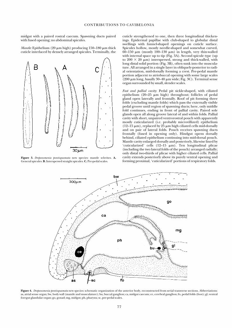
midgut with a paired rostral caecum. Spawning ducts pairedwith fused opening; no abdominal spicules.
Mantle: Epithelium (20 �m high) producing 150–180 �m thickcuticle interlaced by densely arranged spicules. Terminally, the
cuticle strengthened to one, then three longitudinal thicken-ings. Epidermal papillae with club-shaped to globular distalswellings with funnel-shaped openings on cuticle surface.Spicules hollow, mostly needle-shaped and somewhat curved,60–150 �m (mostly 100–130 �m) in length, very thin-walledwith internal space up to tip (Fig. 3A). Second spicule type (upto 200 � 20 �m) interspersed, strong and thick-walled, withlong distal solid portion (Fig. 3B), often sunk into the muscula-ture. All arranged in a single layer in obliquely-posterior to radi-al orientation, mid-dorsally forming a crest. Pre-pedal mantleportion adjacent to atriobuccal opening with some large scales(200 �m long, basally 30–40 �m wide; Fig. 3C). Terminal senseorgan surrounded by small, slender scales.
Foot and pallial cavity: Pedal pit sickle-shaped, with ciliatedepithelium (20–25 �m high) throughout; follicles of pedalgland open laterally and frontally. Roof of pit forming threefolds (excluding mantle folds) which pass the externally visiblepedal groove until region of spawning ducts; here, only middlefold continues, ending in front of pallial cavity. Paired soleglands open all along groove lateral of and within folds. Pallialcavity with short, unpaired ventrorostral pouch with apparentlymostly cuticularized (i.e. probably microvillized) epithelium(12–15 �m), replaced by 25 �m high ciliated cells mid-dorsallyand on pair of lateral folds. Pouch receives spawning ductsfrontally (fused in opening only). Hindgut opens dorsallybehind, ciliated epithelium continuing into mid-dorsal pouch.Mantle cavity enlarged dorsally and posteriorly, likewise lined by‘cuticularized’ cells (12–15 �m). Ten longitudinal plicae(including the two lateral folds of the pouch) arranged radially;only distal two-thirds of plicae with higher ciliated cells. Pallialcavity extends posteriorly above its purely ventral opening andforming proximal, ‘cuticularized’ portions of respiratory folds.
CONTRIBUTIONS TO CAVIBELONIA
77
12345678910123456789201234567893012345678940123456789501234567896012345678
Figure 3. Drepanomenia pontisquamata new species: mantle sclerites. A.General spicules. B. Interspersed straight spicules. C. Pre-pedal scales.
Figure 4. Drepanomenia pontisquamata new species: schematic organization of the anterior body, reconstructed from serial transverse sections. Abbreviations: as, atrial sense organ; bw, body wall (mantle and musculature); bu, buccal ganglion; ca, midgut caecum; ce, cerebral ganglion; fo, pedal folds (foot); gl, ventralforegut glandular organ; go, gonad; mg, midgut; ph, pharynx; sc, pre-pedal scales.

Musculature: Three-partite body wall musculature; inner longitudinal layer reinforced ventrally at both sides of pedalgroove. Dorsoventral muscle bundles prominent, lower/outerones 25–40 �m across, upper/inner bundles 40–50 �m in diameter.
Nervous system: Cerebral ganglion unpaired and relatively small(200 �m wide, 150 �m high, 100 �m long), giving rise to twopairs of lateroventral nerves to atrium, one ventral pair to oralregion. Cerebral connectives at each side with common trunk.Buccal connectives separate first, running dorsolaterally alongthe foregut, buccal ganglia (35–40 �m diam.) at bend offoregut, buccal commissure behind bend ventral to foregutglands. Common base of lateral and ventral connectives givesrise to some nerves before separation. Both pairs of connectivesshow an intermediate swelling with a latero-ventral connectivebefore typical (first) lateral and ventral ganglia; lateral ganglia(40–45 �m diam.) adjacent to body wall. Intermediate swellingof ventral connectives give rise to a more prominent nerve topedal pit; ventral ganglia (70 �m diam.) above pedal pit withstrong commissure. Longitudinal cords consist of fibrous mate-rial with fairly regularly differentiated ganglionic swellings; ven-tral commissures, latero-ventral connectives and dorsal nerves.Posterior-most ventral ganglia (50 �m diam.) with commissureand at each side with latero-ventral connective medially ofspawnings duct, this connective being connected to third poste-rior-most lateral ganglia. Ganglia posterior superiora (100 �m� 75 �m diam.) above terminal portion of spawning ducts andinterconnected by a 350-�m-long, medullary suprarectal com-missure (25 �m diam.), giving rise to a paired nerve (gap150 �m) which innervates terminal sense organ. Lateral cordscontinue and end by splitting between gill folds.
Sense organs: Atrial sense organ (atrium) spacious, posteriorlypaired, with relatively few, elongate papillae intruding into cav-ity. Half length of atrium roof with wide tract of scarcely ciliatedcells; tract divided by mouth opening into paired ciliated ridgeswhich enter medially the lateral extensions of atrium (untilbeside and behind mouth) and unite with horseshoe-shaped,scarcely ciliated border of atrium. Dorsoterminal sense organ inexactly terminal position, formed by 10–12 �m high ‘cuticular-ized’ cells (with microvillous border?) arranged in a bowl andsurrounded by small, slender scales; paired innervation fromsuprarectal commissure.
Alimentary tract: Mouth opening between ciliary tracts at the pos-terior roof of atrium; buccal cavity with folded, cuticularizedepithelium (30–40 �m high). Anterior foregut (about 270 �mdiam.) weakly cuticularized, epithelium 15–20 �m high withnumerous long and slender papillae. Subsequent foregut con-tinuing by means of a curve in dorsofrontally running upper leg,a narrow tube (about 100 �m diam.) opening above pedal cil-iary pit into midgut (Fig. 4). Entire foregut surrounded bystrong musculature (30 �m at lower foregut leg, 35 �m at uppertube) of an inner circular layer and a thicker outer longitudinallayer. Buccal cavity and anterior foregut probably protrusible(‘proboscis’). Foregut with intercellularly opening, small glandular cells (5–10 �m diam.) between periphery and muscu-lature, containing coarsely granular secretion. Lower leg offoregut posteriorly with short sac; proximal portion of upper leg(i.e. in the turn) with short anterior and short posterior pouch.Into the latter the two ventral glandular organs open ventro-rostrally side by side upon a slighly protruding double papilla.Glandular organs multiply wound (due to retracted foregut),110 �m wide with 40–50 �m high glandular epithelium (type Corgans in Salvini-Plawen, 1972, 1978). They include big gran-ules and drops of secretion; a weak muscularis surrounds the
glandular organs. Upper leg of foregut opens at low angle dor-sorostrally into midgut; midgut with paired, spacious anteriorcaecum. Caeca and midgut proper show lateral constrictionsdue to strong dorsoventral muscle bundles. Mid-dorsal ciliarytract of midgut continuous with the entirely ciliated hindgut,which opens dorsally into pallial cavity.
Circulatory system: Heart auricle (atrium) beginning as a dorso-median invagination of posterior pericardium, covered andinternally lined by muscle fibres with connective tissue. Alonghalf of pericardium the auricle is paired; it opens ventrally intoventricle. The ventricle throughout is a dorsally open invagina-tion, internally lined by musculature, and continuous with dor-sal sinus still within pericardium. Free dorsal sinus betweengonopericardioducts, mid-dorsally over gonads and betweenthe two midgut caeca. Ventral sinus below midgut delimited byportions of intercrossing dorsoventral musculature. Haemo-lymph cells of homogeneous, small round bodies (5 �m), and ofgranulated, mostly oval elements (10 �m � 7 �m).
Gonopericardial system: Mid-dorsal gonads paired throughout,medially with developing ova (50 �m diam.), lateroventrallywith sperm; gonads terminally continuous with two ciliatedgonopericardioducts (100 �m diam.) which are encircled byweak musculature and open frontally into pericardium. Gonadsbelow dorsal sinus with ciliation which lines (as a pair of tracts)the dorsolateral pericardium and proceeds into pericardiod-ucts. Pericardium spacious (enclosing some genital products)with paired extension continuous with pericardioducts.Pericardioducts wide with flat cellular lining, medial epitheliumprominently invaginated like a typhlosole; only this is ciliated.Ducts open anteriorly from lateral into the respective spawningduct. Spawning ducts separated throughout until their com-mon opening via short muscular outlet or papilla, and possess-ing a high epithelium of glandular cells as well as slender ciliat-ed cells; part of this epithelium is protruded into stout folds.Neither seminal receptacles, seminal vesicles, copulatory styletsnor abdominal spicules are present.
Remarks: The present organization clearly fits into the frame ofthe genus Drepanomenia. The configuration of the posteriorbend of the foregut suggests its protrusibility. In this connec-tion, the opening of the foregut glands into the posterior por-tion (pouch) of the upper foregut leg and the buccal gangliawith their commissure located below this, argue against the axialblind sac of the lower foregut leg representing a vestigial radularsheath (the radular sheath being presented always above thegland openings and buccal commissure).
Compared with known representatives of Drepanomenia(Table 1), note that the large scales at the mantle bridgebetween atriobuccal opening and pedal pit in D. pontisquamataare paralleled in D. perticata by strong spines (see Salvini-Plawen,1978: 227–230); note also the common trunk of the cerebralconnectives (separate in D. perticata, D. tenuitecta and probablyalso in D. vampyrella; no information for D. incrustata). Other differences include the paired midgut caecum and the lack of adistinct mid-dorsal keel in contrast to D. vampyrella, the thickmantle cuticle and the lack of abdominal spicules in contrast to D. perticata, the specially elaborated spawning ducts as well asthe configuration of the posterior pharynx in contrast to D.incrustata. The latter region in D. incrustata is characterized by a‘thin sub-oesophageal pocket’ (Odhner, 1921: 19), which maysimply point to some protrusibility, whereas the ‘thin’ (thin-walled) differentiation appears to be a true character not foundin D. pontisquamata.
L. VON SALVINI-PLAWEN
78

Simrothiellidae Salvini-Plawen, 1978
Diagnosis: Solenogastres with thick cuticle, sclerites as hollowneedles (Cavibelonia); radula biserial (or vestigial), ventralforegut glandular organs epithelial.
Remarks: See Remarks on Aploradoherpia insolita below.
Genus Simrothiella Pilsbry, 1898
Diagnosis: Cavibelonia with hollow spicules intercrossing in sev-eral layers; with common atrio-buccal opening; radula biserial,plates according to age of different shape according to develop-mental stage, with strongly heterogeneous denticles, olderplates all retained in place in paired, ventrally curved and termi-nally coiled radular sac; ventral foregut glandular organs as epithelial ampullae, midgut with regular constrictions; se-condary genital opening single, with copulatory stylets; terminalsense organ present; with respiratory organs.
Type species: Solenopus margaritaceus Koren & Danielssen, 1877(Salvini-Plawen, 1975; ICZN Opinion 1185, International Com-mission on Zoological Nomenclature, 1981)
Simrothiella margaritacea (Koren & Danielssen, 1877)
Solenopus margaritaceus Koren & Danielssen, 1877: 128. Koren &Danielssen, 1879: 328.
Simrothiella margaritacea — Odhner, 1921: 12–19, figs 1–16.
(Figures 5–7A, 19, 20)
Types: ‘Prototype’ in Odhner (1921), ‘lectotypes’ in Scheltema& Schander (2000) (ZMUB 2075 and 66806), 12 mm in length,incomplete specimens in alcohol; Boknfjord off Kvitingsog/Stavanger (Norway), 75–115 m.
Remarks: Koren & Danielssen’s original material of Solenopusmargaritaceus (ZMUB 2075 and 66806) remains organizationallyundescribed as the only diagnostic species description byOdhner (1921), as well as the present re-examination, refers tospecimens from off Kopervik (Boknfjord) at 75–95 m (ZMUB2078 and SMNH 4731). The latter animals show a ‘body round-ed at both ends’ (Odhner, 1921: 12; Salvini-Plawen, 1968: figs24–25; Scheltema & Schander, 2000: fig. 20A), whereas the typeindividuals are ‘almost transversely cut off at the hinder end’(Koren & Danielssen, 1879: 328). It is therefore questionablewhether Odhner’s material and description of Simrothiella margaritacea (based on SMNH 4731, see below) is actually con-specific with Solenopus margaritaceus Koren & Danielssen. In anycase, Odhner’s section series on slides (SMNH 4731) should be considered as crucial para(lecto)types of S. margaritacea, or(together with ZMUB 2078) as syntypes of a separate species,rather than declared as representatives outside the type material(Scheltema & Schander, 2000).
Diagnosis: Body up to 12 mm long (type material) or 10 mm,respectively (Kopervik material), mantle with epidermal papil-lae, hollow spicules thick-walled; no distinct dorsal foregutgland; radular plates up to 120 � 60 �m, in adults with verystrong lateral denticle up to 180 �m in length and with othersmall heterogeneous denticles at distal border of posterior por-tion, these denticles eventually being reduced; radular appara-tus without bolster; midgut with short, single rostral caecum,regular constrictions weak; seminal receptacles as pouches ofwidened spawning ducts only; one long and one much shortercopulatory stylet per sheath; with a pair of strong, short pallialspicules above rostral border of mantle cavity opening; largespecimens with suprapallial glands.
Material examined: Four specimens from Boknfjord off Kopervikat 75–95 m, sectioned by N. Odhner: SMNH 4731:2 (transversesections at 10 �m); 4731:1 (ts 5 �m anterior end), 4731:3 (ts5 �m posterior body); 4731:4a (longitudinal sections at 5 �m);4731:4b (ls 10 �m). Odhner’s (1921) figures 9, 10, 16 refer tothe series 4731:2 and figures 11–14 to 4731:3; the respective sec-tions are marked each on the slides by an encirclement. Threeadditional specimens from off Kopervik (ZMUB 2078, seeOdhner, 1921: 12; Salvini-Plawen, 1968: figs 24–25): two animalsserially sectioned (ts 10 �m; one not yet mature) and stainedwith azan, third individual (9 mm long) used for in situ prepara-tion of radular apparatus.
Description: Odhner (1921: 11–19) described the organization indetail except for the not quite understood radular apparatus(see Salvini-Plawen, 1978: 215). In some points this descriptioncan be supplemented:
Cuticle about 25 �m thick; hollow spicules generally120–160 �m (up to 200 �m) long, thick-walled and intercross-ing in two or three layers. Epidermal papillae present, but scat-tered. Single pedal fold to opening of mantle cavity; Odhner’spre-pallial pit (1921: 14) is part of the mantle cavity that is irreg-ularly outlined by contraction (and only present in SMNH4731:2a). Longitudinal musculature of body wall ventrally rein-forced. Atrial sense organ with up to 10 bundled papillae.Foregut surrounded by circular and distinct longitudinal mus-culature; along the second quarter of its course (from mouth toopening into midgut) with subepithelial pharyngeal glandsgrouped dorsally and dorsolaterally (no compact gland). Closeto transition into midgut, an additional, differently stained clus-ter of subepithelial pharyngeal glands opens dorsolaterally ateach side into pharynx; Odhner (1921: 17, fig. 1) probably mis-took these as a distinct dorsal foregut gland (not present ineither Odhner’s or in the new specimens at hand). Except forsome musculature, without special radular support (bolster of
CONTRIBUTIONS TO CAVIBELONIA
79
12345678910123456789201234567893012345678940123456789501234567896012345678
Figure 5. Simrothiella margaritacea: developmental sequence (above to below;cf. Fig. 6A) of stages of left radular plates according to serial sections, views ofposterior face.

vesicle or chondroid cells). Ventral foregut glandular organsepithelial (type C in Salvini-Plawen, 1972, 1978), forming a pairof ampullae; they are embedded dorsolaterally of radularsheath (see Odhner, 1921: fig. 10), opening at both sides offront end of radula. Rostral midgut caecum single and short, butdistinct; midgut more or less distant to body wall, dorsoventralmuscles bundles in most individuals not constricting midgut(‘only slightly developed’ diverticula in Odhner, 1921: 17).Midgut without nematocysts, but SMNH 4731:4a and anotherspecimen each containing one or to worm-like fragments ofprey (tentacles of polychaetes or holothurians?) occupyingmost of length of midgut.
Odhner’s specimens (SMNH 4731:2, 4731:3) appear to beprotandrous, because the sperm is well elaborated in volumi-nous lateral sacculations of the gonads, whereas the medianeggs are still small (up to 75 �m diam. in 4731:2); in other speci-mens examined, eggs are even smaller or the gonad is not yetdifferentiated. Heart extends freely through pericardium, witha single auricle. Pericardioducts emerge laterally at the rear ofpericardium and open from dorsal into most anterior portion ofthe spawning ducts. In Odhner’s material (SMNH 4731:2a,4731:3a) spawning ducts at this position with wide lumen(epithelium about 15 �m high) and posteriorly form 2–3pouches (‘three small vesiculae’ in Odhner, 1921: 18, fig. 2),which represent the (not separated) seminal receptacles.Following spawning ducts typically lined (25–30 �m high withslender glandular cells) and continue as somewhat narrowed,lateral portions fusing in their posterior half to a single, bulbous
organ. In histological sections, the pair of long copulatory stylets(over 600 �m long) appear to be solid anteriorly (25 � 30 �mdiam.); adjacent second stylet fairly short and entirely within thesheath. Anterior border of mantle cavity below distal-most styletsheaths. What Odhner describes as ‘a pair of vesiculae or sac-formed glands’ (1921: 19, fig. 2v) is not clear. On the one hand,in larger specimens (SMNH 4731:2a, 4731:4a, 4731:4b) thereare pouchings of the mantle cavity, and all the anterior as well asdorsal wall (as far as to fig. 16 in Odhner, 1921) is underlain byan extensive mass of suprapallial glands; on the other hand, atboth sides above the border of the beginning mantle cavityopening, a strong, short pallial spicule is present within its later-ally invaginated sheath, not noticed by Odhner (‘accessory cop-ulatory spicules’ in Scheltema & Schander 2000: 146). Odhner(1921) counted 20 gill organs, in the new specimens up to about10 respiratory folds are present; due to distinct lateral folds inlarger respiratory organs, these superficially resemble ctenidia.Suprarectal commissure above anal opening long (new material80–110 �m, Odhner’s material 250 �m) and medullary (new �12 �m, Odhner � 22 �m diam.). Terminal sense organ locateddorsally behind mantle cavity.
Radular apparatus: Biserial radula as usual formed in sheath andsubsequently freely exposed within pharynx. Already in juve-niles it continues into two separate, ventrally curved sacs whichextend posteriorly and recurve or coil medially at their end (Fig.6A). During development, the shape of the radular plates orbars changes; plates in adult specimens thus of different outlines,
L. VON SALVINI-PLAWEN
80
Figure 6. Simrothiella margaritacea, radula. A. Sketch of isolated radula, ventral view. B. Early radular plate, from ventral coil of A (between first and second stagein Fig. 5), posterior portion with median border at right. C. Pharyngeal radular plate, anterior face (see Fig. 20). D. Same pharyngeal plate as A, posterior face.E. Plate in anteriormost ventral sac, view from above (anterior portion is above). F. Plate in anterior ventral sac, posterior portion. Abbreviations: re, cap-likereinforcement; wa, weak portion of plate.

with the earliest form at the tip of the coils (Fig. 5) and Odhner’sdrawing (1921: fig. 5) refers to a plate (about 60 �m wide) in the posterior portion of the ventral sac. The in situ preparedspecimen (9 mm long) with a 700 �m long radular apparatus(Fig. 6A) showed a ‘coil’ of 540° (360° � 180°) and was there-fore older (at left about 50 plates, at right about 55 plates intotal). Smallest (i.e. earliest juvenile) plates are about 12 �mwide and bear 5–6 equally sized, tiny distal denticles (Fig. 5);subsequent plates have increased size and denticle number. Inaddition, the denticles become heterogeneous and a strong lateral denticle is added, dominating the outline (Figs 5, 6B, E, F, 7A). Thus in SMNH 4731:4b, the plates exposed to thepharynx (90 �m � 35 �m, denticle 110 �m long) are promi-nently provided with heterogeneous denticles (Figs 7A, 20).Other material (SMNH 4731:4a) shows plates of 100 �m �40 �m with an up to 180 �m long denticle. In the new material,the three anterior-most ventral plates measured 120 �m �40–60 �m and show a 120–160-�m-long denticle; the small heterogeneous denticles, however, are vestigial. The largest pairof plates, exposed to the pharynx (new series sections as well asin situ preparation), were 120 �m wide and 60 �m high; eachvery strong lateral denticle was 180 �m long (basally 28 �mdiam.), whereas the small heterogeneous denticles were notelaborated in these most advanced plates (Figs 6C, D, 20).
Within the sheath (slightly bifid proximally in one specimen),the radular material mostly forms two continuous bands (140–170 �m wide), being subdivided only distally into prospectiveplates (Fig. 6A), 160 �m (new specimens) and 150 �m wide(Odhner) in sections. Within the distal sheath, the free, dentic-ulated rim of each plate is medially directed and points dorso-posteriorly; all denticles emerge from the posterior (!) portionof the about 45-�m-thick plates, whereas the anterior portionlacks special structures except a reinforcement (Figs 6C, 20):
the strong denticle emerges posterio-laterally at the plate with abasal thickening (Fig. 6D–F), which therefore embraces the lat-eral border of the plate and forms a lateral, cap-like reinforce-ment at the anterior face of the radular plate (Figs 6C–E, 20); onthe other hand, the plates are less thick at the medio-basal areaof their anterior portion as well as at the medio-distal area oftheir posterior portion (Figs 6C, D, 20). Within the pharynx, theplates become erect, with the strong denticle pointing into thelumen (Figs 7A, 19). Passing into the ventral sacs, the plates curvedown with the heterogeneously denticulated rim pointing ven-tro-anteriorly and the originally posterior portion of the platesnow facing obliquely upwards. This condition elucidates the dif-ferent aspects of the radular plates: only the posterior portionbears both the strong and the small, heterogeneous denticles dis-tally (well visible in histological sections, see Fig. 5); the anteriorportion and face have lateral thickening only (Figs 6C, 20).
Remarks: The specimens at hand belong to the original materialcollected by G. O. Sars and described by Odhner (1921).Odhner’s (1921) unsatisfactory representation of the radularapparatus in S. margaritacea has already been unravelled in anearlier investigation; its basic configuration is identical to that inKruppomenia, showing a ‘saccoglossan’ condition (as also pre-sent also in several Amphimeniidae), although the ventral radular sac is bifurcate. Therefore, Kruppomenia was put into synonymy (Salvini-Plawen, 1972: 222–223, 1975), althoughrestored by Scheltema & Schander (2000).
Scheltema & Schander (2000) also referred specimens fromthe West European Basin (southwest of Ireland) to the soleknown Simrothiella species S. margaritacea. This new material(also referred to in Ivanov, 1990), however, actually represents adistinct species, characterized below.
Simrothiella abysseuropaea new species(Figure 7B)
Simrothiella margaritacea—Scheltema & Schander, 2000:143–146, figs 20D–J, 21, 22A–G (in part, not Solenopus margari-taceus Koren & Danielssen, 1877).
Types: Syntypes (USNM 894261–894266) up to 9.5 mm inlength; northeastern part of the West European Basin southwestof Ireland (50°58.7� N, 13°01.6� W), Station 316 of RV ChainCruise 106, 2173 m.
Ethymology: Greek abyssos, abyss; europa, mythical Greek princessrepresenting Europe; referring to the provenance of thespecies.
Diagnosis: Body up to 9.5 � 0.8 mm. Radular plates with stronglateral denticle (up to 280 �m in length), small denticles scattered at the posterior face (rather than at distal border);reinforcement (buttress) at plates restricted to the anterior faceonly. Hollow spicules heavy-walled; two copulatory stylets persheath, one long and a much shorter, S-shaped one; with a pairof strong and short, hooked pallial spicules.
Remarks: 26 specimens from the RV Chain Cruise referred toSimrothiella margaritacea by Scheltema & Schander (2000:143–146, figs 20D–J, 21, 22A–G) are defined by the hard partsonly. Among these, the diagnostic characters of the radularapparatus including its size (about 1200 �m long in figs 11B and21A; 450 �m in fig. 21B, wrong scale?), with larger plates, andthe arrangement of denticles and reinforcements, clearly con-tradict conspecifity with S. margaritacea (Fig. 7). Compared withinformation gained from S. digitoradulata (below), these differ-ences exceed mere variations and appear to represent specific
CONTRIBUTIONS TO CAVIBELONIA
81
12345678910123456789201234567893012345678940123456789501234567896012345678
Figure 7. Simrothiella spp., posterior face of radular plates. A. S. margaritacea,one pharyngeal radular plate from Odhner’s section series 4731:4b(redrawn from Fig. 19). B. S. abysseuropaea new species, two radular platesfrom the West European Basin after Scheltema & Schander (2000: fig. 21F).A, B to same scale.

characters. In addition, there is a different distribution. This distribution calls attention to unexpected biodiversity evenwithin a close geographic range. The ‘Armauer Hansen’ material(62°01� N, 0°08� E; at 1400 m), likewise referred to S. margari-tacea (Odhner, 1921; Scheltema & Schander, 2000), has notbeen examined and may possibly belong to another species.The features of S. abysseuropaea distinguishing it from S. margari-tacea are summarized in Table 2.
Simrothiella digitoradulata new species(Figures 8, 9, 17)
Types: Holotype (USNM 1016984), 3 mm length, serial cross-sections on slides; Atacama Trench off northern Peru (08°13� S,81°09� W), Anton Bruun Station 11–101 of South-East PacificBiological Oceanographic Program (SEPBOP), 16 Oct. 1965,1927–1997 m.
Ethymology: Latin digitus, finger; radere, to rasp, hence radula;radular plates reminiscent of a pointing forefinger.
Diagnosis: Body about 3 � 0.5 mm; no distinct dorsal foregutgland, radular apparatus with paired supportive bolster (vesi-cle), radular plates with long para-lateral, slender denticle and5 � 3 small denticles at the face; midgut with short paired rostralcaecum. Copulatory stylets of a long and a short element persheath, posteriorly running within a pouch of mantle cavity.About 20 respiratory folds.
Description: Mantle cuticle depressed with tangential holes ofspicules in several layers; no spicules retained, no epidermalpapillae discernible. One simple pedal fold until the mantle cavity; cavity extends rostrally of its opening and provided withabout 20 radially arranged respiratory folds.
Mouth opening in direct continuation of dorso-posterior atrial cavity, separated merely by a dorsomedial to paired lateral
ciliated tract (Fig. 8); atrial papillae few and blunt, some beingbasally bundled into three.
Foregut with distinct circular and longitudinal musculature;dorsolaterally, pharyngeal glands are concentrated (doubtfullyforming a ‘dorsal gland’). Radular apparatus at least 270 �mlong; supportive apparatus with a paired, distinct bolster, eachof a voluminous lateral vesicle (160 �m long, 70 �m diam.) offew turgid cells and surrounded by musculature, bolsters at theirbeginning with a strong interconnecting transverse bundle.Radula extending into paired lateroventral sac, distally curvingbelow and medially of beginning vesicle (‘coil’ of 270°). Ventralforegut glandular organs as a pair of epithelial ampullae (type Cin Salvini-Plawen, 1972, 1978) dorso-frontally of each bolster vesi-cle and opening laterally at the beginning of radula. Free radularplates in pharynx (Fig. 9) about 50 �m wide and 15 �m high, witha 65–80 �m long, distally curved main denticle and 5 � 3 smalldenticles emerging from the face of the plate (rather than fromthe distal rim). Largest radular plates (in distal portion of sheath)85 � 25 �m, diameter of long denticle 8 �m (fragment of 35 �mlength). Within ventral radular sac, just behind curve, radularplates 40 � 9 �m with about six denticles, the second outer onebeing largest (Fig. 9). Radular sheath with a dorsomedian foldthroughout, not causing bipartition; long denticles clearlyformed by dorsal cells of proximal-most portion.
Midgut with short, paired rostral caecum. Dorsoventral musculature not very prominent, bundles running close to bodywall, scarcely constricting midgut. Ventral longitudinal muscu-lature only slightly reinforced.
Unpaired cerebral ganglion 125 �m wide, 35 �m high, 40 �mlong. Ganglia posteriora superiora interconnected by a shortand medullary suprarectal commissure (35 �m diam.). Supra-pallial sense organ in terminal position.
Gonads form scarcely enlarged tubes with developing eggsand continue posteriorly in two ciliated gonopericardioducts.Heart extends freely through pericardium. Spawning ductunpaired throughout, opening ventrally into mantle cavity;
L. VON SALVINI-PLAWEN
82
Figure 8. Simrothiella digitoradulata new species: schematic organization of the anterior body, reconstructed from serial transverse sections. Abbreviations: as, atrial sense organ; bw, body wall (mantle and musculature); bo, radular bolster; ca, midgut caecum; ce, cerebral ganglion; fo, pedal fold (foot); gl, foregutglandular organ; mg, midgut; ph, pharynx; (ra), space with pharyngeal radula; rs, radular sheath.

seminal receptacles not (not yet?) visible. One pair of copulatorystylets, each proximally accompanied within the sheath by ashort second one. Stylets extend far anteriorly; posteriorly theyrun, adhered medially by a ‘mesenterium’, each within a pouchof the bottom of the mantle cavity. Pallial spicules or hooks notdiscernible (not yet developed?).
Remarks: The organization of this not yet fully mature animalidentifies it as a member of the genus Simrothiella. Up to now,three species of Simrothiella are known: S. margaritacea, S. comoren-
sis Todt & Salvini-Plawen, 2003 and the new S. abysseuropaea. Theshape and structure of the radular plates of the present specimencome close to those of S. abysseuropaea (see discussion above). Itdiffers from this species, however, at least by the more detailedshape of the radular plates and by the geographical distribution.The differences from S. margaritacea refer to the radula, the pres-ence of a paired radular bolster, the paired midgut caecum andthe copulatory stylet apparatus at each side running within apouch of the mantle cavity. The smaller S. comorensis likewiseposses such pouches of the mantle cavity as well as a paired radu-lar bolster (pers. obs.; not mentioned in Todt & Salvini-Plawen,2003), but differs at least in the structure of the radula, the ‘coil’of the ribbon (about 450°; pers. obs.) and the number of copula-tory stylets (Table 2). The present specimen thus clearly definesa new species and enlarges the known geographical distributionof Simrothiella to the Atacama Trench.
Genus Aploradoherpia new genus
Diagnosis: Cavibelonia with hollow spicules arranged oblique-radially in one layer. With common atrio-buccal opening, biseri-al radula, foregut glandular organs as short epithelial ampullae,no regular midgut constrictions. Secondary genital opening sin-gle, no copulatory stylets; with respiratory organs; terminalsense organ present.
Type species: Aploradoherpia insolita new species
Etymology: Greek aploos, simple; herpein, to move slowly and her-peton, a slowly moving animal; Latin radere to rasp, hence radula);referring to the simple radular plates: Feminine gender.
Aploradoherpia insolita new species(Figures 10–12, 18, 21, 22)
Types: Holotype (USNM 1016985 ), 3 mm length, serial cross-sections on slides; Atacama Trench off northern Chile(25°43�–25°44� S, 70°58� W), Menzies-trawl at Station 3-62 of USAntarctic Research Program, 21 June 1962, 1863–1965 m.
Etymology: Latin insolitus, unusual, strange, odd.
CONTRIBUTIONS TO CAVIBELONIA
83
12345678910123456789201234567893012345678940123456789501234567896012345678
Figure 9. Simrothiella digitoradulata new species, radular plates (median border at left): above, radular plate within ventral radular sac; below, radularplate within pharynx (see Figs 21, 22).
Table 2. Specific characters of known Simrothiella species (and treated herein).
Character S. margaritacea S. abysseuropaea S. digitoradulata S. comorensis
Body length (mm) 9–12 9.5 >3 1.9
Epidermal papillae Present ? ? Not present
Dimension of radular 700 1200 >270 200
apparatus (�m)
Ventral ‘coil’ of radular ribbon (°) 540 270 270 450
Number of radular plates 50–55 40–45 ? 20
Dimensions of largest plates (�m) 119 � 60 130 � 90 >85 � 25 90 � 25
Length of strong denticle (�m) 180 280 80 40
Arrangement of small denticles At distal border At posterior face At posterior face At distal border
Reinforcement at plate Cap-like with ‘nose’ Limited to anterior face ? At posterior face
Radular bolster None ? Present, paired Present, paired
Midgut caecum Single, short ? Paired No
Seminal receptacle Pouch of Spawning duct ? ? ?
Spawing ducts Posterior half fused ? Fused throughout Posterior half fused
Copulatory stylet apparatus Embedded ? Within pouch of mantle cavity Within pouch of mantle cavity
No. of stylets Two pairs Two pairs Two pairs One pair
Pallial spicules One pair: short, strong ? ? ?
Suprarectal commissure Long ? Short ?
Provenance, depth (m) Coastal, 75–115 Deep sea, 2173 Deep sea, 1827–1897 Deep sea, 3716
Geographical distribution Norwegian shelf region W European abyssal Trench off N Peru Near Comoro Islands
?, No information.

Diagnosis: Body 3 mm (immature), somewhat tapered posteriorly.Cuticle moderately thick, without epidermal papillae, hollowspicules thin-walled, up to 350 �m in length; pedal groove notcontinuous with mantle cavity. Radular plates low and curved,with four median denticles; squamous pharyngeal cuticle enter-ing radular sheath; radula support consisting of several musclebundles; midgut with unpaired rostral caecum. Ganglia posteri-ora superiora close together; mantle cavity with ventrorostralpouch; four pairs of pre-pallial spines, the two inner ones withmusculature; respiratory organs present as papillae.
Mantle: Epidermis (7–10 �m) with voluminous vacuolizedglands, yet no papillae. Mantle spicules needle-shaped and hol-low with thin wall, up to 350 �m long, all arranged in one layeroblique-tangentially in posterior direction, causing a net-likeperforation of the cuticle (30–50 �m). At each side in front ofmantle cavity opening four strong, solid calcareous spines, fairlyradially invaginated (Fig. 12) and grouped as two outer ones(about 100 �m � 10–13 �m diam.) and two inner ones (about160–200 �m � 25 �m diam.); inner spines terminally curved,and their simply invaginated epithelium is basally and laterallyprovided with musculature (absent in the two outer ones).
Foot and mantle cavity: Ciliated pedal pit shallow, anteriorly withoutlets of pedal gland. Posterior portion forming a single, weakmid-dorsal fold continuous into the externally well visible pedalgroove; groove ending far in front of mantle cavity opening.Paired sole glands emptying laterally into groove as well as intopedal fold (see Drepanomenia pontisquamata).
Mantle cavity generally non-ciliated with a spacious, ventro-rostral pouch with dorsal opening of single outlet of spawningducts. Rectum opens dorsally into mantle cavity; adjacent dorsaland lateral walls forming a total of 10–12 papilla-like respiratoryorgans (up to 225 �m in length). These are locally ciliated andform lateral bulges or weak folds, thus somewhat resemblingctenidia (see also Simrothiella margaritacea and Kruppomenia mini-ma in Nierstrasz, 1905: 655–666; Nierstrasz & Stork, 1940:
26–32); cross-sections show two internal lacunae and a pair ofweak lateral muscle bundles; ciliation restricted to 1–2 cells lat-erally (Fig. 10A).
Musculature: Ventral muscles of three-layered body wall muscu-lature reinforced on both sides above pedal groove; in posteriorbody, these separate and form musculature of the two pairs ofinner abdominal spines. Dorsoventral bundles in usual arrange-ment. Additional musculature: strong foregut coat.
Sensory system: Unpaired cerebral ganglion (150 �m wide, 75 �mhigh, 70 �m long) gives rise dorso-laterally to three pairs of sepa-rate connectives. First lateral ganglia (50 �m � 25 �m diam.)close to cerebral ganglion, oriented towards body wall. First ven-tral ganglia (50 �m � 25 �m diam.) with one commissure abovepedal pit. Buccal system with commissure dorsorostrally ofbucco-pharyngeal sphincter; buccal connectives continue pos-teriorly, running dorsally adjacent to bolster apparatus andforming ganglion (30 �m � 20 �m diam.) at both sides offoregut opening into midgut; commissure of ganglia betweenradular sheath and midgut.
Longitudinal body cords (15–20 �m diam.) with fairly regu-larly spaced swellings (ventrally 40 � 20 �m) interconnected by
L. VON SALVINI-PLAWEN
84
Figure 11. Aploradoherpia insolita new genus, new species: diagrammatic cross-sections through radular apparatus (see Figs 21, 22). A, B. Sections within phar-ynx. C, D. Sections through radular sheath. Abbreviations: cu, scaly cuticle; gl, foregut gland; mg, midgut epithelium; mu, musculature; ph, pharynx; ra, radu-lar plate; rs, radular sheath.
Figure 10. Aploradoherpia insolita new genus, new species. A. Cross-sectionthrough respiratory papilla. B. One radular plate (median border at left).Abbreviations: ci, cilia; la, lacuna; mu, muscle.
Figure 12. Aploradoherpia insolita new genus, new species: cross-sectionthrough posterior body in the region of (inner) pre-pallial spines. Abbre-viations: hg, hindgut; is, inner pre-pallial spine; mu, musculature; nl, lateralnerve cord; pc, (extension of) pericardium; pd, pericardioduct (at left soliddistal end); sd, spawning duct.

ventral commissures and less often by latero-ventral connec-tives. Ganglia posteriora inferiora (50 �m � 25 �m diam.)below spawning ducts and medially of pre-pallial spines, veryclose together, but not fused. Ganglia posteriora superiora(70 �m � 25 �m diam.) next to anus with a strong, medullarycommissure (15 �m diam.).
Atrial sense organ with papillae mostly united into twos. Roofforming two narrow tracts of ciliarly cells, with similar tracts pre-sent ventrolaterally. These tracts are elevated neither in ridgesnor folds, and in contrast to the usual configuration they do notcurve to fuse posteriorly. Three strong muscle bundles (retrac-tors) insert on atrial roof.
Terminal sense organ very big (in relation to body size) andterminally located, protruded and with intruding muscle fibres;distally delimited by a plate of cells 5 �m high. The innervationcould not be traced.
Alimentary tract: Mouth opening dorso-posteriorly in the atriumand opening into spacious buccal cavity of high cells with glandcells between, surrounded by dorso-posteriorly strengthenedmusculature forming a sphincter against pharynx with radularapparatus. The anteriorly high pharyngeal epithelium appearsto be ciliated with gland cells, but becomes flat without glandcells and ciliation becomes scarce near radula.
Radular apparatus intrudes into this spacious pharyngealregion (250 � 190 �m, in cross-section initially nearly rectangu-lar) (Fig. 11A); laterally, bulges (150 �m high) support radulaon both sides; anteriorly, each bulge includes a foregut glandrepresenting compact ampullae with club-shaped, 70–130-�m-high epithelial cells (type C in Salvini-Plawen, 1972, 1978).More posteriorly, the pharynx closes the ventral transversespace (thus becoming V-shaped in cross-section) and the lateralbulges are underlain by a bolster of three muscle bundles (Fig.11B). Foregut opens dorsally into midgut.
Biserial radula covers dorsomedian surface of lateral bulgesand also curves a short distance rostro-ventrally. Radular sheathbegins between bulges behind foregut opening into midgut(Fig. 11C, D). Roof of sheath intrudes a fold, which represents a septum separating the two radular rows. Entire apparatus250 �m in height, rear of sheath diminishes to 80 �m � 100 �macross (Fig. 11D). Within lateral bulges the three-parted muscu-lature of the bolster posteriorly splits further to become severalcompact bundles (Fig. 11). No large turgid cells or vesicles areassociated with the radular support. Anterior, older plates of biserial radula 50 � 8 �m in size, curved, forming four mediandenticles at 10 �m intervals (Fig. 10B); their simple form resem-bles that of Cyclomenia holoserica (Nierstrasz, 1902, 1905). Platescover dorso-median surface of bulges and are medio-ventrallycontinuous with a squamous cuticle of 6–8 �m height built ofrods (4–5 �m wide), as are underlying cells (Fig. 11B). This cuti-cle is very similar to the radula itself, which thus appears to coverthe entire lateral bulge (Figs 11B, 21, 22). Subsequent conditionhighly unusual: the median squamous cuticle enters posteriorsheath together with radular plates (Figs 11C, 21, 22) and is like-wise covered by epithelium of the dorsal septum. Only in thesheath’s final 30 �m is the sheath diminished (see above),restricted to radula-forming area (but not the area of the squa-mous cuticle; Fig. 11D); odontoblasts are about 20 �m high.
Midgut with spacious rostral caecum extending to cerebralganglion and with a narrow, mid-dorsal ciliary tract; latero-ventral pouchings only weak and irregularly shaped. Hindgutwith tall epithelium bearing long cilia; it opens dorsally abovebeginning of mantle cavity opening.
Present animal with swallowed prey occupying rostral caecum,two-thirds of midgut and posterior pharyngeal space betweenradular plates. This elongate prey (about 1.2 � 0.3 mm) coveredby a cuticle(?), shows a strongly ciliated gut embedded in densecell material and muscle fibres (mesenchyme); its body wall
musculature cannot be ascertained, but there are scattered eggs(diam. 15–20 �m) and numerous balls of bundled cilia (sperm?)in the tissues.
Gonopericardial system: Spacious pericardium with mid-dorsallyinvaginated heart shows a paired rostral extension lateral of thehindgut (primordia of gonads). Terminally, the two pericar-dioducts emerge laterally, curve anteriorly and end blindly assolid cords (Fig. 12). Without any connection to the latter, thespawning ducts extend below them; they are voluminous butwith a simple epithelium. Spawning ducts fuse in their posteriorportion and open by a single outlet from dorsal into ventro-ros-tral pouch of mantle cavity.
Remarks: From the juvenile state of the animal it is unclearwhether seminal receptacles are not or not yet present.Although the inner pre-pallial spines show musculature, theirfairly radial orientation (Fig. 12) and size are not comparable togrowing copulatory stylets (see Syngenoherpia intermedia inSalvini-Plawen, 1978). The immature state does underline theheterochronic development of the pericardium and (primor-dia of) gonadial spaces. In addition, it again demonstrates theindependent derivation of the pericardioducts (from the meso-dermal pericardium) and the spawnings ducts (from the ecto-dermal mantle cavity; see also Baba, 1938).
The organization of the present animal clearly places it inSimrothiellidae, but the structure of the radular plates and res-piratory papillae are unique within this taxon.
At the supraspecific level, new findings have resulted inrecent reclassification of the Simrothiellidae. Based on an earli-er re-examination of Simrothiella margaritacea and the particularorganization of the radular apparatus in Simrothiella andKruppomenia, Salvini-Plawen (1972, 1978) synonymized bothgenera. Recently, Scheltema & Schander (2000) restoredKruppomenia, basing separation from Simrothiella on details ofthe radula (simple multi-serrate plates vs. heterogeneously den-ticulated plates with lateral reinforced thickening or ‘buttress’).Further, they removed S. schizoradulata S.-Plawen to a new genusPlawenia, because of the arrangement of tangential spicules inone layer only (versus diagonal-intercrossingly multilayered or‘skeletal’ spicules in Simrothiella and Kruppomenia). Recently,Arnofsky (2000) erected a new genus Spiomenia for a species simi-lar to Plawenia having hollow spicules with an asymmetrical axe-shaped (‘captate’) tip; the special character of the radular platesin Spiomenia (additional denticles lateral of the ‘butress’), how-ever, appears to be relevant only at the specific rather thangeneric level. The genera Uncimenia Nierstrasz and SialoherpiaS.-Plawen, previously classified with some doubt amongSimrothiellidae (Salvini-Plawen, 1978), are now placed in thePararrhopaliidae within the subfamily Unciherpiinae (Garcia-Alvarez, Salvini-Plawen & Urgorri, 2001).
Among recognized genera of the family (Table 3), onlyPlawenia and Spiomenia correspond with the present specimenin exhibiting a thick cuticle and hollow acicular spiculesarranged obliquely (tangentially) in one layer. However, boththese genera are characterized by the presence of complex cop-ulatory stylets. Even if we regard that character as unknown inthe new species, the simply denticulated radular plates are instrong contrast to Plawenia and Spiomenia. The different radularconfiguration between Simrothiella and Kruppomenia (as stressedby Scheltema and Schander, 2000) parallel the differencesbetween Plawenia/Spiomenia and the new species, whose radulais intermediate between that of Cyclomenia and Kruppomenia.Thus, if Kruppomenia is accepted as being generically differentfrom Simrothiella, the new species analogously represents agenus proper (regardless of the condition of copulatory stylets).According to current systematics (Salvini-Plawen, 1978;Scheltema & Schander, 2000; Arnofsky, 2000; Garcia-Alvarez et
CONTRIBUTIONS TO CAVIBELONIA
85
12345678910123456789201234567893012345678940123456789501234567896012345678

al., 2001), nine genera are thus recognized within the Simro-thiellidae (Table 3), making it the most diverse family within theCavibelonia.
Rhipidoherpiidae Salvini-Plawen, 1978
Diagnosis: Cavibelonia with thick cuticle, sclerites generally ashollow needles; radula polystichous, ventral foregut glandularorgans with subepithelially arranged glands; seminal recepta-cles in bundles.
Thieleherpia new genus
Diagnosis: Cavibelonia with hollow spicules intercrossing in several layers; with common atrio-buccal opening; radula poly-stichous; ventral foregut glandular organs tubular with sub-epithelial layer of gland cells in compact arrangement; midgutwith regular lateroventral constrictions; with paired bundle ofseminal receptacles, secondary genital opening single; no copu-latory stylets; dorsoterminal sense organ(s) present; no respira-tory organs.
Type species: Proneomenia thulensis Thiele, 1900.
Ethymology: Greek herpein, to move slowly and herpeton, a slowlymoving animal; dedicated to Johannes Thiele (1860–1935),prominent German malacologist. Female gender.
Thieleherpia thulensis (Thiele, 1900)(Figures 13, 14)
Proneomenia thulensis Thiele, 1900: 111.
Types: Holotype (ZMB Moll. 105.405 b), 25 mm length, sectionseries on slides; Hinlopen Strait, Spitzbergen (80°08� N, 16°55�E), 480 m.
Diagnosis: 13–25 mm long; cuticle 180–220 �m thick, thick-walled spicules tangential and intercrossing in several layers;epidermal papillae present; foregut (protrusible?) with distinctpostradular, anteriodorsal portion; radula with 32–36 equalteeth per transverse row (no particular central teeth); ventralforegut glandular organs with long duct, distal half with subep-ithelial glands in compact arrangement; hindgut with a dorso-lateral bundle of rectal glands; paired bundle of 5–9 seminalreceptacles; 2 dorsoterminal sense organs; abdominal spiculesnot distinct.
Material examined: Holotype and one specimen 13 � 1.5–2 mm,incomplete section series on slides; off northeastern Iceland(67°19� N, 15°52� W), Station 126 of Ingolf Expedition, 29 July1896, 552 m (ZMUC).
Mantle: New animal with all epidermal papillae strictly in radialarrangement, with distal swellings nearly without exception atthe periphery only. Cuticle 180 �m high (holotype 220 �m),spicules measuring 70–210 �m (100–150 �m), thick-walled andtangentially arranged in several intercrossing layers; with inter-spersed similar radial spicules. Abdominal spicules solid, distally(not very distinctly) hooked, up to 115 �m � 8 �m diam. (80 �mlong in Thiele, 1911: 4).
Foot and pallial cavity: Spacious pedal pit with up to five folds,only the median main fold (100 �m high) passes pedal groove(bordered on both sides by mantle fold) until mantle cavity.Voluminous follicles of pedal gland cells open anteriorly intopit, differently staining glands open laterally; smaller sole glandssingle, not very numerous, corresponding in structure to anteri-or pedal gland cells. Epithelium of mantle cavity irregularlyfolded and ciliated, but without formation of respiratory foldsor papillae. No posterior extension of cavity beyond its opening.
Musculature: Body wall musculature strong, inner longitudinallayer reinforced ventrally (M. longitudinalis ventralis). Upper/inner bundles of dorsoventral musculature providing fibres alsointo the mantle folds. Entire foregut with distinct longitudinaland feeble circular musculature; four retractors extend fromthe subradular sac ventro-posteriorly, and two additional retrac-tors insert at the dorsal radular sheath and run to the body wall.
Sensory system: Nervous system (Fig. 13) fused cerebral gangliagiving rise to frontal nerves to atrial sense organ. Lateral andventral connectives medullary, emerging side by side; buccalconnective beginning termino-laterally. Together with the lat-eral connective, a strong nerve leaves to the lateroventral bodywall. First lateral ganglion not prominent (60 �m diam.), subse-quent cord (25 �m diam.) predominantly fibrous with inter-spersed weak swellings giving rise to a dorsal nerve; anterior por-tion of cords with several nerves running to lateroventral bodywall. Each cerebro-ventral connective gives rise to a strong nerve(at left 15 �m diam., at right divided with 10 �m and 7 �mdiam.) running to the pedal pit. First ventral ganglia abovepedal pit prominent (100 �m diam.) with one strong commis-sure and with a ventral nerve to posterior pedal pit. Buccal gan-glia (70 �m diam.; in holotype 105 � 80 �m) with one commis-sure only. Ventral cords with weak swelling at intervals between
L. VON SALVINI-PLAWEN
86
Table 3. Generic characters in Simrothiellidae (Spiomenia differs from Plawenia by the presence of distally axe-like spicules only, which may or may not beaccepted as a generic character).
Tangential Epid. Paired Cop. Go
spicules pap. Mouth Radular plates radula sac Foregut glands styl. sem. Rec. Dts Gills
Simrothiella Inter-crossing ± Fused Heterodented + Ampullae + 1 ? + +
Kruppomenia Inter-crossing ± Fused Serrate + Ampullae + 1 + + +
Cyclomenia Inter-crossing – Separate Simple, wide – Ampullae + 1 – + +
Aploradoherpia One layer – Fused Simple – Ampullae – 1 ? + +
Plawenia One layer – Fused Heterodented + Ampullae + 1 + + +
Spiomenia One layer and axes – Fused Heterodented + Ampullae + 1 + + +
Birasoherpia Inter-crossing + Fused Heterodented + 3 club-shaped tubes + 1 + + +
Biserramenia One layer – Separate Serrate – Tubes – 1 + + –
Helicoradomenia Solid, one layer + Fused Heterodented + Bunches of cells + 1 + + +
Abbreviations: cop. styl., copulatory stylets; dts, dorsoterminal sense organ; epid. pap., epidermal papillae; go, secondary genital opening; rec. sem., seminal receptacle; +, present; –, not present; ±, in part present; ?, unclear or not known.

60 �m and 85 �m, provided with commissures and lateroventralconnectives. Suprarectal commissure dorsally with nuclei, nervecords continuing behind ganglia posteriora and showing twolateroventral connectives. Both the holotype and new specimenwith two dorsoterminal sense organs: one above posterior wallof mantle cavity, second at the rear of body (only this recognizedby Thiele, 1900: 112), each forming a pit (120 �m diam.) sur-rounded by slender lanceolate scales (20–30 �m long) and
enclosing a 25–30 �m high sensory epithelium. Both organswith paired innervation, the anterior one from the suprarectalcommissure, the second from the posterior lateral cords (Fig.13). Atrial sense organ spacious; horseshoe-shaped ciliarly tractdelimiting sensory area begins mid-dorsally as a single plate thatbecomes divided.
Alimentary tract: Mouth dorsocaudally within the atrium posteri-or to ciliary tract. Sphincter at border between buccal cavity andsubsequent foregut weak (in contrast to Thiele, 1900: 113).Foregut with lower leg and upper, anterior-dorsally directedportion; radular apparatus in a blind pouch at bend (see Thiele,1900). Radular teeth all identical: 40–55 �m long and (basally)5–7 �m wide with a 25–35 �m free tip (see also Thiele, 1911: fig.6); 32–40 teeth per transverse row, all inserting upon basal cuti-cle at intervals of 3 �m; epithelium 15–20 �m high. Upper leg offoregut short and opening from ventro-posterior into midgut;rostral caecum extends into atrial region. Dorsoventral musclebundles cause distinct, regular, lateroventral constrictions ofcaecum and midut, the latter with intruding clustered epitheli-um. Mid-dorsal ciliated tract wide and beginning within cae-cum. Anterior midgut containing a piece of an unidentifiableprey with cuticle; no nematocysts observed. Hindgut thoroughlyciliated and receives several tubular outlets of the voluminousrectal gland. The gland cells (20–50 �m diam.) with finely granu-lated, mucus-like contents packed into follicles, which extendfrom just behind the pericardium backwards to above mantlecavity.
Ventral foregut glandular organs of unusual organization:there are long and anteriorly wide ducts of 8–10 �m high, cubiccells with central nuclei (5 �m diam.); this epithelium is coveredby a 5–8 �m thick matrix (secretion or cuticle? See also Baba,1940: 134–135 for Epimenia babai). These portions of the ductsshow an S-loop and extend over half the length of whole organs,then changing apruptly their organization: lumen becomesstrongly narrowed and lined by a 10–20 �m high epithelium ofcylindrical cells with subcentral nuclei, this lining is surroundedby a proper, mainly longitidinal musculature. Simultaneously,subepithelial gland cells organized in follicles of about 150 �mlength open inter-cellularly. Follicles in very compact arrange-ment (Fig. 14), without common surrounding membrane,although individually delimited by a delicate lamina. Theglands thus possess a structure defined as type A (Salvini-Plawen, 1972, 1978).
Circulatory system: Heart auricles begin as paired groove at peri-cardium/pericardioduct transition and continue anteriorly as a
CONTRIBUTIONS TO CAVIBELONIA
87
12345678910123456789201234567893012345678940123456789501234567896012345678
Figure 13. Thieleherpia thulensis: nervous system of anterior body (above) andposterior body (below), reconstructed from serial transverse sections (newspecimen). Abbreviations: bu, buccal ganglia; co, suprarectal commissure;lg, (first) lateral ganglia; so, terminal sense organ; vg, (first) ventral ganglia.
Figure 14. Thieleherpia thulensis: transverse section through foregut ventralglandular organ (type A). Abbreviations: ep, epithelial cells of duct; gl, glandcells arranged in follicles; mu, musculature.
short, single dorsal pericardial invagination that closes up belowventricle. Ventricle invaginates above fused atrium and (in contrast to Thiele, 1900: 115) runs freely until anterior end of the pericardium; it continues a certain distance between the gonopericardioducts before passing into the dorsal sinus(rather than aorta), distinct until the middle of the body.
Three types of blood cells: round granulocytes of differentsize (6–15 �m diam.) with rough granulation and rarely visiblenucleus; oval to spindle-shaped erythrocytes (12–18 �m long)with fine granulation and partly with well-visible central rod (cf.Salvini-Plawen, 1997); round and homogeneous to very finelygranulated cells with distinct nucleus (2–3 �m diam.).
Gonopericardial system: Gonad paired; stalked eggs (100–160 �mdiam.) developing on median walls. Short gonopericardialducts ciliated throughout, joining the pericardium frontally.Pericardium anteriorly ciliated throughout (except at the ventricle) and laterally provided with several small folds of epi-thelium. Folds enter pericardioducts; pericardial ciliation subsequently confined to those folded lateral areas. Pericardio-ducts ciliated throughout, with 5–7 medial, slender epithelialfolds attaining a height of 30–60 �m. Distal third of the ductsprovided with a strong (19 �m thick) circular musculature. Ateach junction of pericardioducts with spawning ducts, a bundleof 5–9 tubular seminal receptacles with sperm (70–100 �m longtails) arranged with their heads (6–8 �m long) side by side at thewall. Spawning ducts of new specimen in their anterior thirdprovided with a high glandular epithelium; second third (stillpaired) and third portion (fused) lined by a flat ciliated epi-thelium. No copulatory stylet apparatus.
Remarks: The new specimen clearly coincides with the holotypeand most of Thiele’s (1900) comprehensive description ofProneomenia thulensis. The special characters of the curvedforegut, the radular apparatus, the ventral foregut glandularorgans, the seminal receptacles and the rectal gland are inagreement, and the new specimen also fits the geographicalrange. Comparative re-examination of both animals, however,revealed some additional information considered above.
With regard to the ventral foregut glandular organs, Thiele(1900: 114) accurately stated that the glandular portion belowthe midgut must be distinguished from a distinct outleadingportion. No description or drawing of the organs themselveswith the interconnecting portion, however, was provided. There-examination of the holotype clearly revealed that the glandu-lar organs proper are simple tubes with subepithelial, elongategland cells in compact arrangement (Fig. 14), i.e. without sur-rounding membrane (in contrast to earlier indication: Salvini-Plawen, 1978: 231). This corresponds with the so-called type A(Salvini-Plawen, 1972, 1978) and contrasts to organs with sur-rounding membrane or musculature (type B; cf. Salvini-Plawen,1978: Abb. 264 in Syngenoherpia).
The polystichous radula agrees with that in Rhipidoherpiidaeand Proneomeniidae (especially of certain Dorymenia species;see Salvini-Plawen, 1978). However, the latter possess purelyepithelial foregut glandular organs (type C in Salvini-Plawen,1972, 1978), whereas Rhipidoherpiidae possess type A glandu-lar organs. Thus, the present species has to be shifted fromProneomenia (cf. Salvini-Plawen, 1978: 231) to close to Rhipido-herpia. Due to the presence of copulatory stylets in Rhipidoherpia(cf. Salvini-Plawen, 1978), however, P. thulensis is consideredgenerically separate and is therefore classified in a new genusThieleherpia. The Rhipidoherpiidae, including both genera, arethus confirmed to represent a valid taxon.
Thiele (1900: 115) reported a high number of about 20 semi-nal receptacles on each side, but the examination of the holo-type revealed 7–9 receptacles. Thiele may thus refer to the totalnumber, or he was misled by the multiple cuts of the curved
tubes and somewhat lobulate receptacles, which simulate amuch higher number of receptacula than actually exist. Fromanother animal (24 mm long), Thiele (1911) reported only afew, short seminal tubes. With respect to the present new animalwith five receptacles at each side, sexual maturity of the speciesis already attained at a body length of 13 mm.
The original record of Thieleherpia thulensis comes from theHinlopen Strait, Spitzbergen and was firmly coiled around aportion of an alcyonarian colony (Thiele, 1900: 111). Anotherfinding was reported by Thiele (1911) from the Laptew Sea(77°01� N, 114°35� E; 60 m). Derjugin (1915) mentioned the(questionable) occurrence of this species in the Kola Fjord(close to Murmansk; 69°15�19�� N, 33°30�50�� E; 91–128 m),also noted by Thiele (1932). The present new record withoutany association to Cnidaria comes from off northeastern Icelandat 552 m depth and thus significantly enlarges the known dis-tribution of the species to the southwest.
Proneomeniidae Simroth, 1893
Diagnosis: Solenogastres with thick cuticle, sclerites as hollowneedles (Cavibelonia); radula polystichous/polyserial, ventralforegut glandular organs epithelial; seminal receptacles not inbundles.
Remarks: Not Proneomenidae Mitchell, 1892. Mitchell (1892: 58)did not introduce a family taxon, elsewhere printed in capitalsand centred. Rather he misleadingly used the term as a collec-tive instead of the genus (hence in italics) for the four ‘P.’species treated by Kowalevsky & Marion (1887); in addition,Proneomenidae would be a misnomer with respect to a non-existent genus Proneomena (rather than Proneomenia Hubrecht,1880).
Genus Dorymenia Heath, 1911
Diagnosis: Cavibelonia with hollow spicules intercrossing in seve-ral layers; with common atrio-buccal opening; radula polystich-ous; ventral foregut glandular organs epithelial, midgut withregular constrictions; with copulatory stylets; dorsoterminalsense organ(s) present; no respiratory organs.Type species: Dorymenia acuta Heath, 1911.
Dorymenia quincarinata (Ponder, 1970)
Proneomenia quincarinata Ponder, 1970: 57.
Types: Holotype (DMW M 23193 ), 31 mm length, intact speci-men in alcohol; station 34 of the 1954 Chatham IslandsExpedition (44°04� S, 175°23.5� E), 1 February 1954, 237–238 m.Paratype 1 (DMW M 23194), 30 mm length, dissected animal inalcohol; from type locality. Paratype 2 (DMW M 23195), 52 mmlength, dissected animal in alcohol; Cook Strait off Lyall Bay,1962, 14–15 m.
Diagnosis: 30–52 mm long; cuticle thick with five longitudinalridges along body; foregut with pharyngeal glands and with dis-tinct postradular, anterio-dorsal portion; radula with about 40teeth per transverse row, with two strong central teeth character-ized by a single hooked cusp. Spawning ducts with separate out-lets (paired secondary genital opening); no sac-like seminalreceptacles. Apart from copulatory stylets, with a pair of pallialspines, abdominal spicules present.
Remarks: The disintegrated condition of the re-examined, dis-sected paratype 2 does not allow more accurate description ofthe animal’s organization. We are thus left with the originaldescription of Proneomenia quincarinata by Ponder (1970), andinformation beyond that needs histological investigation of the
L. VON SALVINI-PLAWEN
88

holotype (or new material). Meanwhile, some of the charactersin the original description can be re-evaluated, leading to reas-signment of the species to the genus Dorymenia.
Proneomenia quincarinata possesses a pair of copulatory stylets.This is the diagnostic character of the genus Dorymenia com-pared with Proneomenia (cf. Thiele, 1913; Odhner, 1921; Salvini-Plawen, 1967, 1978); the present species is thus transferred toDorymenia.
The species is characterized by ‘shell gland paired, with sepa-rate openings into cloacal chamber’ (Ponder, 1970: 47–48).Among Proneomeniidae, including 32 known species, this is thefirst member with separate outlets of the spawning ducts (pairedsecondary genital openings). This condition represents a con-servative state and is interpreted as a generic character (Salvini-Plawen, 1967, 1978), though exceptions among other familiesof Solenogastres (e.g. within Neomenia) or intermediate condi-tions occur (e.g. within Anamenia or Wirenia; Salvini-Plawen,1972; Handl & Salvini-Plawen, 2001).
Ponder (1970: 51) stated that ‘there is no separate seminalreceptacle but the shell glands . . . each communicate with arather diffuse, external strip of (follicular?) tissue’. As all hither-to known Proneomeniidae possess a single, rounded (and most-ly stalked) seminal receptacle at the proximal portion of eachspawning duct, the reported condition is exceptional and out-side the accepted generic definition of either Proneomenia orDorymenia (Salvini-Plawen, 1978).
Ponder described (1970: 50–51) a ‘pair of thick (glandular?)plates . . . attached dorsally and hang vertically into the chamberbehind the cloacal opening’, and discussed (1970: 53) ‘if thesewere interpreted as gills it may result in quincarinata beingremoved from Proneomenia’. According to the drawing (Ponder,1970: fig. 12), however, these two plates are not respiratoryorgans, as these are not ‘plates’ and always elaborated at the pos-terior (rather than dorsal) wall of the mantle cavity. In addition,the pallial cavity of Dorymenia harpagata S.-Plawen shows an ante-rior tri-partition, as a pair of ventral or frontal vertical walls(plates or septa) intrudes into the cavity (Salvini-Plawen, 1978:263). This condition corresponds to the ‘pair of thick plates’ inD. quincarinata.
Ponder compared his new species with Dorymenia tricarinata(Thiele). With respect to the radula, there is even more similarity
to Dorymenia harpagata; this species from close to MacquarieIsland also shows three longitudinal ridges of the mantle cuticle,with indication of another ridge on one side (Salvini-Plawen,1978).
These comparisons indicate that the organization of P. quin-carinata more or less fits into the generic framework of Dorymenia.Based on the separate outlets of the spawning ducts and the ‘cer-tainly unusual’ condition of the seminal receptacles (Ponder,1970: 53), however, a separate taxonomic status (at subgenericlevel: Doryherpia in Salvini-Plawen, 2003) may prove to be justifiedwhen histological investigation of the species is carried out.
Notomeniidae new family
Diagnosis: Cavibelonia with thick cuticle and hollow mantle bodies apparently largely not calcareous and with the internalspace filled by matrix. Type of radula unknown (vestigial); ventral foregut glandular organs are subepithelial follicles withoutleading duct.
Genus Notomenia Thiele, 1897
Diagnosis: Cavibelonia with matrix-filled mantle bodies in onetangential-oblique layer within thick cuticle; mouth openingseparated from atrial sense organ; without radula (vestigial);midgut with lateral pouches. One pair of seminal receptacles,secondary genital opening paired, without copulatory stylets.Presence of respiratory organs and dorsoterminal sense organunknown.
Type species: Notomenia clavigera Thiele, 1897
Notomenia clavigera Thiele, 1897(Figure 15A, B)
Notomenia clavigera Thiele, 1897: 398.
Types: Holotype (ZMB Moll. 105.391), 6 mm length, serial cross-sections on slides; Torres Strait, northeastern Australia, 36 m.
Diagnosis: Body up to 6 mm long and brownish, terminallytapered. Mantle bodies club-shaped (up to 100 �m � 15 �m
CONTRIBUTIONS TO CAVIBELONIA
89
12345678910123456789201234567893012345678940123456789501234567896012345678
Figure 15. Notomenia clavigera: transverse sections of holotype to demonstrate openings of spawning ducts into mantle cavity. A. Section close to body end (afterThiele, 1902: fig. 16, supplemented by original body wall, stippled). B. Section further posterior to A, through opening of left spawning duct. Abbreviations: cu, mantle cuticle; mc, mantle cavity; sd, spawning duct; sdo, opening of left spawning duct into mantle cavity; x, artificial opening (by rupture) of right spawn-ing duct.

diam.), internal matrix-filled space delimited by a circularlyribbed wall. Epidermal papillae present. Pedal groove with onefold, ending in front of the ventral opening of mantle cavity.Radular sheath present but without teeth, flanked by openingsof ventral foregut glandular organs with subepithelial follicles;midgut with single rostral caecum. Spawning ducts at their ros-tral-most portion curved dorsocaudally, each with a seminalreceptacle. Hindgut at transition to mantle cavity with openinginto long dorsorostral pouch (brood pouch?).
Remarks: The holotype (about 4 mm long) was damaged termi-nally before histological treatment; the posterior portion of themantle cavity (and at right side the entire dorsolateral wall) ismissing in the sections. The re-examination of the sections con-firmed most of the description by Thiele (1897, 1902), but somedetails are added below.
In contrast to Thiele (1902), the cuticle measures about75 �m thick, locally ranging between 50 �m and 119 �m; thepeculiar mantle bodies are embedded obliquely, in the cross-sections thus appearing in several layers. Epidermal papillaefragmentarily visible. Mouth opening distinctly separated fromthe atrial sense organ. The relationships of the alimentary tract
L. VON SALVINI-PLAWEN
90
Figure 16. Drepanomenia pontisquamata new species: preserved animal (6 mmin size).
Figure 17. Simrothiella digitoradulata new species: preserved animal (3 mm insize, anterior end at right).
Figure 19. Simrothiella margaritacea: cross-section through (posterior portionof) a radular plate exposed to pharynx (from Odhner’s series 4731:4b; seeFig. 7A). Scale bar � 20 �m.
Figure 18. Aploradoherpia insolita new genus, new species: preserved animal(3 mm in size).

with toothless radular sheath, foregut glandular organs (type Ain Salvini-Plawen, 1972, 1978) and midgut are clearly presentedby Thiele (1902). The heart is an open invagination of the peri-cardium. Unlike the description by Thiele (1902) the pericar-dioducts run rostrally and are continuous with the dorsocaudal-ly curved spawning ducts; each seminal receptacle is connectedto the transition between the pericardioduct and upper portionof the spawning duct (Thiele, 1902: fig. 14).
The missing rear of the body in the sections of the animalincluding the posterior mantle cavity with its ventral opening(Haddon in Thiele, 1902: figs 2, 3) prohibits a definitive state-ment on the respective organs (respiratory organs, dorsotermi-nal sense organ). Whereas Thiele’s fig. 16 shows an artificialopening of the right spawning duct (entire body wall is missing;Fig. 15A), the real (separate) opening of the (left) spawningduct is shown in Fig. 15B. Here, it can be verified that thehindgut/mantle cavity transition (Thiele, 1902: fig. 15) is char-acterized by an extensive dorsorostral pouch (Thiele, 1902: fig.14) extending over the pericardium, possibly representing abrood pouch.
Thiele’s (1902) neglect of the damaged rear of the body inthe sectioned animal led him to somewhat mis-interpret theorganization of the mantle cavity. This is repeated by Scheltema(1998: 157) in characterizing the species on the occasion of anew record from the Bass Strait at 40 m.
Due to the lack of some essential organizational information,Notomenia was classified among Solenogastres as a genus incertaesedis (Salvini-Plawen, 1978; Scheltema, 1998). Based on the pre-sent re-examination of the type species, revealing the presenceof a thick cuticle, of hollow mantle bodies and of epidermalpapillae, the generic diagnosis has been emended and thegenus may now be placed among the Cavibelonia. The unique
type of the mantle bodies and the lack of the radula, however, do not allow the genus to be classified within one of the knownfamilies (cf. Salvini-Plawen, 1978); therefore, a new family Noto-meniidae is proposed.
CONCLUSIONS
The results on new and re-examined species presented hereenlarge our information on the organizational diversity of theCavibelonia and enable more accurate systematic demarcationsat the generic and family level. The pricipal results can be sum-marized as follows.
The organization of two new species of Drepanomenia under-lines the isolated status of this genus and thus supports recogni-tion of the monogeneric family Drepanomeniidae. They lack aradula, and other characters do not allow a classification close toany other family of Cavibelonia.
The investigation of Proneomenia thulensis (new specimen andholotype) revealed a generic organization close to but distinctfrom Rhipidoherpia, recognized by introduction of the newgenus Thieleherpia. Both genera are clearly different from otherfamilies of Cavibelonia, which confirms the hitherto mono-generic family Rhipidoherpiidae.
CONTRIBUTIONS TO CAVIBELONIA
91
12345678910123456789201234567893012345678940123456789501234567896012345678
Figure 20. Simrothiella margaritacea: radular plate exposed to parynx (in situpreparation), view to anterior face with reinforment (see Fig. 6C). Scale bar� 20 �m. Abbreviations: re, reinforcement; wa, weak portion.
Figure 21. Aploradoherpia insolita new genus, new species: cross-sectionthrough radular apparatus in the region of pharynx opening into midgut.Scale bar � 30 �m. Abbreviations: cu, scaly cuticle; ph, pharynx; ra, radularplates.

Another Proneomenia species, P. quincarinata, is transferredwithin the family Proneomeniidae to Dorymenia based on char-acters already described.
A comparative re-examination of Simrothiella margaritaceaunderlines the high systematic diversity of Simrothiellidae. Thedetailed configuration of the radula reveals species-level differ-ences between S. margaritacea and specimens from the WestEuropean Basin leading to the recognition of the new species S. abysseuropaea. In contrast, the radula of the new species S. digi-toradulata from the Atacama Trench is similar to that of S.abysseuropaea. Another simrothiellid record from the AtacamaTrench represents a new genus and species, Aploradoherpiainsolita, and thus enlarges the known diversity among Simro-thiellidae to nine genera (Table 3).
Notomenia clavigera has been re-examined and its organizationplaces it in the Cavibelonia. However, it cannot be related to anyother genus within the framework of the established families,and comparative evaluation leads to its classification within anew family Notomeniidae.
REFERENCESARNOVSKY, P. 2000. Spiomenia spiculata, gen. et sp. nov. (Aplacophora:
Neomeniomorpha) collected from the deep waters of the WestEuropean Basin. Veliger, 43: 110–117.
BABA, K. 1938. The later development of a solenogastre, Epimenia verru-cosa (Nierstrasz). Journal of the Department of Agriculture, KyusyuImperial University, 6: 21–41.
BABA, K. 1940. The mechanisms of absorption and excretion in aSolenogastre, Epimenia verrucosa (NIERSTRASZ), studied by meansof injection methods. Journal of the Department of Agriculture, KyusyuImperial University, 6: 119–165.
DERJUGIN, K. 1915. La faune du Golfe de Kola et les conditions de sonexistence. Mémoires de l’Académie impériale des Sciences St. Petersbourg,Série VIII, 34: 1–929.
GARCIA-ALVAREZ, O., SALVINI-PLAWEN, L. von & URGORRI, V.2001. Unciherpia hirsuta a new genus and species of Aplacophoran(Mollusca Solenogastres: Pararrhopaliidae) from Galicia, NorthwestSpain. Journal of Molluscan Studies, 67: 113–119.
HANDL, C. & SALVINI-PLAWEN, L. von 2001. New records of Soleno-gastres-Pholidoskepia (Mollusca) from Norwegian fjords and shelfwaters including two new species. Sarsia, 86: 367–381.
HANDL, C. & SALVINI-PLAWEN, L. von 2002. New records of Soleno-gastres-Cavibelonia (Mollusca) from Norwegian fjords and shelfwaters including three new species. Sarsia, 87: 423–450.
HASZPRUNAR, G. 2000. Is the Aplacophora monophyletic? A cladisticpoint of view. Malacological Bulletin, 15: 115–130.
HEATH, H. 1911. Reports on the scientific results of the expedition tothe tropical Pacific. XIV. The Solenogastres. Memoirs of the Museum ofComparative Zoology at Harvard College, 45: 1–182.
HOFFMAN, S. 1949. Über das Integument der Solenogastren, nebstBemerkungen über die Verwandtschaft zwischen den Solenogastrenund Placophoren. Zoologiska Bidrag från Uppsala, 27: 293–427.
HOFFMANN, H. 1929. Amphineura (I). In: Bronns Klassen und Ordnun-gen des Tierreichs, 3, Abteilung 1, Suppl.: 1–368. Akademische Verlags-gesellschaft m.b.H., Leipzig.
HYMAN, L.H. 1967. Mollusca I. In: The Invertebrates (L.H. Hyman, ed.),VI: 1–792. McGraw-Hill, New York.
INTERNATIONAL COMMISSION ON ZOOLOGICAL NOMENCU-LATURE 1981. Opinion 1185, Simrothiella Pilsbry, 1898 (Mollusca,Solenogastres): Designation of a type species. Bulletin of ZoologicalNomenclature, 38: 185–187
IVANOV, D. 1990. The radula of the shell-less molluscan classes(Aplacophora). Archives of Zoological Museum Moscow State University,28: 159–198 [in Russian].
KOREN, J. & DANIELSSEN, D.C. 1877. Beskrivelse over nye arter, hen-hørend til slaegten Solenopus, samt nogle oplysninger om dens organisation. Archiv for Mathemetik og Naturvidenskab (Christiana), 2:121–128.
KOREN, J. & DANIELSSEN, D.C. 1879. Descriptions of new speciesbelonging to the genus Solenopus, with some observations on theirorganization. Annals and Magazine of Natural History (Series 5), 3:321–328.
KOWALEVSKY, A. & MARION, A. 1887. Contributions à l’histoire desSolenogastres ou Aplacophores. Annales du Museé d’Histoire Naturellede Marseille, Zoologie III (Mèm. 1): 1–77.
MITCHELL, P. C. 1892. Mollusca (1890). Zoological Record, 27: 1–71.
NIERSTRASZ, H. 1902. The Solenogastres of the Siboga-Expedition.Siboga-Expédition, 47: 1–46.
NIERSTRASZ, H. 1905. Kruppomenia minima und die Radula derSolenogastren. Zoologisches Jahrbuch für Anatomie, 21: 655–702.
NIERSTRASZ, H. & STORK, H. 1940. Monographie der Solenogastrendes Golfes von Neapel. Zoologica (Stuttgart), 99: 1–99.
ODHNER, N. 1921. Norwegian Solenogastres. Bergens Museums Aarbok1918–19, Naturvidenskabelig række, 3: 1–86.
PONDER, W.F. 1970. A new Aplacophoran from New Zealand. Journal ofthe Malacological Society of Australia, 2: 47–54.
SALVINI-PLAWEN, L. von 1967. Kritische Bemerkungen zum Systemder Solenogastres (Mollusca, Aculifera). Zeitschrift für zoologischeSystematik und Evolutionsforschung, 5: 398–444.
SALVINI-PLAWEN, L. von 1968. Über einige Beobachtungen anSolenogastres (Mollusca, Aculifera). Sarsia, 31: 131–142.
L. VON SALVINI-PLAWEN
92
Figure 22. Aploradoherpia insolita new genus, new species: cross-sectionthrough radular sheath (see Fig. 11C). Scale bar � 20 �m. Abbreviations: cu, scaly cuticle; mu, musculature; ra, radular plates.

SALVINI-PLAWEN, L. von 1972. Revision der monegassischen Soleno-gastres (Mollusca, Aculifera). Zeitschrift für zoologische Systematik undEvolutionsforschung, 10: 215–240.
SALVINI-PLAWEN, L. von 1975. Solenopus margaritaceus Koren &Danielssen, 1877 (Mollusca, Solenogastres): proposed designationas type-species of the genus Simrothiella Pilsbry, 1898. Z.N. (S.) 1983.Bulletin of Zoological Nomenclature, 32: 156–157.
SALVINI-PLAWEN, L. von 1978. Antarktische und subantarktischeSolenogastres – Eine Monographie: 1898–1974. Zoologica (Stuttgart),128: 1–315.
SALVINI-PLAWEN, L. von 1997. Systematic revision of the Epimeniidae(Mollusca: Solenogastres). Journal of Molluscan Studies, 63: 131–155.
SALVINI-PLAWEN, L. von 2003. On the phylogenetic significance ofthe aplacophoran Mollusca. Iberus, 21: 67–97.
SALVINI-PLAWEN, L. von & STEINER, G. 1996. Synapomorphies andsymplesiomorphies in higher classification of Mollusca. In: Originand evolutionary radiation of the Mollusca (J. Taylor, ed.): 29–51.Oxford University Press.
SCHELTEMA, A.H. 1998. Class Aplacophora. In: Mollusca: The SouthernSynthesis (P.L. Beesley, G.J.B. Ross & A. Wells, eds). Fauna of Australia,5/A: 145–159. CSIRO Publications, Melbourne.
SCHELTEMA, A.H. 1999. New eastern Atlantic Neomenioid aplaco-phoran molluscs (Neomeniomorpha, Aplacophora). Ophelia, 51:1–28.
SCHELTEMA, A.H. & KUZIRIAN, A.M. 1991. Helicoradomenia juani gen.et sp. nov., a Pacific hydrothermal vent Aplacophora (Mollusca:Meomeniomorpha). Veliger, 34: 195–193.
SCHELTEMA, A.H. & SCHANDER, C. 2000. Discrimination and phylogeny of solenogaster species through the morphology of hardparts (Mollusca, Aplacophora, Neomeniomorpha). BiologicalBulletin, 198: 121–151.
THIELE, J. 1897. Zwei australische Solenogastres. Zoologischer Anzeiger,19: 398–400.
THIELE, J. 1900. Proneomenia thulensis n. sp.. In: Fauna arctica (F. Römer& F. Schaudinn, eds), 1: 111–116. G. Fischer, Jena.
THIELE, J. 1902. Die systematische Stellung der Solenogastren und diePhylogenie der Mollusken. Zeitschrift für wissenschaftliche Zoologie, 72:249–466.
THIELE, J. 1911. Die Solenogastres der russischen Polar-Expedition1900–1903. Mémoires de l’ Académie impériale des Sciences St. Pétersbourg,Série VIII, Classe physico-mathématique, 29(5): 1–4.
THIELE, J. 1913. Solenogastres. Das Tierreich, 38: 1–57.
THIELE, J. 1932. Die Solenogastren des arktischen Gebietes. In: Faunaarctica (F. Römer & F. Schaudinn, eds), 6 (4): 380–382. FrommannDruck (H. Pohle), Jena.
TODT, C. & SALVINI-PLAWEN, L. von 2003. New Simrothiellidae(Mollusca: Solenogastres) from the Mozambique Channel, westernIndian Ocean. Veliger, 46: 263–277.
CONTRIBUTIONS TO CAVIBELONIA
93
12345678910123456789201234567893012345678940123456789501234567896012345678