Embed Size (px)
Citation preview

ORIGINAL ARTICLE
Three new species of Pruvotinidae (Mollusca: Solenogastres)from Antarctica and NW Spain
Maria Zamarro • Oscar Garcıa-Alvarez •
Victoriano Urgorri
Received: 23 July 2010 / Revised: 6 August 2012 / Accepted: 28 September 2012 / Published online: 20 October 2012
� Springer-Verlag Berlin Heidelberg and AWI 2012
Abstract The family Pruvotinidae (Solenogastres, Cav-
ibelonia) includes thirty species of fifteen genera grouped in
five subfamilies. These subfamilies are defined by the
combination of the presence or absence of hollow hook-
shaped sclerites, the presence or absence of a dorsopha-
ryngeal gland and the type of ventrolateral foregut glandular
organs: type A, type C or circumpharyngeal. In this paper,
three new species of the family Pruvotinidae are described:
Pruvotina artabra n. sp. and Gephyroherpia impar n. sp.
from NW Spain, and Pruvotina manifesta n. sp. from
Antarctic Peninsula. These new descriptions increase the
global knowledge of Solenogastres biodiversity.
Keywords Mollusca � Solenogastres � Pruvotinidae �Pruvotina � Gephyroherpia � New species
Introduction
The Solenogastres are a small class of shell-less vermiform
Mollusca with calcareous sclerites, a ventral pedal groove
and a terminal or subterminal pallial cavity. They are a
common, and sometimes abundant, part of the benthic
biotypes, inhabiting marine bottoms from depths of
1–6,850 m (predominantly below 50 m), and they are
carnivorous, feeding chiefly on Cnidaria. The class Sole-
nogastres includes about 260 species, grouped into 23
families within 4 orders.
The ventrolateral foregut glandular organs are present in
most taxa of Solenogastres and their variable configuration
means an important suprageneric taxonomic character
(Salvini-Plawen 1978; Handl and Todt 2005; Garcıa-
Alvarez and Salvini-Plawen 2007); moreover, many spe-
cies show unicellular pharyngeal glands. However, the
dorsopharyngeal gland is restricted to a number of taxa as
some genera within the family Pruvotinidae Heath, 1911
(Garcıa-Alvarez and Salvini-Plawen 2007) and some spe-
cies of Nematomenia Simroth, 1893 and Proneomenia
Hubrecht, 1880 (Pruvot 1890, 1891; Thiele 1902; Heath
1911, 1918; Salvini-Plawen 1978; Garcıa-Alvarez et al.
2009). The dorsopharyngeal gland is a multi-cellular
structure that consists of a dense package of subepithelial
glandular cells limited to the dorsal region of the preradular
pharynx, which open either directly into the lumen of the
pharynx or into a dorsal pharyngeal pouch (papilla) (Handl
and Todt 2005; Garcıa-Alvarez and Salvini-Plawen 2007;
Gil-Mansilla et al. 2011).
Fifteen of the thirty species of the family Pruvotinidae
are known for Antarctic and sub-Antarctic waters; the
genus Pruvotina Cockerell, 1903, was represented by 9
species in this area before the present work. This can be
extended to the global of Solenogastres, in which almost
50 % of the species have been described for the Antarctic
and sub-Antarctic regions. In the European Atlantic, five
species and an innominate species of the family Pruvotin-
idae were known: Eleutheromenia sierra (Pruvot, 1890)
Communicated by Peter Funch.
M. Zamarro (&)
Instituto de Acuicultura, Universidade de Santiago de
Compostela, 15782 Santiago de Compostela, Spain
e-mail: [email protected]
O. Garcıa-Alvarez � V. Urgorri
Departamento de Zooloxıa e Antropoloxıa Fısica,
Facultade de Bioloxıa, Universidade de Santiago de Compostela,
15782 Santiago de Compostela, Spain
V. Urgorri
Estacion de Bioloxıa Marina da Grana, Universidade
de Santiago de Compostela, A Grana, 15590 Ferrol, Spain
123
Helgol Mar Res (2013) 67:423–443
DOI 10.1007/s10152-012-0333-0

from Costa Brava, Roscoff (France), Irish Sea and
Trondheim (Norway); Gephyroherpia (?) triangulata Sal-
vini-Plawen, 2009 and Eleutheromenia sp. from the Irish
Sea; and Pararrhopalia pruvoti Simroth, 1893, Luitfriedia
minuta Garcıa-Alvarez and Urgorri, 2001 and Unciherpia
hirsuta Garcıa-Alvarez, Salvini-Plawen and Urgorri, 2001
from the coast of Galicia (NW Spain) (Pruvot 1897; Gar-
cıa-Alvarez and Urgorri 2001; Garcıa-Alvarez et al. 2001;
Salvini-Plawen 1997, 2003, 2008a, 2009; Todt 2006;
Garcıa-Alvarez and Salvini-Plawen 2007).
The efforts made in the study of the European sea bot-
toms during the last decades have significantly increased
the records of Solenogastres (see Salvini-Plawen 1997;
Garcıa-Alvarez and Salvini-Plawen 2007). Nevertheless,
the continuous appearance of new species, as shown in this
communication, indicates that Solenogastres biodiversity
has been underestimated, even in the areas where previous
research work has been done.
Materials and methods
The specimens studied were collected in the expeditions
DIVA-ARTABRIA I/2002 and 2003 in NW Galicia (NW
Spain) and in the Antarctic expedition BENTART-2006.
Specimens were fixed and preserved in 70 % ethanol.
Animals were photographed and measured, and the exter-
nal anatomy was described. Sclerites were studied directly
on the animal and by separating small pieces of the mantle
from the dorsal and lateral areas of the body. These pieces
were treated with 5 % sodium hypochlorite for 12 h in
order to isolate the sclerites; they were later rinsed with
water, dried in a heater at 40 �C and mounted using Eukitt
or Araldite for examination by light microscopy or shad-
owed with gold–palladium for study under scanning elec-
tron microscope (SEM). For their anatomical study,
specimens were decalcified in an EDTA solution for 12 h,
and then dehydrated and embedded in paraffin or araldite:
The paraffin blocks were cut in 5-lm transverse sections
and stained with Mallory’s trichromic; the araldite blocks
were cut in ribbons of semithin serial sections (ca. 2 lm)
with glass knives and stained with Richardson’s blue.
Histological examination and reconstruction were done
under Olympus microscope.
Systematic descriptions
Order Cavibelonia Salvini-Plawen, 1978
Family Pruvotinidae Heath, 1911
Synonyms Parameniidae Simroth, 1893; Paramenidae
Pruvot, 1902; Perimeniidae Nierstrasz, 1908 (part); Pruvoti-
niidae Heath, 1911; Pararrhopaliidae Salvini-Plawen, 1972.
With hollow acicular sclerites; with or without hollow
hook-shaped sclerites. Radula distichous or missing. With
or without dorsopharyngeal gland. Ventrolateral foregut
glandular organs of ducts with subepithelially/extra-
epithelially arranged gland cells, or circumpharyngeal
subepithelial/extraepithelial follicular glands, or with epi-
thelial/intraepithelial gland cells. With or without respira-
tory organs.
Subfamily Pararrhopaliinae Salvini-Plawen, 1978
With hollow hook-shaped sclerites. Ventrolateral foregut
glandular organs of ducts with subepithelially/extraepithelially
arranged gland cells. With a dorsopharyngeal papilla gland.
Genus Pruvotina Cockerell, 1903
Synonyms Paramenia Pruvot, 1890 (part.) (non Brauer
and Bergenstamm, 1889); Perimenia Nierstrasz, 1908 (part.).
Distichous radula present. Midgut with regular con-
strictions. Secondary genital opening unpaired. Without
copulatory stylets. With dorsoterminal sense organ. With
respiratory organs.
TYPE SPECIES Paramenia impexa Pruvot, 1890.
Pruvotina artabra n. sp. (Figs. 1, 2, 3, 4)
Type material
Mature holotype and paratypes 1–3 (mature) and 7–10
(immature) cut in 5 lm serial sections. Paratype 4 (mature)
cut in 2 lm serial sections. Paratypes 5–6 preserved in 70�ethanol.
The holotype (MHN USC 1008) and paratype 5 (MHN
USC 10009) are deposited in the Museo de Historia Nat-
ural of the University of Santiago de Compostela. Para-
types 1–4 and 6–10 are part of the collection of the
Zoology and P. A. Department at the University of Santi-
ago de Compostela.
Type locality
NW Galicia (NW Spain). Expedition DIVA-ARTABRIA I/2002.
Station DIVA-ARTABRIA I/2002 AT-1000 (43�57.030N;
08�54.7950W–43�57.2480N; 08�54,1330W), 1,132–1,191 m
depth; holotype and paratypes 1–6.
Other localities
NW Galicia (NW Spain). Expedition DIVA-ARTABRIA
I/2003. Station DIVA-ARTABRIA I/2003 AT-1000 (43�53,
8470N–08�57,3240W; 43�54,6210N–08�57,3610W), 993–
1,004 m depth: paratype 7. Station DIVA-ARTABRIA I/2003
424 Helgol Mar Res (2013) 67:423–443
123

DRN-1000 (43�53.5750N; 08�56,8680W–43�54,0150N; 08�56,
9590W), 965–974 m depth: paratypes 8 and 9. Station DIVA-
ARTABRIA I/2003 EBS-800 (43�51.8730N; 08�53,6830W–43�53.1200N; 8�53.3010W), 788–802 m depth: paratype 10.
Etymology
The specific name refers to the area of the Artabro Gulf
(NW Galician, NW Spain) which gave name to the
Oceanographic Expeditions in which the studied specimens
were collected.
Diagnosis
Moderately thick cuticle. With epidermal papillae. Without
longitudinal keels. Seven types of sclerites. Pedal fold ends
at the opening of the pallial cavity. Atrial papillae simple.
With dorsopharyngeal papilla gland. Distichous radula;
Fig. 1 Pruvotina artabra n. sp. a–b habitus; a holotype (mature);
b paratype 8 (immature); c drawings of the sclerites types; d drawing
of the radula teeth. I hollow hook-shaped sclerite, II long and straight
hollow acicular sclerite, III small hollow acicular sclerite, IV sigmoid
hollow acicular sclerite, V medially arched hollow acicular sclerite, VI
distally serrated hollow acicular sclerite, VII pedal-groove scale. See
Fig. 2 for SEM photographs of the sclerites types
Helgol Mar Res (2013) 67:423–443 425
123

teeth with distal hook and four medial denticles. Radular
sheath unpaired. Ventrolateral foregut glandular organs of
ducts with subepithelially/extraepithelially arranged gland
cells (type A according to Salvini-Plawen 1978 or type
Pararrhopalia according to Handl and Todt 2005); ducts
short and wide. Without oesophagus. Long anterodorsal
midgut caecum, paired in most of its length. Seminal
vesicles at gonopericardioducts. Seminal receptacles at
pericardioducts. Secondary genital opening unpaired in the
ventroanterior pouch of the pallial cavity. Thick suprarectal
commissure, posterior to the pericardium. Up to 14 respi-
ratory folds. Without abdominal spicules. Dorsoterminal
sense organ above the anterior region of the pallial cavity.
Habitus
Vermiform specimens up to 3.2 mm long and 0.5 mm wide in
the medial body region, with slightly widened body ends
(Fig. 1a–b). Without lumps or longitudinal keels. Sclerites
obliquely and radially inserted protruding slightly from the
cuticle. Pedal pit and pedal groove externally marked. Pallial
cavity with subterminal opening. Yellowish-white colour
after fixation and preservation in 70� ethanol.
Mantle
Epidermis (5–8 lm) with spherical pedunculate papillae.
Moderately thick cuticle (35–50 lm thick). Blade-shaped
scales (55–90 lm long, 15–18 lm wide) of pedal groove
(Figs. 1c-VII, 2g) and six different types of hollow sclerites
obliquely or radially inserted in 4–5 layers. Hook-shaped
sclerites (90–150 lm long), with a sharp tooth in the hook
curvature and with the proximal end narrow and curved;
they are present along the whole dorsal body, protruding up
to 40 lm (Figs. 1c-I, 2a). Long, narrow and straight acic-
ular sclerites (170–250 lm long) radially inserted in the
posterior body end, from which they protrude up to 160 lm
(Figs. 1c-II, 2b). Small and narrow acicular sclerites,
Fig. 2 SEM photographs of the sclerites types of P. artabra n. sp.
a hollow hook-shaped sclerite; b long and straight hollow acicular
sclerite; c small hollow acicular sclerite; d sigmoid hollow acicular
sclerite; e medially arched hollow acicular sclerite; f distally serrated
hollow acicular sclerite; g pedal-groove scale
426 Helgol Mar Res (2013) 67:423–443
123

slightly curved in their medial region (30–75 lm long)
(Figs. 1c-III, 2c), are obliquely inserted, more abundant in
the ventral body half. Sigmoid acicular sclerites
(90–200 lm long) (Figs. 1c-IV, 2d) obliquely inserted.
Acicular sclerites curved in their medial region
(85–175 lm long) (Figs. 1c-V, 2e) and obliquely inserted.
And acicular sclerites serrated in the convex part of their
distal end, with a variable number of 3–7 small teeth
(100–140 lm long) (Figs. 1c-VI, 2f); they are present
exclusively in the anterior body region, obliquely inserted
and pointing the concave part towards the posterior body
region.
Pedal groove and pallial cavity
Pedal pit (100 lm long, 110 lm high, 120 lm wide)
densely ciliated. Pedal groove with a medial ciliated fold
(45 lm high, 50 lm wide) that extending to opening of the
Fig. 3 Schematic organization
of P. artabra n. sp. a anterior
body; b posterior body. ag
anterior ganglion, ap atrial
papillae, at atrium, bg buccal
ganglion, cg cerebral ganglion,
cu cuticle, dc anterodorsal
midgut caecum, dg
dorsopharyngeal papilla gland,
dp dorsoanterior pouch of the
pallial cavity, dso dorsoterminal
sense organ, go gonad, gp
gonopericardioduct, mg midgut,
mo mouth, pa pallial cavity, pc
pericardium, pd pericardioduct,
ph pharynx, pp pedal pit, psd
paired spawning duct, ra radula,
re rectum, rf respiratory fold, rs
radular sheath, sc suprarectal
commissure, sr seminal
receptacle, sv seminal vesicle,
usd unpaired spawning duct, vfg
ventrolateral foregut glandular
organ, vg ventral ganglion, vp
ventroanterior pouch of the
pallial cavity
Helgol Mar Res (2013) 67:423–443 427
123
pallial cavity. The anterior follicular pedal glands opening
dorsally into the pedal pit and the small posterior pedal
glands along the dorsal wall of the pedal groove.
Pallial cavity with up to 14 respiratory folds radially
arranged in the posterior region; the anterior region of the
pallial cavity is divided into a deep dorsoanterior pouch
428 Helgol Mar Res (2013) 67:423–443
123

where the rectum opens and a ventroanterior one where
the unpaired secondary genital orifice dorsally opens
(Figs. 3b, 4j). Without brood chambers or abdominal spicules.
Nervous system and sense organs
Cerebral ganglion unpaired with transverse section almost
rectangular (70 lm long, 130 lm high, 45 lm wide),
located dorsal to the anterior region of the pharynx. With
two small anterior ganglia (35 lm long, 40 lm high,
20 lm wide) whose nerves innervate the atrium and oral
region (Figs. 3a, 4d). The cerebro-ventral connectives
leave separately from the anterior region and the cerebro-
lateral leave from the posterior region of the cerebral
ganglion. First ventral ganglia (70 lm long, 35 lm high,
30 lm wide) above the posterior region of the pedal pit and
joined by a slender commissure. Buccal ganglia (20 lm
long, 30 lm high, 23 lm maximum wide) arranged later-
ally to the pharynx, posteriorly to the opening of the
dorsopharyngeal papilla gland (Fig. 3a). Last pair of lateral
ganglia arranged laterally to the posterior region of the
rectum and joined by a long suprarectal commissure
(110 lm long, 10 lm high, 20 lm wide) posterior to the
pericardium (Figs. 3b, 4i).
Atrial sense organ with up to 15 simple and thick
papillae (40 lm long, 12 lm wide) (Figs. 3a, 4a). A
dorsoterminal sense organ is located medially above the
anterior region of the pallial cavity, prior to the beginning
of the respiratory folds (Figs. 3b, 4j).
Digestive system
Mouth and atrium functionally separated. The atrial sense
region is anterior and separated from the posterior buccal
region by a groove with a thin epithelium but without
cuticle (Figs. 3a, 4b). Dorsal to this groove there are bun-
dles of peripheral circular muscles. Pharynx with high and
slightly folded epithelium internally covered by a thin
cuticular layer and externally by a thin coat of circular
muscles and subepithelial glandular cells (Fig. 4d). A
dorsopharyngeal papilla gland posterior to the cerebral
ganglion opens dorsally into the pharynx; bodies of the
glandular cells associated with the dorsal papilla are placed
above the cerebral ganglion (except in immature speci-
mens) and extend anteriorly to the atrium (Figs. 3a, 4d, e).
Radula distichous with 14 pairs of radula teeth, each tooth
(20–25 lm long) with a distal hook, four medial denticles
and a reinforcement on its exterior margin (Figs. 1d, 4f, g).
Radular sheath posterior and unpaired. Ventrolateral fore-
gut glandular organs type A (according to Salvini-Plawen
1978) or type Pararrhopalia (corresponding to Handl and
Todt 2005), as a pair of wide and short ducts encircled by
musculature, with subepithelially/extraepithelially arran-
ged gland cells along their entire length (Fig. 4f). These
organs open laterally at the beginning of the radular sheath
and extend posteriorly under the anterior midgut. Just two
immature specimens (paratypes 7 and 8) show a short
postradular oesophagus, which lacks glands and sphincter.
Anterodorsal midgut caecum long and paired in the three
anterior quarters of its length. Midgut with two or three
sphincters in the anterior region that reduce its lumen;
moreover, the midgut has well-developed lateral constric-
tions due to the strong dorsoventral musculature. The cil-
iated rectum opens into the dorsoanterior pouch of the
pallial cavity, anterior to the opening of the genital orifice
(Fig. 3b).
Reproductive system
Oocytes of paired gonads in posterior region (up to 50 lm
diameter) and formative tissue in anterior region. Short and
narrow gonopericardioducts (60 lm long, 30 lm high,
20 lm wide) with a pair of seminal vesicles containing
spermatozoids. Pericardium (140 lm long, 55 lm high,
100 lm wide maximum) dorsal to the rectum before its
opening into the pallial cavity (Fig. 3b). Bicameral heart of
oval transverse section (45 lm high, 50 lm wide), with
ventricle linked with the dorsal wall and auricle free, except
for its posterior end. Blood cells oval, granular and nucle-
ated, filling the blood sinuses, mainly the ventral sinus and
the sinuses that cover the region of the respiratory folds.
Narrow pericardioducts (25 lm high, 15 lm wide) leave
the posterior end of the pericardium, extending posteriorly
onto both sides of the dorsoanterior pouch of the pallial
cavity. Paired seminal receptacles, containing spermato-
zoids, open far distally into pericardioducts (Figs. 3b, 4h, i).
The two spawning ducts (75 lm long, 75 lm high, 70 lm
wide) fuse together into a single and voluminous oval
spawning duct (170 lm long, 120 lm high, 140 lm wide);
this unpaired duct extends anteriorly between the paired
region, making up a small blind sac with unknown function
Fig. 4 Cross sections of P. artabra n. sp. a atrium with sensitive
papillae; b groove between atrium and mouth openings; c mouth
opening; d section through the anterior region of the pharynx,
showing the cerebral ganglion and the beginning of the dorsopha-
ryngeal papilla gland; e opening of the dorsopharyngeal papilla gland
into the pharynx; f–g detail of the radula teeth and the ventrolateral
foregut glandular organs; h section through junction of spawning
ducts; i detail of the unpaired spawning duct, seminal receptacles and
suprarectal commissure; j anterior region of pallial cavity. ag anterior
ganglia, ap atrial papillae, at atrium, cg cerebral ganglion, dc
anterodorsal midgut caecum, dg dorsopharyngeal papilla gland, dp
dorsoanterior pouch of the pallial cavity, dso dorsoterminal sense
organ, he heart, mg midgut, mo mouth, pc pericardium, pd pericar-
dioduct, ph pharynx, psd paired spawning duct, ra radula, re rectum,
sc suprarectal commissure, sr seminal receptacle, usd unpaired
spawning duct, vfg ventrolateral foregut glandular organ, vp ventro-
anterior pouch of the pallial cavity
b
Helgol Mar Res (2013) 67:423–443 429
123

(Figs. 3b, 4h). The unpaired genital opening without
sphincter opens dorsally into the posterior region of the
ventroanterior pouch of the pallial cavity (Figs. 3b).
Pruvotina manifesta n. sp. (Figs. 5, 6, 7)
Type material
Mature holotype in serial sections of 5 lm. Deposited in
the Museo Nacional de Ciencias Naturales of Madrid,
number MNCN 15.02/28.
Type locality
Antarctic Peninsula. Expedition BENTART-2006. Station PA
43-TA (63�361840S; 64�29470W), 254 m depth.
Etymology
Latin, manifestus: clear. In reference to the fact that it
clearly represents the generic characteristics.
Diagnosis
3 mm 9 0.91 mm specimen. Without longitudinal keels.
Moderately thick cuticle (30–40 lm). With epidermal
papillae. Four types of sclerites. Pedal groove with one fold
that does not extend into the pallial cavity. Atrial papillae
simple. With dorsopharyngeal papilla gland. Radula teeth
with distal hook and 5 medial denticles. Radular sheath
partially paired. Ventrolateral foregut glandular organs of
ducts with subepithelially/extraepithelially arranged gland
cells (type A according to Salvini-Plawen 1978 or type
Pararrhopalia according to Handl and Todt 2005); ducts
long and tubular. With oesophagus. Anterodorsal midgut
caecum frontally paired. Seminal vesicles at gonads.
Seminal receptacles at spawning ducts. Secondary genital
opening unpaired and ventral through a duct. Pallial cavity
without pouches and without brood chambers. With 10
respiratory folds. With suprapallial glands. Without
abdominal spicules. Dorsoterminal sense organ in terminal
position.
Fig. 5 Pruvotina manifesta n. sp. a habitus; b drawing of the radula teeth; c drawings of the mantle sclerites. I hollow hook-shaped sclerite, II
slightly curved hollow acicular sclerite, III distally serrated hollow acicular sclerite, IV pedal-groove scale
430 Helgol Mar Res (2013) 67:423–443
123

Habitus
Specimen 3 mm long by 0.91 mm thick in the medial
region, with rounded anterior and posterior ends (Fig. 5a).
No keel or ridge. Atriobuccal cavity, pedal groove and
pallial cavity well-marked externally. Sclerites clearly
protruding from the cuticle and pointing posteriorly. Col-
our white in 70� ethanol.
Mantle
Epidermis 10 lm thick with epidermal papilla. Moderately
thick cuticle (30–40 lm) with four types of calcareous
sclerites (Fig. 5c) in oblique arrangement, leaning 60�–70�,
pointing posteriorly. Hollow hook-shaped sclerites (185 lm
long, 8 lm wide) restricted to dorsal surface. Slightly curved
hollow acicular sclerites with a pointed distal end and
rounded proximally (250 lm long, 10 lm wide). Slightly
curved hollow acicular sclerites distally pointed and serrated
and proximally rounded (325 lm long, 13 lm wide). And
blade-shaped scales (90 lm long) along the pedal groove.
Pedal groove and pallial cavity
Pedal groove with one pedal fold ending before opening of
pallial cavity. Pallial cavity subterminal with 10 long
respiratory folds radially arranged in dorsoposterior
region (Figs. 6b, 7f). Pallial cavity dorsally underlain
Fig. 6 Schematic organization
of P. manifesta n. sp. a anterior
body; b posterior body. ap atrial
papillae, at atrium, bg buccal
ganglion, cg cerebral ganglion,
cu cuticle, dc anterodorsal
midgut caecum, dg
dorsopharyngeal papilla gland,
dso dorsoterminal sense organ,
go gonad, gp
gonopericardioduct, he heart, lg
lateral ganglion, mg midgut, mo
mouth, oe oesophagus, pa
pallial cavity, pc pericardium,
pd pericardioduct, ph pharynx,
pp pedal pit, ra radula, re
rectum, rf respiratory fold, rs
radular sheath, sc suprarectal
commissure, sd spawning duct,
sg suprapallial glands, sr
seminal receptacle, sv seminal
vesicle, vfg ventrolateral foregut
glandular organ, vg ventral
ganglion
Helgol Mar Res (2013) 67:423–443 431
123

by suprapallial glands. Lacking copulatory stylets and
abdominal spicules.
Nervous system and sense organs
Large atrial sense organ with simple, thick papillae.
Cerebral ganglion large (50 lm long, 125 lm wide, 90 lm
high) with a pair of lateral ganglia. First pair of ventral
ganglia (60 lm diameter) ventrolateral to pharynx in pos-
terior region of pedal pit. Buccal ganglia (30 lm diameter)
lateral in anterior radular region with a distinct ventral
commissure. Suprarectal commissure lies above terminal
part of rectum. Dorsoterminal sense organ at the outermost
end of body.
Digestive system
Mouth separated from atrium by a ventral groove without
cuticle, but with peripheral dorsal musculature. Entire
pharynx with a thin circular musculature and thick coat of
subepithelial pharyngeal glandular cells. A globular
dorsopharyngeal papilla gland opens mediodorsally to
pharynx and extends to the posterior region of cerebral
ganglion (Figs. 6a, 7a). Paired ventrolateral foregut glan-
dular organs that open into the anterior radular region; they
are glandular organs type A (according to Salvini-Plawen
1978) or type Pararrhopalia (corresponding to Handl and
Todt 2005), tubular and long ducts with a muscular sheath
and encircled by subepithelial/extraepithelial glands along
its whole length (Fig. 7b). Distichous radula made up of
pairs of hooked teeth (43 lm long) with 5 medial denticles
(Fig. 5b). Radular sheath (25 lm long) divided longitudi-
nally in its two proximal thirds by a septum of connective
tissue. There is a short oesophagus with circular muscula-
ture and oesophageal glandular cells; it opens frontally into
the midgut where it clearly penetrates. Midgut with well-
developed anterodorsal caecum frontally paired and with
serial lateral constrictions. Rectum tubular and narrow
Fig. 7 Cross sections of P. manifesta n. sp. a detail of the anterior
pharynx showing the globular body of the dorsopharyngeal papilla
gland; b section through the posterior pharynx with the ventrolateral
foregut glandular organs and the radular sheath; c paired spawning
duct; d single spawning duct and body of the seminal receptacles;
e ventral opening of the single spawning duct; f posterior region of the
pallial cavity with respiratory folds. dc anterodorsal midgut caecum,
dg dorsopharyngeal papilla gland, he heart, mg midgut, pa pallial
cavity, pc pericardium, pd pericardioduct, ph pharynx, psd paired
spawning duct, re rectum, rf respiratory folds, rs radular sheath, sr
seminal receptacle, usd unpaired spawning duct, vfg ventrolateral
foregut glandular organ
432 Helgol Mar Res (2013) 67:423–443
123

placed dorsal to spawning duct. Anus opening dorsofron-
tally into pallial cavity.
Reproductive system
Gonads narrow and tubular anteriorly, wider and full of
oocytes and spermatozoids medially, spermatozoids more
abundant posteriorly; in the posterior region the gonads
have a pair of seminal vesicles, with are full of sperma-
tozoids. Gonopericardioducts circular in transverse section
opening into pericardium with a tubular heart (Fig. 7d)
linked with the wall of the pericardium only by its anterior
and posterior ends; a constriction divides the heart into an
anterior ventricle and a posterior auricle.
The pericardioducts leave from the posterior region of
the pericardium and join the dorsoanterior region of the
spawning ducts. A pair of seminal receptacles (full of
spermatozoids) is dorsal to and opens dorsoanteriorly into
the paired spawning ducts, in the same region where the
pericardioducts open (Figs. 6b, 7d). The paired spawning
ducts were full of spermatozoids anteriorly; therefore they
can be considered as other seminal receptacles (Fig. 7c).
Medially the spawning duct is unpaired, wide and high
(Fig. 7d); further posteriorly duct is tubular and narrow,
opening ventrally into the pallial cavity (Fig. 7e).
Taxonomic remarks on the two new species
of Pruvotina
The family Pruvotinidae is a diverse group that includes
five subfamilies (see Garcıa-Alvarez and Salvini-Plawen
2007). The subfamily Pararrhopaliinae Salvini-Plawen,
1978 is characterized by the combination of the following
characters: hollow hook-shaped sclerites, a dorsopharyn-
geal papilla gland and ventrolateral foregut glandular
organs of ducts with subepithelially/extraepithelially
arranged gland cells. Three genera are recognized in this
subfamily: Pararrhopalia Simroth, 1893, Pruvotina
Cockerell, 1903 and Labidoherpia Salvini-Plawen, 1978.
P. manifesta n. sp. and P. artabra n. sp. are generically
defined by the presence of midgut constrictions and
respiratory folds and lack of copulatory stylets (see
Table 1).
Pruvotina artabra n. sp. and P. manifesta n. sp., apart
from their geographical locations, have significant differ-
ences from each other: the shape or the size of their ven-
trolateral foregut glandular organs and dorsopharyngeal
papilla glands, as well as the organization of their radular
system, with double-sized radula teeth and an additional
denticle per radula tooth in P. manifesta n. sp. This latter
also has a radular sheath proximally divided by a medial
septum, whereas in P. artabra n. sp., the radular sheath is
unpaired. Both species clearly differ in the organization of
the spawning ducts and pallial cavity, particularly by the
division in pouches in P. artabra n. sp. or the presence of
suprapallial glands in P. manifesta n. sp. Other differences
are the extension of the pedal fold as far as the opening of
the pallial cavity in P. artabra n. sp. as well as the different
positions of their respective dorsoterminal sense organs.
The main characters of the species in Pruvotina are
shown in Table 2. At present, ten species are included
within the genus, nine of which are described for Antarc-
tica or for Tierra del Fuego. The only European species is
Pruvotina impexa (Pruvot, 1890) known for Banyuls-sur
Mer (France; 60–80 m depth). P. artabra n. sp. differs
distinctly from P. impexa by its body size four times
smaller (P. impexa = 12 mm), by the mouth partially
separated from the atrium, by the end of the pedal fold in
the opening of the pallial cavity and by the midgut with
two or three anterior sphincters and a long anterodorsal
caecum frontally paired. Moreover, P. artabra n. sp. has
seminal vesicles at the gonopericardioducts, absent in
P. impexa, and a different arrangement of the pallial cavity
(Pruvot 1890, 1891). In addition, P. artabra n. sp. differs
from P. impexa by the length and the posterior position to
the pericardium of the suprarectal commissure and by the
position of the dorsoterminal sense organ above the ante-
rior region of the pallial cavity (Table 2).
Compared with the other known species, the anatomical
features of P. artabra n. sp. place it close to Pruvotina
peniculata Salvini-Plawen, 1978, with which it shares
characters such as a similar organization of the anterior
region of the midgut, the opening of the genital orifice into
the ventroanterior pouch of the pallial cavity and the
absence of brood chambers (Salvini-Plawen 1978). How-
ever, P. artabra n. sp. differs from P. peniculata in the
absence of a middorsal crest of sclerites, suprapallial
glands and abdominal spicules, with simple atrial papillae
instead of grouped papillae, a completely unpaired radular
sheath, with seminal vesicles at the gonopericardioducts
and more respiratory folds than P. peniculata. Other spe-
cific differences are the anterior position of the dorsoter-
minal sense organ as well as the posterior position of the
suprarectal commissure in P. artabra n. sp. (Table 2).
Finally, P. artabra n. sp. differs from all species of the
genus, except for Pruvotina pallioglandulata Salvini-
Table 1 Main difference among the genus of the subfamily Parar-
rhopaliinae Salvini-Plawen 1978
Genus Midgut
constrictions
Copulatory
stylets
Respiratory
folds
Labidoherpia ? ? ?
Pararrhopalia 2 ? 2
Pruvotina ? 2 ?
1 Presence, 2 absence
Helgol Mar Res (2013) 67:423–443 433
123

Ta
ble
2C
om
par
iso
no
fth
em
ain
spec
ific
char
acte
rsin
the
spec
ies
of
the
gen
us
Pru
voti
na
Co
cker
ell,
19
03
Pru
voti
na
cryo
ph
ila
?P.
cryo
ph
ila
(?)
ga
usz
iim
pex
alo
ng
isp
ino
sam
ega
thec
ata
pa
llio
gla
nd
ula
tap
enic
ula
tap
raeg
na
ns
pro
vid
ens
un
iper
ata
art
ab
ran
.sp
.m
an
ifes
tan
.sp
.
Len
gth
(mm
)5
2.1
?1
25
55
46
8.5
43
.23
Mid
do
rsal
cres
to
f
scle
rite
s
22
22
22
2?
22
22
2
Ped
alfo
ld
exte
nd
ing
to
pal
lial
cav
ity
22
?2
?2
2?
??
2?
2
Atr
ial
pap
illa
eS
imp
leS
imp
le?
?S
imp
leo
r
pai
red
Gro
up
edP
aire
dG
rou
ped
Pai
red
or
gro
up
ed
Gro
up
edP
aire
do
r
gro
up
ed
Sim
ple
Sim
ple
Med
ial
rad
ula
den
ticl
es
44
?3
43
–4
43
3–
43
–4
4–
54
5
Rad
ula
rsh
eath
pai
red
?2
?2
?2
2?
?2
?2
?
Oes
op
hag
us
?2
??
??
2?
??
±±
?
An
tero
do
rsal
mid
gu
tca
ecu
m
Un
pai
red
Un
pai
red
?U
np
aire
dU
np
aire
dU
np
aire
dU
np
aire
dP
aire
d/
un
pai
red
Un
pai
red
Un
pai
red
Un
pai
red
Pai
red
/
un
pai
red
Pai
red
/un
pai
red
Sp
hin
cter
so
nth
e
ante
rio
rm
idg
ut
22
?2
22
2?
22
2?
2
Sem
inal
ves
icle
s2
2?
atp
d2
22
22
22
2?
atg
p?
atg
o
Bro
od
cham
ber
s2
22
22
22
2?
??
22
Gen
ital
ori
fice
op
ens
into
a
ven
tro
ante
rio
r
po
uch
of
the
pal
lial
cav
ity
22
?2
22
2?
??
2?
2
Su
pra
pal
lial
gla
nd
s?
2?
22
??
?2
2±
2?
Res
pir
ato
ryfo
lds
42
–3
71
2–
20
8–
20
Up
to3
08
–1
02
–5
16
–2
81
31
01
41
0
Ab
do
min
al
spic
ule
s
2?
22
??
2?
22
?2
2
Po
siti
on
of
the
dso
resp
ect
toth
e
pal
lial
cav
ity
Do
rso
po
ster
ior
Do
rso
po
ster
ior
Do
rso
po
ster
ior
Do
rso
po
ster
ior
Do
rso
po
ster
ior
Do
rso
po
ster
ior
Do
rso
po
ster
ior
Ter
min
al?
?T
erm
inal
Do
rso
ante
rio
rT
erm
inal
1P
rese
nce
,2
abse
nce
,–
amb
igu
ou
s,?
un
kn
ow
n,d
sod
ors
ote
rmin
alse
nse
org
an,g
pg
on
op
eric
ard
iod
uct
s,g
og
on
ads,
pd
per
icar
dio
du
cts.
P.im
pex
aP
ruv
ot,
18
90
inP
ruv
ot
(18
90
,1
89
1);
P.cr
yop
hil
a(P
else
nee
r,1
90
1)
inP
else
nee
r(1
90
1,
19
03
)an
d?
P.cr
yop
hil
ain
Sal
vin
i-P
law
en(1
97
8);
P.
pro
vid
ens
Th
iele
,1
91
3in
Th
iele
(19
13
)an
dS
alv
ini-
Pla
wen
(19
78
);P
.(?
)g
au
szi
Sal
vin
i-P
law
en1
97
8,
P.lo
ng
isp
ino
saS
alv
ini-
Pla
wen
19
78,
P.m
ega
thec
ata
Sal
vin
i-P
law
en1
97
8,P
.
pa
llio
gla
nd
ula
taS
alv
ini-
Pla
wen
19
78,
P.
pen
icu
lata
Sal
vin
i-P
law
en,
19
78
,P
.p
raeg
na
ns
Sal
vin
i-P
law
en,
19
78
and
P.
un
iper
ata
Sal
vin
i-P
law
en,
19
78
inS
alv
ini-
Pla
wen
(19
78
)
434 Helgol Mar Res (2013) 67:423–443
123

Plawen, 1978 and Pruvotina uniperata Salvini-Plawen,
1978, in lacking an oesophagus (Salvini-Plawen 1978).
However, this character should be taken into account with
some reservation, for there is a short postradular oesoph-
agus in two immature specimens of P. artabra n. sp.; the
variability on the oesophagus presence also has been
reported in other species of Solenogastres as P. uniperata
(Salvini-Plawen 1978).
Pruvotina manifesta n. sp. is distinguished from P. impexa
by geographical distance, by its significantly smaller body
size with the posterior end of the body not truncated, by the
separation of the mouth and atrium and by the bulbous
dorsopharyngeal papilla gland. In addition, P. manifesta
n. sp. has two additional medial denticles per radula tooth, a
radular sheath partially divided, and the anterodorsal mid-
gut caecum is frontally paired. Moreover, P. manifesta
n. sp. but not P. impexa has suprapallial glands.
Of the 9 Antarctic and Subantartic species, Pruvotina
longispinosa Salvini-Plawen, 1978, P. pallioglandulata,
Pruvotina praegnans Salvini-Plawen 1978 and P. uniper-
ata, are present in the South Shetland Islands (Salvini-
Plawen 1978), the same biogeographical area as P. manifesta
n. sp. The new species differs from P. praegnans and
P. uniperata in having an anterodorsal midgut caecum
frontally paired and lacking brood chambers. Moreover,
P. praegnans has fewer medial radula denticles, different
organization of the pallial cavity, and no suprapallial
glands and seminal vesicles present in P. manifesta n. sp.,
and P. uniperata has abdominal spicules absents in
P. manifesta n. sp.
Pruvotina longispinosa and P. pallioglandulata have
foregut ventral glandular organs with short ducts and an
unpaired anterodorsal midgut caecum; besides, P. longi-
spinosa has abdominal spicules and lacks suprapallial
glands, and the pedal fold ends at the opening of the pallial
cavity. In addition, P. pallioglandulata has an undivided
radular sheath and lacks an oesophagus, the opening of the
spawning duct is axial and wide and the pallial cavity has a
dorsoanterior pouch absent in P. manifesta n. sp.
As regards the other 5 species, they have significant
differences with P. manifesta n. sp. in the structure of their
respective pallial cavities. In particular, Pruvotina (?)
gauszi Salvini-Plawen 1978, Pruvotina megathecata Sal-
vini-Plawen 1978, P. peniculata and Pruvotina providens
Thiele, 1913, have a pallial cavity arranged in pouches and
P. providens furthermore has two brood chambers.
P. manifesta n. sp. also differs from P. peniculata in
lacking a middorsal crest of sclerites, having a larger
number of medial radula denticles, and lacking sphincters
on the anterior midgut, possessing seminal vesicles and
lacking abdominal spicules. And finally, Pruvotina cryo-
phila (Pelseneer, 1901) has fewer respiratory folds, an
unpaired anterodorsal midgut caecum and an unpaired
radular sheath.
Subfamily Eleutheromeniinae Salvini-Plawen 1978
With hollow hook-shaped sclerites. Ventrolateral foregut
glandular organs of ducts with subepithelially/extraepithe-
lially arranged gland cells. Without dorsopharyngeal gland.
Genus Gephyroherpia Salvini-Plawen 1978
With epidermal papillae. Mouth opening separated from
the atrium. With distichous radula. Secondary genital
opening unpaired. Without copulatory stylets. With dorso-
terminal sense organ. With respiratory organs.
TYPE SPECIES Gephyroherpia antarctica Salvini-Plawen
1978.
Gephyroherpia impar n. sp. (Figs. 8, 9, 10, 11)
Type material
Mature holotype cut in serial sections of 5 lm. Deposited
in the Museo de Historia Natural of the University of
Santiago de Compostela, number MHN USC 10010.
Type locality
NW Galicia (NW Spain). Expedition DIVA-ARTABRIA
I/2003. Station DIVA-ARTABRIA I/2003 EBS-600 (43�48.
5870N; 08�51.4020W–43�49.5450N; 08�51.4970W), 598–610 m
depth.
Etymology
Latin impar: unpaired. Referring to the unpaired antero-
dorsal midgut caecum.
Diagnosis
Cuticle up to 50 lm thick; spicules in several layers. With
epidermal papillae. With middorsal keel. Atrial sense organ
posteriorly trilobed, central region forming a blind pouch
that exceeds the lateral ones; single or paired atrial papillae.
One pedal fold that not extends into the pallial cavity.
Hooked radula teeth without medial denticles. Radular
sheath unpaired. Ventrolateral foregut glandular organs of
ducts with subepithelially/extraepithelially arranged gland
cells (type A according to Salvini-Plawen 1978 or type
Pararrhopalia according to Handl and Todt 2005); ducts
long. Oesophagus with glands. Unpaired anterodorsal
midgut caecum. Midgut without lateral constrictions. Peri-
cardioducts distally with seminal receptacles. Secondary
Helgol Mar Res (2013) 67:423–443 435
123

Fig. 8 Gephyroherpia impar n. sp. a habitus; b drawings of the
sclerites types; c–f SEM photographs of several types of sclerites;
c hollow hook-shaped sclerite; d long hollow acicular sclerite;
e sigmoid hollow acicular sclerite; f pedal-groove scale. I hollow
hook-shaped sclerite, II long hollow acicular sclerite, III sigmoid
hollow acicular sclerite, IV medially curved hollow acicular sclerite,
V pedal-groove scale
436 Helgol Mar Res (2013) 67:423–443
123

genital opening unpaired and ventral. With 15 respiratory
folds. Without abdominal spicules.
Habitus
Vermiform specimen 7 mm long and 0.8 mm high in the
medial body region. With a well-marked and continuous
middorsal keel which varies somewhat in its height along
its course and shows, in the medial body region, 10 lobu-
lations (Fig. 8a). Sclerites obliquely and radially inserted.
Pedal groove externally marked. Yellowish-white colour
after fixation and preservation in 70� ethanol.
Mantle
Epidermis (10–12.5 lm thick) with spherical epidermal
papillae. Thick cuticle (40–50 lm thick) with sclerites
arranged in 4 layers. Continuous middorsal keel with
isosceles triangle transverse section more marked in the
four anterior lobulations; the keel not extending as far as
the body ends, beginning at the pharynx level and ending at
the level of the medial region of the spawning ducts.
With 5 types of mantle sclerites (Fig. 8b). Hollow hook-
shaped sclerites with curved proximal end and a sharp
tooth in the curvature of the hook (80–100 lm long,
6–8 lm wide) (Fig. 8b-I, c); limited to dorsal body region
and radially inserted. Long hollow acicular sclerites,
straight or slightly curved medially (350–450 lm long,
13–15 lm wide); obliquely inserted in the cuticle (Fig. 8b-
II, d). Hollow acicular sclerites sigmoid near their proximal
end (100–180 lm long, 6–8 lm wide) (Fig. 8b-III, e);
obliquely inserted. Hollow acicular sclerites slightly curved
in their medial region (120–180 lm long, 6–9 lm wide);
obliquely inserted (Fig. 8b-IV). And blade-shaped scales
(90–95 lm long, 15–18 lm maximum wide) on both sides
of the pedal groove (Fig. 8b-V, f).
Fig. 9 Schematic organization
of G. impar n. sp. a anterior
body; b posterior body. ag
anterior ganglion, ap atrial
papillae, at atrium, bg buccal
ganglion, cg cerebral ganglion,
cu cuticle, dc anterodorsal
midgut caecum, dso
dorsoterminal sense organ, go
gonad, gp gonopericardioduct,
he heart, lg lateral ganglion, mg
midgut, mo mouth, oe
oesophagus, og genital orifice,
pa pallial cavity, pc
pericardium, pd pericardioduct,
ph pharynx, pp pedal pit, ra
radula, re rectum, rf respiratory
fold, rs radular sheath, sc
suprarectal commissure, sd
spawning duct, sr seminal
receptacle, vfg ventrolateral
foregut glandular organ, vg
ventral ganglion, vp
ventroanterior pouch of the
pallial cavity
Helgol Mar Res (2013) 67:423–443 437
123

438 Helgol Mar Res (2013) 67:423–443
123

Pedal groove and pallial cavity
Wide pedal pit (170 lm high, 120 lm wide) located below
the anterior region of the pharynx and provided with a
strongly ciliated epithelium (Fig. 10f). Pedal groove
(75 lm high, 80 lm wide) with a ciliated fold (60 lm
high, 50 lm wide) ending before the opening of the pallial
cavity. A pair of follicular anterior pedal glands opens into
the pedal pit and some small posterior pedal glands open
along the pedal groove.
The pallial cavity opens subterminally. Dorsoposterior
region of pallial cavity with 15 respiratory folds radially
arranged (Fig. 11i). In the anterior region, the unpaired
genital orifice opens ventrally whereas the dorsal region
receives the opening of the rectum (Figs. 9b, 11g, h); a short
ventroanterior pouch is formed in front of the genital orifice.
Without suprapallial glands and abdominal spicules.
Nervous system and sense organs
Cerebral ganglion (140 lm long, 120 lm high, 150 lm
wide) with trapezoidal transverse section, dorsally to the
preradular region of the pharynx; with three pairs of small
anterior ganglia of oval transverse section (30 lm diame-
ter), almost fused with the cerebral ganglion (Figs. 9a, 10d).
Connectives leave the cerebral ganglion separately, but
close. Thick cerebro-ventral connectives (20 lm wide)
(Fig. 10e) leave the ventrolateral margins of the cerebral
ganglion to connect with the first pair of ganglia of the
ventral nervous cords (120 lm long, 40 lm high, 50 lm
wide) arranged above the posterior region of the pedal pit
and joined by a 10 lm wide pedal commissure. Short
cerebro-lateral connectives (80 lm long, 10 lm wide)
leave the cerebral ganglion separately, lateroventrally and
join the first pair of ganglia of the lateral nervous cords
(40 lm long, 70 lm high, 40 lm wide) (Figs. 9a, 10e).
Finally, the cerebro-buccal connectives (10 lm wide) leave
ventrolaterally the cerebral ganglion to connect with the
pair of buccal ganglia (50 lm long, 50 lm high, 30 lm
wide) arranged on both sides of the pharyngeal radular
region; buccal ganglia joined by a commissure (5 lm wide)
ventral to the pharynx, anterior to the radular sheath. Last
pair of lateral ganglia above the medial region of the rec-
tum; they are joined by a short suprarectal commissure as
thick as the ganglia themselves (35 lm long, 20 lm wide).
Atrial sense organ represents a separate cavity from the
mouth; it shows numerous papillae (75 lm long, 10 lm
wide) simple or in pairs placed on the lateral and dorsal
walls (Fig. 10a). The posterior region of the atrium has a
trilobed section, whose central part without papillae con-
tinues posteriorly making up a short pouch dorsal to the
anterior pharynx (Fig. 10b, c). Dorsoterminal sense organ
dorsal to the posterior end of the pallial cavity (Fig. 11i).
Digestive system
Mouth opening distinctly separated from atrium by a cutic-
ularized groove with peripheral muscles. Preradular pharynx
with folded epithelium (25 lm thick) surrounded by a thin
envelope of circular muscles and a layer of subepithelial
glandular cells with cytoplasm loaded with granules that get
red with Mallory’s trichromic (Fig. 10f); both increase their
thickness posteriorly. There is no dorsopharyngeal gland. In
the radular region of the pharynx, the muscular envelope gets
thinner and the subepithelial glandular cells are replaced by a
pair of ventrolateral foregut glandular organs made up two
long ducts enveloped by muscle fibres, into which many
subepithelial/extraepithelial gland cells open intercellularly
(type A according to Salvini-Plawen 1978; type Pararrh-
opalia corresponding to Handl and Todt 2005). The ducts of
these ventrolateral foregut glandular organs (30–40 lm
diameter) open ventrally into the pharynx, in front of the
radular sheath and extend posteriorly under the anterior
midgut (Figs. 9a, 10h–k). Distichous radula with 12 pairs of
teeth (30 lm high, 20 lm wide) with a distal hook (12 lm
high) and without medial denticles (Fig. 10g, h). Radular
sheath unpaired along its whole length and encircled by a
weak wrapping of circular muscles. Radular support made
up of 6 pairs of small turgescent cells (Fig. 10h).
Long postradular oesophagus opening ventrally into
anterior midgut; oesophagus lacks sphincter, but shows a
dense envelop of subepithelial glandular cells similar to
those of the preradular pharyngeal region (Fig. 10j, k).
Midgut without serial constrictions but with a long and
wide anterodorsal caecum (225 lm long, 60 lm high,
175 lm maximum width) placed above the oesophagus.
Rectum opens frontodorsally into pallial cavity, anterior to
the secondary genital orifice.
Reproductive system
Long and tubular gonads with oocytes (up to 40 lm
diameter) along its whole length. Posteriorly, the gonads
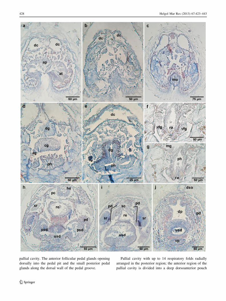
Fig. 10 Cross sections through the anterior body of G. impar n. sp.
a anterior region of the atrium with papillae; b–c posterior region of
the atrium with the central lobe extending above the anterior pharynx;
d detail of the anterior ganglia; e origin of cerebro-ventral connective;
f cross section through preradular pharynx and pedal pit; g–h radula
opening into pharynx; g pair of radula teeth; h radular support cells
and ventrolateral foregut glandular organs; i radular sheath; j–koesophageal region. ag anterior ganglion, ap atrial papillae, at atrium,
bg buccal ganglion, cg cerebral ganglion, cs radular support cells, dc
anterodorsal midgut caecum, eg oesophageal glands, lg lateral
ganglion, mo mouth, oe oesophagus, pg pharyngeal glands, ph
pharynx, pp pedal pit, ra radula tooth, rs radular sheath, vc cerebro-
ventral connective, vfg ventrolateral foregut glandular organ,
vg ventral ganglion
b
Helgol Mar Res (2013) 67:423–443 439
123

440 Helgol Mar Res (2013) 67:423–443
123
form a pair of pouches where spermatozoa accumulate,
functioning as seminal vesicles. Gonopericardioducts lat-
erally ciliated (200 lm long, 50 lm high, 40 lm wide)
opening dorsofrontally into a voluminous pericardium
(675 lm long, 140 lm high, 250 lm wide) whose ven-
troanterior part extends under the gonopericardioducts
(Fig. 9b). Bicameral heart (425 lm long, 40 lm high,
100 lm wide), partially bilobed, as an invagination of the
dorsal wall of the pericardium (Fig. 11d). Pericardioducts
(170 lm long, 45 lm high, 20 lm wide) leave from the
posterior region of the pericardium and opening dorsally
into the anterior region of the spawning ducts; before
opening into the spawning ducts, the pericardioducts widen
and contain spermatozoids, here they functioning as sem-
inal receptacles. Spawning ducts with high and glandular
epithelium along its whole length. The anterior region of
the spawning duct is paired (220 lm long, 85 lm high,
75 lm wide), fusing distally to form a large single duct
(325 lm long, 300 lm high, 350 lm wide) that dislodges
the rest of internal organs towards the dorsal body area
(Fig. 11e, f). Genital orifice with sphincter opens ventrally
at the beginning of the pallial cavity opening (Fig. 11g).
There are no copulatory stylets.
Taxonomic remarks
Gephyroherpia impar n. sp. belongs to the order Cavibe-
lonia Salvini-Plawen 1978, because it presents hollow
acicular sclerites and is included in the family Pruvotinidae
Salvini-Plawen 1978, for having a distichous radula and a
pair of ventrolateral foregut glandular organs of ducts with
subepithelially/extraepithelially arranged gland cells. The
new species is classified within the genus Gephyroherpia
Salvini-Plawen 1978, because having a thick cuticle with
epidermal papillae, a mouth clearly separated from the
atrium and a distichous radula and lacking copulatory
stylets.
Two other Gephyroherpia species are currently known:
Gephyroherpia antarctica Salvini-Plawen 1978 from Ross
Sea and Davis Sea (342–714 m) and Gephyroherpia (?)
triangulata Salvini-Plawen, 2009 from the Irish Sea (78 m)
(Salvini-Plawen 1978, 2009). G. impar n. sp. differs from
both species in having oesophageal glands, an unpaired
anterodorsal midgut caecum and lacking midgut
constrictions.
Gephyroherpia (?) triangulata was described from an
immature specimen and the anatomical characteristics of
its posterior region are still unknown; however, this species
is closer to G. impar n. sp. than to G. antarctica as regards
its biogeographical distribution, although it comes from a
much shallower depth. G. impar n. sp. and G. (?) trian-
gulata have an atrium posteriorly trilobed, radula teeth
without medial denticles and long ventrolateral foregut
glandular organs, characters that differentiate them clearly
from G. antarctica. Moreover, both species exhibit a
middorsal keel that gives them a triangular transverse
section, but in G. impar n. sp. the keel has lobulations
absent in G. (?) triangulata. Nevertheless, there are addi-
tional differences between G. impar n. sp. and G. (?) tri-
angulata. G. impar n. sp. has epidermal papillae and their
pedal fold ends before the opening of pallial cavity; it has
hooked radula teeth; and their radular sheath is not divided
proximally. Besides, G. (?) triangulata lacks respiratory
folds, although according to Salvini-Plawen (2009), the
absence of respiratory folds may be due to the juvenile
state.
Regarding G. antarctica, G. impar n. sp. also differs
because it lacks the abdominal spicules and the suprapallial
glands which presents G. antarctica and by the modifica-
tion of the pericardioducts serving as seminal receptacles in
G. impar n. sp. The diagnostic characters for all species of
Gephyroherpia are shown in Table 3.
General conclusions
In spite of the last reviews made (Salvini-Plawen 1978;
Garcıa-Alvarez and Salvini-Plawen 2007), the systematics
of Solenogastres still presents some problems. The
descriptions are sometimes incomplete, and different
characters that are considered taxonomically relevant at
present were not taken into account in the oldest descrip-
tions. Besides, the histological conditions of the very few
specimens available for research do not often allow making
detailed descriptions, which makes more difficult the study
of the species.
In this context, one of the generic characters used is the
position of the mouth opening, sharing a common cavity
either with the atrium (common atriobuccal cavity) or as an
independent cavity (buccal cavity). In the diagnosis of the
genus Pruvotina Cockerell, 1903, the ‘‘mouth opening
(in part separated from atrium but) within the common
atrio-buccal opening’’ (Garcıa-Alvarez and Salvini-Plawen
2007), is an ambiguous, variable and badly defined char-
acter. In this way, in some species of Pruvotina, the mouth
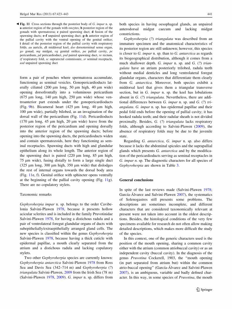
Fig. 11 Cross sections through the posterior body of G. impar n. sp.
a anterior region of the gonads with oocytes; b posterior region of the
gonads with spermatozoa; c paired spawning duct; d fusion of the
spawning ducts; e–f unpaired spawning duct; g–h anterior region of
the pallial cavity with the ventral opening of the genital orifice;
i detail of the posterior region of the pallial cavity with respiratory
folds. au auricle, dk middorsal keel, dso dorsoterminal sense organ,
go gonad, mg midgut, og genital orifice, pa pallial cavity, pc
pericardium, pd pericardioduct, psd paired spawning duct, re rectum,
rf respiratory fold, sc suprarectal commissure, sr seminal receptacle,
usd unpaired spawning duct
b
Helgol Mar Res (2013) 67:423–443 441
123

opens very close to the posterior region of the atrium,
whereas in other species, as it is in the case of P. artabra
n. sp. and P. manifesta n. sp., the atrium and mouth are
functionally separated, but linked by a groove. The clear
separation of mouth and atrium is characterized by the fact
that between both openings there is a cuticular covering
and bundles of circular and retractile musculature above
the separation area. In P. artabra n. sp. and P. manifesta
n. sp. the separation groove between the atrium and the mouth
lacks a cuticular coating but shows the typical musculature,
whereas in the descriptions of P. pallioglandulata and
P. uniperata, it is pointed out that this groove is weakly
cuticularized (Salvini-Plawen 1978). However, the com-
bination of the characters absence of copulatory stylets and
presence of serial lateral constrictions on the midgut and
respiratory folds on the pallial cavity clearly differentiates
the genus Pruvotina from the other two genera of the
subfamily Pararrhopaliinae: Pararrhopalia Simroth, 1903
and Labidoherpia Salvini-Plawen 1978 (Table 1). There-
fore, we consider that the position of the mouth opening
should not be taken into account in the diagnosis of
Pruvotina, as it is an ambiguous generic character.
Concerning the genus Gephyroherpia, both species
known so far have a midgut with serial lateral constrictions,
but G. impar n. sp. lacks them (Table 3); therefore, this
character should be considered as specific and not as generic.
The two new species of Gephyroherpia and Pruvotina
from NW Spain are the first records of both genera in the
geographical Atlantic region of Iberian Peninsula. Despite
the little information on the distribution of Solenogastres,
the available data indicate that many of the genera of the
class could have a global distribution, except for some that
would be limited to a kind of typical habitat, such as
abyssal bottoms or hydrothermal vents (Scheltema and
Kuzirian 1991; Scheltema 2000; Salvini-Plawen 2008b).
Nevertheless, it is necessary to extend the studies to
other geographical areas and bathymetries to get to know
the reality about the biodiversity and distribution of
Solenogastres.
Acknowledgments The authors are very grateful to Prof. Dr. Luitfried
Salvini-Plawen (Vienna University) for his help to improve this work.
This paper is a contribution to the following projects carried out by
the Marine Biological Station of A Grana from the University of
Santiago de Compostela: PGIDT01PXI20008PR, PGIDIT05PXIC
20001P, PGIDIT07PXB000120PR, A Selva-08 and ForSaGal-09
(Xunta de Galicia Regional Government); VEM2003-20070-C04-04,
CGL2004-22429-E and CTM2004-00740 (MEC, Spanish Govern-
ment). The paper is also a part of the research project BENTART
REN2003-01881/ANT (MEC, Spanish Government). The first author
was supported by a scholarship from the FPU Programme (MEC,
Spanish Government).
References
Garcıa-Alvarez O, Salvini-Plawen LV (2007) Species and diagnosis
of the families and genera of Solenogastres (Mollusca). Iberus
25(2):73–143
Table 3 Differences among the
species of the genus
Gephyroherpia Salvini-Plawen
1978
1 Presence, - absence,
? unknown. G. antarctica
Salvini-Plawen 1978 in Salvini-
Plawen (1978); G. (?)
triangulata Salvini-Plawen,
2009 in Salvini-Plawen (2009)
Gephyroherpia antarctica (?) triangulata impar n. sp.
Distribution Antarctica
Ross Sea, 342–714 m
Davis Sea, 385 m
Irish Sea
78 m
NW Spain
598–610 m
Length (mm) 4 2.4 7
Middorsal keel - ? ?
Epidermal papillae ? - ?
Posterior atrium Bilobed Trilobed Trilobed
Radula teeth
Distal hook ? - ?
Medial denticles 6–7 - -
Radular sheath Unpaired Paired/unpaired Unpaired
Ducts of the ventrolateral
foregut glandular organs
Short Long Long
Oesophagus Short Long Long
Oesophageal glands - - ?
Anterodorsal midgut caecum Paired/unpaired Paired/unpaired Unpaired
Midgut constrictions ? ? -
Abdominal spicules ? - -
Suprapallial glands ? ? -
Respiratory folds 10 - 15
Pedal fold reaches pallial cavity - ? -
442 Helgol Mar Res (2013) 67:423–443
123

Garcıa-Alvarez O, Urgorri V (2001) Luitfriedia minuta gen. et sp.
nov. (Mollusca: Solenogastres), a new species from Galicia,
North-West Spain. Cah Biol Mar 42(3):197–202
Garcıa-Alvarez O, LV Salvini-Plawen, Urgorri V (2001) Unciherpia
hirsuta a new genus and species of Aplacophora (Mollusca
Solenogastres: Pararrhopaliidae) from Galicia, Northwest Spain.
J Moll Stud 67:113–119
Garcıa-Alvarez O, Zamarro M, Urgorri V (2009) Proneomeniidae
(Solenogastres, Cavibelonia) from the Bentart-2006 Expedition,
with description of a new species. Iberus 27(1):67–78
Gil-Mansilla E, Garcıa-Alvarez O, Urgorri V (2011) A new genus and
two new species of Cavibelonia (Mollusca: Solenogastres) from
the Abyssal Angola Basin. Cah Biol Mar 52(2):233–243
Handl C, Todt C (2005) Foregut glands of Solenogastres (Mollusca):
anatomy and revised terminology. J Morphol 265(1):28–42
Heath H (1911) Reports on the scientific results of the expedition to
the tropical Pacific, XIV. The Solenogastres. Mem Mus Comp
Zool Harvard Coll 45(1):1–182
Heath H (1918) Solenogastres from the Eastren Coast of North
America. Mem Mus Comp Zool Harvard Coll 45(2):183–263
Pelseneer P (1901) Les Neomeniens de l’Expedition antarctique belge
et la distribution geographique des Aplacophora. Bull Acad R
Sci Lett B-Arts Belg 9–10:528–534
Pelseneer P (1903) Zoologie: Mollusques (Amphineures, Gastro-
podes et Lamellibranches). Resultats Voyage S. Y. Belgica
(1897–1899) sous le commandement de A. Gerlache de Gomery:
Rapports Scient. (1901–1913). Buschmann, Anvers, Belgium
Pruvot G (1890) Sur quelques Neomeniees nouvelles de la
Mediterranee. Arch Zool Exp Gen Ser 2(8):21–24
Pruvot G (1891) Sur l’organisation de quelques Neomeniens des cotes
de France. Arch Zool Exp Gen Ser 2(9):699–810
Pruvot G (1897) Essai sur les fonds et la faune de la Manche
Occidentale (Cotes de Bretagne) compares a ceux du Golfe du
Lion. Arch Zool Exp Gen Ser 3(5):511–660
Salvini-Plawen LV (1978) Antarktische und subantarktische Soleno-
gastres (Eine Monographie: 1898–1974). Zoologica (Stuttgart)
128:1–315
Salvini-Plawen LV (1997) Fragmented knowledge on West-European
and Iberian Caudofoveata and Solenogastres. Iberus 15(2):35–50
Salvini-Plawen LV (2003) Contributions to West-Mediterranean
Solenogastres (Mollusca) with three new species. Iberus
21(2):37–60
Salvini-Plawen LV (2008a) Contributions to West European Cavibe-
lonia (Mollusca, Solenogastres) with two new species. Zoosys-
tema 30(4):873–897
Salvini-Plawen LV (2008b) Three new species of Simrothiellidae
(Solenogastres) associated with the hot-vent biotope. J Moll Stud
74(3):223–238. doi:10.1093/mollus/eyn010
Salvini-Plawen LV (2009) Two immature pruvotinids from the Irish
Sea (Mollusca: Solenogastres). Mar Biod Rec 2:e164. doi:
10.1017/s1755267209990935
Scheltema AH (2000) Two new hydrothermal vent species, Helico-
radomenia bisquama and Helicoradomenia acredema, from the
eastern Pacific Ocean (Mollusca, Aplacophora). Argonauta
14(2):15–25
Scheltema AH, Kuzirian AM (1991) Helicoradomenia juani gen. et
sp. nov., a Pacific hydrothermal vent Aplacophora (Mollusca:
Neomeniomorpha). Veliger 34(2):195–203
Thiele J (1902) Proneomenia valdiviae n. sp. Wissusch Ergednisse
Dtsch Tiefsee-Exped Valdivia, 1898–1899 3:167-174
Thiele J (1913) Antarktische Solenogastren. Dtsch Sudpolar-Exp
1901–1903 14, Zool, Heft 6(1): 35–65
Todt C (2006) Ultrastructure of multicellular foregut glands in
selected Solenogastres (Mollusca). Zoomorphology 125(3):
119–134. doi:10.1007/s00435-006-0016-y
Helgol Mar Res (2013) 67:423–443 443
123