Embed Size (px)
Citation preview
438
Contemporary Management of a Clinical Case by Multidisciplinary Approach – A Case ReportZaid A. Al Jeaidi1, Mohammed Mustafa2
1Assistant Professor, Chairman, Department of Conservative Dental Sciences, College of Dentistry, Prince Sattam bin Abdulaziz University, Al-Kharj, Saudi Arabia. 2Assistant Professor, Head of Endodontic Division, Department of Conservative Dental Sciences, College of Dentistry, Prince Sattam bin Abdulaziz University, Al-Kharj, Saudi Arabia.
AbstractThe purpose of this article is to show how to manage and treat a comprehensive case in advanced and modern ways. A 28 years old Saudi female, came to our specialty clinic at College of Dentistry(COD), Prince Sattam bin Abdulaziz University, AlKharj, complaining of ugly appearance of her upper front teeth that reinforced her to avoid smiling and she cannot eat properly with her teeth, she was asking to improve her look and fix her teeth with a reasonable cost. A detailed clinical examination and investigation was done to reach to our final treatment plan, in regard to periodontal data; probing depths were in the range of 1-3mm, with plaque score of 92% and bleeding score of 78%. This case was treated completely in our speciality clinics with results showing great improvement in patients’ esthetic and functional condition. This case report illustrates the multidisciplinary approach of the different aspects of dentistry to achieve an esthetic, biologic and functional results.
Key Words: Esthetics, Smile, Dentition, Comprehensive treatment
Corresponding author: Dr. Mohammed Mustafa, Head of Endodontic Division, Department of Conservative Dental Sciences, College of Dentistry, P. O. Box: 153, Al-Kharj Postal Code: 11942, Saudi Arabia; e-mail: [email protected]
IntroductionEsthetics has now become an important issue in modern Society and Today’s dental patients are demanding a youthful, attractive smile. The esthetic rehabilitation of the patients with functionally compromised dentitions frequently involves a multi-disciplinary approach. The realization of esthetic objectives in multi-disciplinary cases represents a considerable clinical challenge due to the fact that esthetics is a subjective concept which may create disagreement between dentists and patients [1]. To a great extent, proper case selection and careful multi-disciplinary and treatment planning can govern the predictability of procedures. A team approach that includes the clinicians, the laboratory technicians, and the patient is essential to achieve the desired results.
The successful integration of esthetics and function does not emerge by chance, but rather as a result of meticulous development of clearly defined anatomical parameters and their subsequent incorporation into the design of the prosthesis [2-5].
This clinical case report demonstrates a successful multi-disciplinary approach in recreating an esthetic smile of a patient whose maxillary anterior dentition and even all her posterior teeth of both upper and lower jaws has been functionally and esthetically compromised.
Case Report
Patient description and examination
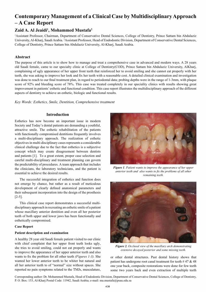
A healthy 28 year old Saudi female patient visited to our clinic with chief complaint that her upper front teeth looks ugly, she tries to avoid smiling, could not eat properly and wants to improve the appearance of her upper anterior teeth and also wants to fix the problem for all other teeth (Figures 1-3). She wanted her lower anterior teeth to be whiter but natural and all her anterior teeth to of “normal” size without spaces. She reported no pain symptoms related to the TMJs, musculature,
or other dental structures. Past dental history shows that patient has undergone root canal treatment for teeth # 47 & 48 one year back, composite restorations were done for few teeth some two years back and even extraction of multiple teeth
Figure 1. Patient wants to improve the appearance of her upper anterior teeth and also wants to fix the problems of all other
remaining teeth.
Figure 2. Occlusal view of the maxillary arch demonstrating extensive decayed posterior and some missing teeth.
439
OHDM - Vol. 14 - No. 6 - December, 20155
Dentition• Carious teeth: # 18, 17, 16, 15, 12, 11, 21, 22, 24, 25,
27, 28, 37, 35, 34, 45 & 47 (total=17).• Missing teeth: # 14, 26, 36, 38 & 46 (total=5).
Etiology• Diet imbalance.• Poor oral hygiene.• Faulty restorations.• Pulpal and periapical pathosis.• Unpleasing esthetics.
Treatment Objectives• To motivate the patient to improve his oral hygiene
& diet.• To educate the patient & enforce his knowledge about
oral & dental awareness. • To eliminate pathology.• To treat caries.• To restore function.• To improve esthetics.
PrognosisGood if the patient maintains good diet control, oral hygiene and attends regular follow- up appointments.Treatment PlanUpon consultation with the patient the following treatment plan was formulated (Tables 1-3).
were done seven years back. Patient is very cooperative and willing to come regularly on her appointment days.
Periodontal evaluation
On Intra-oral examination reveals generalized mild redness of the gingiva and interdental papillae, stippled with rolled gingival margins, localized swelling & blunting of interdental papilla, poor oral hygiene with generalized plaque accumulation, localized supra and sub-gingival calculus, pocket depth ranging between 1-3mm with localized gingival recession. No tooth mobility and no furcation involvement.
Occlusal evaluation
When it comes to patients dentition, there was a Class I canine relationship both on right side and left side, with canine guidance occlusion, Vertical & Horizontal Overlap of 1 mm each, with mild crowding in lower anterior teeth, few missing were teeth # 14, 26, 36, 38 & 46.
Diet analysis• Patient is taking 3 main meals during the day.• Snacks between meals every day.• Sugar consumption is high.• Meals contain mainly of rice, meat, bread and Pepsi.• Minimal vegetable & fruit intake.
Diagnostic toolsRadiographs (FMX)
• Normal maxillary sinus.• Generalized bone level ranges 1-3 mm from Cemento-
enamel junction, with normal bone trabeculation.• Multiple missing teeth # 14, 26, 36, 38 & 46.• Multiple carious Teeth.• Periapical lesion around teeth # 17, 16 &15.
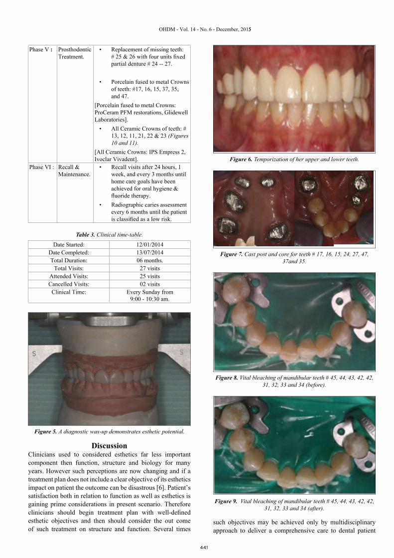
Mounted diagnostic casts (Figure 4)• Class I canine relationship (Right & Left).• Canine guidance.• Vertical overlap 1 mm.• Horizontal overlap 1 mm.
DiagnosisPeriodontium
• Generalized plaque induced gingivitis with localizedgingival recession.
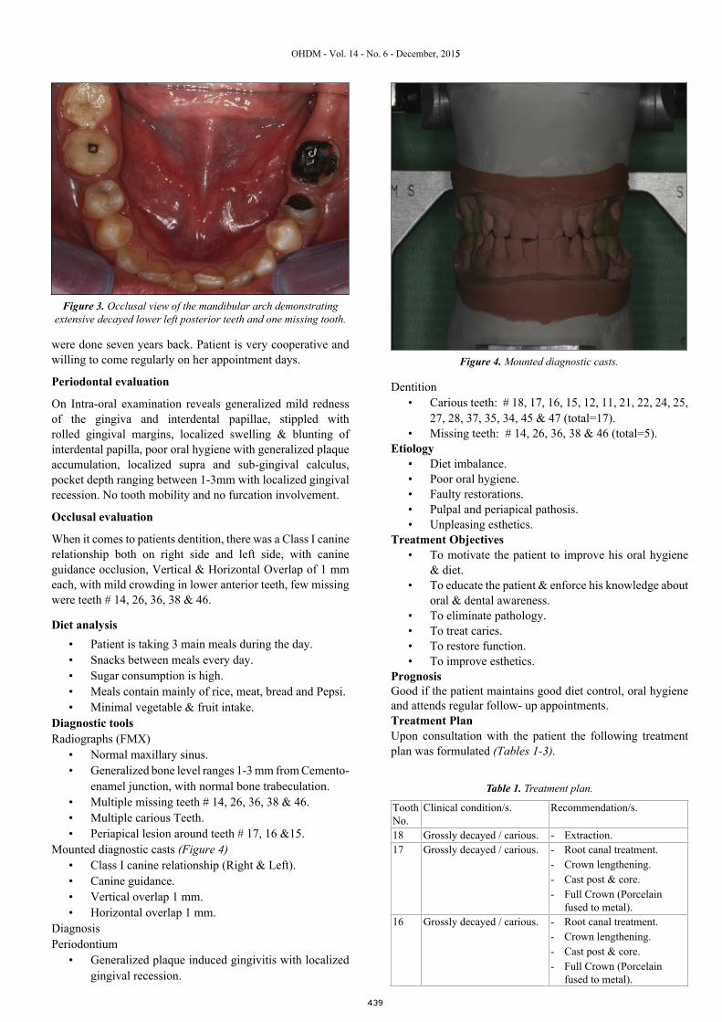
Figure 3. Occlusal view of the mandibular arch demonstrating extensive decayed lower left posterior teeth and one missing tooth.
Figure 4. Mounted diagnostic casts.
Tooth No.
Clinical condition/s. Recommendation/s.
18 Grossly decayed / carious. - Extraction.17 Grossly decayed / carious. - Root canal treatment.
- Crown lengthening.- Cast post & core.- Full Crown (Porcelain
fused to metal).16 Grossly decayed / carious. - Root canal treatment.
- Crown lengthening.- Cast post & core.- Full Crown (Porcelain
fused to metal).
Table 1. Treatment plan.

440
OHDM - Vol. 14 - No. 6 - December, 20155
15 Grossly decayed / carious. - Root canal treatment.- Crown lengthening.- Cast post & core.- Full Crown (Porcelain
fused to metal).14 Missing - No replacement.13 Short clinical crown - Crown lengthening.
- All Ceramic crown.12 Mesial & distal carious
lesions.- Mesial & distal composite
restoration.- All Ceramic crown.
11 Recurrent carious lesion(mesially with labial composite restorations).
- Root canal treatment.- Fiber (post & core) &
composite restoration.- All Ceramic crown.
21 Recurrent carious lesion with labial composite restorations
- Mesial & distal composite restoration.
- All Ceramic crown.22 Mesial composite
restoration and distal carious lesion.
- Mesial & distal composite restoration.
- All Ceramic crown.23 Short clinical crown - Crown lengthening
- All Ceramic crown.24 Grossly decayed / carious. - Root canal treatment.
- Crown lengthening- Cast post & core- Abutment of FPD
(Porcelain fused to metal).25 Grossly decayed / carious. - Extraction.
- Replacement.26 Missing - Replacement.27 Grossly decayed / carious. - Root canal treatment.
- Crown lengthening- Cast post & core- Abutment of FPD
(Porcelain fused to metal).28 Grossly decayed / carious. - Extraction.37 Grossly decayed / carious. - Root canal treatment.
- Cast post & core.- Full Crown (Porcelain
fused to metal).36 Missing - Replacement.35 Grossly decayed / carious. - Root canal treatment.
- Cast post & core.- Full Crown (Porcelain
fused to metal).34 Proximal (distal) carious
lesion.- Composite Restoration.
33, 32, 31, 41, 42, 43, 44.
Supra-gingival plaque & calculus.
- Scaling & oral prophylaxis.- Vital bleaching.
45 Occlusal carious lesion. - Composite restoration.46 Missing - Replacement.47 Sub-standard root canal
treated, screw post & composite restoration with recurrent caries.
- Root canal treatment.- Cast post & core.- Full Crown (Porcelain
fused to metal).48 Composite restoration with
recurrent carious lesion.- Extraction.
Phase I : Emergency/Preventive treatment.
• Scaling and oral prophylaxisdone.
• Oral hygiene instructions given.
• Diet advices and reassessment:
- Encourage intake of 3 well-balanced meals per day.
- Increase fruits and vegetable intake (natural cleansing effect).
- Reduce the intake of refined carbohydrates.
- Reduce the sweet snacks and confine them to meal time.
- Reduce sugar intake.
- Reduce the amount of soft drinks.
• Oral hygiene assessment &reinforcement done.
• Fluoride application done,and prescribed chlorhexidinemouthwash.
Phase II Operative treatment.
• Composite restorations withteeth # 12, 11, 21, 22, 34, 45.
[Composite used: Filtek TM Z350 XT Universal Restorative Material, 3M ESPE Dental Products USA].
Phase III : Endodontic treatment.
• Root canal treatment with teeth# 17, 16, 15, 11, 24, 27, 37, 35,47.
[Single visit / sitting root canal treatment were done. The working length was determined with an electronic apex locater (Root ZX, J. Morita Corp., Kyoto, Japan) and was confirmed radiographically. The Cleaning and Shaping was done using rotary NiTi instrumentation (Protaper, Dentsply company), followed by gutta-percha obturation using endodontic sealer (Sealapex, Kerr, USA).
Phase IV: Pre-Prosth-odontic / Sur-gical Treat-ment.
I. Pre-Prosthodontic:• Diagnostic wax-up (Figure 5).• Temporization (Figure 6).II. Pre-Prosthodontic – Surgical Phase• Crown lengthening: # 17, 16, 15,
13, 23, 24, and 27.• Cast post and core in Teeth: #
17, 16, 15, 24, 27, 37, 35, and 4(Figure 7)
• Fiber post & composite core inTeeth # 11.
[Fibers post used: RelyX™ Fiber Post, 3M ESPE Dental Products, U.S.A).
• Vital bleaching tooth #: 33, 32,32, 42, 42, 43, 44 (Figures 8and 9).
Table 2. Treatment phases.
441
OHDM - Vol. 14 - No. 6 - December, 20155
Phase V : Prosthodontic Treatment.
• Replacement of missing teeth:# 25 & 26 with four units fixedpartial denture # 24 -- 27.
• Porcelain fused to metal Crownsof teeth: #17, 16, 15, 37, 35,and 47.
[Porcelain fused to metal Crowns: ProCeram PFM restorations, Glidewell Laboratories].
• All Ceramic Crowns of teeth: #13, 12, 11, 21, 22 & 23 (Figures 10 and 11).
[All Ceramic Crowns: IPS Empress 2, Ivoclar Vivadent].
Phase VI : Recall & Maintenance.
• Recall visits after 24 hours, 1week, and every 3 months untilhome care goals have beenachieved for oral hygiene &fluoride therapy.
• Radiographic caries assessmentevery 6 months until the patientis classified as a low risk.
Table 3. Clinical time-table.
Date Started: 12/01/2014Date Completed: 13/07/2014Total Duration: 06 months.
Total Visits: 27 visitsAttended Visits: 25 visitsCancelled Visits: 02 visits
Clinical Time: Every Sunday from 9:00 - 10:30 am.
DiscussionClinicians used to considered esthetics far less important component then function, structure and biology for many years. However such perceptions are now changing and if a treatment plan does not include a clear objective of its esthetics impact on patient the outcome can be disastrous [6]. Patient’s satisfaction both in relation to function as well as esthetics is gaining prime considerations in present scenario. Therefore clinicians should begin treatment plan with well-defined esthetic objectives and then should consider the out come of such treatment on structure and function. Several times
such objectives may be achieved only by multidisciplinary approach to deliver a comprehensive care to dental patient
Figure 5. A diagnostic wax-up demonstrates esthetic potential.
Figure 6. Temporization of her upper and lower teeth.
Figure 7. Cast post and core for teeth # 17, 16, 15, 24, 27, 47, 37and 35.
Figure 8. Vital bleaching of mandibular teeth # 45, 44, 43, 42, 42, 31, 32, 33 and 34 (before).
Figure 9. Vital bleaching of mandibular teeth # 45, 44, 43, 42, 42, 31, 32, 33 and 34 (after).
442
OHDM - Vol. 14 - No. 6 - December, 20155
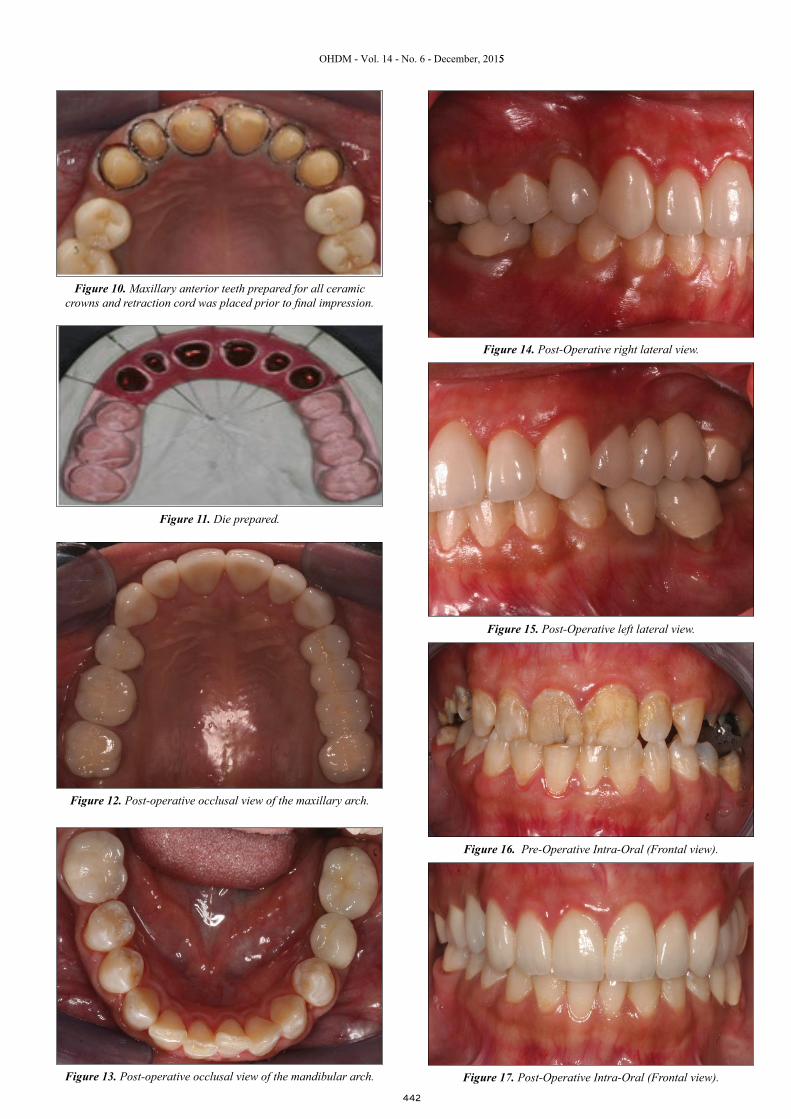
Figure 10. Maxillary anterior teeth prepared for all ceramic crowns and retraction cord was placed prior to final impression.
Figure 13. Post-operative occlusal view of the mandibular arch.
Figure 14. Post-Operative right lateral view.
Figure 15. Post-Operative left lateral view.
Figure 11. Die prepared.
Figure 12. Post-operative occlusal view of the maxillary arch.
Figure 16. Pre-Operative Intra-Oral (Frontal view).
Figure 17. Post-Operative Intra-Oral (Frontal view).
443
OHDM - Vol. 14 - No. 6 - December, 20155
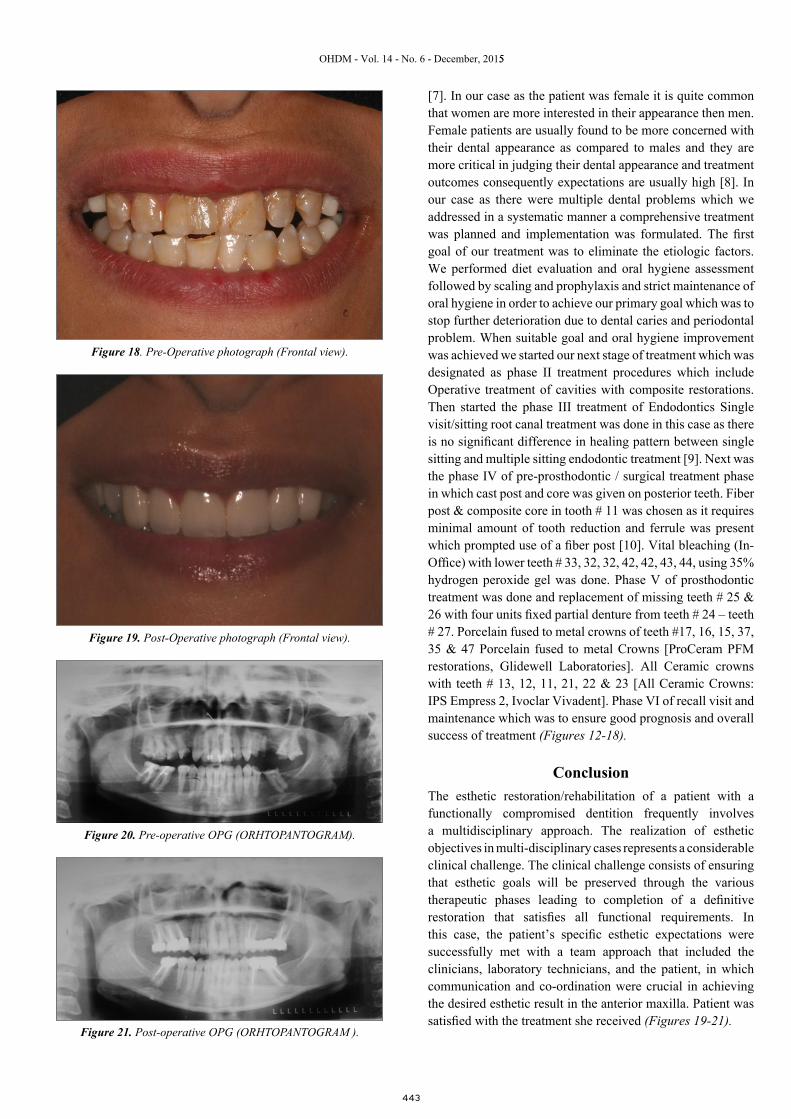
Figure 18. Pre-Operative photograph (Frontal view).
Figure 20. Pre-operative OPG (ORHTOPANTOGRAM).
Figure 21. Post-operative OPG (ORHTOPANTOGRAM ).
Figure 19. Post-Operative photograph (Frontal view).
[7]. In our case as the patient was female it is quite common that women are more interested in their appearance then men. Female patients are usually found to be more concerned with their dental appearance as compared to males and they are more critical in judging their dental appearance and treatment outcomes consequently expectations are usually high [8]. In our case as there were multiple dental problems which we addressed in a systematic manner a comprehensive treatment was planned and implementation was formulated. The first goal of our treatment was to eliminate the etiologic factors. We performed diet evaluation and oral hygiene assessment followed by scaling and prophylaxis and strict maintenance of oral hygiene in order to achieve our primary goal which was to stop further deterioration due to dental caries and periodontal problem. When suitable goal and oral hygiene improvement was achieved we started our next stage of treatment which was designated as phase II treatment procedures which include Operative treatment of cavities with composite restorations. Then started the phase III treatment of Endodontics Single visit/sitting root canal treatment was done in this case as there is no significant difference in healing pattern between single sitting and multiple sitting endodontic treatment [9]. Next was the phase IV of pre-prosthodontic / surgical treatment phase in which cast post and core was given on posterior teeth. Fiber post & composite core in tooth # 11 was chosen as it requires minimal amount of tooth reduction and ferrule was present which prompted use of a fiber post [10]. Vital bleaching (In-Office) with lower teeth # 33, 32, 32, 42, 42, 43, 44, using 35% hydrogen peroxide gel was done. Phase V of prosthodontic treatment was done and replacement of missing teeth # 25 & 26 with four units fixed partial denture from teeth # 24 – teeth # 27. Porcelain fused to metal crowns of teeth #17, 16, 15, 37, 35 & 47 Porcelain fused to metal Crowns [ProCeram PFM restorations, Glidewell Laboratories]. All Ceramic crowns with teeth # 13, 12, 11, 21, 22 & 23 [All Ceramic Crowns: IPS Empress 2, Ivoclar Vivadent]. Phase VI of recall visit and maintenance which was to ensure good prognosis and overall success of treatment (Figures 12-18).
ConclusionThe esthetic restoration/rehabilitation of a patient with a functionally compromised dentition frequently involves a multidisciplinary approach. The realization of esthetic objectives in multi-disciplinary cases represents a considerable clinical challenge. The clinical challenge consists of ensuring that esthetic goals will be preserved through the various therapeutic phases leading to completion of a definitive restoration that satisfies all functional requirements. In this case, the patient’s specific esthetic expectations were successfully met with a team approach that included the clinicians, laboratory technicians, and the patient, in which communication and co-ordination were crucial in achieving the desired esthetic result in the anterior maxilla. Patient was satisfied with the treatment she received (Figures 19-21).
444
OHDM - Vol. 14 - No. 6 - December, 20155
References1. Peck S. Introdução. Secção II: Relevância da estética facial na
Ortodontia. In: Sadowsky PL, (Editor). Atualidades em Ortodontia. 1997: pp.250.
2. Lee EA, Jun Sk. Achieving esthetic excellence through anoutcome-based restorative treatment rationale. Pract Periodontics Aesthet Dent. 2000; 12: 641-648.
3. Lee EA, Jun Sk. Aesthetic design preservation inmultidisciplinary therapy: Philosophy and clinical execution. Pract Proced Aesthet Dent. 2001; 14: 561-569.
4. Reddy MS. Achieving gingival esthetics. J Am Dent Assoc.2003; 134: 295-304.
5. Lambardi R. The principles of visual perception and theirclinical application to denture esthetics. J Prosthet Dent. 1973; 29: 358-382.
6. Spear FM, Kokich VG, Mathews DP. Interdisciplinarymanagement of anterior dental esthetics. J Am Dent Assoc. 2006; 137: 160-169.
7. Samorodnitzky-Naveh GR, Geiger SB, Levin L. Patients'satisfaction with dental esthetics. J Am Dent Assoc. 2007; 138: 805-808.
8. Gozalo-Diaz D, Johnston WM, Wee AG. Estimating the color of maxillary central incisors based on age and gender. J Prosthet Dent. 2008; 100: 93-98.
9. Ashken ZPJ. One visit endodontics. Dent clinics North Amer.1984; 28: 853.
10. Soares CJ, Valdivia AD, da Silva GR, Santana FR, Menezes Mde S. Longitudinal evaluation of post systems- A Literature review, Brazil Dent J. 2012; 23: 135-140.