Embed Size (px)
Citation preview

Aalborg Universitet
Complement C3 opsonization of Chlamydia trachomatis facilitates uptake in humanmonocytes
Lausen, Mads; Christiansen, Gunna; Karred, Nichlas; Winther, Robert; Poulsen, ThomasBouet Guldbæk; Palarasah, Yaseelan; Birkelund, SvendPublished in:Microbes and Infection
DOI (link to publication from Publisher):10.1016/j.micinf.2018.04.004
Creative Commons LicenseCC BY-NC-ND 4.0
Publication date:2018
Document VersionAccepted author manuscript, peer reviewed version
Link to publication from Aalborg University
Citation for published version (APA):Lausen, M., Christiansen, G., Karred, N., Winther, R., Poulsen, T. B. G., Palarasah, Y., & Birkelund, S. (2018).Complement C3 opsonization of Chlamydia trachomatis facilitates uptake in human monocytes. Microbes andInfection, 20(6), 328-336. https://doi.org/10.1016/j.micinf.2018.04.004
General rightsCopyright and moral rights for the publications made accessible in the public portal are retained by the authors and/or other copyright ownersand it is a condition of accessing publications that users recognise and abide by the legal requirements associated with these rights.
- Users may download and print one copy of any publication from the public portal for the purpose of private study or research. - You may not further distribute the material or use it for any profit-making activity or commercial gain - You may freely distribute the URL identifying the publication in the public portal -
Take down policyIf you believe that this document breaches copyright please contact us at [email protected] providing details, and we will remove access tothe work immediately and investigate your claim.

Accepted Manuscript
Complement C3 opsonization of Chlamydia trachomatis facilitates uptake in humanmonocytes
Mads Lausen, Gunna Christiansen, Nichlas Karred, Robert Winther, Thomas BouetGuldbæk Poulsen, Yaseelan Palarasah, Svend Birkelund
PII: S1286-4579(18)30096-0
DOI: 10.1016/j.micinf.2018.04.004
Reference: MICINF 4581
To appear in: Microbes and Infection
Received Date: 11 June 2017
Revised Date: 13 March 2018
Accepted Date: 23 April 2018
Please cite this article as: M. Lausen, G. Christiansen, N. Karred, R. Winther, T.B.G. Poulsen, Y.Palarasah, S. Birkelund, Complement C3 opsonization of Chlamydia trachomatis facilitates uptake inhuman monocytes, Microbes and Infection (2018), doi: 10.1016/j.micinf.2018.04.004.
This is a PDF file of an unedited manuscript that has been accepted for publication. As a service toour customers we are providing this early version of the manuscript. The manuscript will undergocopyediting, typesetting, and review of the resulting proof before it is published in its final form. Pleasenote that during the production process errors may be discovered which could affect the content, and alllegal disclaimers that apply to the journal pertain.

MANUSCRIP
T
ACCEPTED
ACCEPTED MANUSCRIPT1
1
Complement C3 opsonization of Chlamydia trachomatis facilitates 2
uptake in human monocytes 3
4
Mads Lausena, Gunna Christiansenb, Nichlas Karreda, Robert Winthera, Thomas Bouet 5
Guldbæk Poulsena, Yaseelan Palarasahc, Svend Birkelunda,* 6
7
aLaboratory for Medical Mass Spectrometry. Department of Health Science and Technology, 8
Fredrik Bajers Vej 3b, 9220 Aalborg Ø, Denmark 9
10
bDepartment of Biomedicine, Aarhus University, Wilhelms Meyers Allé 4, 8000 Aarhus, 11
Denmark 12
13
cUnit for Thrombosis Research, Institute of Public Health, University of Southern Denmark, 14
Esbjerg, Denmark 15
16
*To whom correspondence should be addressed: 17
Svend Birkelund, Professor, MD, PhD, DMSc, Department of Health Science and 18
Technology, Aalborg University, Fredrik Bajers Vej 3b, 9220 Aalborg, Denmark. Email: 19
[email protected], Phone: +45 4096 1916 20
21
Word count 22
Abstract: 200 words 23
Text: 4950 words 24

MANUSCRIP
T
ACCEPTED
ACCEPTED MANUSCRIPT2
Abstract 25
Chlamydia trachomatis is an obligate intracellular bacterium that causes severe infections, 26
which can lead to infertility and ectopic pregnancy. Although both innate and adaptive 27
immune responses are elicited during chlamydial infection the bacterium succeeds to evade 28
host defense mechanisms establishing chronic infections. Thus, studying the host-pathogen 29
interaction during chlamydial infection is of importance to understand how C. trachomatis 30
can cause chronic infections. Both the complement system and monocytes play essential roles 31
in anti-bacterial defense, and, therefore, we investigated the interaction between the 32
complement system and the human pathogens C. trachomatis D and L2. 33
34
Complement competent serum facilitated rapid uptake of both chlamydial serovars into 35
monocytes. Using immunoelectron microscopy, we showed that products of complement C3 36
were loosely deposited on the bacterial surface in complement competent serum and further 37
characterization demonstrated that the deposited C3 product was the opsonin iC3b. Using C3-38
depleted serum we confirmed that complement C3 facilitates rapid uptake of chlamydiae into 39
monocytes in complement competent serum. Complement facilitated uptake did not influence 40
intracellular survival of C. trachomatis or C. trachomatis-induced cytokine secretion. 41
Hence, C. trachomatis D and L2 activate the complement system leading to chlamydial 42
opsonization by iC3b and subsequent phagocytosis, activation and bacterial elimination by 43
human monocytes. 44
45
Keywords 46
monocytes; Chlamydia trachomatis; complement C3

MANUSCRIP
T
ACCEPTED
ACCEPTED MANUSCRIPT3
1. Introduction 47
Chlamydia trachomatis is estimated to infect 100 million people annually causing chronic 48
genital and ocular infections [1]. The course of genital infection is mostly asymptomatic 49
leaving the infection undiagnosed and untreated. Untreated genital chlamydial infection can 50
cause severe tissue damage and lead to pelvic inflammatory disease, ectopic pregnancy, and 51
infertility [2]. 52
C. trachomatis is an obligate intracellular Gram-negative bacterium with a unique biphasic 53
developmental cycle. The infectious, but metabolic inactive elementary body (EB) infects 54
epithelial cells in the genital mucosa. Intracellularly, the EB transforms to a larger non-55
infectious but metabolic active reticulate body (RB) [3]. During entry, C. trachomatis inhibits 56
phagosome-lysosome fusion and resides in a modified vacuole called an inclusion, which 57
provides a niche for bacterial replication [4]. 58
Chlamydial infections tend to be chronic even though both humoral and cell-mediated 59
immunity are elicited [5]. Monocytes and macrophages play essential roles in anti-bacterial 60
immunity in general, but little is known about the exact role of monocytes during C. 61
trachomatis infections. During infection, epithelial cells respond by secreting several 62
cytokines and chemokines creating a local inflammatory condition that recruits monocytes to 63
the site of infection [6]. In vitro studies show that several C. trachomatis serovars infect 64
human monocytes inducing cellular activation with secretion of inflammatory cytokines, such 65
as IL-1β, IL-6, and IL-8 [7]. Chlamydial uptake into host cells is supposedly carried out by 66
phagocytosis or by receptor-mediated endocytosis, but the exact mechanisms and the 67
receptors involved remain elusive [8]. An involvement of plasma membrane lipid rafts and 68
the mannose receptor have been suggested, but also complement receptors could be involved 69
since these receptors facilitate uptake of other intracellular bacteria such as Mycobacterium 70
tuberculosis and Legionella pneumophila [9–12]. 71

MANUSCRIP
T
ACCEPTED
ACCEPTED MANUSCRIPT4
The complement system consists of more than 30 different proteins comprising both soluble 72
factors and cell surface receptors [13]. Complement activation initiates a cascade of 73
proteolytic cleavages leading to both direct and indirect anti-microbial effects. The direct anti-74
microbicidal functions are carried out by the membrane attack complex (MAC), a pore-75
formed structure consisting of repetitive membrane-spanning complement factors that causes 76
membrane permeability and cellular lysis. Another function of the complement system is 77
mediated by the so-called opsonins, which bind to the surface of pathogens tagging them for 78
uptake and degradation in professional phagocytes. 79
The complement system is mainly activated through three distinct pathways which are 80
triggered by different structural motifs and involve different intermediate complement 81
products, but they all converge at the common downstream effector C3 convertase. C3 82
convertase cleaves complement factor C3 into the anaphylatoxin C3a and the opsonin C3b. 83
C3b may be further cleaved into additional opsonins called iC3b and C3dg. C3b, iC3b, and 84
C3dg are all recognized by surface receptors expressed on different host immune cells and 85
opsonin-receptor engagement leads to receptor-mediated phagocytosis of the opsonized 86
organism. It has been shown that C3b and iC3b are recognized by complement receptor (CR) 87
1 and CR3, respectively, and both receptors are ubiquitously expressed on monocytes and 88
macrophages and are important for mononuclear phagocytosis of infectious bacteria [14]. 89
C. trachomatis is able to activate the complement system and it has been demonstrated that C. 90
trachomatis induced complement activation leads to binding of C3 to the bacterium [15]. 91
To further explore the interaction between the complement system and C. trachomatis, we 92
investigated how complement deposition on C. trachomatis affects the uptake of chlamydial 93
EBs into human monocytes and how complement modulates the intracellular fate of C. 94
trachomatis in monocytes. Uncovering new aspects of the interaction between complement, 95
MANUSCRIP
T
ACCEPTED
ACCEPTED MANUSCRIPT5
monocytes, and C. trachomatis are important to understand how chlamydial infections are 96
controlled by the innate immune system. 97
98
2. Materials and methods 99
2.1. Antibodies 100
The following primary antibodies were used in this study: Anti-human CD11b (MEM-174) 101
(ImmunoTools GmbH, Friesoythe, Germany), Polyclonal Rabbit Anti-Human C3c 102
Complement (Agilent Technologies, Glostrup, Denmark), Mab32.3 against C. trachomatis 103
MOMP [16], and PAb17 against C. trachomatis outer membrane [17]. FITC-, Alexa Flour® 104
488-, and rhodamine-conjugated secondary antibodies were purchased from Jackson 105
ImmunoResarch (Jackson ImmunoResearch, PA, USA). Anti-Rabbit IgG Alkaline 106
Phosphatase was purchased from Sigma-Aldrich (Sigma-Aldrich, St. Louis, MO, USA). Goat 107
anti-rabbit antibody conjugated with 10 nm colloidal gold (British BioCell, Cardiff, UK) was 108
used for immunoelectron microscopy. 109
110
2.2. Bacteria strains and culture 111
C. trachomatis D/UW-3/cx and L2/434/Bu were obtained from the American Type Culture 112
Collection (ATCC, VA, USA) and propagated in McCoy cells according to Ripa and Mårdh 113
[18]. Chlamydia were tested free of mycoplasma by PCR according to Huniche et al. [19]. 114
McCoy cells were obtained from ATCC and tested free of mycoplasma by Hoechst 33342 115
staining and PCR according to[19]. 116
C. trachomatis D and L2 EB were purified by density gradient centrifugation essentially 117
according to Caldwell et al. 1981 [20] and purity was estimated using negative staining and 118
transmission electron microscopy (TEM), (see 2.7) 119
120

MANUSCRIP
T
ACCEPTED
ACCEPTED MANUSCRIPT6
2.3. Cell isolation and culture 121
Blood samples were obtained from C. trachomatis seronegative donors at Aalborg University 122
(Approved by The Ethics Committee of Region Nordjylland, case no. N-20150073). 123
Peripheral blood mononuclear cells (PBMCs) were isolated from heparinized blood by 124
density gradient centrifugation on LymphoPrep™ (STEMCELL Technologies™, Vancouver, 125
Canada) according to Carlsen et al. [21]. The cells were seeded in 8 well Lab-Tek® Chamber 126
Slide™ Permanox slides (Thermo Scientific, MA, USA) at a density of 5x10⁵ cells/well and 127
cultured in standard medium containing RPMI 1640 supplemented with 10% heat inactivated 128
fetal calf serum (FCS), and 0.01 mg/ml gentamicin. Cells were allowed to adhere for 90 129
minutes at 37 °C and 5% CO2 and non-adherent cells were subsequently removed by washing 130
the cells twice in PBS. 131
132
2.4. Monocyte infection 133
C. trachomatis was suspended in RPMI 1640 (Biowest, Nuaillé, France) containing either 134
10% human autologous serum (NHS) or 10% heat-inactivated human autologous serum 135
(HIHS) and added immediately to adherent monocytes. Serum was heat-inactivated by 136
incubating serum for 30 minutes at 56 °C. Infection was carried out for 1, 4 or 24. For 24 137
hours infection, extracellular bacteria were removed after 4 hours and infection medium was 138
replaced by standard medium for the remaining incubation period. In some experiments, 139
medium was supplemented with 10% C3-depleted human serum alone or added 5 µg purified 140
C3 (Sigma Aldrich) to a final concentration of 20 µg/ml. 141
For some experiments lipopolysaccharide (LPS) from Escherichia coli (026:B6, Sigma-142
Aldrich) was used as positive controls at a concentration of 1 µg/ml. 143
144
2.5. Immunofluorescence microscopy 145

MANUSCRIP
T
ACCEPTED
ACCEPTED MANUSCRIPT7
Immunofluorescence staining was carried out essentially according to Carlsen et al. [21]. 146
Extracellular bacteria and surface bound CD11b were stained prior to fixation by incubating 147
cells for 30 minutes at 37 °C with Pab17 (1:200) or anti-CD11b (5 µg/ml) diluted in PBS 148
containing 0.1% bovine serum albumin (BSA) and 0.05% sodium azide. Cells were washed 149
twice in PBS and fixed for 20 minutes in 3.7% formaldehyde at 4 °C. Cells were 150
permeabilized for 7 minutes in 0.2% Triton-X 100 at room temperature and blocked in 0.1% 151
BSA for 15 minutes at 37 °C. Primary antibodies were diluted in antibody buffer containing 152
0.1% BSA in PBS (Mab32.3: 5 µg/ml) and cells were incubated with primary antibody for 30 153
minutes at 37 °C. Cells were washed three times in antibody buffer and incubated with 154
secondary antibodies diluted 1:200 in antibody buffer. Cells were washed three times in 155
antibody buffer and counter-stained with either 2 µM To-Pro-3 Iodide or 2 µM DAPI for 10 156
minutes at room temperature. Finally, mounting medium was added to each well and slides 157
were mounted with cover slips. 158
Cells were visualized and imaged using a Leica SP5 confocal microscope or a Leica DM 159
5500 B fluorescence microscope. 160
161
2.6. Immunoelectron microscopy 162
Purified EBs were mixed with 1/10 volume of NHS or HIHS. Five microL purified EB was 163
added to the surface of carbon-coated glow discharged 400 mesh nickel grids as described 164
(20). The grids were washed on three drops of PBS (pH 6.5) and blocked on one drop of 1% 165
ovalbumin (Sigma-Aldrich) in PBS. The grids were then incubated for 30 min at 37 °C with 166
1/200 rabbit anti-C3c antibody (Agilent Technologies) diluted in ovalbumin. The grids were 167
then washed on three drops PBS and incubated for 30 min at 37 °C in goat anti-rabbit 168
antibodies conjugated with 10 nm colloidal gold (1:25) in ovalbumin. Following this, the 169
grids were washed on three drops of PBS, incubated on three drops 0.5% cold fish gelatin 170

MANUSCRIP
T
ACCEPTED
ACCEPTED MANUSCRIPT8
(Sigma-Aldrich) in PBS (10 min each), washed on three drops of PBS, one drop of H2O and 171
stained with one drop of 0.5% phosphotungstic acid and blotted dry on filter paper. Electron 172
microscopy was done at 60 keV on a JEOL 1010 transmission electron microscope (Jeol, 173
Tokyo, Japan). Images were obtained using a KeenView digital camera (Olympus Soft 174
Imaging Solutions GmbH, Münster, Germany). 175
176
2.7. SDS-PAGE and immunoblotting 177
Purified EBs from C. trachomatis D and L2 were incubated with an equal volume of either 178
NHS or HIHS for 30 minutes at 37 °C. EBs were washed twice in PBS with centrifugation at 179
20000 x g for 15 minutes between each wash. Samples were boiled in RunBlue LDS Sample 180
Buffer (Expedeon, CA, USA) containing 5% v/v β-mercaptoethanol and proteins were 181
separated on a 7,5% SDS polyacrylamide gel according to Laemmli (Laemmli 1970). Proteins 182
were blotted on a nitrocellulose membrane according to Drasbek et al. (Drasbek 2004). The 183
membrane was blocked in Tris buffered saline (TBS) with 3% gelatin. Polyclonal Rabbit 184
Anti-Human C3c Complement (Agilent Technologies) (1:1000) was used as primary antibody 185
and Anti-Rabbit IgG Alkaline Phosphatase (Sigma-Aldrich) (1:20,000) was used as secondary 186
antibody. Protein bands were developed by adding BCIP/NBT alkaline phosphatase substrate 187
(Kem-En-Tec Diagnostics, Taastrup, Denmark). 188
189
2.8. Reinfection assay 190
Monocytes were cultured and infected according to section 2.1 except PBMCs were seeded in 191
24-well plates at a density of 2x10⁶ cells/well. After 4 and 24 hours, adherent monocytes were 192
washed thoroughly three times in PBS and detached by scrabing of cells in 2SP buffer (0.2 M 193
sucrose, 0.02 M phosphate, pH = 7.2). Monocytes were lysed by ultrasonication and lysates 194
from two wells were pooled and diluted 1:2 in standard medium (see 2.3) and added to 195

MANUSCRIP
T
ACCEPTED
ACCEPTED MANUSCRIPT9
confluent McCoy cells. McCoy cells were incubated for 1 hour at 37 °C and 5% CO2 and 196
subsequently washed three times in PBS and cultured for additional 23 hours in standard 197
medium containing 2 µg/ml cyclohexamide. 198
Cells were processed for immunofluorescence staining as described in 2.5. 199
200
2.9. Enzyme-linked immunosorbent assay (ELISA) 201
IL-6 and IL-8 ELISA kits were purchased from ImmunoTools GmbH and the analyses were 202
performed according to manufacture’s protocol with minor changes. Briefly, MaxiSorp plates 203
(NUNC) were coated with capture antibody diluted in PBS over night at 4 °C. Excess binding 204
was blocked with 1% BSA in PBS for one hour at room temperature. Monocyte culture 205
supernatants were diluted in 0.1% BSA + 0.05% Tween-20 in PBS and added to the wells and 206
left for incubation for one hour at room temperature. Captured IL-6 and IL-8 were detected 207
using a biotinylated detector antibody and subsequently streptavidin conjugated to horseradish 208
peroxidase (HRP). The enzymatic reaction was initiated by adding the HRP substrate TMB-209
ONE (Kem-En-Tec Diagnostics) and stopped after 30 minutes by adding 1M HCl. 210
211
2.10. Statistics 212
Statistical differences between two independent groups were calculated using Student’s t-test. 213
Multiple comparisons were analyzed by One-way ANOVA with Tukey’s multiple 214
comparison test. All statistical analyses were performed in GraphPad Prism 7 (GraphPad 215
Software Inc., CA, USA). P-values < 0.05 were considered statistically significant. 216
217
3. Results 218
3.1. Investigating the role of complement components in C. trachomatis uptake 219

MANUSCRIP
T
ACCEPTED
ACCEPTED MANUSCRIPT10
We aimed to investigate whether complement components affect the uptake of C. trachomatis 220
into monocytes. First, dilutions of C. trachomatis D and L2 were titrated to obtain an average 221
Chlamydia-to-monocyte ratio of 1. Intracellular chlamydiae were visualized using a 222
monoclonal antibody against chlamydial MOMP (Fig. 1A). The intracellular localization in 223
monocytes were confirmed both by membrane staining against CD11b and by differential 224
staining of intracellular and extracellular bacteria. Monocytes were infected in media 225
containing either normal autologous serum (NHS) or heat-inactivated autologous serum 226
(HIHS). Heat-inactivation of serum was done to denature complement factors, abrogating a 227
functional complement system. Cells were fixed after 1 hour of infection and chlamydial 228
uptake was quantified by counting the percentage of infected cells. 229
Fig. 1B shows that the percentage of infected cells was statistically significantly higher for 230
both serovars after 1 hour of infection in NHS samples compared to HIHS samples. Fig. 1B 231
also shows that there was no difference between uptake efficiency between serovars. These 232
findings suggest that C. trachomatis D and L2 are taken up by monocytes with the same 233
efficiency and that complement-competent serum facilitates rapid uptake of C. trachomatis D 234
and L2 into human monocytes. 235
236
3.2. Complement deposition on C. trachomatis D and L2 237
Our observations suggest that complement opsonization of C. trachomatis D and L2 238
facilitates uptake into monocytes. Monocytes express different receptors recognizing the C3 239
opsonins, C3b and iC3b, and it was previously demonstrated that these complement proteins 240
bind to C. trachomatis L2 [15]. We therefore used a polyclonal antibody against C3c to 241
visualize possible opsonizing complement by immuno-gold electron microscopy, since C3c is 242
a common component found in both C3b and iC3b. Purified C. trachomatis D and L2 EBs 243
were incubated with NHS and subsequently stained against C3c and with gold-conjugated 244

MANUSCRIP
T
ACCEPTED
ACCEPTED MANUSCRIPT11
IgG as secondary antibody [22]. Fig. 2A+E show that C3 complement fragments were 245
deposited in patchy areas on the surface of both serovars when incubated in NHS. In contrast, 246
when chlamydial EBs were incubated with HIHS, only few gold particles were observed on 247
the EB surface (Fig. 2B, F). No gold was observed on the EB surface when EBs were 248
incubated with NHS and anti-C3c was omitted (Fig. 2C, G). To quantify complement 249
deposition, bacteria associated gold particles and gold particles associated with the 250
background were enumerated and these numbers were expressed as a ratio. Fig. 2D+H show 251
that more gold particles are deposited on the bacterial surface when incubated with NHS 252
compared to HIHS. Thus, complement factors containing the C3c domain bind to the surface 253
of C. trachomatis D and L2 in the presence of NHS, but not HIHS, and this may account for 254
the observed differences in uptake efficiency. 255
256
3.3. Investigation of Chlamydia-bound C3 257
By immune-gold electron microscopy we confirmed that complement C3 fragments bind to 258
the surface of C. trachomatis D and L2 EBs. However, since C3c is a common structure 259
found in different C3 fragments we could not elucidate exactly which fragments were bound 260
to the EBs or if activation of the cleavage cascade had occurred. 261
We therefore conducted an immunoblot analysis of purified chlamydial EBs incubated in 262
either NHS or HIHS. Western blotting was performed three times using different sera with 263
similar results and a representative blot is shown in Fig. 3A. Fig. 3A shows that uncleaved α 264
and β chains of C3 (119 and 74 kDa, respectively) were present on EB after incubation with 265
either NHS or HIHS, though much stronger when incubated with NHS. Several other C3 266
protein bands were bound to both C. trachomatis D and L2 when incubated in NHS. The 267
protein band observed around 45 kDa corresponds to the α’2 fragment of C3 which is only 268
found in complement iC3b (Fig. 3B). These findings showed that the complement cascade is 269

MANUSCRIP
T
ACCEPTED
ACCEPTED MANUSCRIPT12
only activated in NHS leading to production and binding of iC3b to both C. trachomatis D 270
and L2. In addition to the protein bands just described, two protein bands were present in the 271
high molecular area (165 and 250 kDa, respectively) in the lanes in which EB were incubated 272
with NHS (Fig. 3A). These bands represent fragments of either the α’ chain of C3b or the α´1 273
chain of iC3b covalently linked to unidentified proteins. Both chains contain an exposed 274
thioester site that allows covalent interactions between C3b/iC3b and target proteins (Fig. 275
3B). 276
Thus, iC3b binds to the surface of both C. trachomatis D and L2 and may be involved in 277
covalent interactions with chlamydial surface proteins. 278
279
3.4. Complement C3 facilitated uptake of C. trachomatis into monocytes 280
The above results suggest that C3 opsonization of C. trachomatis could explain the 281
differential uptake efficiency observed using complement-competent serum and heat-282
inactivated serum, respectively. To elucidate whether C3 in fact facilitates uptake into 283
monocytes, we investigated the monocyte uptake of C. trachomatis L2 in the presence C3-284
depleted human serum after one hour of incubation Fig. 4. shows that using C3-depleted 285
serum reduces the uptake of chlamydia into monocytes compared to bacteria incubated in the 286
presence of NHS. Adding purified human C3 to the C3-depleted serum restored the monocyte 287
uptake efficiency, demonstrating that complement C3 facilitates uptake of C. trachomatis into 288
monocytes. 289
290
3.4. Intracellular fate of C. trachomatis after complement-mediated monocyte ingestion 291
As early uptake of C. trachomatis in monocytes is facilitated by complement C3 292
opsonization, we analyzed the fate of C. trachomatis when ingested by monocytes to 293
elucidate the biological significance of the rapid uptake. 294

MANUSCRIP
T
ACCEPTED
ACCEPTED MANUSCRIPT13
We have previously observed that both serovar D and L2 detection diminishes over time in 295
monocytes (data not shown), suggesting that both serovars are eradicated in monocytes. 296
One previous study demonstrated that C. trachomatis D and L2 can survive intracellularly in 297
monocytes for up three days post infection [23]. These results conflict with our initial 298
observations, and we speculated whether the observed differences may be due to the 299
presence/absence of functional complement. To test this, we evaluated the viability and 300
growth potential of complement-opsonized and non-opsonized C. trachomatis L2 using a 301
reinfection assay. Monocytes containing C. trachomatis L2 were lysed by ultrasonication and 302
the lysates were applied to confluent McCoy cells. The viability of ingested bacteria was 303
evaluated by quantifying the percentage of McCoy cells containing mature inclusions (Fig. 304
5A, right image). As demonstrated in Fig 5A (table), only few McCoy cells contained mature 305
inclusions when C. trachomatis was incubated with monocytes for 4 hours. No differences in 306
chlamydial viability were observed between NHS and HIHS, suggesting that complement-307
mediated uptake of C. trachomatis does not affect intracellular degradation of C. trachomatis 308
in monocytes. When C. trachomatis was incubated within monocytes for 24 hours no mature 309
inclusions were observed in either condition demonstrating that C. trachomatis is efficiently 310
killed in monocytes independently of complement. 311
312
3.6. Complement modulation of C. trachomatis induced cytokine production 313
We showed that complement C3 potentiates the chlamydial uptake, and that uptake leads to 314
efficient intracellular killing of the bacteria, which is one of the primary roles of monocytes 315
during infection. Another key role of monocytes during infection is to produce and secrete 316
inflammatory cytokines potentiating anti-microbial immune mechanisms. Thus, to further 317
extend our understanding of the functional consequences of bacterial opsonization we 318

MANUSCRIP
T
ACCEPTED
ACCEPTED MANUSCRIPT14
explored if complement affects secretion of IL-6 and IL-8 in monocytes incubated with C. 319
trachomatis. 320
Monocytes were cultured for 4 hours with C. trachomatis L2 in either NHS or HIHS, LPS or 321
media alone. After 4 hours of incubation cells were washed and new medium was added and 322
cells were incubated for further 20 hours. The conditioned monocyte medium was harvested 323
and the concentration of IL-6 and IL-8 was determined by ELISA. 324
As shown in Fig. 5B. C. trachomatis induces the secretion of both IL-6 and IL-8 as reported 325
previously [7,24]. Neither IL-6 or IL-8 secretion were significantly affected by the presence 326
of functional complement since monocytes incubated in NHS and HIHS demonstrates similar 327
concentrations of the cytokine. However, for both cytokines a small non-significant difference 328
was observed between NHS and HIHS with a higher concentration in monocytes incubated 329
with HIHS. Thus, complement opsonization does not affect C. trachomatis induced secretion 330
of IL-6 and IL-8 in monocytes. 331
332
4. Discussion 333
We demonstrated that purified EBs of the two serovars, D and L2, of C. trachomatis activated 334
the complement system leading to deposition of C3 fragments on the chlamydial surface and 335
that complement C3 facilitates rapid chlamydial uptake into human primary monocytes 336
leading to bacterial elimination and cytokine production. 337
Complement activation and complement-mediated phagocytosis of bacterial agents by 338
monocytes and macrophages have been demonstrated for different intracellular bacteria such 339
as M. tuberculosis and Listeria monocytogenes [25,26]. Other studies have demonstrated the 340
ability of C. trachomatis to activate the proteolytic complement cascade leading to activation 341
of both the C3- and C5-convertase [15,27]. 342

MANUSCRIP
T
ACCEPTED
ACCEPTED MANUSCRIPT15
To our knowledge, we are the first to demonstrate direct involvement of complement C3 in 343
monocyte ingestion of C. trachomatis and that complement C3 is deposited on the surface of 344
C. trachomatis D EBs. Additionally, using immunoelectron microscopy, we directly 345
visualized complement deposition on serovar L2 previously reported by Hall et al. [15]. We 346
found that complement was activated generating iC3b which was bound to the surface of both 347
serovar D and L2. iC3b is a potent opsonin that has been involved in opsonization and 348
phagocytosis of other intracellular bacteria such as M. tuberculosis [25]. 349
iC3b is recognized by complement receptor 3 (CR3), a heterodimeric integrin consisting of 350
CD11b and CD18 that is ubiquitously expressed on the surface of monocytes [28]. CR3 351
participates in phagocytosis of other intracellular bacteria, such as Mycobacteria spp. and 352
Peyron et al. [29] showed that CR3 is involved in lipid raft-dependent internalization of M. 353
kanasii. In line with these findings, it has been demonstrated that the integrity of lipid rafts is 354
important for host cell entry of several C. trachomatis serovars [9,30]. Additionally, binding 355
and internalization of Borellia burgdoferi was shown to be dependent on complement C3 and 356
CD14-dependent recruitment of CR3 to lipid rafts, suggesting that CD14 may also be 357
involved in the enhanced uptake, since CD14 is widely expressed on monocytes [31,32]. 358
Thus, it seems likely that the enhanced uptake of C. trachomatis observed in NHS is due to 359
iC3b-mediated phagocytosis by CR3 engagement. However, iC3b is not exclusively 360
recognized by CR3. CR1 and CR4 can also bind iC3b leading to iC3b-mediated phagocytosis 361
[33]. 362
An important parameter to discuss in this context is the involvement of the complement 363
anaphylatoxins C3a and C5a, which are generated by proteolytic cleavage of C3 and C5 364
during complement activation. These inflammatory mediators were not investigated in the 365
current study, but we demonstrate C3 cleavage and, therefore, we know that C3a is generated. 366
Although some degree of C3b inactivation was observed, it is likely that the complement 367

MANUSCRIP
T
ACCEPTED
ACCEPTED MANUSCRIPT16
cascade proceeds to C5 cleavage. This was previously shown by Megran and colleagues who 368
demonstrated that C. trachomatis L2 induced cleavage of C5 to C5a [27]. Both 369
anaphylatoxins are recognized by G-protein coupled receptors expressed on monocytes. 370
These mediators could likely contribute to the increased monocytic phagocytosis, since C5aR 371
antagonists were shown to reduce phagocytosis of heat-killed Staphylococcus aureus in 372
monocytes [34]. Supporting this observation, it was demonstrated that both C3a and C5a 373
upregulates CD11b surface expression in neutrophils and monocytes [35]. The contribution of 374
the anaphylatoxins was not addressed in our study, but literature suggests that anaphylatoxins 375
likely contribute to the observed effects presented in this study [27,34,35] 376
377
A unique feature of C3 opsonins is their ability to covalently attach to target structures 378
through a thioester site located in the α’ chain of C3b and in the α’1 chain of iC3b (Fig. 3B). 379
Our data suggest that iC3b is covalently attached to protein structures on both serovars since 380
several high molecular protein bands are observed under both denaturing and reducing 381
conditions. Under reducing conditions iC3b will split into three protein fragments: α’1 (63 382
kDa), α’2 (39 kDa), and β (75 kDa) [36]. We observe the latter two, but not the α’1 fragment. 383
The α’1 fragment is likely located in the observed high molecular bands covalently attached 384
to other proteins. It was previously proposed that C3 fragments interact with MOMP on the 385
chlamydial surface, but it was not conclusively determined due to antibody cross-reactivity 386
[15]. We observed anti-C3c reactive protein bands migrating approximately at 165 and 250 387
kDa, which does not correspond to the summed molecular mass of MOMP and the α’1 388
fragment (40 kDa + 110 kDa, respectively). The protein bands observed around 250 kDa 389
suggest that iC3b interacts with high molecular weight surface structures. Potential high 390
molecular candidates to interact with C3 are the polymorphic membrane proteins, which 391
ranges in size from 95 kDa to 187 kDa, however this was not further investigated. 392

MANUSCRIP
T
ACCEPTED
ACCEPTED MANUSCRIPT17
We used immunoelectron microscopy to directly visualize protein deposition on the EB 393
surface. Interestingly, we observed that the C3 fragments were loosely bound to the 394
chlamydial surface and this observation does not fit with the idea that C3 is covalently linked 395
to chlamydial outer membrane proteins, however, several chlamydial-complement bindings 396
may be involved. It has been demonstrated that LPS is loosely bound in the chlamydial outer 397
membrane and we observed a very similar gold-labelling pattern that could suggest that C3 398
also interacts with non-protein structures like LPS, which has been demonstrated for other 399
bacteria previously [22,37]. 400
The loosely attachment of complement to the bacterial surface may be advantageous to the 401
bacterium allowing some degree of complement shedding which can reduce the rapid 402
recognition and ingestion by phagocytes. Thus, complement binding to C. trachomatis EBs 403
may involve interactions with both protein structures and LPS on the bacterial surface. 404
405
We showed that uptake of C. trachomatis was accompanied by rapid inactivation and 406
elimination of the bacteria inside monocytes. In our experiments, no viable chlamydiae were 407
recovered after 24 hours inside monocytes even though chlamydia could still be detected by 408
immunofluorescence staining against MOMP at this time (data not shown). This demonstrates 409
the limitation of antibodies as a detection tool when questions regarding bacterial viability is 410
addressed. Under these circumstances, it is important to include functional assays or include 411
analyses of bacterial metabolites that can highlight important differences in bacterial viability. 412
There is some ambiguity related to the fate of C. trachomatis in monocytes and macrophages. 413
In murine macrophages C. trachomatis L2 is rapidly directed to destructive intracellular 414
compartments including both lysosomes and autophagosomes [38]. However, a study using 415
primary human monocytes showed that C. trachomatis can remain viable and infectious after 416
48 hours in monocytes [23]. In addition, the authors found no reduction in the number of 417

MANUSCRIP
T
ACCEPTED
ACCEPTED MANUSCRIPT18
infected cells over a 72-hour period which conflicts with our observations. This discrepancy 418
may be explained by the infection method, since Datta et al. [23] used centrifugation for 419
monocyte infection. It was previously demonstrated that centrifugation of C. psittaci on 420
McCoy cells reduced the bacterial association with cell lysosomes compared to static 421
infection [39]. Thus, using centrifugation instead of static infection the normal endo-422
lysosomal pathway may be omitted leading to increased chlamydial survival and growth. This 423
remains to be demonstrated in monocytes, but it is well-known that the mechanisms and 424
receptors involved in the uptake process influence the subsequent intracellular fate of the 425
ingested organism in monocytes and macrophages. Beside altered intracellular trafficking 426
induced by complement receptor signaling also complement anaphylatoxins may affect the 427
intracellular fate of C. trachomatis in monocytes. Anaphylatoxins can modulate the 428
production of reactive oxygen species (ROS) in monocytes, which have been proposed to be 429
important for intracellular degradation of C. trachomatis [35,40]. Mollnes et al. showed that 430
an antibody directed against C5a was able to inhibit E.coli-induced ROS production in both 431
monocytes and neutrophils [35]. Therefore, it is important to consider possible effects of 432
anaphylatoxins when looking at intracellular survival of C. trachomatis in monocyte cultures 433
supplemented with fresh serum. These observations, together with our results, emphasizes the 434
need to carefully revise the methods used for cell-chlamydia culture/infection used in many in 435
vitro studies on host-chlamydial interactions, since method parameters such as centrifugation 436
and culture supplements have important implications for the observed biological effects. 437
438
In this study, we did not observe any statistically significant effect of complement on 439
chlamydia-induced cytokine secretion. Several studies, however, suggest that monocyte 440
cytokine secretion can be triggered and/or potentiated by the presence of complement. Both 441
C3a and C5a was demonstrated to induce IL-8 secretion in human neutrophils and that 442

MANUSCRIP
T
ACCEPTED
ACCEPTED MANUSCRIPT19
specific antibodies against these anaphylatoxins reduced PAMP-induced cytokine secretion 443
(Vecchiarelli 1998). Similar effects were later observed in monocytes when Cheng et al. 444
showed that complement C5a potentiates Candida albicans-induced cytokine production in 445
human PBMCs [41]. Asgari and colleagues [42] further demonstrated that C5a directly 446
induces IL-6 secretion and that C3a receptor ligation potentiates LPS-induced IL-1β 447
production in human primary monocytes. Thus, both anaphylatoxins may influence the 448
cytokine profiles observed in this study. 449
450
Both macrophages and complement are present in the genital mucosal lining, and during 451
infection-induced inflammation additional circulating monocytes are recruited [6,43,44]. 452
Thus, complement activation and complement-directed phagocytosis by monocytes may 453
provide an important innate mechanism to restrict chlamydial infection. This is further 454
supported by in vivo studies using knock-out mice infection models. C3-/- mice displayed 455
decreased survival following intranasal infection with different chlamydial species compared 456
to wild-type mice and this reduced survival was not attributed to differences in antibody titers 457
[44,45]. Thus, our findings could provide a mechanistic explanation for the observed 458
differences between C3-/- and wild-type mice, but generally it is difficult to translate 459
complement-mediated effector functions demonstrated in vitro to the complex in vivo 460
environment. This was highlighted by a study by Yang et al. who demonstrated that 461
Chlamydia-induced pathology were C5-dependent, but occurred independently of C3 [46]. 462
This observation conflicts with the normal paradigm of complement activation where C5 463
functions downstream of C3 activation and cleavage. Thus, several in vivo factors can 464
modulate complement functions, and these were not addressed in our “clean” in vitro system. 465
These factors could include other cell-types expressing complement receptors or soluble 466
factors, such as coagulation factors neither of which were included in our experimental setup. 467
MANUSCRIP
T
ACCEPTED
ACCEPTED MANUSCRIPT20
Thus, during initial infection, before adaptive immunity is developed, complement 468
opsonization with C3 and subsequent monocytic phagocytosis may be a key process for 469
controlling bacterial dissemination until adaptive immunity is developed. 470
471
Conflict of interest 472
The authors declare no conflicts of interest. 473
474
Acknowledgements 475
This study was supported by the Beckett Foundation, The Obel Family Foundation (grant 476
25508), The Hertha Christensen Foundation, Aase and Ejner Danielsens Foundation (grant 477
10-001785), and Danish Rheumatism Association (grant R116-Rp4652). Svend Birkelund 478
received all grants. The funders were not involved in designing and performing the 479
experiments or involved in analyzing and interpreting the data. 480
481
References 482
.[1] World Health Organization. Global incidence and prevalence of selected curable 483
sexually transmitted infections-2008. WHO 2012:1–28. doi:10.1016/S0968-484
8080(12)40660-7. 485
[2] Byrne GI. Chlamydia trachomatis straind and virulence: rethinking links to infection 486
prevalence and disease severity. J Infect Dis 2011;201:126–33. 487
doi:10.1086/652398.Chlamydia. 488
[3] Brunham RC, Rey-Ladino J. Immunology of Chlamydia infection: implications for a 489
Chlamydia trachomatis vaccine. Nat Rev Immunol 2005;5:149–61. 490
doi:10.1038/nri1551. 491
[4] Scidmore MA, Fischer ER, Hackstadt T. Restricted fusion of Chlamydia trachomatis 492
MANUSCRIP
T
ACCEPTED
ACCEPTED MANUSCRIPT21
vesicles with endocytic compartments during the initial stages of infection. Infect 493
Immun 2003;71:973–84. 494
[5] Vasilevsky S, Greub G, Nardelli-Haefliger D, Baud D. Genital Chlamydia trachomatis: 495
Understanding the roles of innate and adaptive immunity in vaccine research. Clin 496
Microbiol Rev 2014;27:346–70. doi:10.1128/CMR.00105-13. 497
[6] Morrison SG, Morrison RP. In situ analysis of the evolution of the primary immune 498
response in murine Chlamydia trachomatis genital tract infection. Infect Immun 499
2000;68:2870–9. doi:10.1128/IAI.68.5.2870-2879.2000. 500
[7] Mpiga P, Mansour S, Morisset R, Beaulieu R, Ravaoarinoro M. Sustained interleukin-6 501
and interleukin-8 expression following infection with Chlamydia trachomatis serovar 502
L2 in a HeLa/THP-1 cell co-culture model. Scand J Immunol 2006;63:199–207. 503
doi:10.1111/j.1365-3083.2006.01734.x. 504
[8] Beagley KW, Huston WM, Hansbro PM, Timms P. Chlamydial infection of immune 505
cells: altered function and implications for disease. Crit Rev Immunol 2009;29:275–506
305. doi:10.1615/CritRevImmunol.v29.i4.10. 507
[9] Norkin LC, Wolfrom SA, Stuart ES. Association of caveolin with Chlamydia 508
trachomatis inclusions at early and late stages of infection. Exp Cell Res 509
2001;266:229–38. doi:10.1006/excr.2001.5202. 510
[10] Kuo C, Puolakkainen M, Lin T-M, Witte M, Campbell LA. Mannose-receptor positive 511
and negative mouse macrophages differ in their susceptibility to infection by 512
Chlamydia species. Microb Pathog 2002;32:43–8. doi:10.1006/mpat.2001.0479. 513
[11] Hmama Z, Peña-Díaz S, Joseph S, Av-Gay Y. Immunoevasion and 514
immunosuppression of the macrophage by Mycobacterium tuberculosis. Immunol Rev 515
2015;264:220–32. doi:10.1111/imr.12268. 516
[12] Payne NR, Horwitz MA. Phagocytosis of Legionella pneumophila is mediated by 517

MANUSCRIP
T
ACCEPTED
ACCEPTED MANUSCRIPT22
human monocyte complement receptors. J Exp Med 1987;166:1377–89. 518
[13] Sarma JV, Ward PA. The complement system. Cell Tissue Res 2011;343:227–35. 519
doi:10.1007/s00441-010-1034-0. 520
[14] van Lookeren Campagne M, Wiesmann C, Brown EJ. Macrophage complement 521
receptors and pathogen clearance. Cell Microbiol 2007;9:2095–102. 522
doi:10.1111/j.1462-5822.2007.00981.x. 523
[15] Hall RT, Strugnell T, Wu X, Devine D V., Stiver HG. Characterization of kinetics and 524
target proteins for binding of human complement component C3 to the surface-exposed 525
outer membrane of Chlamydia trachomatis serovar L2. Infect Immun 1993;61:1829–526
34. 527
[16] Birkelund S, Lundemose AG, Christiansen G. Chemical cross-linking of Chlamydia 528
trachomatis. Infect Immun 1988;56:654–9. 529
[17] Birkelund S, Johnsen H, Christiansen G. Chlamydia trachomatis serovar L2 induces 530
protein tyrosine phosphorylation during uptake by HeLa cells. Infect Immun 531
1994;62:4900–8. 532
[18] Ripa KT, Mårdh P a. Cultivation of Chlamydia trachomatis in cycloheximide-treated 533
mccoy cells. J Clin Microbiol 1977;6:328–31. 534
[19] Huniche BS, Jensen LT, Birkelund S, Christiansen G. Mycoplasma contamination of 535
Chlamydia pneumoniae isolates. Scand J Infect Dis 1998;30:181–7. 536
[20] Caldwell HD, Kromhout J, Schachter J. Purification and partial characterization of the 537
major outer membrane protein of Chlamydia trachomatis. Infect Immun 538
1981;31:1161–76. 539
[21] Carlsen TG, Kjaersgaard P, Jorgensen TL, Foldbjerg R, Nielsen ML, Poulsen TBG, et 540
al. Interleukin-1alpha activation and localization in lipopolysaccharide-stimulated 541
human monocytes and macrophages. J Immunol Methods 2015;422:59–71. 542
MANUSCRIP
T
ACCEPTED
ACCEPTED MANUSCRIPT23
doi:10.1016/j.jim.2015.03.025. 543
[22] Birkelund S, Lundemose a. G, Christiansen G. Immunoelectron microscopy of 544
lipopolysaccharide in Chlamydia trachomatis. Infect Immun 1989;57:3250–3. 545
[23] Datta B, Njau F, Thalmann J, Haller H, Wagner AD. Differential infection outcome of 546
Chlamydia trachomatis in human blood monocytes and monocyte-derived dendritic 547
cells. BMC Microbiol 2014;14:209. doi:10.1186/s12866-014-0209-3. 548
[24] Bas S, Neff L, Vuillet M, Spenato U, Seya T, Matsumoto M, et al. The 549
proinflammatory cytokine response to Chlamydia trachomatis elementary bodies in 550
human macrophages is partly mediated by a lipoprotein, the macrophage infectivity 551
potentiator, through TLR2/TLR1/TLR6 and CD14. J Immunol 2008;180:1158–68. 552
doi:180/2/1158 [pii]. 553
[25] Schlesinger LS, Bellinger-Kawahara CG, Payne NR, Horwitz MA. Phagocytosis of 554
Mycobacterium tuberculosis is mediated by human monocyte complement receptors 555
and complement component C3. J Immunol 1990;144:2771–80. 556
[26] Drevets DA, Leenen PJ, Campbell PA. Complement receptor type 3 (CD11b/CD18) 557
involvement is essential for killing of Listeria monocytogenes by mouse macrophages. 558
J Immunol 1993;151:5431–9. 559
[27] Megran DW, Stiver HG, Bowie WR. Complement activation and stimulation of 560
chemotaxis by Chlamydia trachomatis. Infect Immun 1985;49:670–3. 561
[28] Preynat-Seauve O, Villiers CL, Jourdan G, Richard MJ, Plumas J, Favier A, et al. An 562
interaction between CD16 and CR3 enhances iC3b binding to CR3 but is lost during 563
differentiation of monocytes into dendritic cells. Eur J Immunol 2004;34:147–55. 564
doi:10.1002/eji.200324260. 565
[29] Peyron P, Bordier C, N’Diaye E-N, Maridonneau-Parini I. Nonopsonic phagocytosis of 566
Mycobacterium kansasii by human neutrophils depends on cholesterol and is mediated 567
MANUSCRIP
T
ACCEPTED
ACCEPTED MANUSCRIPT24
by CR3 associated with glycosylphosphatidylinositol-anchored proteins. J Immunol 568
2000;165:5186–91. doi:10.4049/jimmunol.165.9.5186. 569
[30] Stuart ES, Webley WC, Norkin LC. Lipid rafts, caveolae, caveolin-1, and entry by 570
Chlamydiae into host cells. Exp Cell Res 2003;287:67–78. doi:10.1016/S0014-571
4827(03)00059-4. 572
[31] Hawley KL, Martín-Ruiz I, Iglesias-Pedraz JM, Berwin B, Anguita J. CD14 targets 573
complement receptor 3 to lipid rafts during phagocytosis of Borrelia burgdorferi. Int J 574
Biol Sci 2013;9:803–10. doi:10.7150/ijbs.7136. 575
[32] Hawley KL, Olson CMJ, Carreras-Gonzalez A, Navasa N, Anguita J. Serum C3 576
enhances complement receptor 3-mediated phagocytosis of Borrelia burgdorferi. Int J 577
Biol Sci 2015;11:1269–71. doi:10.7150/ijbs.13395. 578
[33] Zipfel PF, Skerka C. Complement regulators and inhibitory proteins. Nat Rev Immunol 579
2009;9:729–40. doi:10.1038/nri2620. 580
[34] Skjeflo EW, Christiansen D, Espevik T, Nielsen EW, Mollnes TE. Combined 581
inhibition of complement and CD14 efficiently attenuated the inflammatory response 582
induced by Staphylococcus aureus in a human whole blood model. J Immunol 583
2014;192:2857–64. doi:10.4049/jimmunol.1300755. 584
[35] Mollnes TE, Brekke O-L, Fung M, Fure H, Christiansen D, Bergseth G, et al. Essential 585
role of the C5a receptor in E. coli-induced oxidative burst and phagocytosis revealed 586
by a novel lepirudin-based human whole blood model of inflammation. Blood 587
2002;100:1869–77. 588
[36] Thurman JM, Kulik L, Orth H, Wong M, Renner B, Sargsyan SA, et al. Detection of 589
complement activation using monoclonal antibodies against C3d. J Clin Invest 590
2013;123:2218–30. doi:10.1172/JCI65861. 591
[37] Joiner KA, Goldman R, Schmetz M, Berger M, Hammer CH, Frank MM, et al. A 592

MANUSCRIP
T
ACCEPTED
ACCEPTED MANUSCRIPT25
quantitative analysis of C3 binding to O-antigen capsule, lipopolysaccharide, and outer 593
membrane protein of E. coli 0111B4. J Immunol 1984;132:369–75. 594
[38] Sun HSHS, Eng EWY, Jeganathan S, Sin AT-WT-W, Patel PC, Gracey E, et al. 595
Chlamydia trachomatis vacuole maturation in infected macrophages. J Leukoc Biol 596
2012;92:815–27. doi:10.1189/jlb.0711336. 597
[39] Prain CJ, Pearce JH. Ultrastructural studies on the intracellular fate of Chlamydia 598
psittaci (strain guinea pig inclusion conjunctivitis) and Chlamydia trachomatis (strain 599
lymphogranuloma venereum 434): modulation of intracellular events and relationship 600
with endocytic mechan. J Gen Microbiol 1989;135:2107–23. doi:10.1099/00221287-601
135-7-2107. 602
[40] Marangoni A, Bergamini C, Fato R, Cavallini C, Donati M, Nardini P, et al. Infection 603
of human monocytes by Chlamydia pneumoniae and Chlamydia trachomatis: an in 604
vitro comparative study. BMC Res Notes 2014;7:230. doi:10.1186/1756-0500-7-230. 605
[41] Cheng S-C, Sprong T, Joosten LAB, van der Meer JWM, Kullberg B-J, Hube B, et al. 606
Complement plays a central role in Candida albicans-induced cytokine production by 607
human PBMCs. Eur J Immunol 2012;42:993–1004. doi:10.1002/eji.201142057. 608
[42] Asgari E, Le Friec G, Yamamoto H, Perucha E, Sacks SS, Köhl J, et al. C3a modulates 609
IL-1β secretion in human monocytes by regulating ATP efflux and subsequent NLRP3 610
inflammasome activation. Blood 2013;122:3473–81. doi:10.1182/blood-2013-05-611
502229. 612
[43] Price RJ, Boettcher B. The presence of complement in human cervical mucus and its 613
possible relevance to infertility in women with complement-dependent sperm-614
immobilizing antibodies. Fertil Steril 1979;32:61–6. 615
[44] Bode J, Dutow P, Sommer K, Janik K, Glage S, Tummler B, et al. A new role of the 616
complement system: C3 provides protection in a mouse model of lung infection with 617

MANUSCRIP
T
ACCEPTED
ACCEPTED MANUSCRIPT26
intracellular Chlamydia psittaci. PLoS One 2012;7:e50327. 618
doi:10.1371/journal.pone.0050327. 619
[45] Dutow P, Wask L, Bothe M, Fehlhaber B, Laudeley R, Rheinheimer C, et al. An 620
optimized, fast-to-perform mouse lung infection model with the human pathogen 621
Chlamydia trachomatis for in vivo screening of antibiotics, vaccine candidates and 622
modified host-pathogen interactions. Pathog Dis 2016;74. doi:10.1093/femspd/ftv120. 623
[46] Yang Z, Conrad T, Zhou Z, Chen J, Dutow P, Klos A, et al. Complement factor C5 but 624
not C3 contributes significantly to hydrosalpinx development in mice infected with 625
Chlamydia muridarum. Infect Immun 2014;82:3154–63. doi:10.1128/IAI.01833-14. 626
627
628
629
630
631
632
633
634
635
636
637
638
639
640

MANUSCRIP
T
ACCEPTED
ACCEPTED MANUSCRIPT27
Figure legends 641
Fig. 1. Immunofluorescence assay of C. trachomatis uptake into human monocytes. Primary 642
human monocytes were incubated with C. trachomatis D and L2 in media supplemented with 643
either normal autologous human serum (NHS) or heat-inactivated autologous human serum 644
(HIHS) for 1 hour. Cells were fixed, immunestained using a chlamydial MOMP antibody and 645
the number of infected cells was quantified. A-D) Confocal microscopy of monocytes fixed 646
after 1 hour post infection and stained against MOMP (green). E) Intracellular location of EBs 647
(green) in monocytes was confirmed by CD11b surface staining (red). F) Extracellular and 648
intracellular bacteria were distinguished by successive staining of extracellular and 649
intracellular bacteria (red and green, respectively). G) Percentage of infected cells at 1 hour 650
post infection. Data were from three independent experiments with duplicate samples in each. 651
All data are presented as means ± SEM. * indicates P < 0.05. Scale bars indicate 10 µm. 652
653
Fig. 2. Transmission electron microscopy of immunostained purified C. trachomatis serovar 654
D and L2 EBs. Primary antibody: rabbit anti C3c and secondary antibody: goat anti rabbit IgG 655
conjugated with 10 nm colloidal gold. A) Serovar D EB incubated with NHS subsequently 656
stained for C3c. B) Serovar D EB incubated with HIHS and thereafter stained for C3c. C) 657
Serovar D EB incubated with NHS and thereafter secondary colloidal gold conjugated 658
antibody. D) Serovar L2 EB incubated with NHS and thereafter stained for C3c. E) Serovar 659
L2 EB incubated with HIHS and thereafter stained for C3c. F) Serovar L2 EB incubated with 660
NHS and thereafter secondary colloidal gold conjugated antibody. G+H) Chlamydia-661
associated gold particles were counted from three chlamydial EBs from two independent 662
experiments (6 cells for each condition). Gold particles per area was estimated for the bacteria 663
and the background, respectively, and a ratio of these numbers was used as quantitative 664
measure of gold particle deposition. The data are represented as means ± SEM. Scale bar 665
indicates 200 nm. 666

MANUSCRIP
T
ACCEPTED
ACCEPTED MANUSCRIPT28
667
Fig. 3. Immunoblot analysis of complement C3 deposition on C. trachomatis EBs. C. 668
trachomatis D and L2 were incubated in either NHS and HIHS, washed and proteins were 669
separated under reduced conditions on a 7.5% SDS gel, transferred to a nitrocellulose 670
membrane and stained with anti-C3c. A) Different fragments of complement C3 are deposited 671
on chlamydial EBs when incubated in NHS. The blot shows C3 (119 kDa and 74 kDa), iC3b 672
(74 kDa and 45 kDa) depositions, and in addition bands of higher molecular size (165 and 673
250 kDa) were seen after incubation with NHS. B) Diagram showing the consecutive 674
cleavage of C3 with theoretical molecular sizes of the cleavage products. 675
676
Fig. 4. Effect of complement C3 on chlamydial uptake into monocytes. Monocytes were 677
incubated with C. trachomatis L2 for 1 hour in the presence of either NHS, HIHS, C3-678
depleted serum (∆C3) or C3-depleted serum + purified human C3 (∆C3+C3). The cells were 679
fixed after 1 hour, stained against chlamydial MOMP, and the percentage of infected cells 680
were quantified. Statistically significantly more cells were infected in presence of NHS 681
compared to C3-depleted serum. Adding C3 to C3-depleted serum causes a statistical 682
significant increase in percentage of infected cells. Data were from four biologically 683
independent experiments with duplicate samples in each. All data are presented as means ± 684
SEM. * indicates P < 0.05. n.s.: non-significant difference. 685
686
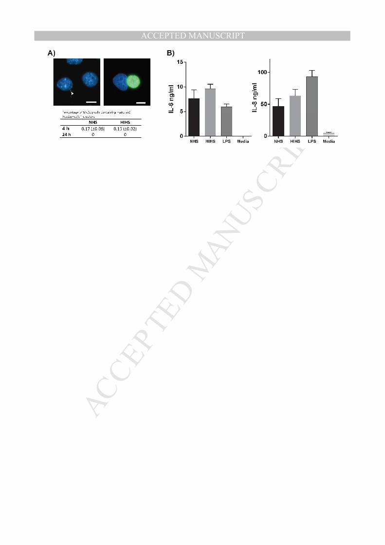
Fig. 5. Functional consequences of complement-mediated uptake of C. trachomatis into 687
monocytes. A) Intracellular survival of C. trachomatis L2 in monocytes. Monocytes were 688
incubated with C. trachomatis L2 for 4 or 24 hours in media containing either NHS or HIHS 689
and subsequently lysed by ultrasonication. Monocyte lysates were added to confluent McCoy 690
cells for one hour and McCoy cells were incubated for additional 23 hours. Chlamydial 691
inclusions were identified by immunofluorescence staining against MOMP. Left image: 692

MANUSCRIP
T
ACCEPTED
ACCEPTED MANUSCRIPT29
McCoy cells with EB that had not developed to an inclusion (arrowhead). Right image: 693
McCoy cell with an inclusion (green) from monocytes incubated in HIHS. Table: Mean 694
percentage (± SEM) of McCoy cells containing mature inclusions quantified from duplicate 695
samples from three biological independent experiments. Scalebars indicate 10 µm. 696
B) IL-6 and IL-8 concentrations in media from monocytes cultured with C. trachomatis L2. 697
Monocytes were incubated with C. trachomatis L2 for 4 hours in media supplemented with 698
either NHS or HIHS. After 4 hours, extracellular bacteria were removed and the monocytes 699
were incubated for further 20 hours. The culture supernatants were harvested and used for 700
ELISA. Standard medium and standard medium supplemented with 1 µg/ml LPS were used 701
as negative and positive controls, respectively. No statistically significant differences were 702
observed between and NHS and HIHS groups. Each condition was analyzed in triplicates and 703
three biologically independent experiments were performed. Data are presented as means ± 704
SEM. 705
706

MANUSCRIP
T
ACCEPTED
ACCEPTED MANUSCRIPT

MANUSCRIP
T
ACCEPTED
ACCEPTED MANUSCRIPT

MANUSCRIP
T
ACCEPTED
ACCEPTED MANUSCRIPT

MANUSCRIP
T
ACCEPTED
ACCEPTED MANUSCRIPT
MANUSCRIP
T
ACCEPTED
ACCEPTED MANUSCRIPT