Embed Size (px)
Citation preview

Comparative Bioinformatic Analysis of Proteome and Transcriptome Derived from a Single Cell TypeMartin Damsbo, Jacob Poder, Erik Nielsen, Christian Ravnsborg, and Alexandre Podtelejnikov
Thermo Fisher Scientific, Odense, Denmark
2 Comparative Bioinformatic Analysis of Proteome and Transcriptome Derived from a Single Cell Type
Conclusion We demonstrated the comparison of proteome and transcriptome data followed
by gene ontology, over-represented analysis, pathways analysis, PPI networks analysis and overall statistics
The state-of-art mass spectrometric technique overcomes the major limitation of peptide-centric proteomics, which is biased towards more-abundant peptides. The data show that the detected proteome is closed to almost the entire functional transcriptome and covers a very large part of functional pathways.
Due to the high proteome coverage and high quality of data, the current study can be used as a reference proteome data set similar to HUPO brain, liver and plasma studies.
References1. Nagaraj, N.; Wisniewski, J.R.; Geiger, T.; Cox, J.; Kircher, M.; Kelso, J.; Pääbo,
S.; Mann, M. Deep proteome and transcriptome mapping of a human cancer cell line. Mol Syst Biol. 2011, Nov 8;7:548.
2. Schwanhausser, B.; Busse, D.; Li, N.; Dittmar, G.; Schuchhardt, J.; Wolf, J.; Chen, W.; Selbach, M. Global quantification of mammalian gene expression control. Nature 2011, 473, 337–342.
3. Cline, M.S., et al. Integration of biological networks and gene expression data using Cytoscape. Nat Protoc. 2007, 2(10), 2366-82.
4. de Sousa Abreu, R.; Penalva, L.O.; Marcotte, E.M.; Vogel, C. Global signaturesof protein and mRNA expression levels. Mol Biosyst. 2009 5(12):1512-26.
OverviewPurpose: Demonstration of fast and effective way to perform statistical, comparative and bioinformatic analysis of large scale proteome and transcriptome studies
Methods: Set of bioinformatic data-mining tools
Results: Comprehensive bioinformatic analysis of data derived from large-scale LC-MS/MS proteomics study of HeLa cells and comparison to transcriptome data
IntroductionWith the current development of mass spectrometry-based proteomics techniques, it is becoming possible to perform in-depth analyses of the entire proteome. With protein identifications of more than ten thousand proteins, it is essential to be able to perform sophisticated bioinformatic analyses of such datasets and compare to gene expression data. Here we present such a comparison for HeLa cell analysis and a new development in web-based Thermo Scientific ProteinCenter software—an integration with Cytoscape®—that enables visualization of molecular interaction networks.
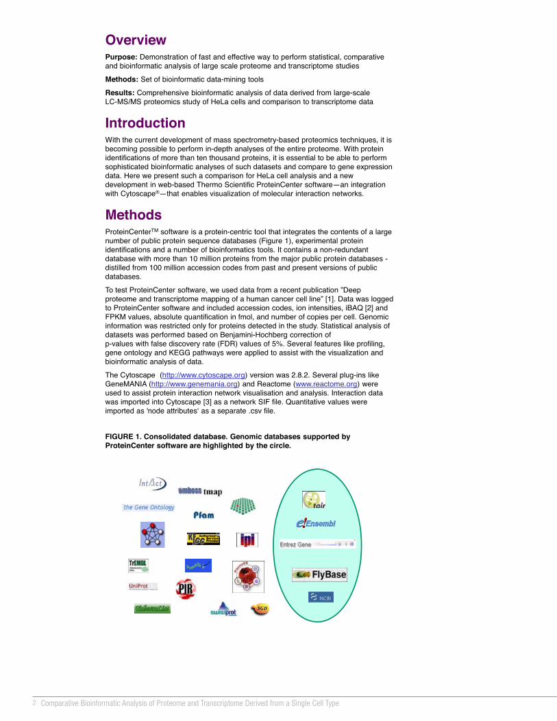
MethodsProteinCenterTM software is a protein-centric tool that integrates the contents of a large number of public protein sequence databases (Figure 1), experimental protein identifications and a number of bioinformatics tools. It contains a non-redundant database with more than 10 million proteins from the major public protein databases -distilled from 100 million accession codes from past and present versions of public databases.
To test ProteinCenter software, we used data from a recent publication ”Deep proteome and transcriptome mapping of a human cancer cell line” [1]. Data was logged to ProteinCenter software and included accession codes, ion intensities, iBAQ [2] and FPKM values, absolute quantification in fmol, and number of copies per cell. Genomic information was restricted only for proteins detected in the study. Statistical analysis of datasets was performed based on Benjamini-Hochberg correction of p-values with false discovery rate (FDR) values of 5%. Several features like profiling, gene ontology and KEGG pathways were applied to assist with the visualization and bioinformatic analysis of data.
The Cytoscape (http://www.cytoscape.org) version was 2.8.2. Several plug-ins like GeneMANIA (http://www.genemania.org) and Reactome (www.reactome.org) were used to assist protein interaction network visualisation and analysis. Interaction data was imported into Cytoscape [3] as a network SIF file. Quantitative values were imported as 'node attributesʻ as a separate .csv file.
FIGURE 1. Consolidated database. Genomic databases supported by ProteinCenter software are highlighted by the circle.
FIGURE 2. General statistics
ResultsStatistical Analysis of HeLa Proteome
Very often proteomics and transcriptomics experiments are performed in different laboratories at different times and using different experiment set-ups. As a result, the degree of correlation between the protein and mRNA levels can be difficult to interpret. While ProteinCenter software removes the obstacles of database entry dynamics, the values observed during these experiments can introduce a level of uncertainty. In the best case scenario, both experiments are performed at one place in the same experimental set-up. That is why we selected the very well controlled study of HeLa cells. A total of 10,255 proteins with 99% confidence were identified; the average identification of the fragmentation spectra was 43%. The average sequence coverage was close to 39%. Currently it is the most exhaustive proteome analysis of human single cell type and we consider to use it as a reference dataset.
Statistical analysis of transcriptome and proteome based on gene ontology, signal peptides, transmembrane regions did not reveal significant differences and was in close proximity to statistical analysis of reference databases, IPI and Swiss-Prot®(Figure 2). This can be explained by high proteome coverage; high mass accuracy mass spectrometry based analysis with a FASP purification method covers nearly the entire functional proteome including membrane proteins. That is why we focused on analysis of pathways in the study.
In total, 242 pathways were detected out of 251 available from KEGG for all human proteins. Among under-represented pathways were asthma, autoimmune thyroid disease, allograft rejection, olfactory transduction and others. All of them are not expected to be functionally relevant in HeLa cells. At the same time, some of the over-represented pathways (Figure 3) like nucleotide excision repair (Figure 4) have more than 92% coverage. All in all, average pathway coverage is around 46% and suggests that the detected proteome covers a very large part of functional pathways.
FIGURE 3. Over-representation analysis of KEGG pathways using Swiss-Prot as a reference database
FIGURE 6. Gene Ontology Biological Processes analysis of low abundant proteins
FIGURE 8. Mitochondrial ribosomal interactome analysis by GeneMANIA
Comparative Bioinformatic Analysis of HeLa Proteome and Transcriptome
Historically, mRNA and protein levels correlate poorly partially because of the inability to sequence significant number of proteins. Until recently, the proteomics dataset from a single LC/MS experiment was in the range of several hundreds of proteins identified. With the improvement of proteomics techniques, like sub-ppm mass accuracy and attomole sensitivity of modern hybrid mass spectrometers, it is becoming possible to achieve identification levels up to ten thousand proteins as it was shown in the selected study of HeLa cells.
A single gene can give rise to multiple distinct proteins due to alternative splicing, sequence polymorphisms and post-translational modifications. As shown in Figure 5, the numbers of protein counts in a set of particular KEGG pathways were higher than the numbers of genes. This can be explained by the different protein isoforms detected in the study.
Recent developments in labelling technology have provided useful techniques for the quantitative study of protein expression. The quantitative values FPKM (fragments per kilobase of exon per million fragments mapped) and absolute protein quantification by intensity-based absolute quantification (iBAQ) approach were used for comparison of HeLa proteome and transciptome. iBAQ protein intensities were translated to protein copy number estimates based on the overall protein amount in the analyzed sample. While most publications found that the overall correlation between quantified mRNA and protein abundances is modest or poor [4], the current study showed relatively good correlation. Partially this can be explained as the datasets were generated from the same set of cells under the same experimental conditions.
Cellular protein abundances span 8 orders of magnitude from approximately 0.2 copy per cell for Centriolin up to 3.3E+7 for Cofilin-1. Highly abundant proteins (top 100) are mainly of cytoplasmic origin and involved in cell development, response to chemical stimulus and cell death. On the level of molecular function they are involved in ATP binding, structural molecule activity, catalytic activity.
FIGURE 5 Proteome versus transcriptome
FIGURE 4. Visualisation of nucleotide excision repair KEGG pathway. (Proteins highlighted in red were detected in the proteome analysis.)
FIGURE 7. Heat map generated from quantitative values of proteome and transcriptome comparison
A heat map can assist the visualization of the quantitative data. Figure 7 shows a comparison of FPKM (AQR5) and iBAQ values for trypsin, LysC and GluC digestions (GD1,GD2 and GD3 correspondingly) for a fraction of a 60S large ribosomal subunit that were summarized on the level of pathway analysis.
While the knowledge of over-represented GO terms or pathways is helpful, it does not always explain the molecular mechanism relevant to the explored proteins and investigation of protein–protein interaction (PPI) networks can be useful. To complete the bioinformatic analysis of the data set we performed PPI analysis using Cytoscape graph algorithms and the VizMapper visualization tool, where ProteinCenter software data were used to colour-code nodes according to the protein quantitative values. Integration of ProteinCenter software with Cytoscape also allows access to a significant number of plug-ins and provides exhaustive analysis of interactome. For example, Figure 8 shows the interactome of mitochondrial ribosomal proteins on the level of physical interactions, prediction and co-localizations. All of these interactions are well curated and have the links to the corresponding publications. Nodes highlighted in grey were detected in the study.
FIGURE 9. Visualization of cancer related genes by Reactome FI
In-depth proteome coverage opens an opportunity to link pathway profiles to disease phenotypes. An excellent tool for such an analysis is Reactome FI plug-in for Cytoscape. Figure 9 shows the fragment of network where the genes highlighted by yellow reflect the association with a cervical carcinoma.
Comparative Bioinformatic Analysis of Proteome and Transcriptome Derived from a Single Cell TypeMartin Damsbo, Jacob Poder, Erik Nielsen, Christian Ravnsborg, Alexandre PodtelejnikovThermo Fisher Scientific, Odense, Denmark
Bioinformatic Analysis of Low Abundant Proteins
We also extracted the list of proteins with the lowest iBAQ values to create a small dataset of low-abundance proteins. These proteins were expressed on the level up to 50 copies per cell in the range of 0.2 to 40 attomoles. Among these proteins 54 have at least one transmembrane domain, 20 contain signal peptide signature and more than 60% of their sequences contain alternative spliced variants. To characterize that pool of proteins we performed the Gene Ontology analysis on three levels: Molecular Function (GO MF), Cellular Components (GO CC), and Biological Processes (GO BP). GO MF showed serine-type exopeptidase activity, GTPase binding, and transmembrane receptor protein serine/threonine kinase activity. Among GO CC were gap junction, Golgi-associated vesicle and ruffle. GO BP analysis showed that these proteins are heavily involved in regulation of signal transduction (Figure 6).
Cytoscape is a registered trademark of The Cytoscape Consortium. Swiss-Prot is a registered trademark of Institut Suisse de Bioinformatique (Sib) Foundation Switzerland. All other trademarks are the property of Thermo Fisher Scientific and its subsidiaries.
This information is not intended to encourage use of these products in any manners that might infringe the intellectual property rights of others.

3Thermo Scientific Poster Note • PN63605_E 06/12S
Conclusion We demonstrated the comparison of proteome and transcriptome data followed
by gene ontology, over-represented analysis, pathways analysis, PPI networks analysis and overall statistics
The state-of-art mass spectrometric technique overcomes the major limitation of peptide-centric proteomics, which is biased towards more-abundant peptides. The data show that the detected proteome is closed to almost the entire functional transcriptome and covers a very large part of functional pathways.
Due to the high proteome coverage and high quality of data, the current study can be used as a reference proteome data set similar to HUPO brain, liver and plasma studies.
References1. Nagaraj, N.; Wisniewski, J.R.; Geiger, T.; Cox, J.; Kircher, M.; Kelso, J.; Pääbo,
S.; Mann, M. Deep proteome and transcriptome mapping of a human cancer cell line. Mol Syst Biol. 2011, Nov 8;7:548.
2. Schwanhausser, B.; Busse, D.; Li, N.; Dittmar, G.; Schuchhardt, J.; Wolf, J.; Chen, W.; Selbach, M. Global quantification of mammalian gene expression control. Nature 2011, 473, 337–342.
3. Cline, M.S., et al. Integration of biological networks and gene expression data using Cytoscape. Nat Protoc. 2007, 2(10), 2366-82.
4. de Sousa Abreu, R.; Penalva, L.O.; Marcotte, E.M.; Vogel, C. Global signaturesof protein and mRNA expression levels. Mol Biosyst. 2009 5(12):1512-26.
OverviewPurpose: Demonstration of fast and effective way to perform statistical, comparative and bioinformatic analysis of large scale proteome and transcriptome studies
Methods: Set of bioinformatic data-mining tools
Results: Comprehensive bioinformatic analysis of data derived from large-scale LC-MS/MS proteomics study of HeLa cells and comparison to transcriptome data
IntroductionWith the current development of mass spectrometry-based proteomics techniques, it is becoming possible to perform in-depth analyses of the entire proteome. With protein identifications of more than ten thousand proteins, it is essential to be able to perform sophisticated bioinformatic analyses of such datasets and compare to gene expression data. Here we present such a comparison for HeLa cell analysis and a new development in web-based Thermo Scientific ProteinCenter software—an integration with Cytoscape®—that enables visualization of molecular interaction networks.
MethodsProteinCenterTM software is a protein-centric tool that integrates the contents of a large number of public protein sequence databases (Figure 1), experimental protein identifications and a number of bioinformatics tools. It contains a non-redundant database with more than 10 million proteins from the major public protein databases -distilled from 100 million accession codes from past and present versions of public databases.
To test ProteinCenter software, we used data from a recent publication ”Deep proteome and transcriptome mapping of a human cancer cell line” [1]. Data was logged to ProteinCenter software and included accession codes, ion intensities, iBAQ [2] and FPKM values, absolute quantification in fmol, and number of copies per cell. Genomic information was restricted only for proteins detected in the study. Statistical analysis of datasets was performed based on Benjamini-Hochberg correction of p-values with false discovery rate (FDR) values of 5%. Several features like profiling, gene ontology and KEGG pathways were applied to assist with the visualization and bioinformatic analysis of data.
The Cytoscape (http://www.cytoscape.org) version was 2.8.2. Several plug-ins like GeneMANIA (http://www.genemania.org) and Reactome (www.reactome.org) were used to assist protein interaction network visualisation and analysis. Interaction data was imported into Cytoscape [3] as a network SIF file. Quantitative values were imported as 'node attributesʻ as a separate .csv file.
FIGURE 1. Consolidated database. Genomic databases supported by ProteinCenter software are highlighted by the circle.
FIGURE 2. General statistics
ResultsStatistical Analysis of HeLa Proteome
Very often proteomics and transcriptomics experiments are performed in different laboratories at different times and using different experiment set-ups. As a result, the degree of correlation between the protein and mRNA levels can be difficult to interpret. While ProteinCenter software removes the obstacles of database entry dynamics, the values observed during these experiments can introduce a level of uncertainty. In the best case scenario, both experiments are performed at one place in the same experimental set-up. That is why we selected the very well controlled study of HeLa cells. A total of 10,255 proteins with 99% confidence were identified; the average identification of the fragmentation spectra was 43%. The average sequence coverage was close to 39%. Currently it is the most exhaustive proteome analysis of human single cell type and we consider to use it as a reference dataset.
Statistical analysis of transcriptome and proteome based on gene ontology, signal peptides, transmembrane regions did not reveal significant differences and was in close proximity to statistical analysis of reference databases, IPI and Swiss-Prot®(Figure 2). This can be explained by high proteome coverage; high mass accuracy mass spectrometry based analysis with a FASP purification method covers nearly the entire functional proteome including membrane proteins. That is why we focused on analysis of pathways in the study.
In total, 242 pathways were detected out of 251 available from KEGG for all human proteins. Among under-represented pathways were asthma, autoimmune thyroid disease, allograft rejection, olfactory transduction and others. All of them are not expected to be functionally relevant in HeLa cells. At the same time, some of the over-represented pathways (Figure 3) like nucleotide excision repair (Figure 4) have more than 92% coverage. All in all, average pathway coverage is around 46% and suggests that the detected proteome covers a very large part of functional pathways.
FIGURE 3. Over-representation analysis of KEGG pathways using Swiss-Prot as a reference database
FIGURE 6. Gene Ontology Biological Processes analysis of low abundant proteins
FIGURE 8. Mitochondrial ribosomal interactome analysis by GeneMANIA
Comparative Bioinformatic Analysis of HeLa Proteome and Transcriptome
Historically, mRNA and protein levels correlate poorly partially because of the inability to sequence significant number of proteins. Until recently, the proteomics dataset from a single LC/MS experiment was in the range of several hundreds of proteins identified. With the improvement of proteomics techniques, like sub-ppm mass accuracy and attomole sensitivity of modern hybrid mass spectrometers, it is becoming possible to achieve identification levels up to ten thousand proteins as it was shown in the selected study of HeLa cells.
A single gene can give rise to multiple distinct proteins due to alternative splicing, sequence polymorphisms and post-translational modifications. As shown in Figure 5, the numbers of protein counts in a set of particular KEGG pathways were higher than the numbers of genes. This can be explained by the different protein isoforms detected in the study.
Recent developments in labelling technology have provided useful techniques for the quantitative study of protein expression. The quantitative values FPKM (fragments per kilobase of exon per million fragments mapped) and absolute protein quantification by intensity-based absolute quantification (iBAQ) approach were used for comparison of HeLa proteome and transciptome. iBAQ protein intensities were translated to protein copy number estimates based on the overall protein amount in the analyzed sample. While most publications found that the overall correlation between quantified mRNA and protein abundances is modest or poor [4], the current study showed relatively good correlation. Partially this can be explained as the datasets were generated from the same set of cells under the same experimental conditions.
Cellular protein abundances span 8 orders of magnitude from approximately 0.2 copy per cell for Centriolin up to 3.3E+7 for Cofilin-1. Highly abundant proteins (top 100) are mainly of cytoplasmic origin and involved in cell development, response to chemical stimulus and cell death. On the level of molecular function they are involved in ATP binding, structural molecule activity, catalytic activity.
FIGURE 5 Proteome versus transcriptome
FIGURE 4. Visualisation of nucleotide excision repair KEGG pathway. (Proteins highlighted in red were detected in the proteome analysis.)
FIGURE 7. Heat map generated from quantitative values of proteome and transcriptome comparison
A heat map can assist the visualization of the quantitative data. Figure 7 shows a comparison of FPKM (AQR5) and iBAQ values for trypsin, LysC and GluC digestions (GD1,GD2 and GD3 correspondingly) for a fraction of a 60S large ribosomal subunit that were summarized on the level of pathway analysis.
While the knowledge of over-represented GO terms or pathways is helpful, it does not always explain the molecular mechanism relevant to the explored proteins and investigation of protein–protein interaction (PPI) networks can be useful. To complete the bioinformatic analysis of the data set we performed PPI analysis using Cytoscape graph algorithms and the VizMapper visualization tool, where ProteinCenter software data were used to colour-code nodes according to the protein quantitative values. Integration of ProteinCenter software with Cytoscape also allows access to a significant number of plug-ins and provides exhaustive analysis of interactome. For example, Figure 8 shows the interactome of mitochondrial ribosomal proteins on the level of physical interactions, prediction and co-localizations. All of these interactions are well curated and have the links to the corresponding publications. Nodes highlighted in grey were detected in the study.
FIGURE 9. Visualization of cancer related genes by Reactome FI
In-depth proteome coverage opens an opportunity to link pathway profiles to disease phenotypes. An excellent tool for such an analysis is Reactome FI plug-in for Cytoscape. Figure 9 shows the fragment of network where the genes highlighted by yellow reflect the association with a cervical carcinoma.
Comparative Bioinformatic Analysis of Proteome and Transcriptome Derived from a Single Cell TypeMartin Damsbo, Jacob Poder, Erik Nielsen, Christian Ravnsborg, Alexandre PodtelejnikovThermo Fisher Scientific, Odense, Denmark
Bioinformatic Analysis of Low Abundant Proteins
We also extracted the list of proteins with the lowest iBAQ values to create a small dataset of low-abundance proteins. These proteins were expressed on the level up to 50 copies per cell in the range of 0.2 to 40 attomoles. Among these proteins 54 have at least one transmembrane domain, 20 contain signal peptide signature and more than 60% of their sequences contain alternative spliced variants. To characterize that pool of proteins we performed the Gene Ontology analysis on three levels: Molecular Function (GO MF), Cellular Components (GO CC), and Biological Processes (GO BP). GO MF showed serine-type exopeptidase activity, GTPase binding, and transmembrane receptor protein serine/threonine kinase activity. Among GO CC were gap junction, Golgi-associated vesicle and ruffle. GO BP analysis showed that these proteins are heavily involved in regulation of signal transduction (Figure 6).
Cytoscape is a registered trademark of The Cytoscape Consortium. Swiss-Prot is a registered trademark of Institut Suisse de Bioinformatique (Sib) Foundation Switzerland. All other trademarks are the property of Thermo Fisher Scientific and its subsidiaries.
This information is not intended to encourage use of these products in any manners that might infringe the intellectual property rights of others.

4 Comparative Bioinformatic Analysis of Proteome and Transcriptome Derived from a Single Cell Type
Conclusion We demonstrated the comparison of proteome and transcriptome data followed
by gene ontology, over-represented analysis, pathways analysis, PPI networks analysis and overall statistics
The state-of-art mass spectrometric technique overcomes the major limitation of peptide-centric proteomics, which is biased towards more-abundant peptides. The data show that the detected proteome is closed to almost the entire functional transcriptome and covers a very large part of functional pathways.
Due to the high proteome coverage and high quality of data, the current study can be used as a reference proteome data set similar to HUPO brain, liver and plasma studies.
References1. Nagaraj, N.; Wisniewski, J.R.; Geiger, T.; Cox, J.; Kircher, M.; Kelso, J.; Pääbo,
S.; Mann, M. Deep proteome and transcriptome mapping of a human cancer cell line. Mol Syst Biol. 2011, Nov 8;7:548.
2. Schwanhausser, B.; Busse, D.; Li, N.; Dittmar, G.; Schuchhardt, J.; Wolf, J.; Chen, W.; Selbach, M. Global quantification of mammalian gene expression control. Nature 2011, 473, 337–342.
3. Cline, M.S., et al. Integration of biological networks and gene expression data using Cytoscape. Nat Protoc. 2007, 2(10), 2366-82.
4. de Sousa Abreu, R.; Penalva, L.O.; Marcotte, E.M.; Vogel, C. Global signaturesof protein and mRNA expression levels. Mol Biosyst. 2009 5(12):1512-26.
OverviewPurpose: Demonstration of fast and effective way to perform statistical, comparative and bioinformatic analysis of large scale proteome and transcriptome studies
Methods: Set of bioinformatic data-mining tools
Results: Comprehensive bioinformatic analysis of data derived from large-scale LC-MS/MS proteomics study of HeLa cells and comparison to transcriptome data
IntroductionWith the current development of mass spectrometry-based proteomics techniques, it is becoming possible to perform in-depth analyses of the entire proteome. With protein identifications of more than ten thousand proteins, it is essential to be able to perform sophisticated bioinformatic analyses of such datasets and compare to gene expression data. Here we present such a comparison for HeLa cell analysis and a new development in web-based Thermo Scientific ProteinCenter software—an integration with Cytoscape®—that enables visualization of molecular interaction networks.
MethodsProteinCenterTM software is a protein-centric tool that integrates the contents of a large number of public protein sequence databases (Figure 1), experimental protein identifications and a number of bioinformatics tools. It contains a non-redundant database with more than 10 million proteins from the major public protein databases -distilled from 100 million accession codes from past and present versions of public databases.
To test ProteinCenter software, we used data from a recent publication ”Deep proteome and transcriptome mapping of a human cancer cell line” [1]. Data was logged to ProteinCenter software and included accession codes, ion intensities, iBAQ [2] and FPKM values, absolute quantification in fmol, and number of copies per cell. Genomic information was restricted only for proteins detected in the study. Statistical analysis of datasets was performed based on Benjamini-Hochberg correction of p-values with false discovery rate (FDR) values of 5%. Several features like profiling, gene ontology and KEGG pathways were applied to assist with the visualization and bioinformatic analysis of data.
The Cytoscape (http://www.cytoscape.org) version was 2.8.2. Several plug-ins like GeneMANIA (http://www.genemania.org) and Reactome (www.reactome.org) were used to assist protein interaction network visualisation and analysis. Interaction data was imported into Cytoscape [3] as a network SIF file. Quantitative values were imported as 'node attributesʻ as a separate .csv file.
FIGURE 1. Consolidated database. Genomic databases supported by ProteinCenter software are highlighted by the circle.
FIGURE 2. General statistics
ResultsStatistical Analysis of HeLa Proteome
Very often proteomics and transcriptomics experiments are performed in different laboratories at different times and using different experiment set-ups. As a result, the degree of correlation between the protein and mRNA levels can be difficult to interpret. While ProteinCenter software removes the obstacles of database entry dynamics, the values observed during these experiments can introduce a level of uncertainty. In the best case scenario, both experiments are performed at one place in the same experimental set-up. That is why we selected the very well controlled study of HeLa cells. A total of 10,255 proteins with 99% confidence were identified; the average identification of the fragmentation spectra was 43%. The average sequence coverage was close to 39%. Currently it is the most exhaustive proteome analysis of human single cell type and we consider to use it as a reference dataset.
Statistical analysis of transcriptome and proteome based on gene ontology, signal peptides, transmembrane regions did not reveal significant differences and was in close proximity to statistical analysis of reference databases, IPI and Swiss-Prot®(Figure 2). This can be explained by high proteome coverage; high mass accuracy mass spectrometry based analysis with a FASP purification method covers nearly the entire functional proteome including membrane proteins. That is why we focused on analysis of pathways in the study.
In total, 242 pathways were detected out of 251 available from KEGG for all human proteins. Among under-represented pathways were asthma, autoimmune thyroid disease, allograft rejection, olfactory transduction and others. All of them are not expected to be functionally relevant in HeLa cells. At the same time, some of the over-represented pathways (Figure 3) like nucleotide excision repair (Figure 4) have more than 92% coverage. All in all, average pathway coverage is around 46% and suggests that the detected proteome covers a very large part of functional pathways.
FIGURE 3. Over-representation analysis of KEGG pathways using Swiss-Prot as a reference database
FIGURE 6. Gene Ontology Biological Processes analysis of low abundant proteins
FIGURE 8. Mitochondrial ribosomal interactome analysis by GeneMANIA
Comparative Bioinformatic Analysis of HeLa Proteome and Transcriptome
Historically, mRNA and protein levels correlate poorly partially because of the inability to sequence significant number of proteins. Until recently, the proteomics dataset from a single LC/MS experiment was in the range of several hundreds of proteins identified. With the improvement of proteomics techniques, like sub-ppm mass accuracy and attomole sensitivity of modern hybrid mass spectrometers, it is becoming possible to achieve identification levels up to ten thousand proteins as it was shown in the selected study of HeLa cells.
A single gene can give rise to multiple distinct proteins due to alternative splicing, sequence polymorphisms and post-translational modifications. As shown in Figure 5, the numbers of protein counts in a set of particular KEGG pathways were higher than the numbers of genes. This can be explained by the different protein isoforms detected in the study.
Recent developments in labelling technology have provided useful techniques for the quantitative study of protein expression. The quantitative values FPKM (fragments per kilobase of exon per million fragments mapped) and absolute protein quantification by intensity-based absolute quantification (iBAQ) approach were used for comparison of HeLa proteome and transciptome. iBAQ protein intensities were translated to protein copy number estimates based on the overall protein amount in the analyzed sample. While most publications found that the overall correlation between quantified mRNA and protein abundances is modest or poor [4], the current study showed relatively good correlation. Partially this can be explained as the datasets were generated from the same set of cells under the same experimental conditions.
Cellular protein abundances span 8 orders of magnitude from approximately 0.2 copy per cell for Centriolin up to 3.3E+7 for Cofilin-1. Highly abundant proteins (top 100) are mainly of cytoplasmic origin and involved in cell development, response to chemical stimulus and cell death. On the level of molecular function they are involved in ATP binding, structural molecule activity, catalytic activity.
FIGURE 5 Proteome versus transcriptome
FIGURE 4. Visualisation of nucleotide excision repair KEGG pathway. (Proteins highlighted in red were detected in the proteome analysis.)
FIGURE 7. Heat map generated from quantitative values of proteome and transcriptome comparison
A heat map can assist the visualization of the quantitative data. Figure 7 shows a comparison of FPKM (AQR5) and iBAQ values for trypsin, LysC and GluC digestions (GD1,GD2 and GD3 correspondingly) for a fraction of a 60S large ribosomal subunit that were summarized on the level of pathway analysis.
While the knowledge of over-represented GO terms or pathways is helpful, it does not always explain the molecular mechanism relevant to the explored proteins and investigation of protein–protein interaction (PPI) networks can be useful. To complete the bioinformatic analysis of the data set we performed PPI analysis using Cytoscape graph algorithms and the VizMapper visualization tool, where ProteinCenter software data were used to colour-code nodes according to the protein quantitative values. Integration of ProteinCenter software with Cytoscape also allows access to a significant number of plug-ins and provides exhaustive analysis of interactome. For example, Figure 8 shows the interactome of mitochondrial ribosomal proteins on the level of physical interactions, prediction and co-localizations. All of these interactions are well curated and have the links to the corresponding publications. Nodes highlighted in grey were detected in the study.
FIGURE 9. Visualization of cancer related genes by Reactome FI
In-depth proteome coverage opens an opportunity to link pathway profiles to disease phenotypes. An excellent tool for such an analysis is Reactome FI plug-in for Cytoscape. Figure 9 shows the fragment of network where the genes highlighted by yellow reflect the association with a cervical carcinoma.
Comparative Bioinformatic Analysis of Proteome and Transcriptome Derived from a Single Cell TypeMartin Damsbo, Jacob Poder, Erik Nielsen, Christian Ravnsborg, Alexandre PodtelejnikovThermo Fisher Scientific, Odense, Denmark
Bioinformatic Analysis of Low Abundant Proteins
We also extracted the list of proteins with the lowest iBAQ values to create a small dataset of low-abundance proteins. These proteins were expressed on the level up to 50 copies per cell in the range of 0.2 to 40 attomoles. Among these proteins 54 have at least one transmembrane domain, 20 contain signal peptide signature and more than 60% of their sequences contain alternative spliced variants. To characterize that pool of proteins we performed the Gene Ontology analysis on three levels: Molecular Function (GO MF), Cellular Components (GO CC), and Biological Processes (GO BP). GO MF showed serine-type exopeptidase activity, GTPase binding, and transmembrane receptor protein serine/threonine kinase activity. Among GO CC were gap junction, Golgi-associated vesicle and ruffle. GO BP analysis showed that these proteins are heavily involved in regulation of signal transduction (Figure 6).
Cytoscape is a registered trademark of The Cytoscape Consortium. Swiss-Prot is a registered trademark of Institut Suisse de Bioinformatique (Sib) Foundation Switzerland. All other trademarks are the property of Thermo Fisher Scientific and its subsidiaries.
This information is not intended to encourage use of these products in any manners that might infringe the intellectual property rights of others.

5Thermo Scientific Poster Note • PN63605_E 06/12S
Conclusion We demonstrated the comparison of proteome and transcriptome data followed
by gene ontology, over-represented analysis, pathways analysis, PPI networks analysis and overall statistics
The state-of-art mass spectrometric technique overcomes the major limitation of peptide-centric proteomics, which is biased towards more-abundant peptides. The data show that the detected proteome is closed to almost the entire functional transcriptome and covers a very large part of functional pathways.
Due to the high proteome coverage and high quality of data, the current study can be used as a reference proteome data set similar to HUPO brain, liver and plasma studies.
References1. Nagaraj, N.; Wisniewski, J.R.; Geiger, T.; Cox, J.; Kircher, M.; Kelso, J.; Pääbo,
S.; Mann, M. Deep proteome and transcriptome mapping of a human cancer cell line. Mol Syst Biol. 2011, Nov 8;7:548.
2. Schwanhausser, B.; Busse, D.; Li, N.; Dittmar, G.; Schuchhardt, J.; Wolf, J.; Chen, W.; Selbach, M. Global quantification of mammalian gene expression control. Nature 2011, 473, 337–342.
3. Cline, M.S., et al. Integration of biological networks and gene expression data using Cytoscape. Nat Protoc. 2007, 2(10), 2366-82.
4. de Sousa Abreu, R.; Penalva, L.O.; Marcotte, E.M.; Vogel, C. Global signaturesof protein and mRNA expression levels. Mol Biosyst. 2009 5(12):1512-26.
OverviewPurpose: Demonstration of fast and effective way to perform statistical, comparative and bioinformatic analysis of large scale proteome and transcriptome studies
Methods: Set of bioinformatic data-mining tools
Results: Comprehensive bioinformatic analysis of data derived from large-scale LC-MS/MS proteomics study of HeLa cells and comparison to transcriptome data
IntroductionWith the current development of mass spectrometry-based proteomics techniques, it is becoming possible to perform in-depth analyses of the entire proteome. With protein identifications of more than ten thousand proteins, it is essential to be able to perform sophisticated bioinformatic analyses of such datasets and compare to gene expression data. Here we present such a comparison for HeLa cell analysis and a new development in web-based Thermo Scientific ProteinCenter software—an integration with Cytoscape®—that enables visualization of molecular interaction networks.
MethodsProteinCenterTM software is a protein-centric tool that integrates the contents of a large number of public protein sequence databases (Figure 1), experimental protein identifications and a number of bioinformatics tools. It contains a non-redundant database with more than 10 million proteins from the major public protein databases -distilled from 100 million accession codes from past and present versions of public databases.
To test ProteinCenter software, we used data from a recent publication ”Deep proteome and transcriptome mapping of a human cancer cell line” [1]. Data was logged to ProteinCenter software and included accession codes, ion intensities, iBAQ [2] and FPKM values, absolute quantification in fmol, and number of copies per cell. Genomic information was restricted only for proteins detected in the study. Statistical analysis of datasets was performed based on Benjamini-Hochberg correction of p-values with false discovery rate (FDR) values of 5%. Several features like profiling, gene ontology and KEGG pathways were applied to assist with the visualization and bioinformatic analysis of data.
The Cytoscape (http://www.cytoscape.org) version was 2.8.2. Several plug-ins like GeneMANIA (http://www.genemania.org) and Reactome (www.reactome.org) were used to assist protein interaction network visualisation and analysis. Interaction data was imported into Cytoscape [3] as a network SIF file. Quantitative values were imported as 'node attributesʻ as a separate .csv file.
FIGURE 1. Consolidated database. Genomic databases supported by ProteinCenter software are highlighted by the circle.
FIGURE 2. General statistics
ResultsStatistical Analysis of HeLa Proteome
Very often proteomics and transcriptomics experiments are performed in different laboratories at different times and using different experiment set-ups. As a result, the degree of correlation between the protein and mRNA levels can be difficult to interpret. While ProteinCenter software removes the obstacles of database entry dynamics, the values observed during these experiments can introduce a level of uncertainty. In the best case scenario, both experiments are performed at one place in the same experimental set-up. That is why we selected the very well controlled study of HeLa cells. A total of 10,255 proteins with 99% confidence were identified; the average identification of the fragmentation spectra was 43%. The average sequence coverage was close to 39%. Currently it is the most exhaustive proteome analysis of human single cell type and we consider to use it as a reference dataset.
Statistical analysis of transcriptome and proteome based on gene ontology, signal peptides, transmembrane regions did not reveal significant differences and was in close proximity to statistical analysis of reference databases, IPI and Swiss-Prot®(Figure 2). This can be explained by high proteome coverage; high mass accuracy mass spectrometry based analysis with a FASP purification method covers nearly the entire functional proteome including membrane proteins. That is why we focused on analysis of pathways in the study.
In total, 242 pathways were detected out of 251 available from KEGG for all human proteins. Among under-represented pathways were asthma, autoimmune thyroid disease, allograft rejection, olfactory transduction and others. All of them are not expected to be functionally relevant in HeLa cells. At the same time, some of the over-represented pathways (Figure 3) like nucleotide excision repair (Figure 4) have more than 92% coverage. All in all, average pathway coverage is around 46% and suggests that the detected proteome covers a very large part of functional pathways.
FIGURE 3. Over-representation analysis of KEGG pathways using Swiss-Prot as a reference database
FIGURE 6. Gene Ontology Biological Processes analysis of low abundant proteins
FIGURE 8. Mitochondrial ribosomal interactome analysis by GeneMANIA
Comparative Bioinformatic Analysis of HeLa Proteome and Transcriptome
Historically, mRNA and protein levels correlate poorly partially because of the inability to sequence significant number of proteins. Until recently, the proteomics dataset from a single LC/MS experiment was in the range of several hundreds of proteins identified. With the improvement of proteomics techniques, like sub-ppm mass accuracy and attomole sensitivity of modern hybrid mass spectrometers, it is becoming possible to achieve identification levels up to ten thousand proteins as it was shown in the selected study of HeLa cells.
A single gene can give rise to multiple distinct proteins due to alternative splicing, sequence polymorphisms and post-translational modifications. As shown in Figure 5, the numbers of protein counts in a set of particular KEGG pathways were higher than the numbers of genes. This can be explained by the different protein isoforms detected in the study.
Recent developments in labelling technology have provided useful techniques for the quantitative study of protein expression. The quantitative values FPKM (fragments per kilobase of exon per million fragments mapped) and absolute protein quantification by intensity-based absolute quantification (iBAQ) approach were used for comparison of HeLa proteome and transciptome. iBAQ protein intensities were translated to protein copy number estimates based on the overall protein amount in the analyzed sample. While most publications found that the overall correlation between quantified mRNA and protein abundances is modest or poor [4], the current study showed relatively good correlation. Partially this can be explained as the datasets were generated from the same set of cells under the same experimental conditions.
Cellular protein abundances span 8 orders of magnitude from approximately 0.2 copy per cell for Centriolin up to 3.3E+7 for Cofilin-1. Highly abundant proteins (top 100) are mainly of cytoplasmic origin and involved in cell development, response to chemical stimulus and cell death. On the level of molecular function they are involved in ATP binding, structural molecule activity, catalytic activity.
FIGURE 5 Proteome versus transcriptome
FIGURE 4. Visualisation of nucleotide excision repair KEGG pathway. (Proteins highlighted in red were detected in the proteome analysis.)
FIGURE 7. Heat map generated from quantitative values of proteome and transcriptome comparison
A heat map can assist the visualization of the quantitative data. Figure 7 shows a comparison of FPKM (AQR5) and iBAQ values for trypsin, LysC and GluC digestions (GD1,GD2 and GD3 correspondingly) for a fraction of a 60S large ribosomal subunit that were summarized on the level of pathway analysis.
While the knowledge of over-represented GO terms or pathways is helpful, it does not always explain the molecular mechanism relevant to the explored proteins and investigation of protein–protein interaction (PPI) networks can be useful. To complete the bioinformatic analysis of the data set we performed PPI analysis using Cytoscape graph algorithms and the VizMapper visualization tool, where ProteinCenter software data were used to colour-code nodes according to the protein quantitative values. Integration of ProteinCenter software with Cytoscape also allows access to a significant number of plug-ins and provides exhaustive analysis of interactome. For example, Figure 8 shows the interactome of mitochondrial ribosomal proteins on the level of physical interactions, prediction and co-localizations. All of these interactions are well curated and have the links to the corresponding publications. Nodes highlighted in grey were detected in the study.
FIGURE 9. Visualization of cancer related genes by Reactome FI
In-depth proteome coverage opens an opportunity to link pathway profiles to disease phenotypes. An excellent tool for such an analysis is Reactome FI plug-in for Cytoscape. Figure 9 shows the fragment of network where the genes highlighted by yellow reflect the association with a cervical carcinoma.
Comparative Bioinformatic Analysis of Proteome and Transcriptome Derived from a Single Cell TypeMartin Damsbo, Jacob Poder, Erik Nielsen, Christian Ravnsborg, Alexandre PodtelejnikovThermo Fisher Scientific, Odense, Denmark
Bioinformatic Analysis of Low Abundant Proteins
We also extracted the list of proteins with the lowest iBAQ values to create a small dataset of low-abundance proteins. These proteins were expressed on the level up to 50 copies per cell in the range of 0.2 to 40 attomoles. Among these proteins 54 have at least one transmembrane domain, 20 contain signal peptide signature and more than 60% of their sequences contain alternative spliced variants. To characterize that pool of proteins we performed the Gene Ontology analysis on three levels: Molecular Function (GO MF), Cellular Components (GO CC), and Biological Processes (GO BP). GO MF showed serine-type exopeptidase activity, GTPase binding, and transmembrane receptor protein serine/threonine kinase activity. Among GO CC were gap junction, Golgi-associated vesicle and ruffle. GO BP analysis showed that these proteins are heavily involved in regulation of signal transduction (Figure 6).
Cytoscape is a registered trademark of The Cytoscape Consortium. Swiss-Prot is a registered trademark of Institut Suisse de Bioinformatique (Sib) Foundation Switzerland. All other trademarks are the property of Thermo Fisher Scientific and its subsidiaries.
This information is not intended to encourage use of these products in any manners that might infringe the intellectual property rights of others.

6 Comparative Bioinformatic Analysis of Proteome and Transcriptome Derived from a Single Cell Type
Conclusion We demonstrated the comparison of proteome and transcriptome data followed
by gene ontology, over-represented analysis, pathways analysis, PPI networks analysis and overall statistics
The state-of-art mass spectrometric technique overcomes the major limitation of peptide-centric proteomics, which is biased towards more-abundant peptides. The data show that the detected proteome is closed to almost the entire functional transcriptome and covers a very large part of functional pathways.
Due to the high proteome coverage and high quality of data, the current study can be used as a reference proteome data set similar to HUPO brain, liver and plasma studies.
References1. Nagaraj, N.; Wisniewski, J.R.; Geiger, T.; Cox, J.; Kircher, M.; Kelso, J.; Pääbo,
S.; Mann, M. Deep proteome and transcriptome mapping of a human cancer cell line. Mol Syst Biol. 2011, Nov 8;7:548.
2. Schwanhausser, B.; Busse, D.; Li, N.; Dittmar, G.; Schuchhardt, J.; Wolf, J.; Chen, W.; Selbach, M. Global quantification of mammalian gene expression control. Nature 2011, 473, 337–342.
3. Cline, M.S., et al. Integration of biological networks and gene expression data using Cytoscape. Nat Protoc. 2007, 2(10), 2366-82.
4. de Sousa Abreu, R.; Penalva, L.O.; Marcotte, E.M.; Vogel, C. Global signaturesof protein and mRNA expression levels. Mol Biosyst. 2009 5(12):1512-26.
OverviewPurpose: Demonstration of fast and effective way to perform statistical, comparative and bioinformatic analysis of large scale proteome and transcriptome studies
Methods: Set of bioinformatic data-mining tools
Results: Comprehensive bioinformatic analysis of data derived from large-scale LC-MS/MS proteomics study of HeLa cells and comparison to transcriptome data
IntroductionWith the current development of mass spectrometry-based proteomics techniques, it is becoming possible to perform in-depth analyses of the entire proteome. With protein identifications of more than ten thousand proteins, it is essential to be able to perform sophisticated bioinformatic analyses of such datasets and compare to gene expression data. Here we present such a comparison for HeLa cell analysis and a new development in web-based Thermo Scientific ProteinCenter software—an integration with Cytoscape®—that enables visualization of molecular interaction networks.
MethodsProteinCenterTM software is a protein-centric tool that integrates the contents of a large number of public protein sequence databases (Figure 1), experimental protein identifications and a number of bioinformatics tools. It contains a non-redundant database with more than 10 million proteins from the major public protein databases -distilled from 100 million accession codes from past and present versions of public databases.
To test ProteinCenter software, we used data from a recent publication ”Deep proteome and transcriptome mapping of a human cancer cell line” [1]. Data was logged to ProteinCenter software and included accession codes, ion intensities, iBAQ [2] and FPKM values, absolute quantification in fmol, and number of copies per cell. Genomic information was restricted only for proteins detected in the study. Statistical analysis of datasets was performed based on Benjamini-Hochberg correction of p-values with false discovery rate (FDR) values of 5%. Several features like profiling, gene ontology and KEGG pathways were applied to assist with the visualization and bioinformatic analysis of data.
The Cytoscape (http://www.cytoscape.org) version was 2.8.2. Several plug-ins like GeneMANIA (http://www.genemania.org) and Reactome (www.reactome.org) were used to assist protein interaction network visualisation and analysis. Interaction data was imported into Cytoscape [3] as a network SIF file. Quantitative values were imported as 'node attributesʻ as a separate .csv file.
FIGURE 1. Consolidated database. Genomic databases supported by ProteinCenter software are highlighted by the circle.
FIGURE 2. General statistics
ResultsStatistical Analysis of HeLa Proteome
Very often proteomics and transcriptomics experiments are performed in different laboratories at different times and using different experiment set-ups. As a result, the degree of correlation between the protein and mRNA levels can be difficult to interpret. While ProteinCenter software removes the obstacles of database entry dynamics, the values observed during these experiments can introduce a level of uncertainty. In the best case scenario, both experiments are performed at one place in the same experimental set-up. That is why we selected the very well controlled study of HeLa cells. A total of 10,255 proteins with 99% confidence were identified; the average identification of the fragmentation spectra was 43%. The average sequence coverage was close to 39%. Currently it is the most exhaustive proteome analysis of human single cell type and we consider to use it as a reference dataset.
Statistical analysis of transcriptome and proteome based on gene ontology, signal peptides, transmembrane regions did not reveal significant differences and was in close proximity to statistical analysis of reference databases, IPI and Swiss-Prot®(Figure 2). This can be explained by high proteome coverage; high mass accuracy mass spectrometry based analysis with a FASP purification method covers nearly the entire functional proteome including membrane proteins. That is why we focused on analysis of pathways in the study.
In total, 242 pathways were detected out of 251 available from KEGG for all human proteins. Among under-represented pathways were asthma, autoimmune thyroid disease, allograft rejection, olfactory transduction and others. All of them are not expected to be functionally relevant in HeLa cells. At the same time, some of the over-represented pathways (Figure 3) like nucleotide excision repair (Figure 4) have more than 92% coverage. All in all, average pathway coverage is around 46% and suggests that the detected proteome covers a very large part of functional pathways.
FIGURE 3. Over-representation analysis of KEGG pathways using Swiss-Prot as a reference database
FIGURE 6. Gene Ontology Biological Processes analysis of low abundant proteins
FIGURE 8. Mitochondrial ribosomal interactome analysis by GeneMANIA
Comparative Bioinformatic Analysis of HeLa Proteome and Transcriptome
Historically, mRNA and protein levels correlate poorly partially because of the inability to sequence significant number of proteins. Until recently, the proteomics dataset from a single LC/MS experiment was in the range of several hundreds of proteins identified. With the improvement of proteomics techniques, like sub-ppm mass accuracy and attomole sensitivity of modern hybrid mass spectrometers, it is becoming possible to achieve identification levels up to ten thousand proteins as it was shown in the selected study of HeLa cells.
A single gene can give rise to multiple distinct proteins due to alternative splicing, sequence polymorphisms and post-translational modifications. As shown in Figure 5, the numbers of protein counts in a set of particular KEGG pathways were higher than the numbers of genes. This can be explained by the different protein isoforms detected in the study.
Recent developments in labelling technology have provided useful techniques for the quantitative study of protein expression. The quantitative values FPKM (fragments per kilobase of exon per million fragments mapped) and absolute protein quantification by intensity-based absolute quantification (iBAQ) approach were used for comparison of HeLa proteome and transciptome. iBAQ protein intensities were translated to protein copy number estimates based on the overall protein amount in the analyzed sample. While most publications found that the overall correlation between quantified mRNA and protein abundances is modest or poor [4], the current study showed relatively good correlation. Partially this can be explained as the datasets were generated from the same set of cells under the same experimental conditions.
Cellular protein abundances span 8 orders of magnitude from approximately 0.2 copy per cell for Centriolin up to 3.3E+7 for Cofilin-1. Highly abundant proteins (top 100) are mainly of cytoplasmic origin and involved in cell development, response to chemical stimulus and cell death. On the level of molecular function they are involved in ATP binding, structural molecule activity, catalytic activity.
FIGURE 5 Proteome versus transcriptome
FIGURE 4. Visualisation of nucleotide excision repair KEGG pathway. (Proteins highlighted in red were detected in the proteome analysis.)
FIGURE 7. Heat map generated from quantitative values of proteome and transcriptome comparison
A heat map can assist the visualization of the quantitative data. Figure 7 shows a comparison of FPKM (AQR5) and iBAQ values for trypsin, LysC and GluC digestions (GD1,GD2 and GD3 correspondingly) for a fraction of a 60S large ribosomal subunit that were summarized on the level of pathway analysis.
While the knowledge of over-represented GO terms or pathways is helpful, it does not always explain the molecular mechanism relevant to the explored proteins and investigation of protein–protein interaction (PPI) networks can be useful. To complete the bioinformatic analysis of the data set we performed PPI analysis using Cytoscape graph algorithms and the VizMapper visualization tool, where ProteinCenter software data were used to colour-code nodes according to the protein quantitative values. Integration of ProteinCenter software with Cytoscape also allows access to a significant number of plug-ins and provides exhaustive analysis of interactome. For example, Figure 8 shows the interactome of mitochondrial ribosomal proteins on the level of physical interactions, prediction and co-localizations. All of these interactions are well curated and have the links to the corresponding publications. Nodes highlighted in grey were detected in the study.
FIGURE 9. Visualization of cancer related genes by Reactome FI
In-depth proteome coverage opens an opportunity to link pathway profiles to disease phenotypes. An excellent tool for such an analysis is Reactome FI plug-in for Cytoscape. Figure 9 shows the fragment of network where the genes highlighted by yellow reflect the association with a cervical carcinoma.
Comparative Bioinformatic Analysis of Proteome and Transcriptome Derived from a Single Cell TypeMartin Damsbo, Jacob Poder, Erik Nielsen, Christian Ravnsborg, Alexandre PodtelejnikovThermo Fisher Scientific, Odense, Denmark
Bioinformatic Analysis of Low Abundant Proteins
We also extracted the list of proteins with the lowest iBAQ values to create a small dataset of low-abundance proteins. These proteins were expressed on the level up to 50 copies per cell in the range of 0.2 to 40 attomoles. Among these proteins 54 have at least one transmembrane domain, 20 contain signal peptide signature and more than 60% of their sequences contain alternative spliced variants. To characterize that pool of proteins we performed the Gene Ontology analysis on three levels: Molecular Function (GO MF), Cellular Components (GO CC), and Biological Processes (GO BP). GO MF showed serine-type exopeptidase activity, GTPase binding, and transmembrane receptor protein serine/threonine kinase activity. Among GO CC were gap junction, Golgi-associated vesicle and ruffle. GO BP analysis showed that these proteins are heavily involved in regulation of signal transduction (Figure 6).
Cytoscape is a registered trademark of The Cytoscape Consortium. Swiss-Prot is a registered trademark of Institut Suisse de Bioinformatique (Sib) Foundation Switzerland. All other trademarks are the property of Thermo Fisher Scientific and its subsidiaries.
This information is not intended to encourage use of these products in any manners that might infringe the intellectual property rights of others.

Thermo Fisher Scientific, San Jose, CA USA is ISO Certified.
PN63605_E 06/12S
Africa-Other +27 11 570 1840Australia +61 3 9757 4300Austria +43 1 333 50 34 0Belgium +32 53 73 42 41Canada +1 800 530 8447China +86 10 8419 3588Denmark +45 70 23 62 60
Europe-Other +43 1 333 50 34 0Finland/Norway/Sweden +46 8 556 468 00France +33 1 60 92 48 00Germany +49 6103 408 1014India +91 22 6742 9434Italy +39 02 950 591
Japan +81 45 453 9100Latin America +1 561 688 8700Middle East +43 1 333 50 34 0Netherlands +31 76 579 55 55New Zealand +64 9 980 6700Russia/CIS +43 1 333 50 34 0South Africa +27 11 570 1840
Spain +34 914 845 965Switzerland +41 61 716 77 00UK +44 1442 233555USA +1 800 532 4752
www.thermoscientific.com©2012 Thermo Fisher Scientific Inc. All rights reserved. ISO is a trademark of the International Standards Organization. All other trademarks are the property of Thermo Fisher Scientific Inc. and its subsidiaries. This information is presented as an example of the capabilities of Thermo Fisher Scientific Inc. products. It is not intended to encourage use of these products in any manners that might infringe the intellectual property rights of others. Specifications, terms and pricing are subject to change. Not all products are available in all countries. Please consult your local sales representative for details.