Embed Size (px)
Citation preview
Comparative Biochemistry and Physiology, Part D 14 (2015) 16–25
Contents lists available at ScienceDirect
Comparative Biochemistry and Physiology, Part D
j ourna l homepage: www.e lsev ie r .com/ locate /cbpd
Identification and expression analysis of 26 oncogenes of the receptortyrosine kinase family in channel catfish after bacterial infection andhypoxic stress
Yi Tian a,b,1, Jun Yao a,1, Shikai Liu a,1, Chen Jiang a, Jiaren Zhang a, Yun Li a, Jianbin Feng a, Zhanjiang Liu a,⁎a FishMolecular Genetics and Biotechnology Laboratory, Aquatic Genomics Unit, School of Fisheries, Aquaculture andAquatic Sciences, and Programof Cell andMolecular Biosciences, AuburnUniversity,Auburn, AL 36849, USAb College of Fisheries and Life Science, Dalian Ocean University, Dalian 116023, China
⁎ Corresponding author. Tel.: +1 334 844 4784; fax: +E-mail address: [email protected] (Z. Liu).
1 These authors contributed equally.
http://dx.doi.org/10.1016/j.cbd.2015.02.0011744-117X/© 2015 Elsevier Inc. All rights reserved.
a b s t r a c t
a r t i c l e i n f oArticle history:Received 17 November 2014Received in revised form 9 February 2015Accepted 9 February 2015Available online 14 February 2015
Keywords:FishRTKsExpressionHypoxic stressDisease infection
Receptor tyrosine kinases (RTKs) are high-affinity cell surface receptors for many polypeptide growth factors,cytokines and hormones. RTKs are not only key regulators of normal cellular processes, but are also involvedin the progression of many types of tumors, and responses to various biotic and abiotic stresses. Catfish is aprimary aquaculture species in the United States, while its industry is drastically hindered by several major dis-eases including enteric septicemia of catfish (ESC) that is caused by Edwardsiella ictaluri. Disease outbreaks areoften accompanied by hypoxic stress, which affects the performance and survival of fish by reducing disease re-sistance. In this study, we identified 26 RTK oncogenes in the channel catfish genome, and determined their ex-pression profiles after ESC infection and hypoxic stress. The 26 RTK genes were divided into four subfamiliesaccording to phylogenetic analysis, including TIE (2 genes), ErbB (6 genes), EPH (14 genes), and INSR (4genes). All identified RTKs possess a similar molecular architecture including ligand-binding domains, a singletransmembrane helix and a cytoplasmic region, which suggests that these genes could play conserved biologicalroles. The expression analysis revealed that eight RTKs were significantly regulated after bacterial infection, withdramatic induction of insulin receptor genes including INSRb, IGF1Ra, and IGF1Rb. Upon hypoxic stress, EPHB3a,EGFR, ErbB4b, and IGF1Rbwere expressed at higher levels in the tolerant catfish, while EPHA2a, EPHA2, TIE1 andINSRa were expressed at higher levels in the intolerant catfish. These results suggested the involvement of RTKsin immune responses and hypoxic tolerance.
© 2015 Elsevier Inc. All rights reserved.
1. Introduction
Receptor tyrosine kinases (RTKs) are high-affinity cell surface recep-tors for many polypeptide growth factors, cytokines, and hormones(Schlessinger, 2000). The human RTK family contains 58 members thatare grouped into 20 subfamilies (Blume-Jensen and Hunter, 2001). Theoverall topology of RTKs, the mechanism of activation, and the keycomponents of the intracellular signaling pathways that they trigger,are evolutionarily conserved from nematodes to humans, suggestingthat they play critical regulatory roles (Lemmon and Schlessinger,2010). The RTK family includes a group of oncogenes and candidateproto-oncogenes, including epidermal growth factor receptor (EGFR/ErbB family), ephrin receptors (EPHs), angiopoietin receptors (TIEs)and insulin receptors (INSRs).
RTKs are activated by induced receptor dimerization through thebinding of their cognate ligands (Ullrich and Schlessinger, 1990).
1 334 844 9208.
Following activation, RTKs selectively form phosphotyrosine residuesby autocatalytic modifications. Trans-autophosphorylation then initi-ates further responses through cascades of posttranslational modifica-tions and generation of lipid second messengers. RTKs not only playkey roles in regulating normal cellular processes such as proliferationand differentiation, cell survival, cell migration, and cell-cycle control(Ullrich and Schlessinger, 1990; Blume-Jensen and Hunter, 2001), butalso play critical roles in regulating development and progressionofmany types of cancers. For instance, ErbB2 (human epidermal growthfactor receptor 2) is a known proto-oncogene. Approximately 20% ofbreast cancers exhibit ErbB2 gene amplification/overexpression,resulting in an aggressive tumor phenotype and reduced survival(Slamon et al., 1987, 1989). TIEs (TIE1 and TIE2) are involved in modu-lating cell–cell and cell–matrix interactions which are required forvascular remodeling and maturation (Pawson, 1995; Shewchuk et al.,2000). The cellular responses to EPH receptor stimulation by theirephrin ligands are important inmediating a wide range of biological ac-tivities, including angiogenesis, cell segregation, cell attachment, shape,and motility. Several EPH/ephrin molecules are expressed in vascularsystems, with the EPHB4 and its ligand EPHRINB2 being found as the
Table 1Identification of RTK genes in the channel catfish genome.
Gene name CDS (bp) Deduced protein CDS status Accession number
Length (aa) pI
EPHA2 2937 978 6.00 Complete JT339582EPHA2a 2958 985 6.37 Complete JT340542EPHA3 2916 971 6.97 Partial JT414026EPHA4 2970 989 6.84 Complete JT409635EPHA4a 2961 986 6.25 Complete JT474057EPHA4b 2937 978 6.16 Complete JT415792EPHA5 2472 823 7.92 Partial JT343803EPHB1 3048 1015 6.27 Complete JT410849EPHB2a 1877 625 6.06 Partial KP126799EPHB2b 2961 986 5.43 Complete JT408020EPHB3a 1452 483 5.63 Complete JT400475EPHB3 2946 981 5.74 Complete JT477737EPHB4b 2946 981 6.77 Complete JT416389EPHB4a 2982 993 7.47 Complete JT410332TIE2 3432 1143 7.38 Complete JT406054TIE1 2772 923 8.45 Partial JT464126INSRa 4026 1341 6.3 Complete JT478824INSRb 4029 1342 6.18 Complete JT407578IGF1RB 4074 1357 5.89 Complete JT410602IGF1RA 4149 1382 5.65 Complete JT410011EGFR 3600 1199 6.36 Complete JT406164ErbB3b 4257 1418 6.10 Complete JT414000ErbB3a 4335 1444 6.17 Complete JT407415ErbB4 2202 733 7.68 Partial JT402114ErbB4b 1857 618 6.71 Partial JT268945ErbB4a 1832 610 6.52 Partial KP126798
17Y. Tian et al. / Comparative Biochemistry and Physiology, Part D 14 (2015) 16–25
most significantly expressed genes (Hasina et al., 2013). EPH/ephrin sig-naling has been identified to play roles in many human cancers, such aslung cancer, breast cancer, and prostate cancer, as well as melanomaand leukemia (Kullander and Klein, 2002). Moreover, a number ofrecent studies provide an updated picture of INSR/IGF1R-mediatedsignaling events (Taniguchi et al., 2006; LeRoith and Accilli, 2008).INSR is a vital mediator of metabolic responses, whereas IGF1R isprimarily involved in mitogenesis, differentiation, and anti-apoptoticactivities. Cross-talk between insulin, IGFs, and their receptors is a com-mon event in many organs and processes. Hence, the role of INSR inmitogenesis and cell motility lays the foundation for its involvementin cancer development and progression (Belfiore et al., 2009).
A common phenomenon in human solid tumors and inflamedtissues is hypoxia. Hypoxia occurs when the balance between oxygensupply and demand is disturbed, which is an important feature at sitesof acute and chronic tissue inflammation under pathogen challenge,such aswound healing and tumor growth (Lund-Olesen, 1970). Clinicalstudies in humanhead andneck, soft tissue, and uterine cervical cancershave demonstrated that patientswith highly hypoxic tumors have a sig-nificantly higher risk of developing metastases and often have a poorerclinical outcome than patients with fewer or non-hypoxic tumors(Hockel et al., 1993, 1996; Brizel et al., 1996, 1997). The lack of oxygenin the inner core of tumors, primarily due to increased distance oftumor cells from blood vessels and aberrant formation of blood vesselsresulting in poor blood flow, is believed to contribute to tumor progres-sion, as well as resistance to chemotherapy and radiotherapy (Janssenet al., 2005). In tumors, irregular tumor vessel formation is the maincause of tumor hypoxia. Studies have shown that various hypoxia-induced proteins are capable of regulating tumor cell growth, autophagy,angiogenesis (e.g., vascular endothelial growth factor [VEGF]), metastasis(e.g., EGFR and c-MET), tumor cell metabolism (e.g., glucose transporter[GLUT1]), and pH (carbonic anhydrase IX [CA9]). These molecularchanges during hypoxic conditionsmay aid tumor cell adaptation to hyp-oxic stress and possibly lead to disease progression (Hong et al., 2013).
Teleost fish are natural model systems for the study of hypoxia be-cause oxygen availability is frequently altered in waters, both spatiallyand temporally. Life or death for fish depends on their ability to adaptto rapidly changing levels of oxygen in the aquatic environment. Indeed,much of the diversity of fishes can be attributed to the adoption of spe-cialized anatomic, behavioral, and physiological strategies to compen-sate for particular aquatic oxygen conditions (Powell and Hahn, 2002;Nikinmaa et al., 2004; Geng et al., 2014). Channel catfish, Ictaluruspunctatus, is highly tolerant to low oxygen. However, under intenseaquaculture conditions, hypoxia frequently causes heavy mortalities.In addition, hypoxic stress often triggers major disease breakouts, suchas enteric septicemia of catfish (ESC) disease, the most serious catfishdisease that is caused by a bacterial pathogen, Edwardsiella ictaluri. Assuch, channel catfish have been extensively used for studies of abioticstresses such as hypoxia (Feng et al., 2013; Geng et al., 2014), heat stress(Liu et al., 2013; Feng et al., 2014), and immune responses to bacterialinfections (Li et al., 2012; Sun et al., 2012; Zhang et al., 2012, 2013). Inthis study, we sought to identify and characterize the RTK genes in thechannel catfish genome, and determine their expression profiles afterESC disease infection and hypoxic stress.
2. Materials and methods
2.1. Identification and analysis of RTK genes
RTK genes were identified from the channel catfish genome byscreening an RNA-Seq database (Liu et al., 2012) and whole genomesequence assembly of the channel catfish (unpublished data). Thereported RTKs of various vertebrates including teleost fish (cod, tilapia,zebrafish, fugu, medaka, and stickleback), amphibian, turtle, chicken,mouse and human were collected and used as query sequences tosearch against the RNA-Seq database using local TBLASTN alignment
tool with E-value cutoff of 1e−5. The RNA-Seq databasewas generatedfrom the transcriptome assembly of expressed short reads of a doubledhaploid channel catfish, providing highly accurate assembled transcriptsequences (Liu et al., 2012). The initial pool of transcript sequences ofRTK genes was first obtained with E-value cutoff of 1e−5 to includeall potential homologous sequences in channel catfish, and then verifiedby aligning with the whole genome sequence assembly using BLASTNwith E-value cutoff of 1e−10. ORF (open reading frame) finder(http://www.ncbi.nlm.nih.gov/gorf/gorf.html) was used to predict cod-ing sequences from transcript sequences. FGENESH program (Solovyevet al., 2006) was used to predict coding sequences from the genome as-sembly. The predicted amino acid sequenceswere confirmed by BLASTPagainst NCBI non-redundant protein sequence database with E-valuecutoff of 1e−10. The theoretical isoelectric point (pI) values of RTKproteins were predicted using ExpAsy online tools (http://expasy.org/tools/). The simple modular architecture research tool (SMART,Letunic et al., 2012) was used to identify conserved domains based onsequence homology. Protein domains were further confirmed by con-served domain prediction from BLAST.
2.2. Sequence alignment and phylogenetic analysis
Phylogenetic analyses were conducted using the amino acidsequences of RTK genes identified from channel catfish, togetherwith se-quences from other species (including zebrafish, cod, tilapia, stickleback,chicken and human) retrieved from the Ensembl genome database(Release 68). Multiple protein sequence alignments were performedby ClustalW (Thompson et al., 2002). The phylogenetic trees wereconstructed using the maximum likelihood method with MEGA 5.2.1(Tamura et al., 2011). Bootstrap testingwas performedwith 1000 resam-pling events to provide statistical support of the phylogenetic tree.
2.3. Conserved synteny analysis
To identify conserved syntenic blocks, genes upstream and down-stream of RTK genes were identified. Ensembl Compara Database(Flicek et al., 2013) and Genomicus (Louis et al., 2013) were utilized
18 Y. Tian et al. / Comparative Biochemistry and Physiology, Part D 14 (2015) 16–25
to determine the conserved syntenic pattern in RTK gene loci amonghuman, chicken, zebrafish, tilapia and channel catfish. The upstreamand downstream genes surrounding the putative RTK genes were iden-tified from the channel catfish scaffolds by FGENESHprogram (Solovyevet al., 2006). BLASTP was used to annotate these neighboring genes bysearching against NCBI nr database.
2.4. Expression analysis after bacterial infection and hypoxic stress
Expression profiles of RTK genes in response to bacterial infectionand hypoxic stress were determined using currently available RNA-Seq datasets. These included RNA-Seq data generated from the intes-tines collected at 3 h, 24 h and 3 days after E. ictaluri infection(Li et al., 2012), and the RNA-Seq data generated from gill tissues of cat-fish that were tolerant or intolerant to hypoxic stress (Feng et al., 2013).RNA-Seq analyses were conducted using CLC Genomics Workbenchpackage. The RNA-Seq short readswere firstmappedwith the referencetranscript sequences (Liu et al., 2012) including RTKs with at least 95%of the reads being aligned with the reference sequence and amaximumof two mismatches. The total number of mapped reads for eachtranscript was determined, and then normalized to calculate RPKM(Reads Per Kilobase of exon model per Million mapped reads). Theproportion-based Kal's tests were conducted to identify differentiallyexpressed genes between control and treatment groups with a statisti-cal significance of P-value b 0.05 after false discovery rate (FDR) adjust-ment. Transcripts with total number of mapped reads greater than5 and absolute fold change values greater than 1.5 (FDR adjustedP-value b 0.05) were determined as significant differentially expressedgenes.
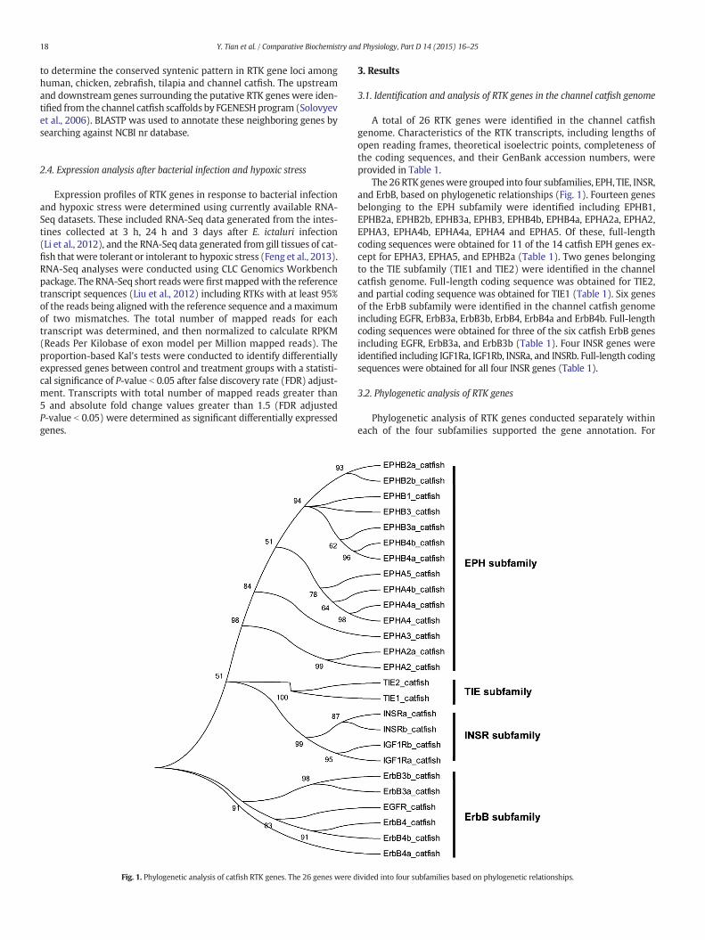
Fig. 1. Phylogenetic analysis of catfish RTK genes. The 26 genes were d
3. Results
3.1. Identification and analysis of RTK genes in the channel catfish genome
A total of 26 RTK genes were identified in the channel catfishgenome. Characteristics of the RTK transcripts, including lengths ofopen reading frames, theoretical isoelectric points, completeness ofthe coding sequences, and their GenBank accession numbers, wereprovided in Table 1.
The 26RTKgeneswere grouped into four subfamilies, EPH, TIE, INSR,and ErbB, based on phylogenetic relationships (Fig. 1). Fourteen genesbelonging to the EPH subfamily were identified including EPHB1,EPHB2a, EPHB2b, EPHB3a, EPHB3, EPHB4b, EPHB4a, EPHA2a, EPHA2,EPHA3, EPHA4b, EPHA4a, EPHA4 and EPHA5. Of these, full-lengthcoding sequences were obtained for 11 of the 14 catfish EPH genes ex-cept for EPHA3, EPHA5, and EPHB2a (Table 1). Two genes belongingto the TIE subfamily (TIE1 and TIE2) were identified in the channelcatfish genome. Full-length coding sequence was obtained for TIE2,and partial coding sequence was obtained for TIE1 (Table 1). Six genesof the ErbB subfamily were identified in the channel catfish genomeincluding EGFR, ErbB3a, ErbB3b, ErbB4, ErbB4a and ErbB4b. Full-lengthcoding sequences were obtained for three of the six catfish ErbB genesincluding EGFR, ErbB3a, and ErbB3b (Table 1). Four INSR genes wereidentified including IGF1Ra, IGF1Rb, INSRa, and INSRb. Full-length codingsequences were obtained for all four INSR genes (Table 1).
3.2. Phylogenetic analysis of RTK genes
Phylogenetic analysis of RTK genes conducted separately withineach of the four subfamilies supported the gene annotation. For
ivided into four subfamilies based on phylogenetic relationships.
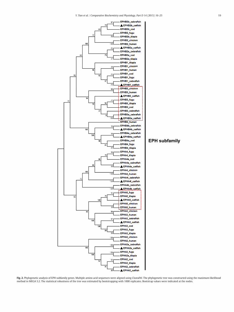
Fig. 2. Phylogenetic analysis of EPH subfamily genes. Multiple amino acid sequences were aligned using ClustalW. The phylogenetic tree was constructed using the maximum likelihoodmethod in MEGA 5.2. The statistical robustness of the tree was estimated by bootstrapping with 1000 replicates. Bootstrap values were indicated at the nodes.
19Y. Tian et al. / Comparative Biochemistry and Physiology, Part D 14 (2015) 16–25
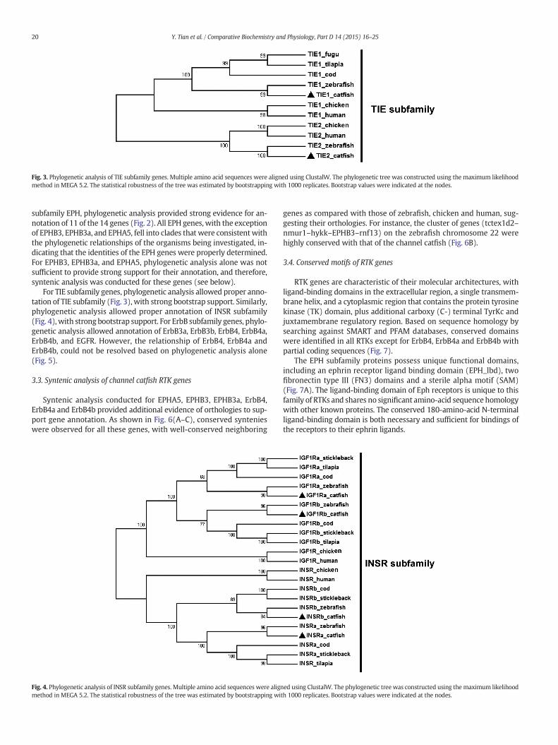
Fig. 3. Phylogenetic analysis of TIE subfamily genes. Multiple amino acid sequences were aligned using ClustalW. The phylogenetic tree was constructed using the maximum likelihoodmethod in MEGA 5.2. The statistical robustness of the tree was estimated by bootstrapping with 1000 replicates. Bootstrap values were indicated at the nodes.
20 Y. Tian et al. / Comparative Biochemistry and Physiology, Part D 14 (2015) 16–25
subfamily EPH, phylogenetic analysis provided strong evidence for an-notation of 11 of the 14 genes (Fig. 2). All EPH genes, with the exceptionof EPHB3, EPHB3a, and EPHA5, fell into clades that were consistentwiththe phylogenetic relationships of the organisms being investigated, in-dicating that the identities of the EPH genes were properly determined.For EPHB3, EPHB3a, and EPHA5, phylogenetic analysis alone was notsufficient to provide strong support for their annotation, and therefore,syntenic analysis was conducted for these genes (see below).
For TIE subfamily genes, phylogenetic analysis allowed proper anno-tation of TIE subfamily (Fig. 3), with strong bootstrap support. Similarly,phylogenetic analysis allowed proper annotation of INSR subfamily(Fig. 4), with strong bootstrap support. For ErbB subfamily genes, phylo-genetic analysis allowed annotation of ErbB3a, ErbB3b, ErbB4, ErbB4a,ErbB4b, and EGFR. However, the relationship of ErbB4, ErbB4a andErbB4b, could not be resolved based on phylogenetic analysis alone(Fig. 5).
3.3. Syntenic analysis of channel catfish RTK genes
Syntenic analysis conducted for EPHA5, EPHB3, EPHB3a, ErbB4,ErbB4a and ErbB4b provided additional evidence of orthologies to sup-port gene annotation. As shown in Fig. 6(A–C), conserved syntenieswere observed for all these genes, with well-conserved neighboring
Fig. 4. Phylogenetic analysis of INSR subfamily genes. Multiple amino acid sequences were alignmethod in MEGA 5.2. The statistical robustness of the tree was estimated by bootstrapping wi
genes as compared with those of zebrafish, chicken and human, sug-gesting their orthologies. For instance, the cluster of genes (tctex1d2–nmur1–hykk–EPHB3–rnf13) on the zebrafish chromosome 22 werehighly conserved with that of the channel catfish (Fig. 6B).
3.4. Conserved motifs of RTK genes
RTK genes are characteristic of their molecular architectures, withligand-binding domains in the extracellular region, a single transmem-brane helix, and a cytoplasmic region that contains the protein tyrosinekinase (TK) domain, plus additional carboxy (C-) terminal TyrKc andjuxtamembrane regulatory region. Based on sequence homology bysearching against SMART and PFAM databases, conserved domainswere identified in all RTKs except for ErbB4, ErbB4a and ErbB4b withpartial coding sequences (Fig. 7).
The EPH subfamily proteins possess unique functional domains,including an ephrin receptor ligand binding domain (EPH_lbd), twofibronectin type III (FN3) domains and a sterile alpha motif (SAM)(Fig. 7A). The ligand-binding domain of Eph receptors is unique to thisfamily of RTKs and shares no significant amino-acid sequence homologywith other known proteins. The conserved 180-amino-acid N-terminalligand-binding domain is both necessary and sufficient for bindings ofthe receptors to their ephrin ligands.
ed using ClustalW. The phylogenetic tree was constructed using the maximum likelihoodth 1000 replicates. Bootstrap values were indicated at the nodes.
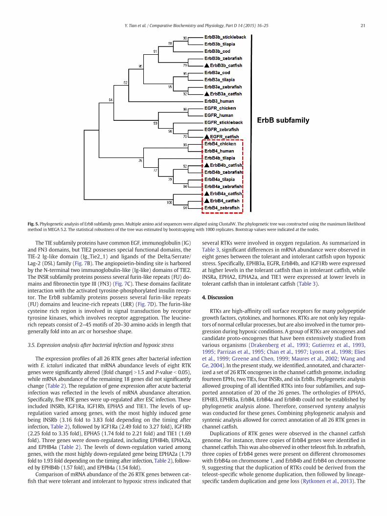
Fig. 5. Phylogenetic analysis of ErbB subfamily genes. Multiple amino acid sequences were aligned using ClustalW. The phylogenetic tree was constructed using the maximum likelihoodmethod in MEGA 5.2. The statistical robustness of the tree was estimated by bootstrapping with 1000 replicates. Bootstrap values were indicated at the nodes.
21Y. Tian et al. / Comparative Biochemistry and Physiology, Part D 14 (2015) 16–25
The TIE subfamily proteins have common EGF, immunoglobulin (IG)and FN3 domains, but TIE2 possesses special functional domains, theTIE-2 Ig-like domain (Ig_Tie2_1) and ligands of the Delta/Serrate/Lag-2 (DSL) family (Fig. 7B). The angiopoietin-binding site is harboredby the N-terminal two immunoglobulin-like (Ig-like) domains of TIE2.The INSR subfamily proteins possess several furin-like repeats (FU) do-mains and fibronectin type III (FN3) (Fig. 7C). These domains facilitateinteraction with the activated tyrosine-phosphorylated insulin recep-tor. The ErbB subfamily proteins possess several furin-like repeats(FU) domains and leucine-rich repeats (LRR) (Fig. 7D). The furin-likecysteine rich region is involved in signal transduction by receptortyrosine kinases, which involves receptor aggregation. The leucine-rich repeats consist of 2–45 motifs of 20–30 amino acids in length thatgenerally fold into an arc or horseshoe shape.
3.5. Expression analysis after bacterial infection and hypoxic stress
The expression profiles of all 26 RTK genes after bacterial infectionwith E. ictaluri indicated that mRNA abundance levels of eight RTKgenes were significantly altered (|fold change| N1.5 and P-value b 0.05),while mRNA abundance of the remaining 18 genes did not significantlychange (Table 2). The regulation of gene expression after acute bacterialinfection was reflected in the levels of mRNA abundance alteration.Specifically, five RTK genes were up-regulated after ESC infection. Theseincluded INSRb, IGF1Ra, IGF1Rb, EPHA5 and TIE1. The levels of up-regulation varied among genes, with the most highly induced genebeing INSRb (3.16 fold to 3.83 fold depending on the timing afterinfection, Table 2), followed by IGF1Ra (2.49 fold to 3.27 fold), IGF1Rb(2.25 fold to 3.35 fold), EPHA5 (1.74 fold to 2.21 fold) and TIE1 (1.69fold). Three genes were down-regulated, including EPHB4b, EPHA2a,and EPHB4a (Table 2). The levels of down-regulation varied amonggenes, with the most highly down-regulated gene being EPHA2a (1.79fold to 1.93 fold depending on the timing after infection, Table 2), follow-ed by EPHB4b (1.57 fold), and EPHB4a (1.54 fold).
Comparison of mRNA abundance of the 26 RTK genes between cat-fish that were tolerant and intolerant to hypoxic stress indicated that
several RTKs were involved in oxygen regulation. As summarized inTable 3, significant differences in mRNA abundance were observed ineight genes between the tolerant and intolerant catfish upon hypoxicstress. Specifically, EPHB3a, EGFR, ErbB4b, and IGF1Rb were expressedat higher levels in the tolerant catfish than in intolerant catfish, whileINSRa, EPHA2, EPHA2a, and TIE1 were expressed at lower levels intolerant catfish than in intolerant catfish (Table 3).
4. Discussion
RTKs are high-affinity cell surface receptors for many polypeptidegrowth factors, cytokines, and hormones. RTKs are not only key regula-tors of normal cellular processes, but are also involved in the tumor pro-gression during hypoxic conditions. A group of RTKs are oncogenes andcandidate proto-oncogenes that have been extensively studied fromvarious organisms (Drakenberg et al., 1993; Gutierrez et al., 1993,1995; Parrizas et al., 1995; Chan et al., 1997; Lyons et al., 1998; Elieset al., 1999; Greene and Chen, 1999; Maures et al., 2002; Wang andGe, 2004). In the present study, we identified, annotated, and character-ized a set of 26 RTK oncogenes in the channel catfish genome, includingfourteen EPHs, two TIEs, four INSRs, and six ErbBs. Phylogenetic analysisallowed grouping of all identified RTKs into four subfamilies, and sup-ported annotation of 20 of the 26 genes. The orthologies of EPHA5,EPHB3, EPHB3a, ErbB4, ErbB4a and ErbB4b could not be established byphylogenetic analysis alone. Therefore, conserved synteny analysiswas conducted for these genes. Combining phylogenetic analysis andsyntenic analysis allowed for correct annotation of all 26 RTK genes inchannel catfish.
Duplications of RTK genes were observed in the channel catfishgenome. For instance, three copies of ErbB4 genes were identified inchannel catfish. This was also observed in other teleostfish. In zebrafish,three copies of ErbB4 genes were present on different chromosomeswith ErbB4a on chromosome 1, and ErbB4b and ErbB4 on chromosome9, suggesting that the duplication of RTKs could be derived from theteleost-specific whole genome duplication, then followed by lineage-specific tandem duplication and gene loss (Rytkonen et al., 2013). The
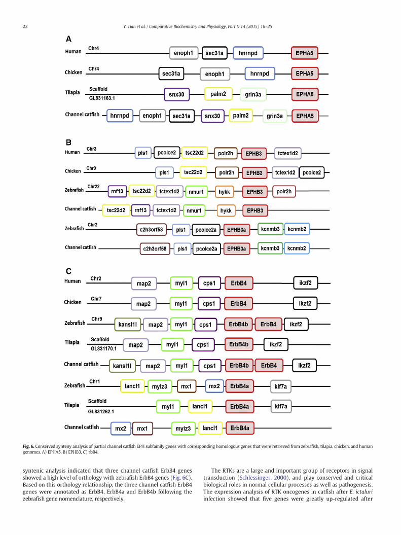
Fig. 6. Conserved synteny analysis of partial channel catfish EPH subfamily genes with corresponding homologous genes that were retrieved from zebrafish, tilapia, chicken, and humangenomes. A) EPHA5, B) EPHB3, C) rbB4.
22 Y. Tian et al. / Comparative Biochemistry and Physiology, Part D 14 (2015) 16–25
syntenic analysis indicated that three channel catfish ErbB4 genesshowed a high level of orthology with zebrafish ErbB4 genes (Fig. 6C).Based on this orthology relationship, the three channel catfish ErbB4genes were annotated as ErbB4, ErbB4a and ErbB4b following thezebrafish gene nomenclature, respectively.
The RTKs are a large and important group of receptors in signaltransduction (Schlessinger, 2000), and play conserved and criticalbiological roles in normal cellular processes as well as pathogenesis.The expression analysis of RTK oncogenes in catfish after E. ictaluriinfection showed that five genes were greatly up-regulated after
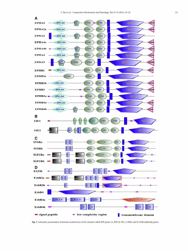
Fig. 7. Schematic presentation of domain architectures of the channel catfish RTK genes. A) EPH, B) TIE, C) INSR, and D) ErbB subfamily genes.
23Y. Tian et al. / Comparative Biochemistry and Physiology, Part D 14 (2015) 16–25
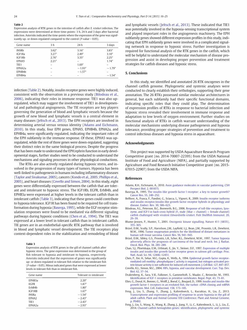
Table 2Expression analysis of RTK genes in the intestine of catfish after E. ictaluri infection. Theexpressions were determined at three time-points: 3 h, 24 h and 3 days after bacterialinfection. Asterisks indicated the time-points where the expression of the genewas signif-icantly up- or down-regulated compared to the control (P-value b 0.05).
Gene name 3 h 24 h 3 days
INSRb 3.62* 3.16* 3.83*IGF1Ra 3.27* 2.49* 3.10*IGF1Rb 2.76* 3.35* 2.25*EPHA5 2.21* – 1.74*TIE1 – 1.69* –
EPHA2a – −1.93* −1.79*EPHB4b – −1.57* –
EPHB4a – – −1.54*
24 Y. Tian et al. / Comparative Biochemistry and Physiology, Part D 14 (2015) 16–25
infection (Table 2). Notably, insulin receptor geneswere highly induced,consistent with the observation in a previous study (Bilodeau et al.,2006), indicating their roles in immune response. TIE1 was also up-regulated, which may suggest the involvement of TIE1 in developmen-tal and pathological angiogenesis. The TIE receptors are key playersgoverning the generation of blood and lymphatic vessels because thegrowth of new blood and lymphatic vessels is a central element inmany diseases (Jeltsch et al., 2013). The EPH receptors are involved indetermining arterial versus venous identity (Adams and Eichmann,2010). In this study, four EPH genes, EPHA5, EPHB4b, EPHA2a, andEPHB4a, were significantly regulated, indicating the important roles ofthe EPH subfamily in the immune response. Of these, EPHA5 was up-regulated, while the rest of three geneswere down-regulated, suggestingtheir distinct roles in the same biological process. Despite the progressthat has beenmade tounderstand theEPH/ephrin function in early devel-opmental stages, further studies need to be conducted to understand themechanisms and signaling processes in other physiological conductions.
The RTKs are also actively regulated during hypoxic stress, and in-volved in the progression of many types of tumors. Hypoxia has beenwell-linked to pathogenesis in humans including inflammatory diseases(Taylor and Sivakumar, 2005), cancers (Kondo et al., 2005; Phillips et al.,2005), and heart diseases (Covello and Simon, 2004). In this study, eightgenes were differentially expressed between the catfish that are toler-ant and intolerant to hypoxic stress. The IGF1Rb, EGFR, ErbB4b, andEPHB3a were expressed at higher levels in the tolerant catfish than inintolerant catfish (Table 3), indicating that these genes could contributeto hypoxia tolerance. IGF1R has been found to be required for cell trans-formation during hypoxia (Baserga, 1995), while the EGF receptor stim-ulation responses were found to be mediated via different signalingpathways during hypoxic conditions (Chen et al., 1994). The TIE1 wasexpressed at a lower level in tolerant catfish than in intolerant catfish.TIE genes are in an endothelial-specific RTK pathway that is involvedin blood and lymphatic vessel development. The TIE receptors playcontext-dependent roles in the stabilization and remodeling of blood
Table 3Expression analysis of RTK genes in the gill of channel catfish afterhypoxic stress. The gene expression was determined in the group offish tolerant to hypoxia and intolerant to hypoxia, respectively.Asterisks indicated that the expression of genes was significantlyup- or down-regulated in tolerant fish relative to the intolerant fish(P-value b 0.05).Minus indicated genes that were expressed at lowerlevels in tolerant fish than in intolerant fish.
Gene name Tolerant vs intolerant
EPHB3a 2.20*EGFR 1.87*ErbB4b 1.80*IGF1RB 1.67*INSRa −1.54*EPHA2 −2.43*TIE1 −2.43*EPHA2a −2.93*
and lymphatic vessels (Jeltsch et al., 2013). These indicated that TIE1was potentially involved in the hypoxia-sensing transcriptional systemand played important roles in the angiogenesis machinery. The EPHsubfamily genes showed different expression profiles in this study, indi-cating that EPH subfamily genes were involved in a complicated signal-ing network in response to hypoxic stress. Further investigation isrequired for functional analysis of the RTK genes in the catfish, whichwill be helpful to understand the molecular mechanism of disease pro-gression and assist in developing proper prevention and treatmentstrategies for catfish diseases and hypoxic stress.
5. Conclusions
In this study, we identified and annotated 26 RTK oncogenes in thechannel catfish genome. Phylogenetic and syntenic analyses wereconducted to clearly establish their orthologies, supporting their geneannotation. The 26 RTKs possessed similar molecular architectures ingeneral, but each subfamily had their specific functional domains,indicating specific roles that they could play. The determinationof expression profiles of RTKs in response to bacterial infection andhypoxic stress revealed their involvement in immune response andadaptation to low levels of oxygen environment. Further studies onfunctional analysis of RTKs in catfish warrant understanding of themolecular mechanisms underlying disease pathogenesis and hypoxiatolerance, providing proper strategies of prevention and treatment tocontrol infectious diseases and hypoxia stress in aquaculture.
Acknowledgments
This project was supported by USDA Aquaculture Research ProgramCompetitive grant (no. 2014-70007-22395) from the USDA NationalInstitute of Food and Agriculture (NIFA), and partially supported byAgriculture and Food Research Initiative Competitive grant (no. 2015-67015-22907) from the USDA NIFA.
References
Adams, R.H., Eichmann, A., 2010. Axon guidance molecules in vascular patterning. CSHPerspect Biol. 2, 001875.
Baserga, R., 1995. The insulin-like growth factor I receptor: a key to tumor growth?Cancer Res. 55, 249–252.
Belfiore, A., Frasca, F., Pandini, G., Sciacca, L., Vigneri, R., 2009. Insulin receptor isoformsand insulin receptor/insulin-like growth factor receptor hybrids in physiology anddisease. Endocr. Rev. 30, 586–623.
Bilodeau, A.L., Peterson, B.C., Bosworth, B.G., 2006. Response of toll-like receptors, lyso-zyme, and IGF-I in back-cross hybrid (F1 male (blue × channel) × female channel)catfish challenged with virulent Edwardsiella ictaluri. Fish Shellfish Immunol. 20,29–39.
Blume-Jensen, P., Hunter, T., 2001. Oncogenic kinase signalling. Nature 411 (6835),355–365.
Brizel, D.M., Scully, S.P., Harrelson, J.M., Layfield, L.J., Bean, J.M., Prosnitz, L.R., Dewhirst,M.W., 1996. Tumor oxygenation predicts for the likelihood of distant metastases inhuman soft tissue sarcoma. Cancer Res. 56, 941–943.
Brizel, D.M., Sibley, G.S., Prosnitz, L.R., Scher, R.L., Dewhirst, M.W., 1997. Tumor hypoxiaadversely affects the prognosis of carcinoma of the head and neck. Int. J. Radiat.Oncol. Biol. Phys. 38, 285–289.
Chan, S.J., Plisetskaya, E.M., Urbinati, E., Jin, Y., Steiner, D.F., 1997. Expression of multipleinsulin and insulin-like growth factor receptor genes in salmon gill cartilage. Proc.Natl. Acad. Sci. 94, 12446–12451.
Chen, P., Xie, H., Sekar, M.C., Gupta, K., Wells, A., 1994. Epidermal growth factor receptor-mediated cell motility: phospholipase C activity is required, but mitogen-activated pro-tein kinase activity is not sufficient for induced cellmovement. J. Cell Biol. 127, 847–857.
Covello, K.L., Simon, M.C., 2004. HIFs, hypoxia, and vascular development. Curr. Top. Dev.Biol. 62, 37–54.
Drakenberg, K., Sara, V.R., Falkmer, S., Gammeltoft, S., Maake, C., Reinecke, M., 1993.Identification of IGF-1 receptors in primitive vertebrates. Regul. Pept. 43, 73–81.
Elies, G., Duval, H., Bonnec, G., Wolff, J., Boeuf, G., Boujard, D., 1999. Insulin and insulin-likegrowth factor-1 receptors in an evoluted fish, the turbot: cDNA cloning and mRNAexpression. Mol. Cell. Endocrinol. 158, 173–185.
Feng, J., Liu, S., Zhang, Y., Zhang, J., Kaltenboeck, L., Kucuktas, H., Liu, Z., 2013.Transcriptome and expression profiling of response to acute hypoxia in the gill ofadult catfish. Plant and Animal Genome XXI Conference. Plant and Animal Genome,p. P0492.
Feng, J., Liu, S., Wang, X., Wang, R., Zhang, J., Jiang, Y., Li, C., Kaltenboeck, L., Li, J., Liu, Z.,2014. Channel catfish hemoglobin genes: identification, phylogenetic and syntenic
25Y. Tian et al. / Comparative Biochemistry and Physiology, Part D 14 (2015) 16–25
analysis, and specific induction in response to heat stress. Comp. Biochem. Physiol.Part D Genomics Proteomics 9, 11–22.
Flicek, P., Ahmed, I., Amode, M.R., Barrell, D., Beal, K., Brent, S., Carvalho-Silva, D., Clapham,P., Coates, G., Fairley, S., Fitzgerald, S., Gil, L., Garcia-Giron, C., Gordon, L., Hourlier, T.,Hunt, S., Juettemann, T., Kahari, A.K., Keenan, S., Komorowska, M., Kulesha, E.,Longden, I., Maurel, T., McLaren, W.M., Muffato, M., Nag, R., Overduin, B., Pignatelli,M., Pritchard, B., Pritchard, E., Riat, H.S., Ritchie, G.R., Ruffier, M., Schuster, M.,Sheppard, D., Sobral, D., Taylor, K., Thormann, A., Trevanion, S., White, S., Wilder, S.P.,Aken, B.L., Birney, E., Cunningham, F., Dunham, I., Harrow, J., Herrero, J., Hubbard, T.J.,Johnson, N., Kinsella, R., Parker, A., Spudich, G., Yates, A., Zadissa, A., Searle, S.M., 2013.Ensembl 2013. Nucleic Acids Res. 41, 48–55.
Geng, X., Feng, J., Liu, S., Wang, Y., Arias, C., Liu, Z., 2014. Transcriptional regulation ofhypoxia inducible factors alpha (HIF-α) and their inhibiting factor (FIH-1) of channelcatfish (Ictalurus punctatus) under hypoxia. Comp. Biochem. Physiol. B 169, 38–50.
Greene, M.W., Chen, T.T., 1999. Characterization of teleost insulin receptor familymembers II. Developmental expression of insulin-like growth factor type I receptormessenger RNAs in rainbow trout. Gen. Comp. Endocrinol. 115, 270–281.
Gutierrez, J., Parrizas, M., Carneiro, N., Maestro, M., Planas, J., 1993. Insulin and IGF-I recep-tors and tyrosine kinase activity in carp ovaries: changes with reproductive stage.Fish Physiol. Biochem. 11, 247–254.
Gutierrez, J., Parrizas, M., Maestro, M.A., Plisetskaya, E.M., 1995. Insulin and IGF-I bindingand tyrosine kinase activity in fish heart. J. Endocrinol. 146, 35–44.
Hasina, R., Mollberg, N., Kawada, I., Mutreja, K., Kanade, G., Yala, S., Surati, M., Liu, R., Li, X.,Zhou, Y., Ferguson, B.D., Nallasura, V., Cohen, K.S., Hyjek, E., Mueller, J., Kanteti, R.,Hashani, E., Kane, D., Shimada, Y., Lingen, M.W., Husain, A.N., Posner, M.C.,Waxman, I., Villaflor, V.M., Ferguson, M.K., Varticovski, L., Vokes, E.E., Gill, P., Salgia,R., 2013. Critical role for the receptor tyrosine kinase EPHB4 in esophageal cancers.Cancer Res. 73, 184–194.
Hockel, M., Knoop, C., Schlenger, K., 1993. Intratumoral pO2 predicts survival in advancedcancer of the uterine cervix. Radiother. Oncol. 26, 45–50.
Hockel, M., Schlenger, K., Aral, B., Mitze, M., Schaffer, U., Vaupel, P., 1996. Associationbetween tumor hypoxia andmalignant progression in advanced cancer of the uterinecervix. Cancer Res. 56, 4509–4515.
Hong, B.V., Lui, W., Hashiguchi, M., Edwin, B., Hui, P., Chan, M.D., 2013. Targeting tumorhypoxia in nasopharyngeal carcinoma. Head Neck 35, 133–145.
Janssen, H.L., Haustermans, K.M., Balm, A.J., Begg, A.C., 2005. Hypoxia in head and neckcancer: how much, how important? Head Neck 27, 622–638.
Jeltsch, M., Leppänen, V., Saharinen, P., Alitalo, K., 2013. Receptor tyrosine kinase-mediated angiogenesis. CSH Perspect. Biol. 5, 009183.
Kondo, Y., Hamada, J., Kobayashi, C., Nakamura, R., Suzuki, Y., Kimata, R., Nishimura, T.,Kitagawa, T., Kunimoto, M., Imura, N., Hara, S., 2005. Over expression of hypoxia-inducible factor-1alpha in renal and bladder cancer cells increases tumorigenicpotency. J. Urol. 173, 1762–1766.
Kullander, K., Klein, R., 2002. Mechanisms and functions of Eph and ephrin signalling. Nat.Rev. Mol. Cell Biol. 3, 475–486.
Lemmon, M.A., Schlessinger, J., 2010. Cell signaling by receptor tyrosine kinases. Cell 141,1117–1134.
LeRoith, D., Accilli, D., 2008. Mechanisms of disease: using genetically altered mice tostudy concepts of type 2 diabetes. Nat. Clin. Pract. Endocrinol. Metab. 4, 164–172.
Letunic, I., Doerks, T., Bork, P., 2012. SMART 7: recent updates to the protein domainannotation resource. Nucleic Acids Res. 40, 302–305.
Li, C., Zhang, Y., Wang, R., Lu, J., Nandi, S., Mohanty, S., Terhune, J., Liu, Z., Peatman, E.,2012. RNA-seq analysis of mucosal immune responses reveals signatures of intestinalbarrier disruption and pathogen entry following Edwardsiella ictaluri infection inchannel catfish, Ictalurus punctatus. Fish Shellfish Immunol. 32, 816–827.
Liu, S., Zhang, Y., Zhou, Z., Waldbieser, G., Sun, F., Lu, J., Zhang, J., Jiang, Y., Zhang, H., Wang,X., Rajendran, K., Khoo, L., Kucuktas, H., Peatman, E., Liu, Z., 2012. Efficient assemblyand annotation of the transcriptome of catfish by RNA-Seq analysis of a doubled hap-loid homozygote. BMC Genomics 13, 595.
Liu, S., Wang, X., Sun, F., Zhang, J., Feng, J., Liu, H., Rajendran, K.V., Sun, L., Zhang, Y., Jiang,Y., Peatman, E., Kaltenboeck, L., Kucuktas, H., Liu, Z., 2013. RNA-Seq reveals expressionsignatures of genes involved in oxygen transport, protein synthesis, folding, anddegradation in response to heat stress in catfish. Physiol. Genomics 45, 462–476.
Louis, A., Muffato, M., Crollius, H.R., 2013. Genomicus: five genome browsers forcomparative genomics in eukaryota. Nucleic Acids Res. 41, 700–705.
Lund-Olesen, K., 1970. Oxygen tension in synovial fluids. Arthritis Rheum. 13, 769–776.Lyons, M.S., Bell, B., Stainier, D., Kevin, K.G., 1998. Isolation of the zebrafish homologues
for the tie-1 and tie-2 endothelium-specific receptor tyrosine kinases. Dev. Dynam.212, 133–140.
Maures, T., Chan, S.J., Xu, B., Sun, H., Ding, J., Duan, A., 2002. Structural, biochemical, andexpression analysis of two distinct insulin-like growth factor i receptors and theirligands in zebrafish. Endocrinology 143, 1858–1871.
Nikinmaa, M., Pursiheimo, S., Soitamo, A.J., 2004. Redox state regulates HIF-1alpha and itsDNA binding and phosphorylation in salmonid cells. J. Cell Sci. 117, 3201–3206.
Parrizas, M., Maestro, M.A., Banos, N., Navarro, I., Planas, J., Gutierrez, J., 1995. Insulin/IGF-Ibinding ratio in skeletal and cardiac muscles of vertebrates: a phylogenetic approach.Am. J. Physiol. 269, 1370–1377.
Pawson, T., 1995. Protein modules and signalling networks. Nature 373, 573–580.Phillips, R.J., Mestas, J., Gharaee-Kermani, M., Burdick, M.D., Sica, A., Belperio, J.A., Keane,
M.P., Strieter, R.M., 2005. Epidermal growth factor and hypoxia-induced expressionof CXC chemokine receptor 4 on non-small cell lung cancer cells is regulated by thephosphatidylinositol 3-kinase/PTEN/AKT/mammalian target of rapamycin signalingpathway and activation of hypoxia inducible factor-1alpha. J. Biol. Chem. 280,22473–22481.
Powell, W.H., Hahn, M.E., 2002. Identification and functional characterization of hypoxia-inducible factor 2alpha from the estuarine teleost, Fundulus heteroclitus: interactionof HIF-2alpha with two ARNT2 splice variants. J. Exp. Zool. 294, 17–29.
Rytkonen, K.T., Akbarzadeh, A., Miandare, H.K., Kamei, H., Duan, C., Leder, E.H., Williams,T.A., Nikinmaa, M., 2013. Subfunctionalization of cyprinid hypoxia-inducible factorsfor roles in development and oxygen sensing. Evolution 67, 873–882.
Schlessinger, J., 2000. Cell signaling by receptor tyrosine kinases. Cell 103, 211–225.Shewchuk, L.M., Hassell, A.M., Ellis, B., Holmes, W.D., Davis, R., Horne, E.L., Kadwell, S.H.,
Mckee, D.D., Moore, J.T., 2000. Structure of the Tie2 RTK domain: self-inhibition bythe nucleotide binding loop, activation loop, and C-terminal tail. Structure 8,1105–1113.
Slamon, D.J., Clark, G.M., Wong, S.G., 1987. Human breast cancer: correlation ofrelapse and survival with amplification of the HER-2/neu oncogene. Science 235,177–182.
Slamon, D.J., Godolphin,W., Jones, L.A., 1989. Studies of the HER-2/neu proto-oncogene inhuman breast and ovarian cancer. Science 244, 707–712.
Solovyev, V., Kosarev, P., Seledsov, I., Vorobyev, D., 2006. Automatic annotation ofeukaryotic genes, pseudogenes and promoters. Genome Biol. 7 (Suppl. 1), 10.
Sun, F., Peatman, E., Li, C., Liu, S., Jiang, Y., Zhou, Z., Liu, Z., 2012. Transcriptomic signaturesof attachment, NF-κB suppression and IFN stimulation in the catfish gill followingcolumnaris bacterial infection. Dev. Comp. Immunol. 38, 169–180.
Tamura, K., Peterson, D., Peterson, N., Stecher, G., Nei, M., Kumar, S., 2011. MEGA5: molec-ular evolutionary genetics analysis using maximum likelihood, evolutionary distance,and maximum parsimony methods. Mol. Biol. Evol. 28, 2731–2739.
Taniguchi, C.M., Emanuelli, B., Kahn, C.R., 2006. Critical nodes in signaling pathways:insights into insulin action. Nat. Rev. Mol. Cell Biol. 7, 85–96.
Taylor, P.C., Sivakumar, B., 2005. Hypoxia and angiogenesis in rheumatoid arthritis. Curr.Opin. Rheumatol. 17, 293–298.
Thompson, J.D., Gibson, T., Higgins, D.G., 2002. Multiple sequence alignment usingClustalW and ClustalX. Curr. Protoc. Bioinforma. 2.3.1–2.3.22.
Ullrich, A., Schlessinger, J., 1990. Signal transduction by receptors with tyrosine kinaseactivity. Cell 61, 203–212.
Wang, Y., Ge, W., 2004. Cloning of Epidermal Growth Factor (EGF) and EGF receptorfrom the zebrafish ovary: evidence for EGF as a potential paracrine factor from theoocyte to regulate activin/follistatin system in the follicle cells. Biol. Reprod. 71,749–760.
Zhang, H., Peatman, E., Liu, H., Feng, T., Chen, L., Liu, Z., 2012. Molecular characterization ofthree L-type lectin genes from channel catfish, Ictalurus punctatus and their responsesto Edwardsiella ictaluri challenge. Fish Shellfish Immunol. 32, 598–608.
Zhang, J., Liu, S., Rajendran, K.V., Sun, L., Zhang, Y., Sun, F., Kucuktas, H., Liu, H., Liu, Z.,2013. Pathogen recognition receptors in channel catfish: III phylogeny and expres-sion analysis of Toll-like receptors. Dev. Comp. Immunol. 40, 185–194.