Embed Size (px)
Citation preview

This is a sample from COMPACT CLINICAL GUIDE TO ARRHYTHMIA AND 12-LEAD EKG INTERPRETATION: FOUNDATIONS OF PRACTICE FOR CRITICAL CARE NURSES
Visit This Book’s Web Page / Buy Now / Request an Exam/Review Copy
© Springer Publishing Company
© Springer Publishing Company
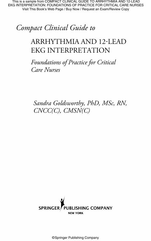
Compact Clinical Guide to
ARRHYTHMIA AND 12-LEAD EKG INTERPRETATION

This is a sample from COMPACT CLINICAL GUIDE TO ARRHYTHMIA AND 12-LEAD EKG INTERPRETATION: FOUNDATIONS OF PRACTICE FOR CRITICAL CARE NURSES
Visit This Book’s Web Page / Buy Now / Request an Exam/Review Copy
© Springer Publishing Company
© Springer Publishing Company
Sandra Goldsworthy, PhD, MSc, RN, CNCC(C), CMSN(C), is a recognized critical care expert, researcher, and author. She is the author or editor of six books, including Stimulation Simplified: A Practical Guide for Nurse Educators, Simulation Simplified: Student Lab Manual for Critical Care Nursing, and The Compact Clinical Guide for Mechanical Ventilation. She is an associate professor in the faculty of nursing at the University of Calgary, where she holds a research professorship in simulation education. She holds two national Canadian Nurses Association credentials in critical care and medical–surgical nursing. She is a member of a number of national and international committees, including serving on the board of directors for the World Federation of Critical Care Nurses Association. Her research focus is on simulation and transfer of learning, job readiness, and transition of new graduates into critical care, and she has conducted and published research involving the use of simulation and technology in nursing education. Her recent publications and national and international presentations have concentrated on critical care nurse retention, critical care nurse work environments, and the use of simulation to build confidence and competence among nurses.
This is a sample from COMPACT CLINICAL GUIDE TO ARRHYTHMIA AND 12-LEAD EKG INTERPRETATION: FOUNDATIONS OF PRACTICE FOR CRITICAL CARE NURSES
Visit This Book’s Web Page / Buy Now / Request an Exam/Review Copy
© Springer Publishing Company
© Springer Publishing Company
Compact Clinical Guide to
ARRHYTHMIA AND 12-LEAD EKG INTERPRETATION
Foundations of Practice for Critical Care Nurses
Sandra Goldsworthy, PhD, MSc, RN, CNCC(C), CMSN(C)
This is a sample from COMPACT CLINICAL GUIDE TO ARRHYTHMIA AND 12-LEAD EKG INTERPRETATION: FOUNDATIONS OF PRACTICE FOR CRITICAL CARE NURSES
Visit This Book’s Web Page / Buy Now / Request an Exam/Review Copy
© Springer Publishing Company
© Springer Publishing Company
Copyright © 2016 Springer Publishing Company, LLCAll rights reserved.
No part of this publication may be reproduced, stored in a retrieval system, or transmitted in any form or by any means, electronic, mechanical, photocopying, recording, or otherwise, without the prior permission of Springer Publishing Company, LLC, or authorization through payment of the appropriate fees to the Copyright Clearance Center, Inc., 222 Rosewood Drive, Danvers, MA 01923, 978-750-8400, fax 978-646-8600, [email protected] or on the Web at www.copyright.com.
Springer Publishing Company, LLC11 West 42nd StreetNew York, NY 10036www.springerpub.com
Acquisitions Editor: Elizabeth NieginskiComposition: diacriTech
ISBN: 978-0-8261-9846-4e-book ISBN: 978-0-8261-9847-1PowerPoint ISBN: 978-0-8261-9629-3
Student supplements are available from www.springerpub.com/goldsworthy
16 17 18 19 / 5 4 3 2 1
The author and the publisher of this Work have made every effort to use sources believed to be reliable to provide information that is accurate and compatible with the standards generally accepted at the time of publication. Because medical science is continually advancing, our knowledge base continues to expand. Therefore, as new information becomes available, changes in procedures become necessary. We recommend that the reader always consult current research and specific institutional policies before performing any clinical procedure. The author and publisher shall not be liable for any special, consequential, or exemplary damages resulting, in whole or in part, from the readers’ use of, or reliance on, the information contained in this book. The publisher has no responsibility for the persistence or accuracy of URLs for external or third-party Internet websites referred to in this publication and does not guarantee that any content on such websites is, or will remain, accurate or appropriate.
Library of Congress Cataloging-in-Publication Data
Names: Goldsworthy, Sandra, 1961- author.Title: Compact clinical guide to arrhythmia and 12-lead ekg interpretation: foundations of practice for critical care nurses / Sandra Goldsworthy, PhD, RN, MSc, CNCC(C), CMSN(C).Description: New York, NY : Springer Publishing Company, LLC, [2016] | Includes bibliographical references and index.Identifiers: LCCN 2016014653| ISBN 9780826198464 | ISBN 9780826198471 (ebook)Subjects: LCSH: Arrhythmia—Diagnosis. | Electrocardiography—Interpretation. | Heart—Electric properties.Classification: LCC RC685.A65 G65 2016 | DDC 616.1/28—dc23 LC record available at https://lccn.loc.gov/2016014653
Special discounts on bulk quantities of our books are available to corporations, professional associations, pharmaceutical companies, health care organizations, and other qualifying groups. If you are interested in a custom book, including chapters from more than one of our titles, we can provide that service as well.For details, please contact:Special Sales Department, Springer Publishing Company, LLC11 West 42nd Street, 15th Floor, New York, NY 10036-8002Phone: 877-687-7476 or 212-431-4370; Fax: 212-941-7842E-mail: [email protected]
Printed in the United States of America by Gasch Printing.
This is a sample from COMPACT CLINICAL GUIDE TO ARRHYTHMIA AND 12-LEAD EKG INTERPRETATION: FOUNDATIONS OF PRACTICE FOR CRITICAL CARE NURSES
Visit This Book’s Web Page / Buy Now / Request an Exam/Review Copy
© Springer Publishing Company
© Springer Publishing Company
I dedicate this book to all the wonderful nursing students whom I have had the privilege to teach over the past 28 years. It has truly enriched
my life and it has been a pleasure.

This is a sample from COMPACT CLINICAL GUIDE TO ARRHYTHMIA AND 12-LEAD EKG INTERPRETATION: FOUNDATIONS OF PRACTICE FOR CRITICAL CARE NURSES
Visit This Book’s Web Page / Buy Now / Request an Exam/Review Copy
© Springer Publishing Company
© Springer Publishing Company
This is a sample from COMPACT CLINICAL GUIDE TO ARRHYTHMIA AND 12-LEAD EKG INTERPRETATION: FOUNDATIONS OF PRACTICE FOR CRITICAL CARE NURSES
Visit This Book’s Web Page / Buy Now / Request an Exam/Review Copy
© Springer Publishing Company
© Springer Publishing Company
vii
Preface ix
Acknowledgments xi
1. Basic Anatomy and Physiology: Highlights 1 Wendy Preiano
2. Arrhythmias: Sinus and Atrial 21
3. Arrhythmias: Junctional and Ventricular 43
4. Arrhythmias: Atrioventricular Blocks 67
5. Arrhythmias: Asystole, Pulseless Electrical Activity, and Paced Rhythms 79
6. 12-Lead EKG Overview 97
7. Special Situations 113
Answers 127
Index 133
Contents

This is a sample from COMPACT CLINICAL GUIDE TO ARRHYTHMIA AND 12-LEAD EKG INTERPRETATION: FOUNDATIONS OF PRACTICE FOR CRITICAL CARE NURSES
Visit This Book’s Web Page / Buy Now / Request an Exam/Review Copy
© Springer Publishing Company
© Springer Publishing Company
This is a sample from COMPACT CLINICAL GUIDE TO ARRHYTHMIA AND 12-LEAD EKG INTERPRETATION: FOUNDATIONS OF PRACTICE FOR CRITICAL CARE NURSES
Visit This Book’s Web Page / Buy Now / Request an Exam/Review Copy
© Springer Publishing Company
© Springer Publishing Company
ix
One of the foundational competencies required of critical care nurses is the ability to interpret cardiac rhythms systematically. Failure to recognize and respond to dangerous arrhythmias could lead to serious life-threatening consequences for the patient.
Through my teaching of cardiac arrhythmia courses and advanced cardiac life support (ACLS) to nurses and other health professionals over the past three decades, I have developed strategies to help bedside practitioners rapidly interpret abnor-mal cardiac rhythms and to determine how to prioritize interventions quickly. The amount of information on this topic can be overwhelming, and there are many different types of education available on the market about arrhythmia/12-lead EKGs. The goal of this text is to be user friendly by striking a balance between cue cards or a basic overview and text-dense lengthy explanations. This text is aimed at being a bedside ref-erence small enough to carry in a nurse’s pocket, yet still con-taining enough detail to provide the nurse with critical information needed at the point of care.
The text begins with a brief overview of the anatomy and physiology related to the heart and its conduction system. The chapters that follow align with specific pacemaker sites related to arrhythmias (i.e., sinus, atrial, junctional, ventricular, atrio-ventricular [AV] heart blocks, and paced rhythms). In addition,
Preface
This is a sample from COMPACT CLINICAL GUIDE TO ARRHYTHMIA AND 12-LEAD EKG INTERPRETATION: FOUNDATIONS OF PRACTICE FOR CRITICAL CARE NURSES
Visit This Book’s Web Page / Buy Now / Request an Exam/Review Copy
© Springer Publishing Company
© Springer Publishing Company
x Preface
an introduction to 12-lead EKGs is presented with content related to myocardial infarction.
The Compact Clinical Guide to Arrhythmia and 12-Lead EKG Interpretation: Foundations of Practice for Critical Care Nurses specifically includes a systematic approach to basic arrhythmia interpretation, examples, and explanations of all of the basic sinus, atrial, ventricular, and AV heart block arrhyth-mias; a systematic approach to 12-lead EKG interpretation; and a systematic approach to pacemaker rhythm interpretation and malfunction. To further consolidate your learning, each chapter ends with practice questions and a case study you can use to test yourself. Associated electronic resources that accompany this text are cue cards of systematic approaches for arrhythmia, 12-lead EKG, and pacemaker interpreta-tion provided as a PowerPoint presentation that can be accessed from www.springerpub.com/goldsworthy. The most current guidelines are incorporated into this text, includ-ing the newest 2015 American Heart Association Guidelines for Cardiac and Emergency Care.
Most important, I hope you will benefit from the clinical tips integrated into each chapter. Because we do not see all rhythms regularly in clinical practice, and some are very rare, you may find it helpful to review the content in this text at regular intervals.
I hope you will find this resource helpful as you care for critically ill patients in your practice. It has been a pleasure creating it!
Sandra Goldsworthy

This is a sample from COMPACT CLINICAL GUIDE TO ARRHYTHMIA AND 12-LEAD EKG INTERPRETATION: FOUNDATIONS OF PRACTICE FOR CRITICAL CARE NURSES
Visit This Book’s Web Page / Buy Now / Request an Exam/Review Copy
© Springer Publishing Company
© Springer Publishing Company
xi
I would like to acknowledge the team at Springer Publishing Company, especially executive editor extraordinaire, Elizabeth Nieginski, who is always a pleasure to work with and a true and gracious professional. I appreciate all of their efforts in bringing this book to fruition. I would also like to acknowledge the con-tribution of Wendy Preiano, nursing professor at Georgian College, for writing the first chapter of the book. Her efforts are greatly appreciated.
Acknowledgments

This is a sample from COMPACT CLINICAL GUIDE TO ARRHYTHMIA AND 12-LEAD EKG INTERPRETATION: FOUNDATIONS OF PRACTICE FOR CRITICAL CARE NURSES
Visit This Book’s Web Page / Buy Now / Request an Exam/Review Copy
© Springer Publishing Company
© Springer Publishing Company

This is a sample from COMPACT CLINICAL GUIDE TO ARRHYTHMIA AND 12-LEAD EKG INTERPRETATION: FOUNDATIONS OF PRACTICE FOR CRITICAL CARE NURSES
Visit This Book’s Web Page / Buy Now / Request an Exam/Review Copy
© Springer Publishing Company
© Springer Publishing Company
21
2
SINUS RHYTHMS
In this chapter, sinus and atrial rhythms are discussed. Specifi cally, the following rhythms are presented: normal sinus rhythm, sinus bradycardia, sinus tachycardia, sinus arrhythmia, and sinus arrest/block. Th e atrial rhythms dis-cussed are premature atrial contractions (PACs) , atrial fi bril-lation, atrial fl utter, and supraventricular tachycardia (SVT) . Unique characteristics, causes, treatment, and tips for inter-pretation are provided. In addition, practice examples, “test yourself” questions, and an integrated case study are provided at the end of the chapter to help you retain the need-to-know information and build your confi dence in interpreting arrhythmias.
ti0010
p0005
Arrhythmias : Sinus and Atrial ti0005
Unique characteristics of each rhythm that help diff erentiate it from others are in boldface.
p0460
Helpful Tips
This is a sample from COMPACT CLINICAL GUIDE TO ARRHYTHMIA AND 12-LEAD EKG INTERPRETATION: FOUNDATIONS OF PRACTICE FOR CRITICAL CARE NURSES
Visit This Book’s Web Page / Buy Now / Request an Exam/Review Copy
© Springer Publishing Company
© Springer Publishing Company
22 2. Arrhythmias: Sinus and Atrial
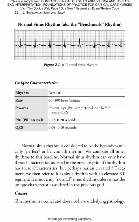
Normal Sinus Rhythm (aka the “Benchmark” Rhythm)ti0015
Normal sinus rhythm is considered to be the hemodynami-cally “perfect” or benchmark rhythm. We compare all other rhythms to this baseline. Normal sinus rhythm can only have these characteristics, as listed in the previous grid. If the rhythm has these characteristics, but perhaps has an elevated ST seg-ment, we then refer to it as sinus rhythm with an elevated ST segment. It is not truly “normal” sinus rhythm unless it has the unique characteristics as listed in the previous grid.
Causes
This rhythm is normal and does not have underlying pathology.
p0010
ti0025
p0015
Unique Characteristics
ti0020
Rhythm Regular
Rate 60–100 beats/minute
P waves Present, upright, symmetrical, one before every QRS
PRi (PR interval) 0.12–0.20 seconds
QRS 0.06–0.10 seconds
td0005td0010td0015td0020td0025td0030td0035td0040td0045td0050
Figure 2.1 n Normal sinus rhythm.

This is a sample from COMPACT CLINICAL GUIDE TO ARRHYTHMIA AND 12-LEAD EKG INTERPRETATION: FOUNDATIONS OF PRACTICE FOR CRITICAL CARE NURSES
Visit This Book’s Web Page / Buy Now / Request an Exam/Review Copy
© Springer Publishing Company
© Springer Publishing Company
Sinus Rhythms 23
Treatment
No treatment is required for normal sinus rhythm.
Tips for Interpretation
Remember, if the rhythm does not have all of the characteris-tics listed earlier, then it is not normal sinus rhythm. Be careful to actually measure regularity with this rhythm because it can closely mimic other rhythms such as sinus arrhythmia. Never fall into the trap of “estimating” rhythms with the naked eye, make sure to always use the systematic approach.
ti0030
p0020
ti0035
p0025
Clinical Pearls
ti0040 How to Recognize Stable Versus Unstable
What defines unstable?
n Vital organ function is acutely impaired or cardiac arrest is imminent
✓ Respiratory failure ✓ Profound hypotension ✓ Severe hypoxemia ✓ Ischemic chest pain ✓ Acute heart failure ✓ Other signs of shock ✓ Altered mental status
n Treatment is required, immediately, without delay for unstable rhythms
What defines stable?
n The arrhythmia is causing symptoms but the patient is stable and there is time to consider pharmacologi-cal interventions.
The patient may exhibit such symptoms as: ✓ Palpitations ✓ Lightheadedness ✓ dyspnea
(American Heart Association [AHA], 2015)
ti0045
p0030
p0035
p0040p0045p0050p0055p0060p0065p0070p0075
p0080p0085
p0090p0095p0100p0105
This is a sample from COMPACT CLINICAL GUIDE TO ARRHYTHMIA AND 12-LEAD EKG INTERPRETATION: FOUNDATIONS OF PRACTICE FOR CRITICAL CARE NURSES
Visit This Book’s Web Page / Buy Now / Request an Exam/Review Copy
© Springer Publishing Company
© Springer Publishing Company
24 2. Arrhythmias: Sinus and Atrial
Unique Characteristicsti0055
Rhythm Regular
Rate 40–60 beats/minute (heart rate may be lower than 40 beats/minute)
P waves Upright, symmetrical, one before every QRS
PRi 0.12–0.20 seconds
QRS 0.06–0.10 seconds
td0425td0430td0435td0440
td0445td0450td0455td0460td0465td0470
Sinus Bradycardiati0050
Figure 2.2 n Sinus bradycardia.
Causes
There are a number of causes of sinus bradycardia but the most common include parasympathetic (vagal) stimulation (e.g., vomiting, endotracheal suctioning, and carotid sinus pressure) and hypoxemia. Carotid sinus pressure can be inadvertently caused by a shirt collar that is too tight or a tie that is too snug. Carotid sinus pressure (i.e., carotid sinus massage) can also be intentionally applied by experienced medical personnel in the instance of rapid SVTs to slow the heart rate. Sinus bradycardia can also occur in myocardial infarctions (MIs) especially those involving the inferior or the posterior portions of the heart. It is important to remember
ti0060
p0110
This is a sample from COMPACT CLINICAL GUIDE TO ARRHYTHMIA AND 12-LEAD EKG INTERPRETATION: FOUNDATIONS OF PRACTICE FOR CRITICAL CARE NURSES
Visit This Book’s Web Page / Buy Now / Request an Exam/Review Copy
© Springer Publishing Company
© Springer Publishing Company
Sinus Rhythms 25
that sinus bradycardia can be normal and can occur while sleeping or in conditioned athlete’s hearts, which are exceedingly efficient.
Treatment
Because sinus bradycardia can be a normal aberration, it is only treated if the patient is unstable or symptomatic. The first-line drug of choice for symptomatic sinus bradycardia is atropine. Atropine is a vagolytic drug that “lyses” or “breaks” the effect of the parasympathetic nervous system to increase the heart rate. The dosage is 0.5 mg every 3 to 5 minutes (to a maximum of 3 mg) and it is administered via intravenous (IV) push (AHA, 2010). Should the patient become unstable, transcutaneous pac-ing or a vasopressor infusion, such as dopamine or epinephrine, is the treatment of choice (AHA, 2010). The recommended dosage for dopamine is 2 to 10 mcg/kg/minute and the dosage for epinephrine is 2 to 10 mcg/minute. Pacemakers and paced rhythms will be discussed further in Chapter 5.
Tips for Interpretation
Sinus bradycardia looks very similar to normal sinus rhythm with one exception: the rate is slower.
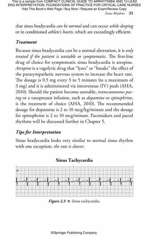
Sinus Tachycardia
ti0065
p0115
ti0070
p0120
ti0075
II
Figure 2.3 n Sinus tachycardia.
This is a sample from COMPACT CLINICAL GUIDE TO ARRHYTHMIA AND 12-LEAD EKG INTERPRETATION: FOUNDATIONS OF PRACTICE FOR CRITICAL CARE NURSES
Visit This Book’s Web Page / Buy Now / Request an Exam/Review Copy
© Springer Publishing Company
© Springer Publishing Company
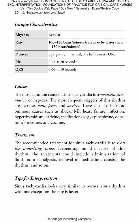
26 2. Arrhythmias: Sinus and Atrial
Unique Characteristicsti0080
Causes
The most common cause of sinus tachycardia is sympathetic stim-ulation or hypoxia. The most frequent triggers of this rhythm are exercise, pain, fever, and anxiety. There can also be more ominous causes such as shock, MI, heart failure, infection, hyperthyroidism, caffeine, medications (e.g., epinephrine, dopa-mine), nicotine, and cocaine.
Treatment
The recommended treatment for sinus tachycardia is to treat the underlying cause. Depending on the cause of this rhythm, the treatments could include administration of fluid and an analgesic, removal of medications causing the rhythm, and so on.
Tips for Interpretation
Sinus tachycardia looks very similar to normal sinus rhythm with one exception: the rate is faster.
ti0085
p0125
ti0090
p0130
ti0095
p0135
Rhythm Regular
Rate 100–150 beats/minute (rate may be faster than 150 beats/minute)
P waves Upright, symmetrical, one before every QRS
PRi 0.12–0.20 seconds
QRS 0.06–0.10 seconds
td0055td0060td0065td0070
td0075td0080td0085td0090td0095td0100

This is a sample from COMPACT CLINICAL GUIDE TO ARRHYTHMIA AND 12-LEAD EKG INTERPRETATION: FOUNDATIONS OF PRACTICE FOR CRITICAL CARE NURSES
Visit This Book’s Web Page / Buy Now / Request an Exam/Review Copy
© Springer Publishing Company
© Springer Publishing Company
Sinus Rhythms 27
Sinus Arrhythmiati0100
Causes
This rhythm can be caused by changes in intrathoracic pressure, hence the changes in rate, with some faster beats and other periods with slower beats corresponding to inspiration and expiration, respectively. Sinus arrhythmia can also be seen in healthy hearts, especially in children or young adults (< 30 years of age). Other causes include inferior MI, increased intracranial pressure, and medi-cations (e.g., digoxin or morphine).
ti0110
p0140
Unique Characteristicsti0105
Rhythm Irregular
Rate 60–100 beats/minute
P waves Upright, symmetrical, one before every QRS
PRi 0.12–0.20 seconds
QRS 0.06–0.10 seconds
td0105td0110td0115td0120td0125td0130td0135td0140td0145td0150
II
Figure 2.4 n Examples of sinus arrhythmia.
This is a sample from COMPACT CLINICAL GUIDE TO ARRHYTHMIA AND 12-LEAD EKG INTERPRETATION: FOUNDATIONS OF PRACTICE FOR CRITICAL CARE NURSES
Visit This Book’s Web Page / Buy Now / Request an Exam/Review Copy
© Springer Publishing Company
© Springer Publishing Company
28 2. Arrhythmias: Sinus and Atrial
Treatment
No treatment is required for this benign rhythm.
Tips for Interpretation
Sinus arrhythmia looks very similar to normal sinus rhythm with one exception: The rhythm is irregular. In fact, you will notice the rhythm can often have a pattern of speeding up and slowing down (sometimes in relation to inspiration and expiration), therefore you will see that part of the rhythm has a quicker pace and then it slows and the process repeats itself.
ti0115
p0145
ti0120
p0150
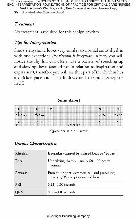
Sinus Arrestti0125
03:21:4944
NN NN NN NNMM
Unique Characteristicsti0130
Rhythm Irregular (caused by missed beat or “pause”)
Rate Underlying rhythm usually 60–100 beats/ minute
P waves Present, upright, symmetrical, and preceding every QRS except in missed beat
PRi 0.12–0.20 seconds
QRS 0.06–0.10 seconds
td0475td0480td0485td0490
td0495td0500td0505td0510
td0515td0520
Figure 2.5 n Sinus arrest.
This is a sample from COMPACT CLINICAL GUIDE TO ARRHYTHMIA AND 12-LEAD EKG INTERPRETATION: FOUNDATIONS OF PRACTICE FOR CRITICAL CARE NURSES
Visit This Book’s Web Page / Buy Now / Request an Exam/Review Copy
© Springer Publishing Company
© Springer Publishing Company
Sinus Rhythms 29
Causes
There are a number of potential causes of sinus block, which include MI; medications, such as digoxin; myocarditis; heart failure; carotid sinus sensitivity; and increased vagal tone. Sinus arrest is caused by similar factors but may also be caused by hyperkalemia, beta blockers, or calcium channel blockers.
Treatment
There is typically no treatment required for sinus block or sinus arrest unless there is hemodynamic compromise (e.g., signifi-cant hypotension).
Tips for Interpretation
Sinus arrest and sinus block appear as a missed beat or pause in the rhythm. This is how to tell them apart: Sinus block drops the beat and resets the rhythm right on time, whereas sinus arrest drops a beat but does not reset to the underlying rhythm at exactly two R–R intervals as sinus block does. In the case of a sinus block, the impulse arises normally in the sinoatrial (SA) node but is not transmitted. With a sinus arrest, the impulse fails to arise in the SA node and, therefore, there is also no transmission of the impulse.
ti0135
p0155
ti0140
p0160
ti0145
p0165
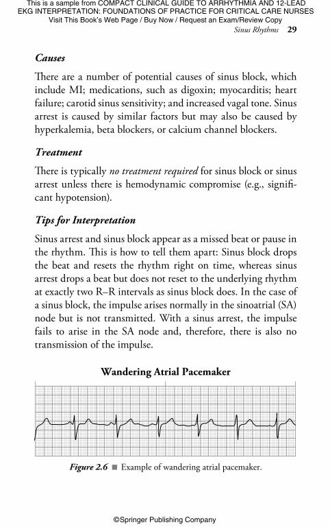
Wandering Atrial Pacemaker
ti0150
Figure 2.6 n Example of wandering atrial pacemaker.
This is a sample from COMPACT CLINICAL GUIDE TO ARRHYTHMIA AND 12-LEAD EKG INTERPRETATION: FOUNDATIONS OF PRACTICE FOR CRITICAL CARE NURSES
Visit This Book’s Web Page / Buy Now / Request an Exam/Review Copy
© Springer Publishing Company
© Springer Publishing Company
30 2. Arrhythmias: Sinus and Atrial
Causes
This rhythm can be seen in normal hearts or during sleep. Occasionally, the wandering atrial pacemaker (WAP) rhythm can be caused by digoxin toxicity.
Treatment
No treatment required.
Tips for Interpretation
In the case of a wandering pacemaker, the pacemaker “ wanders” between the SA node and the atria causing the impulses to be generated from a variety of locations in the atria. As the origin of where the impulse varies, this causes a change in the shape and size of the P waves. P waves in this rhythm are not the perfectly upright, rounded, and symmetrical P waves seen when all impulses arise directly from the SA node.
ATRIAL RHYTHMS
Atrial rhythms arise from an irritable focus in the atria. These rhythms can have significant effects for the patient hemody-namically and can result in the loss of atrial kick or a
ti0160
p0170
ti0165
p0175
ti0170
p0180
ti0175
p0185
Unique Characteristicsti0155
Rhythm Regular or slightly irregular
Rate 60–100 beats/minute
P waves Vary in shape and size, one for every QRS
PRi 0.12–0.20 seconds
QRS 0.06–0.10 seconds
td0155td0160td0165td0170td0175td0180td0185td0190
td0195td0200
This is a sample from COMPACT CLINICAL GUIDE TO ARRHYTHMIA AND 12-LEAD EKG INTERPRETATION: FOUNDATIONS OF PRACTICE FOR CRITICAL CARE NURSES
Visit This Book’s Web Page / Buy Now / Request an Exam/Review Copy
© Springer Publishing Company
© Springer Publishing Company
Atrial Rhythms 31
synchronized atrial contraction. The result of no atrial kick, for example, in the case of atrial flutter and atrial fibrillation, can result in a decrease in cardiac output of up to 30%. This drop in cardiac output can have serious effects on a patient’s blood pres-sure, level of consciousness, and overall hemodynamic stability. In addition, when the atria are fluttering or fibrillating, mini clots can form in the atria and can cause serious consequences (e.g., stroke) if released. The first sign of trouble in the atria’s electrical conduction system is when PACs begin to appear. These early ectopic beats superimposed on the underlying rhythm can transition very quickly into more dangerous atrial arrhythmias such as atrial flutter, atrial fibrillation, and SVT.
The primary cause of atrial arrhythmias includes factors such as overstretch or understretch (e.g., as with fluid overload or hypovolemia) of the atria and ischemia over the conduc-tion system. Atrial arrhythmias must be identified and treated urgently to prevent further deterioration or sequelae (e.g., stroke, severe hypotension) in the patient.
p0190
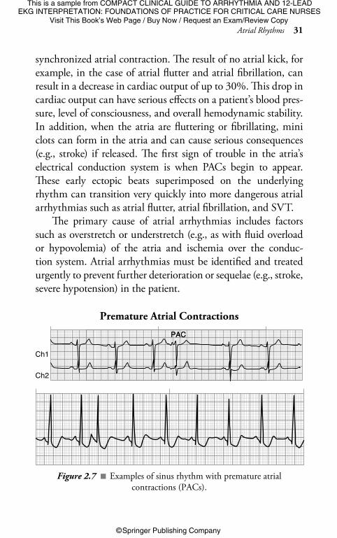
Premature Atrial Contractionsti0180
Ch1
Ch2
PACPAC
Figure 2.7 n Examples of sinus rhythm with premature atrial contractions (PACs).
This is a sample from COMPACT CLINICAL GUIDE TO ARRHYTHMIA AND 12-LEAD EKG INTERPRETATION: FOUNDATIONS OF PRACTICE FOR CRITICAL CARE NURSES
Visit This Book’s Web Page / Buy Now / Request an Exam/Review Copy
© Springer Publishing Company
© Springer Publishing Company
32 2. Arrhythmias: Sinus and Atrial
Causes
Overall, atrial arrhythmias are caused by one of three underly-ing mechanisms: altered automaticity, triggered activity, or reentry issues. Altered automaticity can be caused by ischemia, drug toxicity, hypocalcemia, or hypoxia. Triggers can include such factors as ischemia, hypoxia, or low levels of magnesium. Reentry issues that cause atrial arrhythmias can be initiated by hyperkalemia and some antiarrhythmics. PACs are the first sign that there is irritability in the atria.
Treatment
Typically, there is no treatment required for PACs. The underlying cause should be treated and the frequency of PACs monitored because an increase in PACs indicates an increase in irritability of the atria and this could lead to a deterioration of the rhythm into a more dangerous atrial arrhythmia such as atrial fibrillation or SVT.
Tips for Interpretation
Watch for early beats with an upright P wave that looks slightly different than the underlying rhythm. Also, in some cases, the
ti0190
p0195
ti0195
p0200
ti0200
p0205
Unique Characteristicsti0185
Rhythm Irregular because of the early beat (PAC) superimposed on the underlying rhythm
Rate Usually 60–100 beats/minute in the underlying rhythm
P waves P waves appear differently in the PAC versus the underlying sinus rhythm (as their origin is from the atria vs. the SA node).
PRi 0.12–0.20 seconds
QRS 0.06–0.10 seconds
td0205td0210td0215
td0220
td0225td0230
td0235
td0240td0245td0250
This is a sample from COMPACT CLINICAL GUIDE TO ARRHYTHMIA AND 12-LEAD EKG INTERPRETATION: FOUNDATIONS OF PRACTICE FOR CRITICAL CARE NURSES
Visit This Book’s Web Page / Buy Now / Request an Exam/Review Copy
© Springer Publishing Company
© Springer Publishing Company
Atrial Rhythms 33
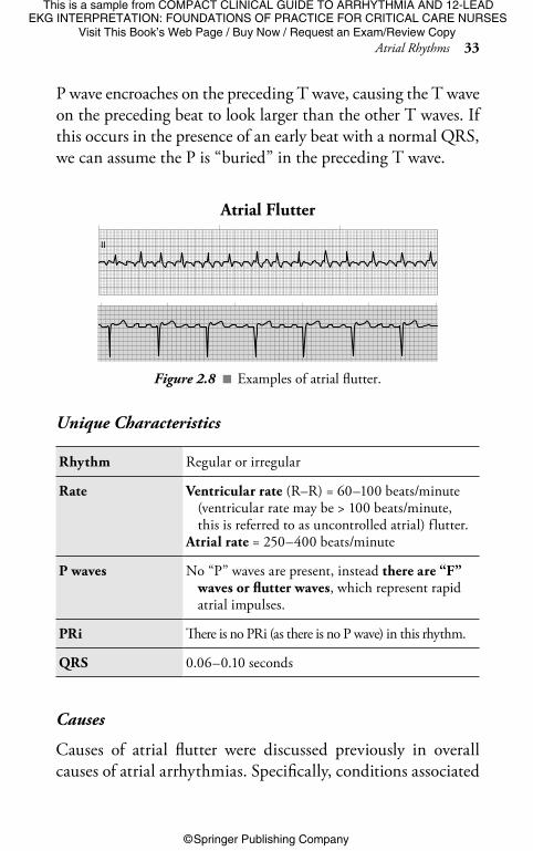
Atrial Flutterti0205
II
P wave encroaches on the preceding T wave, causing the T wave on the preceding beat to look larger than the other T waves. If this occurs in the presence of an early beat with a normal QRS, we can assume the P is “buried” in the preceding T wave.
Figure 2.8 n Examples of atrial flutter.
Unique Characteristicsti0210
Rhythm Regular or irregular
Rate Ventricular rate (R–R) = 60–100 beats/minute (ventricular rate may be > 100 beats/minute, this is referred to as uncontrolled atrial) f lutter.
Atrial rate = 250–400 beats/minute
P waves No “P” waves are present, instead there are “F” waves or flutter waves, which represent rapid atrial impulses.
PRi There is no PRi (as there is no P wave) in this rhythm.
QRS 0.06–0.10 seconds
td0525td0530td0535td0540
td0545td0550td0555
td0560td0565td0570td0575
Causes
Causes of atrial flutter were discussed previously in overall causes of atrial arrhythmias. Specifically, conditions associated
ti0215
p0210

This is a sample from COMPACT CLINICAL GUIDE TO ARRHYTHMIA AND 12-LEAD EKG INTERPRETATION: FOUNDATIONS OF PRACTICE FOR CRITICAL CARE NURSES
Visit This Book’s Web Page / Buy Now / Request an Exam/Review Copy
© Springer Publishing Company
© Springer Publishing Company
34 2. Arrhythmias: Sinus and Atrial
with atrial flutter include hypoxia, pulmonary embolism, pneumonia, chronic lung disease, coronary artery disease, MI, digoxin toxicity, myocarditis, or pericarditis. Patients who experience atrial flutter have a loss of atrial kick that can subsequently cause them to have palpitations, shortness of breath, fatigue, and chest discomfort caused by hypotension.
Treatment
Rate control is typically indicated as the priority treatment if the patient is stable. Rate control is typically achieved through use of beta blockers and calcium channel block-ers such as diltiazem. Amiodarone may also be used, espe-cially in the patient with heart failure. If the patient is unstable (particularly with accompanying high ventricular rates), cardioversion is recommended as the first-line treat-ment to halt the rhythm. Instability can manifest itself as severe hypotension, heart failure, or signs of shock. A discus-sion of cardioversion is found in Table 2.1. In addition, if the patient has been experiencing atrial flutter for more than 48 hours, the risk of emboli must be considered. Anticoagulants are recommended prior to cardioversion unless the patient is unstable and immediate electrical inter-vention is required (cardioversion).
Tips for Interpretation
In atrial flutter, there are no P waves; therefore, there is no PRi. Instead, there are flutter waves that are continuous and carry on right through the QRS. One of the key features of atrial flutter is the “ sawtooth” pattern of the flutter waves making it a very distinctive looking rhythm. In order to
ti0220
p0215
ti0225
p0220
This is a sample from COMPACT CLINICAL GUIDE TO ARRHYTHMIA AND 12-LEAD EKG INTERPRETATION: FOUNDATIONS OF PRACTICE FOR CRITICAL CARE NURSES
Visit This Book’s Web Page / Buy Now / Request an Exam/Review Copy
© Springer Publishing Company
© Springer Publishing Company
Atrial Rhythms 35
calculate the ratio of flutter waves (e.g., atrial flutter with a 4:1 conduction), you must first calculate the ventricular rate and then calculate the atrial rate. If the R–R is irregular, this is referred to as a varying conduction. When the R–R is regular, the ventricular rate is divided into the atrial rate to calculate the conduction ratio. For example, if the ventricular (R–R) rate is 75 and the atrial rate is 300 (beginning of one flutter wave to beginning of the next) the conduction ratio is 4:1. Please note: You do not calculate the conduction ratio by counting the number of F (flutter) waves between R waves because flutter waves are continuous.
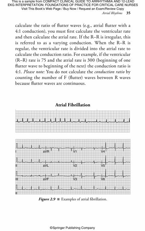
Atrial Fibrillationti0230
I aVR V1 V4
II
II
aVL V2 V5
III aVF V3 V6
Figure 2.9 n Examples of atrial fibrillation.

This is a sample from COMPACT CLINICAL GUIDE TO ARRHYTHMIA AND 12-LEAD EKG INTERPRETATION: FOUNDATIONS OF PRACTICE FOR CRITICAL CARE NURSES
Visit This Book’s Web Page / Buy Now / Request an Exam/Review Copy
© Springer Publishing Company
© Springer Publishing Company
36 2. Arrhythmias: Sinus and Atrial
Unique Characteristicsti0235
Rhythm Irregular
Rate Ventricular rate: 60–100 beats/minute (ventricular rate may be > 100 beats/minute, this is referred to as uncontrolled atrial fibrillation)
Atrial rate: 400–600 beats/minute (This cannot be measured on the rhythm strip because the waveform is so chaotic.)
P waves There are no P waves, instead there are fibrillatory waves or “f” waves.
PRi There is no PRi since there is no P wave.
QRS 0.06–0.10 seconds
td0255td0260td0265td0270
td0275
td0280td0285td0290td0295
td0300
td0305
Causes
Atrial fibrillation is the most common type of arrhythmia. Causes of atrial fibrillation are similar to those of atrial flutter and produce similar signs and symptoms. Additional causes or triggers of atrial fibrillation can include excessive caffeine inges-tion, idiopathic (no clear cause), increased age, stress, hypoka-lemia, infection, and hyperthyroidism.
Treatment
The treatment of atrial fibrillation is similar to the treatment for atrial flutter (see “Atrial Flutter” section). Ablation may be required for persistent, recurring atrial fibrillation. Ablation refers to a procedure that eliminates tissue through an energy source, such as a laser or cryothermy, with the goal of stopping the cause of the arrhythmia.
ti0240
p0225
ti0245
p0230

This is a sample from COMPACT CLINICAL GUIDE TO ARRHYTHMIA AND 12-LEAD EKG INTERPRETATION: FOUNDATIONS OF PRACTICE FOR CRITICAL CARE NURSES
Visit This Book’s Web Page / Buy Now / Request an Exam/Review Copy
© Springer Publishing Company
© Springer Publishing Company
Atrial Rhythms 37
Tips for Interpretation
In atrial fibrillation, it is important to remember that the ventricular rhythm (R–R) is always irregular. Also, when naming the rhythm remember there is no such rhythm as atrial “ fib/ flutter.” The patient may have runs of atrial flutter and then atrial fibrilla-tion but they do not occur at the same time; therefore, each needs to be identified separately on the strip during interpretation.
ti0250
p0235
Unique Characteristicsti0260
Rhythm Regular
Rate 150–250 beats/minute (usually closer to 200 and over)
P waves Can be difficult to see when the rate is this fast
PRi If you can see them, 0.12–0.20 seconds
QRS 0.06–0.10 seconds
td0310td0315td0320td0325
td0330td0335td0340td0345td0350td0355
Causes
Most SVTs are caused by reentry mechanisms which refer to the fact that the rhythm takes an abnormal repetitive circuit of
ti0265
p0240
Atrial Tachycardia (aka SVT or PSVT)ti0255
Figure 2.10 n Supraventricular tachycardia (SVT).
This is a sample from COMPACT CLINICAL GUIDE TO ARRHYTHMIA AND 12-LEAD EKG INTERPRETATION: FOUNDATIONS OF PRACTICE FOR CRITICAL CARE NURSES
Visit This Book’s Web Page / Buy Now / Request an Exam/Review Copy
© Springer Publishing Company
© Springer Publishing Company
38 2. Arrhythmias: Sinus and Atrial
cardiac tissue. Triggers can include stimulants (e.g., caffeine, albuterol), infection, electrolyte imbalance, or MI.
Treatment
Treatment for SVT includes consideration of vagal maneuvers (e.g., carotid sinus massage or having the patient bear down) and the administration of adenosine. Adenosine is initially given in a rapid 6-mg IV injection quickly followed by a 12-mg dose if the first was not successful. This medication creates almost a chemical type of “cardioversion” and has a very short half-life of only 10 to 15 seconds therefore the need for rapid administration. Upon admin-istration of adenosine, it is advisable to inform the patient that they may initially feel uncomfortable and anxious but that this only lasts for a short period. Reassure the patient that the rhythm is being monitored closely and the reason for giving this medication. Should adenosine and vagal maneuvers fail to convert the rhythm, calcium channel blockers or beta blockers may be considered. If the patient is unstable, immediate cardioversion is indicated.
Tips for Interpretation
SVT typically starts suddenly, is preceded by a PAC, and the patient is often symptomatic (e.g., light headed, feeling “palpitations,” flushed, or hypotensive). SVT is a broad term that can encompass paroxysmal atrial tachycardia or paroxysmal junctional tachycardia and refers to the fact that the “ runaway” accessory path impulses are “supra,” “ventricular,” or originating above the ventricles.
ti0270
p0245
ti0275
p0250
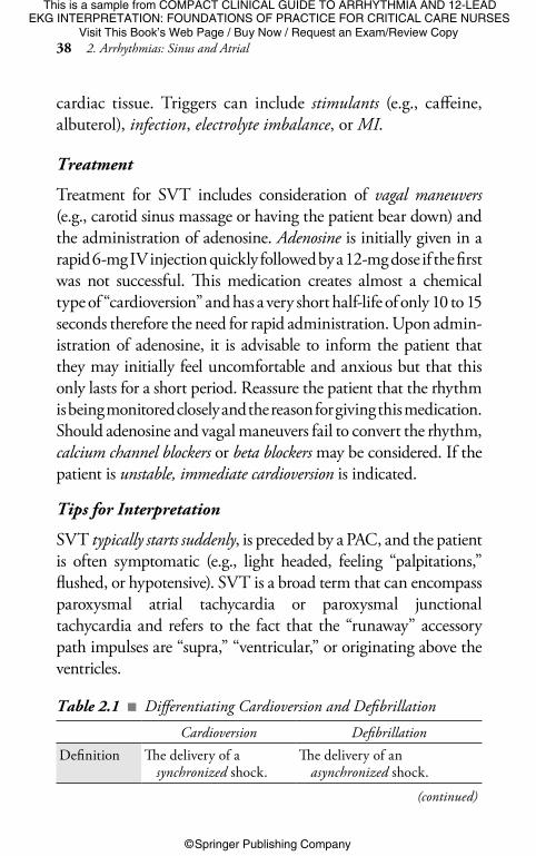
Table 2.1 n Differentiating Cardioversion and Defibrillation
Cardioversion DefibrillationDefinition The delivery of a
synchronized shock.The delivery of an
asynchronized shock.
t0040p0255
td0360td0365td0370td0375td0380
(continued)
This is a sample from COMPACT CLINICAL GUIDE TO ARRHYTHMIA AND 12-LEAD EKG INTERPRETATION: FOUNDATIONS OF PRACTICE FOR CRITICAL CARE NURSES
Visit This Book’s Web Page / Buy Now / Request an Exam/Review Copy
© Springer Publishing Company
© Springer Publishing Company
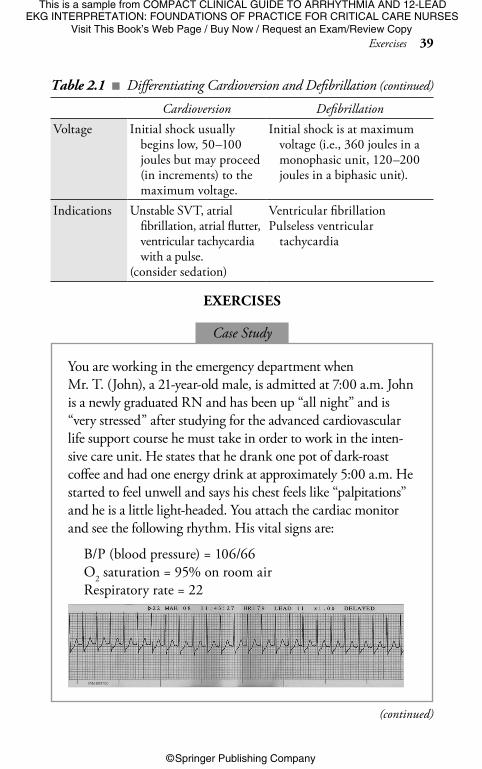
Exercises 39
You are working in the emergency department when Mr. T. (John), a 21-year-old male, is admitted at 7:00 a.m. John is a newly graduated RN and has been up “all night” and is “very stressed” after studying for the advanced cardiovascular life support course he must take in order to work in the inten-sive care unit. He states that he drank one pot of dark-roast coffee and had one energy drink at approximately 5:00 a.m. He started to feel unwell and says his chest feels like “palpitations” and he is a little light-headed. You attach the cardiac monitor and see the following rhythm. His vital signs are:
B/P (blood pressure) = 106/66O2 saturation = 95% on room airRespiratory rate = 22
p0260p0265
p0270p0275p0280
Case Study
(continued)
Cardioversion DefibrillationVoltage Initial shock usually
begins low, 50–100 joules but may proceed (in increments) to the maximum voltage.
Initial shock is at maximum voltage (i.e., 360 joules in a monophasic unit, 120–200 joules in a biphasic unit).
Indications Unstable SVT, atrial fibrillation, atrial flutter, ventricular tachycardia with a pulse.
(consider sedation)
Ventricular fibrillationPulseless ventricular
tachycardia
td0360td0365td0385td0390td0395td0400td0405td0410
td0415td0420
Table 2.1 n Differentiating Cardioversion and Defibrillation (continued)
ti0280 EXERCISES
This is a sample from COMPACT CLINICAL GUIDE TO ARRHYTHMIA AND 12-LEAD EKG INTERPRETATION: FOUNDATIONS OF PRACTICE FOR CRITICAL CARE NURSES
Visit This Book’s Web Page / Buy Now / Request an Exam/Review Copy
© Springer Publishing Company
© Springer Publishing Company
40 2. Arrhythmias: Sinus and Atrial
HR (heart rate) = 180Temp = 37.1°C
1. Would you consider John to be stable or unstable at this point?2. What rhythm is he in?3. What is the priority treatment for this rhythm according to
the current AHA guidelines?
Approximately 15 minutes after the initial treatment, John remains in the same rhythm and upon reassessment of his vital signs, his B/P is now 80/56 and he is losing consciousness.
4. What is the priority treatment for John at this point?
After this treatment is administered, John reverts to a rhythm that is regular, 80 beats/minute; PRi of 0.12 seconds; and QRS of 0.08 seconds.
5. What rhythm is John in now?6. What are the likely causes of John’s initial rhythm (on
admission)?
p0285p0290
p0295p0300p0305
p0310
p0315
p0320
p0325p0330
(continued)
1. What is the most common type of arrhythmia (and also one of the hardest to treat)?a. Sinus tachycardiab. Wandering atrial pacemakerc. Sinus blockd. Atrial fibrillation
2. The drug of choice in a symptomatic bradycardia is:a. Adenosineb. Amiodarone
p0335
p0340p0345p0350p0355p0360p0365p0370
Test Yourself !ti0285
(continued)
This is a sample from COMPACT CLINICAL GUIDE TO ARRHYTHMIA AND 12-LEAD EKG INTERPRETATION: FOUNDATIONS OF PRACTICE FOR CRITICAL CARE NURSES
Visit This Book’s Web Page / Buy Now / Request an Exam/Review Copy
© Springer Publishing Company
© Springer Publishing Company
Exercises 41
c. Atropined. Beta blocker
3. Treatment of sinus tachycardia includes:a. Beta blockersb. Pacingc. Treating the caused. Cardioversion
4. Sinus arrhythmia is a benign rhythm and no treatment is required.a. Trueb. False
5. The definitive characteristic in WAP is:a. An irregular rhythmb. The P wavesc. The rated. The QRS duration
p0375p0380p0385p0390p0395p0400p0405p0410
p0415p0420p0425p0430p0435p0440p0445
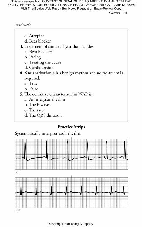
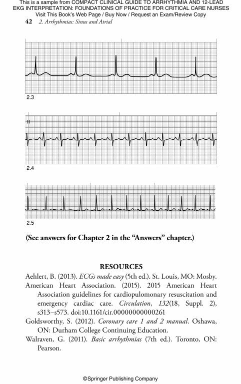
Practice StripsSystematically interpret each rhythm.
ti0290
2.1
2.2
(continued)
p0450
This is a sample from COMPACT CLINICAL GUIDE TO ARRHYTHMIA AND 12-LEAD EKG INTERPRETATION: FOUNDATIONS OF PRACTICE FOR CRITICAL CARE NURSES
Visit This Book’s Web Page / Buy Now / Request an Exam/Review Copy
© Springer Publishing Company
© Springer Publishing Company
42 2. Arrhythmias: Sinus and Atrial
(See answers for Chapter 2 in the “Answers” chapter.)p0455
RESOURCESAehlert, B. (2013). ECGs made easy (5th ed.). St. Louis, MO: Mosby.American Heart Association. (2015). 2015 American Heart
Association guidelines for cardiopulomonary resuscitation and emergency cardiac care. Circulation, 132(18, Suppl. 2), s313–s573. doi:10.1161/cir.00000000000261
Goldsworthy, S. (2012). Coronary care 1 and 2 manual. Oshawa, ON: Durham College Continuing Education.
Walraven, G. (2011). Basic arrhythmias (7th ed.). Toronto, ON: Pearson.
ti0295
2.3
II
2.4
2.5