Embed Size (px)
Citation preview

Color Plates

Plate 1. Steps required for gene delivery. Four basic steps are required for effective gene delivery leading to successful
transgene expression. The cellular and nuclear membrane barriers must be penetrated, and cytoplasmic degredation must
be avoided. (See also Fig. 1 in article by Garrity-Moses et al.)
Plate 2. Schematic representation of polyethylenimine (PEI)-enhanced delivery. Cationic polymers, such as PEI, provide
an alternative method of enhanced DNA plasmid delivery in gene therapy. Their ability to condense plasmid DNA and
cause lysosomal osmotic disruption contributes to increased transfection efficiency. (See also Fig. 2 in article by Garrity-
Moses et al.)
Plate 3. Components necessary for production of a viral vector. Illustration of the mechanisms involved in the
conversion of a wild-type virus into a vector for gene transfer. Key components include the promoter, transgene, and
polyadenylation (PolyA) sequence in the expression cassette. (See also Fig. 3 in article by Garrity-Moses et al.)
Plate 4. Retrograde axonal transport. Tissue demonstrating positive adenoviral gene transfer via retrograde axonal
transport to dorsal root ganglia primary sensory neurons. (See also Fig. 4 in article by Garrity-Moses et al.)
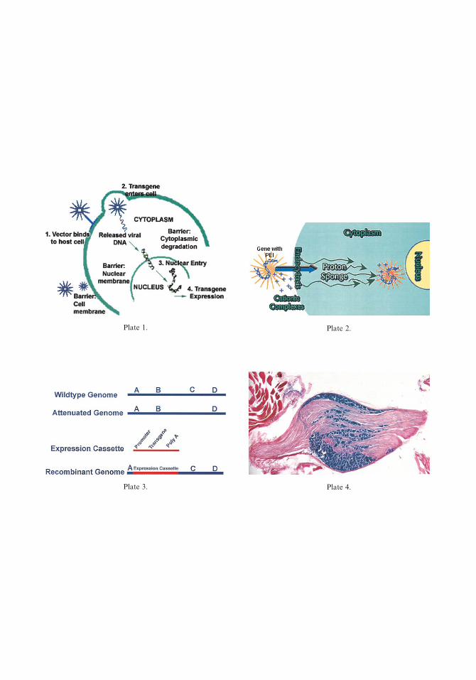
Plate 2.Plate 1.
Plate 3. Plate 4.

Plate 5. Green fluorescent protein (GFP) expressing dorsal root ganglia (DRG) neurons. Peripheral nerve injection of
adenoassociated virus GFP resulted in DRG expression of GFP. (See also Fig. 5 in article by Garrity-Moses et al.)
Plate 6. Gene-based neuromodulation. (1) Neuronal vector uptake. (2) Transgene transcription. (3) Transgene
translation. (4) Neurotransmitter precursor or neurotransmitter synthetic enzyme. (5) Ion channel. (6) Vesicle docking
protein. (7) Neurotransmitter reuptake protein. (8) Receptor. (9) Second messenger. (See also Fig. 6 in article by Garrity-
Moses et al.)
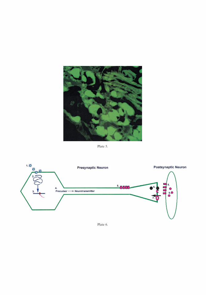
Plate 5.
Plate 6.

Plate 7. Green fluorescent protein (GFP) distribution in lateral hypothalamus after stereotactic injection. This figure
illustrates positive GFP expression in the lateral hypothalamus after injection of 500 nL of adenoassociated virus
glutamate decarboxylase GFP (left) and the Paxinos and Watson [103] atlas illustration of they targeted area (right).
(See also Fig. 7 in article by Garrity-Moses et al.)
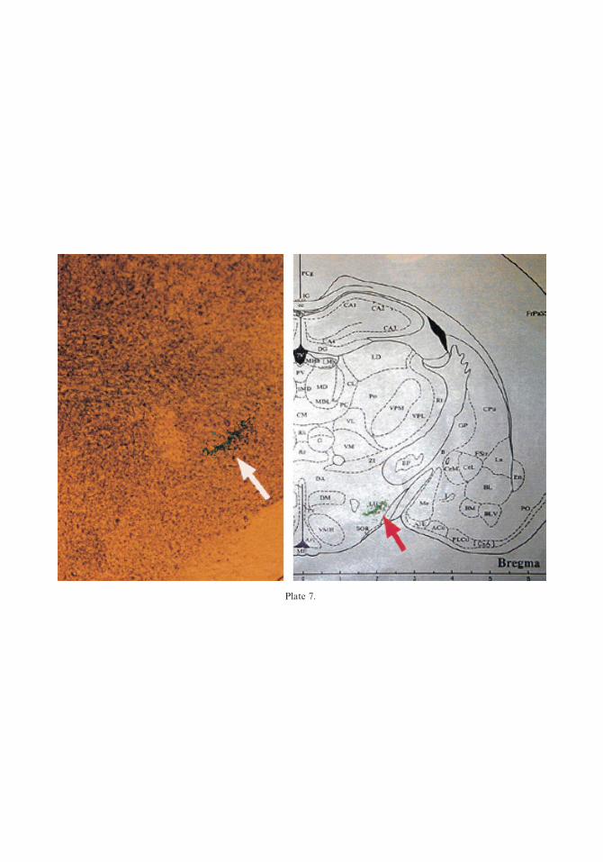
Plate 7.

Plate 8. Slope of average weight in rats. A postoperative comparison of the slope of the weight gain in rats injected with
adenoassociated virus glutamate in decarboxylase (GAD) versus rats injected with placebo and controls. A significant
decrease in weight gain is evident in the GAD rats. (See also Fig. 8 in article by Garrity-Moses et al.)
Plate 9. Ex vivo gene transfer. The delivery of analgesic proteins can be affected through gene transfer to cell lines that
are subsequently transplanted into the nervous system. Ex vivo gene therapy involves removing tissue from the patient,
transfecting the cells in culture, and then reimplanting the genetically altered cells to the patient. (See also Fig. 9 in article
by Garrity-Moses et al.)
Plate 10. Gene expression in dorsal root ganglia (DRG) after intraneural recombinant adenoassociated virus injection in
diabetic rats. The presence of green fluorescent protein in DRG sensory neurons serves as a positive indication of gene
expression. (See also Fig. 10 in article by Garrity-Moses et al.)
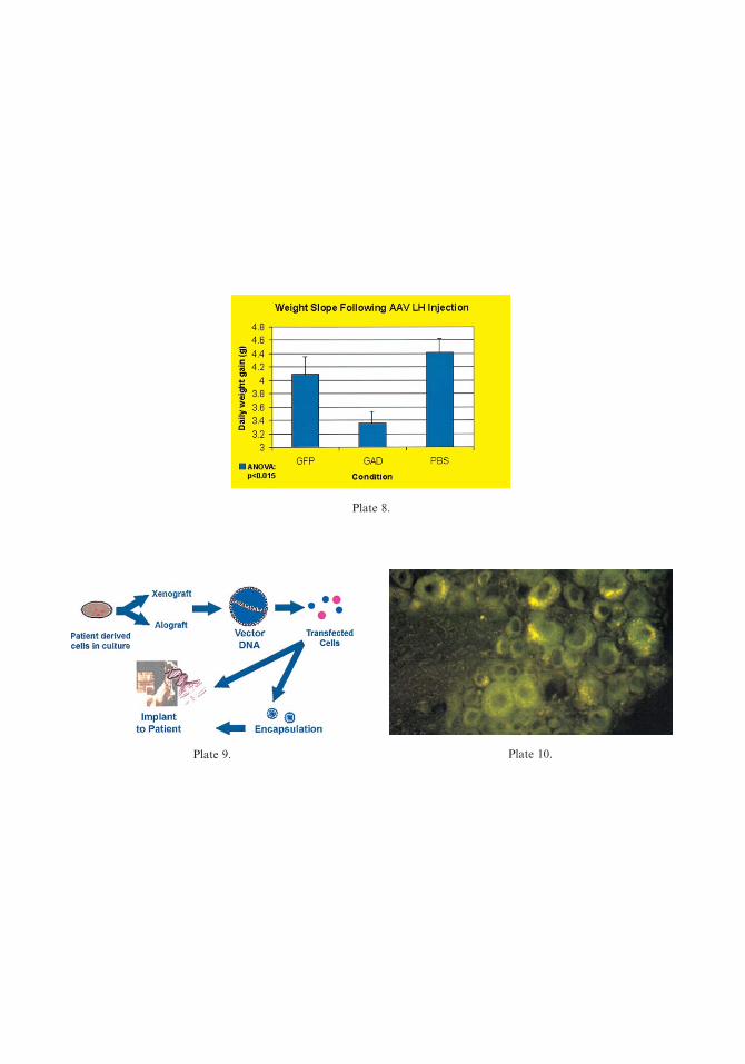
Plate 8.
Plate 9. Plate 10.


![Studies in Color Sensitive Photographic Plates and Methods ... · 5ja a,J£"] ColorSensitivePhotographicPlates 355 difficultywashaduntilthenotablediscoveryin1882ofeosin2 whichmadesilverbromidegelatineplatesmuchmoresensitiveto](https://img.dokumen.tips/doc/110x75/5fa2b12a0e101719c1678b25/studies-in-color-sensitive-photographic-plates-and-methods-5ja-aj.jpg)