Embed Size (px)
Citation preview
Closing a Large Maxillary Median Diastema using Bapat Power Arm
International Journal of Clinical Pediatric Dentistry, April-June 2017;10(2):201-204 201
IJCPD
Closing a Large Maxillary Median Diastema using Bapat Power Arm1Shirish M Bapat, 2Chanchal Singh, 3Prashant Bandejiya
IJCPD
CASE REPORT10.5005/jp-journals-10005-1435
1Director, Principal, Professor and Head, 2Professor and Head 3Postgraduate Student1,3Department of Orthodontics and Dentofacial Orthopedics K.D. Dental College & Hospital, Mathura, Uttar Pradesh, India2Department of Pedodontics and Preventive Dentistry, K.D. Dental College & Hospital, Mathura, Uttar Pradesh, India
Corresponding Author: Shirish M Bapat, Director, Principal Professor and Head, Department of Orthodontics and Dentofacial Orthopedics, K.D. Dental College & Hospital Mathura, Uttar Pradesh, India, Phone: +9102026853382, e-mail: [email protected]
ABSTRACT
Aim: The aim of this study is to present a case of large maxil-lary median diastema closed by bodily movement of central incisors using Bapat power arm (BPA).
Materials and methods: After extraction of mesiodens, a power chain with a force of 120 gm was applied to BPA ligated to preadjusted edgewise brackets bonded to maxillary central incisors to move them over round steel wire for closure of resultant diastema. Bonded retainer was placed after the closure of median diastema.
Results: The median diastema was completely closed in 5 months period with almost bodily movement of incisors, which was confirmed by periapical X-ray.
Conclusion: Bapat power arm was efficient in closing dia-stema without any discomfort or injury and was well accepted by the patient.
Keywords: Bapat power arm, Bodily movement, Diastema closure.
How to cite this article: Bapat SM, Singh C, Bandejiya P. Closing a Large Maxillary Median Diastema using Bapat Power Arm. Int J Clin Pediatr Dent 2017;10(2):201-204.
Source of support: Nil
Conflict of interest: None
INTRODUCTION
The median diastema is a condition of having central incisors with intervening space between them. Such a space, called diastema, is certainly annoying for affected individuals as it is easily noticed and esthetically dis-pleasing. Therefore, it is a matter of great concern for the child as well as parents.1 Such spaces are usually seen in early mixed dentition period, around 6 to 9 years, but
spontaneously disappear by the time maxillary perma-nent canines erupt and often require no intervention.2-4 However, in some children, diastema continues to persist till adult age.
Occurrence of such diastemas is attributed to mul-tifactorial etiological reasons like normal physiological event, genetic and racial predisposition, developmental defects and congenital anomalies, local physical impedi-ments, muscular imbalance, pernicious habits, dental anomalies, or iatrogenic result of orthodontic procedures like rapid expansion.5,6 Prevalence of median diastema is high around age 6 to 7 years (40–50%) but diminishes by 15 years (5–7%).7,8 It is reported more in females at the age of 6 years but more males present with diastemas by 14 years of age than females.6 Diastemas of less than 2 mm in 9-year-old children generally close spontaneously.6,9,10
AIM
The aim of this study is to present a case of a large median diastema closed by bodily movement of maxillary central incisors using Bapat power arm (BPA).11
CASE REPORT
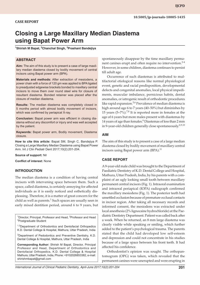
A 9-year-old male child was brought to the Department of Paediatric Dentistry of K.D. Dental College and Hospital, Mathura, Uttar Pradesh, India, by his parents with a com-plaint of an ugly looking small tooth between maxillary permanent central incisors (Fig. 1). Intraoral examination and intraoral periapical (IOPA) radiograph confirmed the maxillary mesiodens (Fig. 1). The posterior teeth had unsettled occlusion because of premature occlusal contacts in incisor region. After taking all necessary records and informed consent, the mesiodens was extracted under local anesthesia (2% lignocaine hydrochloride) at the Pae-diatric Dentistry Department. Patient was called back after a week. When he returned, an 8 mm large diastema was clearly visible while speaking or smiling, which further added to the patient’s psychological trauma. The parents stated that the child had developed low self-esteem and depression and could not concentrate in his studies because of a large space between his front teeth. It had affected his confidence.
Orthodontist’s opinion was sought. The orthopan-tomogram (OPG) was taken, which revealed that the permanent canines were unerupted and were erupting in
Shirish M Bapat et al
202
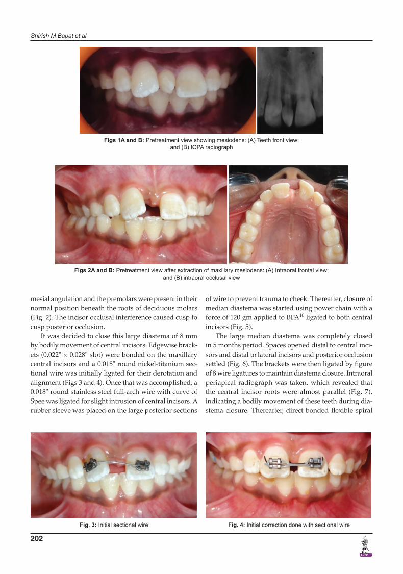
mesial angulation and the premolars were present in their normal position beneath the roots of deciduous molars (Fig. 2). The incisor occlusal interference caused cusp to cusp posterior occlusion.
It was decided to close this large diastema of 8 mm by bodily movement of central incisors. Edgewise brack-ets (0.022" × 0.028" slot) were bonded on the maxillary central incisors and a 0.018" round nickel-titanium sec-tional wire was initially ligated for their derotation and alignment (Figs 3 and 4). Once that was accomplished, a 0.018" round stainless steel full-arch wire with curve of Spee was ligated for slight intrusion of central incisors. A rubber sleeve was placed on the large posterior sections
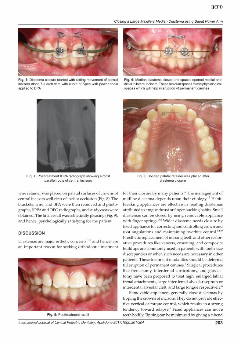
of wire to prevent trauma to cheek. Thereafter, closure of median diastema was started using power chain with a force of 120 gm applied to BPA10 ligated to both central incisors (Fig. 5).
The large median diastema was completely closed in 5 months period. Spaces opened distal to central inci-sors and distal to lateral incisors and posterior occlusion settled (Fig. 6). The brackets were then ligated by figure of 8 wire ligatures to maintain diastema closure. Intraoral periapical radiograph was taken, which revealed that the central incisor roots were almost parallel (Fig. 7), indicating a bodily movement of these teeth during dia-stema closure. Thereafter, direct bonded flexible spiral
Figs 1A and B: Pretreatment view showing mesiodens: (A) Teeth front view; and (B) IOPA radiograph
Figs 2A and B: Pretreatment view after extraction of maxillary mesiodens: (A) Intraoral frontal view; and (B) intraoral occlusal view
Fig. 3: Initial sectional wire Fig. 4: Initial correction done with sectional wire
Closing a Large Maxillary Median Diastema using Bapat Power Arm
International Journal of Clinical Pediatric Dentistry, April-June 2017;10(2):201-204 203
IJCPD
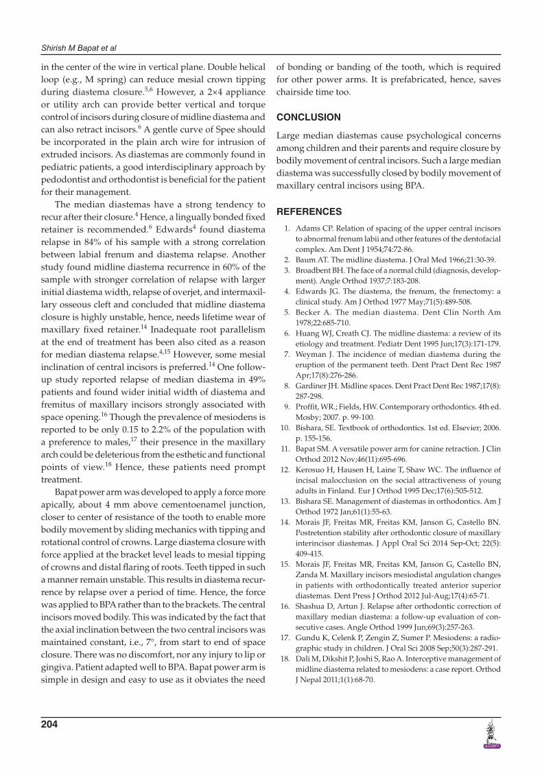
wire retainer was placed on palatal surfaces of crowns of central incisors well clear of incisor occlusion (Fig. 8). The brackets, wire, and BPA were then removed and photo-graphs, IOPA and OPG radiographs, and study casts were obtained. The final result was esthetically pleasing (Fig. 9), and hence, psychologically satisfying for the patient.
DISCUSSION
Diastemas are major esthetic concerns1,12 and hence, are an important reason for seeking orthodontic treatment
for their closure by many patients.6 The management of midline diastema depends upon their etiology.13 Habit-breaking appliances are effective in treating diastemas attributed to tongue thrust or finger-sucking habits. Small diastemas can be closed by using removable appliance with finger springs.5,6 Wider diastema needs closure by fixed appliance for correcting and controlling crown and root angulations and maintaining overbite control.5,6,9 Prosthetic replacement of missing teeth and other restor-ative procedures like veneers, crowning, and composite buildups are commonly used in patients with tooth size discrepancies or when such needs are necessary in other patients. These treatment modalities should be deferred till eruption of permanent canines.6 Surgical procedures like frenectomy, interdental corticotomy, and glossec-tomy have been proposed to treat high, enlarged labial frenal attachments, large interdental alveolar septum or interdental alveolar cleft, and large tongue respectively.6
Removable appliances generally close diastemas by tipping the crowns of incisors. They do not provide effec-tive vertical or torque control, which results in a strong tendency toward relapse.6 Fixed appliances can move teeth bodily. Tipping can be minimized by giving a v-bend
Fig. 5: Diastema closure started with sliding movement of central incisors along full arch wire with curve of Spee with power chain applied to BPA
Fig. 6: Median diastema closed and spaces opened mesial and distal to lateral incisors. These residual spaces mimic physiological spaces which will help in eruption of permanent canines
Fig. 7: Posttreatment IOPA radiograph showing almost parallel roots of central incisors
Fig. 8: Bonded palatal retainer was placed after diastema closure
Fig. 9: Posttreatment result
Shirish M Bapat et al
204
in the center of the wire in vertical plane. Double helical loop (e.g., M spring) can reduce mesial crown tipping during diastema closure.5,6 However, a 2×4 appliance or utility arch can provide better vertical and torque control of incisors during closure of midline diastema and can also retract incisors.6 A gentle curve of Spee should be incorporated in the plain arch wire for intrusion of extruded incisors. As diastemas are commonly found in pediatric patients, a good interdisciplinary approach by pedodontist and orthodontist is beneficial for the patient for their management.
The median diastemas have a strong tendency to recur after their closure.4 Hence, a lingually bonded fixed retainer is recommended.6 Edwards4 found diastema relapse in 84% of his sample with a strong correlation between labial frenum and diastema relapse. Another study found midline diastema recurrence in 60% of the sample with stronger correlation of relapse with larger initial diastema width, relapse of overjet, and intermaxil-lary osseous cleft and concluded that midline diastema closure is highly unstable, hence, needs lifetime wear of maxillary fixed retainer.14 Inadequate root parallelism at the end of treatment has been also cited as a reason for median diastema relapse.4,15 However, some mesial inclination of central incisors is preferred.14 One follow-up study reported relapse of median diastema in 49% patients and found wider initial width of diastema and fremitus of maxillary incisors strongly associated with space opening.16 Though the prevalence of mesiodens is reported to be only 0.15 to 2.2% of the population with a preference to males,17 their presence in the maxillary arch could be deleterious from the esthetic and functional points of view.18 Hence, these patients need prompt treatment.
Bapat power arm was developed to apply a force more apically, about 4 mm above cementoenamel junction, closer to center of resistance of the tooth to enable more bodily movement by sliding mechanics with tipping and rotational control of crowns. Large diastema closure with force applied at the bracket level leads to mesial tipping of crowns and distal flaring of roots. Teeth tipped in such a manner remain unstable. This results in diastema recur-rence by relapse over a period of time. Hence, the force was applied to BPA rather than to the brackets. The central incisors moved bodily. This was indicated by the fact that the axial inclination between the two central incisors was maintained constant, i.e., 7°, from start to end of space closure. There was no discomfort, nor any injury to lip or gingiva. Patient adapted well to BPA. Bapat power arm is simple in design and easy to use as it obviates the need
of bonding or banding of the tooth, which is required for other power arms. It is prefabricated, hence, saves chairside time too.
CONCLUSION
Large median diastemas cause psychological concerns among children and their parents and require closure by bodily movement of central incisors. Such a large median diastema was successfully closed by bodily movement of maxillary central incisors using BPA.
REFERENCES
1. Adams CP. Relation of spacing of the upper central incisors to abnormal frenum labii and other features of the dentofacial complex. Am Dent J 1954;74:72-86.
2. Baum AT. The midline diastema. J Oral Med 1966;21:30-39. 3. Broadbent BH. The face of a normal child (diagnosis, develop-
ment). Angle Orthod 1937;7:183-208. 4. Edwards JG. The diastema, the frenum, the frenectomy: a
clinical study. Am J Orthod 1977 May;71(5):489-508. 5. Becker A. The median diastema. Dent Clin North Am
1978;22:685-710. 6. Huang WJ, Creath CJ. The midline diastema: a review of its
etiology and treatment. Pediatr Dent 1995 Jun;17(3):171-179. 7. Weyman J. The incidence of median diastema during the
eruption of the permanent teeth. Dent Pract Dent Rec 1987 Apr;17(8):276-286.
8. Gardiner JH. Midline spaces. Dent Pract Dent Rec 1987;17(8): 287-298.
9. Proffit, WR.; Fields, HW. Contemporary orthodontics. 4th ed. Mosby; 2007. p. 99-100.
10. Bishara, SE. Textbook of orthodontics. 1st ed. Elsevier; 2006. p. 155-156.
11. Bapat SM. A versatile power arm for canine retraction. J Clin Orthod 2012 Nov;46(11):695-696.
12. Kerosuo H, Hausen H, Laine T, Shaw WC. The influence of incisal malocclusion on the social attractiveness of young adults in Finland. Eur J Orthod 1995 Dec;17(6):505-512.
13. Bishara SE. Management of diastemas in orthodontics. Am J Orthod 1972 Jan;61(1):55-63.
14. Morais JF, Freitas MR, Freitas KM, Janson G, Castello BN. Postretention stability after orthodontic closure of maxillary interincisor diastemas. J Appl Oral Sci 2014 Sep-Oct; 22(5): 409-415.
15. Morais JF, Freitas MR, Freitas KM, Janson G, Castello BN, Zanda M. Maxillary incisors mesiodistal angulation changes in patients with orthodontically treated anterior superior diastemas. Dent Press J Orthod 2012 Jul-Aug;17(4):65-71.
16. Shashua D, Artun J. Relapse after orthodontic correction of maxillary median diastema: a follow-up evaluation of con-secutive cases. Angle Orthod 1999 Jun;69(3):257-263.
17. Gundu K, Celenk P, Zengin Z, Sumer P. Mesiodens: a radio-graphic study in children. J Oral Sci 2008 Sep;50(3):287-291.
18. Dali M, Dikshit P, Joshi S, Rao A. Interceptive management of midline diastema related to mesiodens: a case report. Orthod J Nepal 2011;1(1):68-70.