Embed Size (px)
Citation preview
130 | march 2011 | volume 41 | number 3 | journal of orthopaedic & sports physical therapy
[ literature review ]
Low back pain (LBP) continues to be a major health problem and burden for individuals and society.16,63 As LBP is a heterogeneous condition, its classification into specific subgroups or syndromes has been suggested to aid the
diagnosis of specific pathologies, assist in management decisions, and improve outcomes.8,25 Structural lumbar segmental instability
(LSI) is universally recognized as an identifiable subgroup of individuals with LBP and is suggested to be a significant
TT STUDY DESIGN: Systematic literature review.
TT OBJECTIVES: To evaluate the diagnostic accu-racy of clinical tests used to diagnose patients with structural lumbar segmental instability (LSI).
TT BACKGROUND: Patients with structural LSI represent an important, identifiable subgrouping of individuals with low back pain. Numerous clinical tests have been proposed to diagnose structural LSI; however, data on the diagnostic accuracy of these tests have not yet been evaluated through a systematic review of the literature.
TT METHODS: A systematic review was conducted in 6 electronic databases for diagnostic accuracy studies, published between January 1950 and March 2010, that evaluated clinical tests against radiological diagnosis of structural LSI. The diagnostic accuracy of the clinical tests from the retrieved articles was independently evaluated, re-viewed, and quality scored using the QUADAS tool.
TT RESULTS: Four articles and a total of 11 clinical tests used in the diagnosis of structural LSI met the study inclusion criteria. The majority of tests had high specificity but low sensitivity, with posi-
tive likelihood ratios ranging from very small to moderate. QUADAS scores ranged from 16 to 25 out of a possible 26. The passive lumbar extension test was the most accurate clinical test, with high sensitivity (84%), specificity (90%), and a positive likelihood ratio of 8.8 (95% CI: 4.5, 17.3), indicating that this clinical test may be useful in the differen-tial diagnosis of structural LSI.
TT CONCLUSION: This systematic review found that the majority of clinical tests routinely em-ployed to diagnose structural LSI demonstrated only limited ability to do so. The results do, how-ever, indicate that the passive lumbar extension test may be useful in orthopaedic clinical practice to diagnose structural LSI. Additional research is required to further validate its use for diagnosing structural LSI in all populations of those with low back pain.
TT LEVEL OF EVIDENCE: Diagnosis, level 2a. J Orthop Sports Phys Ther 2011;41(3):130-140, Epub 2 February 2011. doi:10.2519/jospt.2011.3457
TT KEY WORDS: accuracy, low back pain, physical examination, validity
1Physical Therapist, Centre for Physiotherapy Research, University of Otago, Dunedin, New Zealand. 2Senior Lecturer, Centre for Physiotherapy Research, University of Otago, Dunedin, New Zealand. 3Professional Practice Fellow, Centre for Physiotherapy Research, University of Otago, Dunedin, New Zealand. Address correspondence to Dr Anthony G. Schneiders, School of Physiotherapy, University of Otago, 325 Great King Street, Dunedin, New Zealand. E-mail: [email protected]
ABDULLAH M. ALQARNI, PT, MPhty1 • ANTHONY G. SCHNEIDERS, PT, PhD2 • PAUL A. HENDRICK, PT, MPhty3
Clinical Tests to Diagnose Lumbar Segmental Instability:
A Systematic Review
cause of morbidity associated with spinal dysfunction.23,42,48
The concept of structural LSI was
first proposed by Knutsson,32 who advocated the assessment of LSI from the retrodisplacement (anterior-to-posterior transla-tion) of lumbar vertebrae on lateral radiographs taken at end
range spinal flexion and extension. Sub-sequently, White and Panjabi60 defined the related concept of “functional” lum-bar instability as loss of the spine’s ability to maintain its pattern of displacement under normal physiological loads. Pan-jabi44 further described functional LSI in relation to the neutral and elastic zones of the functional spinal unit. Functional LSI is proposed to exist throughout spi-nal motion; but assessment is specifically focused on midrange spinal movements, where the neutral zone is suggested to be more manifest.17,19,50 While radiographic diagnosis of structural LSI is considered quantifiable, traditionally by assessment of vertebral translation at the end range spinal motion,52 the discrimination of functional LSI has not been consistently characterized in the literature and clini-cal diagnostic tests for functional LSI have not been specifically evaluated. It is, however, recognized that functional instability can exist in the absence of ra-diological evidence of LSI.10
Cited causes of structural LSI include disc degeneration,7 postoperative spinal fusion40 (purported to produce abnormal
41-03 Alqarni.indd 130 2/24/2011 4:35:07 PM
journal of orthopaedic & sports physical therapy | volume 41 | number 3 | march 2011 | 131
pain,”42 an “instability catch” sensation during return from a flexed position,41,55 and a sensation of “slipping out” during the normal demands of spinal mobility.41 Additionally, subjective patient reports of pain exacerbation in the morning, on standing or rolling over, and with wors-ening weather, are all considered to be as-sociated with structural LSI.31 However, to date, none of these clinical features have been validated as accurate diagnos-tic signs of structural LSI.1
Orthopaedic clinical tests for structur-al LSI have also been routinely described in the literature and can be broadly di-vided into either passive or active tests. Passive tests include the posterior shear
agement strategies.8,14,23 The diagnosis of structural LSI currently relies on radio-graphic confirmation, which exposes the patient to radiation and has limitations associated with access and cost.47 Effec-tive noninvasive clinical tests to identify structural LSI would aid differential di-agnosis, as well as improve understand-ing of the aetiology and management of this condition.
Numerous clinical examination find-ings have been proposed and promoted as signs and symptoms of structural LSI. These signs include palpation of vertebral malalignment, excessive passive interver-tebral motion,46 subjective patient reports of “giving away,” “locking,” “through range
stresses on adjacent cephalad or caudal nonfused segments), postoperative disk excision or extensive decompression, and a history of trauma or recurrent LBP.14 Furthermore, a number of coexisting morphologies have been identified, in-cluding traction spurs,6,40 facet joint hy-pertrophy, and osteophytic formation.7 The estimated prevalence of structural LSI reported in the literature ranges from 12% of patients attending physical therapy for nonspecific LBP1 to 57% of patients with LBP who are suspected of having structural LSI.23
The differential diagnosis of LBP subgroups, such as structural LSI, is considered vital for more effective man-
Search strategy:1. "low back pain" OR "instability" OR
"lumbar spine” OR "spondylolisthesis" OR "anterolisthesis " OR "posterolisthesis" OR "spondylolysis"
2. "validity" OR "sensitivity" OR "specificity"3. "diagnosis" OR "clinical tests" OR
"provocation tests" OR "palpation" OR "physical examination"
4. (1 AND 2), (1 AND 3), (1 AND 2 AND 3)
Databases:1. CINAHL (n = 483)2. MEDLINE (n = 3741)3. PubMed (n = 4512)4. Scopus (n = 3343)5. SPORTDiscus (n = 1331)6. AMED (n = 311)
Electronic database searched (n = 13721)
Included after title screen (n = 230)
Full abstracts (n = 80)
Full texts (n = 6)
Full texts (n = 6)
Included in the review (n = 4)
Quality assessment
Excluded after abstracts screen (n = 74)
Hand search reference lists
Excluded after full text screen (n = 2)
Duplications removed (n = 150)
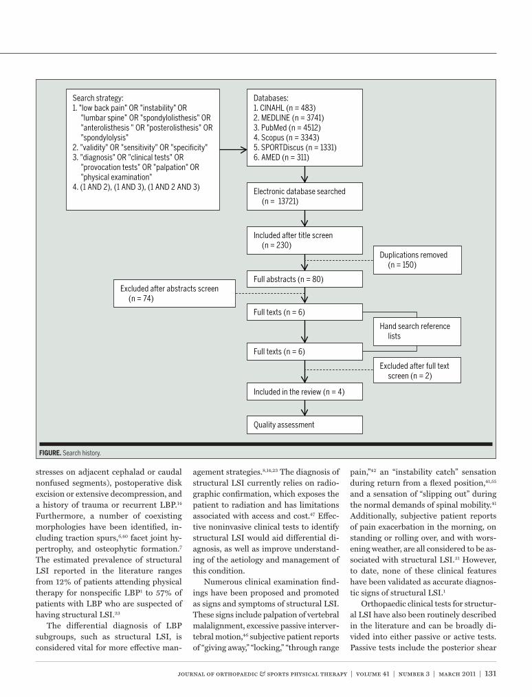
FIGURE. Search history.
41-03 Alqarni.indd 131 2/24/2011 4:35:08 PM
132 | march 2011 | volume 41 | number 3 | journal of orthopaedic & sports physical therapy
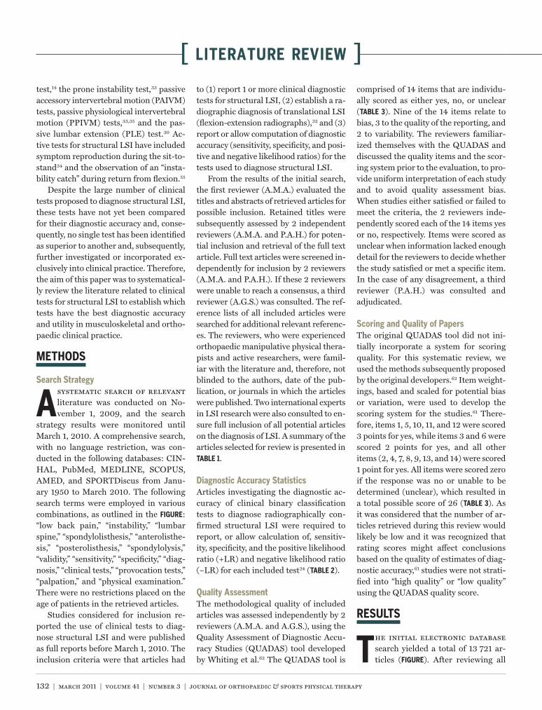
[ literature review ]test,14 the prone instability test,33 passive accessory intervertebral motion (PAIVM) tests, passive physiological intervertebral motion (PPIVM) tests,33,35 and the pas-sive lumbar extension (PLE) test.30 Ac-tive tests for structural LSI have included symptom reproduction during the sit-to-stand34 and the observation of an “insta-bility catch” during return from flexion.55
Despite the large number of clinical tests proposed to diagnose structural LSI, these tests have not yet been compared for their diagnostic accuracy and, conse-quently, no single test has been identified as superior to another and, subsequently, further investigated or incorporated ex-clusively into clinical practice. Therefore, the aim of this paper was to systematical-ly review the literature related to clinical tests for structural LSI to establish which tests have the best diagnostic accuracy and utility in musculoskeletal and ortho-paedic clinical practice.
METHODS
Search Strategy
A systematic search of relevant literature was conducted on No-vember 1, 2009, and the search
strategy results were monitored until March 1, 2010. A comprehensive search, with no language restriction, was con-ducted in the following databases: CIN-HAL, PubMed, MEDLINE, SCOPUS, AMED, and SPORTDiscus from Janu-ary 1950 to March 2010. The following search terms were employed in various combinations, as outlined in the FIGURE: “low back pain,” “instability,” “lumbar spine,” “spondylolisthesis,” “anterolisthe-sis,” “posterolisthesis,” “spondylolysis,” “validity,” “sensitivity,” “specificity,” “diag-nosis,” “clinical tests,” “provocation tests,” “palpation,” and “physical examination.” There were no restrictions placed on the age of patients in the retrieved articles.
Studies considered for inclusion re-ported the use of clinical tests to diag-nose structural LSI and were published as full reports before March 1, 2010. The inclusion criteria were that articles had
to (1) report 1 or more clinical diagnostic tests for structural LSI, (2) establish a ra-diographic diagnosis of translational LSI (flexion-extension radiographs),32 and (3) report or allow computation of diagnostic accuracy (sensitivity, specificity, and posi-tive and negative likelihood ratios) for the tests used to diagnose structural LSI.
From the results of the initial search, the first reviewer (A.M.A.) evaluated the titles and abstracts of retrieved articles for possible inclusion. Retained titles were subsequently assessed by 2 independent reviewers (A.M.A. and P.A.H.) for poten-tial inclusion and retrieval of the full text article. Full text articles were screened in-dependently for inclusion by 2 reviewers (A.M.A. and P.A.H.). If these 2 reviewers were unable to reach a consensus, a third reviewer (A.G.S.) was consulted. The ref-erence lists of all included articles were searched for additional relevant referenc-es. The reviewers, who were experienced orthopaedic manipulative physical thera-pists and active researchers, were famil-iar with the literature and, therefore, not blinded to the authors, date of the pub-lication, or journals in which the articles were published. Two international experts in LSI research were also consulted to en-sure full inclusion of all potential articles on the diagnosis of LSI. A summary of the articles selected for review is presented in TABLE 1.
Diagnostic Accuracy StatisticsArticles investigating the diagnostic ac-curacy of clinical binary classification tests to diagnose radiographically con-firmed structural LSI were required to report, or allow calculation of, sensitiv-ity, specificity, and the positive likelihood ratio (+LR) and negative likelihood ratio (–LR) for each included test24 (TABLE 2).
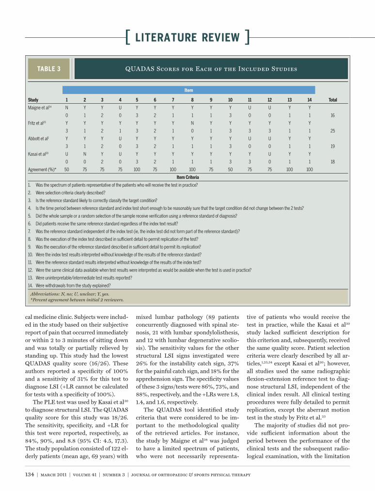
Quality AssessmentThe methodological quality of included articles was assessed independently by 2 reviewers (A.M.A. and A.G.S.), using the Quality Assessment of Diagnostic Accu-racy Studies (QUADAS) tool developed by Whiting et al.62 The QUADAS tool is
comprised of 14 items that are individu-ally scored as either yes, no, or unclear (TABLE 3). Nine of the 14 items relate to bias, 3 to the quality of the reporting, and 2 to variability. The reviewers familiar-ized themselves with the QUADAS and discussed the quality items and the scor-ing system prior to the evaluation, to pro-vide uniform interpretation of each study and to avoid quality assessment bias. When studies either satisfied or failed to meet the criteria, the 2 reviewers inde-pendently scored each of the 14 items yes or no, respectively. Items were scored as unclear when information lacked enough detail for the reviewers to decide whether the study satisfied or met a specific item. In the case of any disagreement, a third reviewer (P.A.H.) was consulted and adjudicated.
Scoring and Quality of PapersThe original QUADAS tool did not ini-tially incorporate a system for scoring quality. For this systematic review, we used the methods subsequently proposed by the original developers.62 Item weight-ings, based and scaled for potential bias or variation, were used to develop the scoring system for the studies.61 There-fore, items 1, 5, 10, 11, and 12 were scored 3 points for yes, while items 3 and 6 were scored 2 points for yes, and all other items (2, 4, 7, 8, 9, 13, and 14) were scored 1 point for yes. All items were scored zero if the response was no or unable to be determined (unclear), which resulted in a total possible score of 26 (TABLE 3). As it was considered that the number of ar-ticles retrieved during this review would likely be low and it was recognized that rating scores might affect conclusions based on the quality of estimates of diag-nostic accuracy,61 studies were not strati-fied into “high quality” or “low quality” using the QUADAS quality score.
RESULTS
The initial electronic database search yielded a total of 13 721 ar-ticles (FIGURE). After reviewing all
41-03 Alqarni.indd 132 2/24/2011 4:35:09 PM
journal of orthopaedic & sports physical therapy | volume 41 | number 3 | march 2011 | 133
titles for key words and context, 230 articles were selected for possible inclu-sion in the review. After title duplica-tions were removed, 80 article abstracts were screened, based on the inclusion criteria. After full text examination, 4 articles1,23,30,34 met the inclusion crite-ria and were vetted by 2 international experts who confirmed that they knew of no other published literature on this topic. All 4 articles were subsequently as-sessed using the QUADAS tool.62 A total of 11 clinical tests used in the diagnosis
of structural LSI were reviewed from the 4 retrieved articles. The quality scores on the QUADAS tool ranged from 1634 to 25 out of a possible 26.23 Sensitivity, specific-ity, likelihood ratios (LRs), and associated confidence intervals (95% CIs) were cal-culated from the study data if they were not specifically provided in the original articles.24
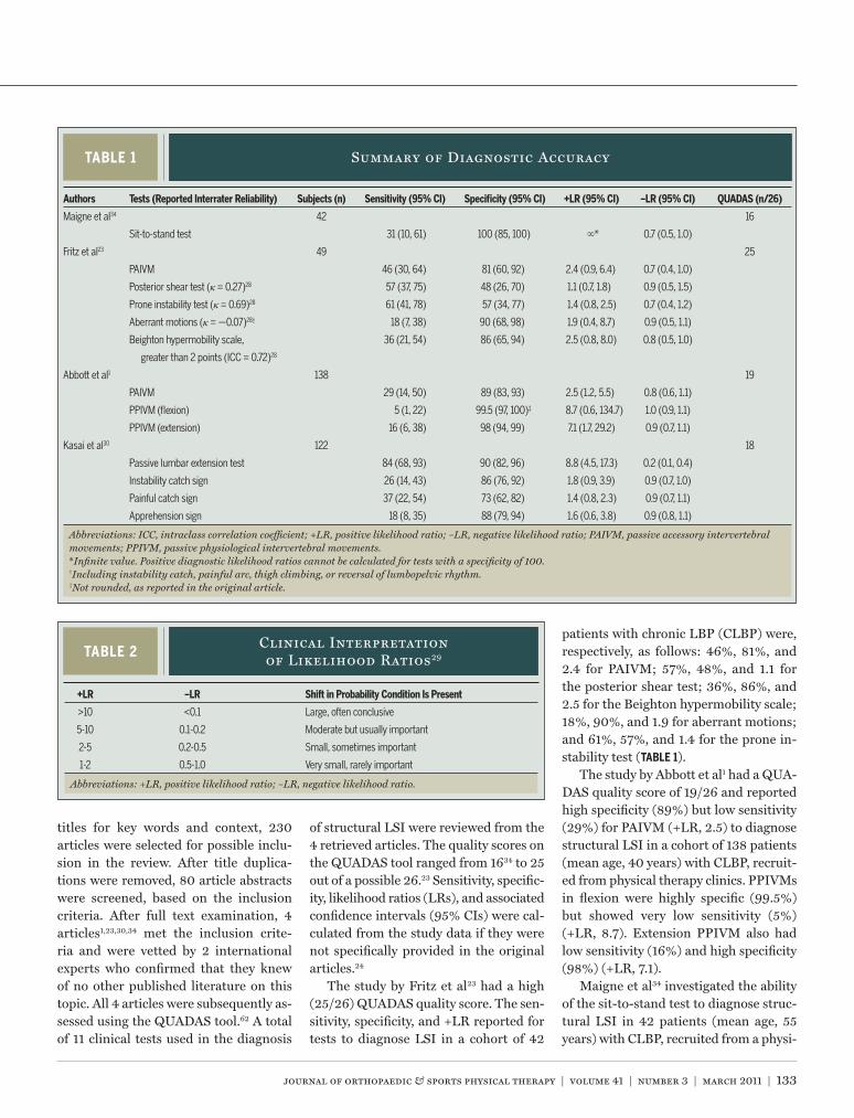
The study by Fritz et al23 had a high (25/26) QUADAS quality score. The sen-sitivity, specificity, and +LR reported for tests to diagnose LSI in a cohort of 42
patients with chronic LBP (CLBP) were, respectively, as follows: 46%, 81%, and 2.4 for PAIVM; 57%, 48%, and 1.1 for the posterior shear test; 36%, 86%, and 2.5 for the Beighton hypermobility scale; 18%, 90%, and 1.9 for aberrant motions; and 61%, 57%, and 1.4 for the prone in-stability test (TABLE 1).
The study by Abbott et al1 had a QUA-DAS quality score of 19/26 and reported high specificity (89%) but low sensitivity (29%) for PAIVM (+LR, 2.5) to diagnose structural LSI in a cohort of 138 patients (mean age, 40 years) with CLBP, recruit-ed from physical therapy clinics. PPIVMs in flexion were highly specific (99.5%) but showed very low sensitivity (5%) (+LR, 8.7). Extension PPIVM also had low sensitivity (16%) and high specificity (98%) (+LR, 7.1).
Maigne et al34 investigated the ability of the sit-to-stand test to diagnose struc-tural LSI in 42 patients (mean age, 55 years) with CLBP, recruited from a physi-
TABLE 1 Summary of Diagnostic Accuracy
Abbreviations: ICC, intraclass correlation coefficient; +LR, positive likelihood ratio; –LR, negative likelihood ratio; PAIVM, passive accessory intervertebral movements; PPIVM, passive physiological intervertebral movements.*Infinite value. Positive diagnostic likelihood ratios cannot be calculated for tests with a specificity of 100.†Including instability catch, painful arc, thigh climbing, or reversal of lumbopelvic rhythm.‡Not rounded, as reported in the original article.
Authors Tests (Reported Interrater Reliability) Subjects (n) Sensitivity (95% CI) Specificity (95% CI) +LR (95% CI) –LR (95% CI) QUADAS (n/26)
Maigne et al34 42 16
Sit-to-stand test 31 (10, 61) 100 (85, 100) * 0.7 (0.5, 1.0)
Fritz et al23 49 25
PAIVM 46 (30, 64) 81 (60, 92) 2.4 (0.9, 6.4) 0.7 (0.4, 1.0)
Posterior shear test (κ = 0.27)28 57 (37, 75) 48 (26, 70) 1.1 (0.7, 1.8) 0.9 (0.5, 1.5)
Prone instability test (κ = 0.69)28 61 (41, 78) 57 (34, 77) 1.4 (0.8, 2.5) 0.7 (0.4, 1.2)
Aberrant motions (κ = —0.07)28† 18 (7, 38) 90 (68, 98) 1.9 (0.4, 8.7) 0.9 (0.5, 1.1)
Beighton hypermobility scale, 36 (21, 54) 86 (65, 94) 2.5 (0.8, 8.0) 0.8 (0.5, 1.0)
greater than 2 points (ICC = 0.72)28
Abbott et al1 138 19
PAIVM 29 (14, 50) 89 (83, 93) 2.5 (1.2, 5.5) 0.8 (0.6, 1.1)
PPIVM (flexion) 5 (1, 22) 99.5 (97, 100)‡ 8.7 (0.6, 134.7) 1.0 (0.9, 1.1)
PPIVM (extension) 16 (6, 38) 98 (94, 99) 7.1 (1.7, 29.2) 0.9 (0.7, 1.1)
Kasai et al30 122 18
Passive lumbar extension test 84 (68, 93) 90 (82, 96) 8.8 (4.5, 17.3) 0.2 (0.1, 0.4)
Instability catch sign 26 (14, 43) 86 (76, 92) 1.8 (0.9, 3.9) 0.9 (0.7, 1.0)
Painful catch sign 37 (22, 54) 73 (62, 82) 1.4 (0.8, 2.3) 0.9 (0.7, 1.1)
Apprehension sign 18 (8, 35) 88 (79, 94) 1.6 (0.6, 3.8) 0.9 (0.8, 1.1)
TABLE 2Clinical Interpretation of Likelihood Ratios29
+LR –LR Shift in Probability Condition Is Present
>10 <0.1 Large, often conclusive
5-10 0.1-0.2 Moderate but usually important
2-5 0.2-0.5 Small, sometimes important
1-2 0.5-1.0 Very small, rarely important
Abbreviations: +LR, positive likelihood ratio; –LR, negative likelihood ratio.
41-03 Alqarni.indd 133 2/24/2011 4:35:10 PM
134 | march 2011 | volume 41 | number 3 | journal of orthopaedic & sports physical therapy
[ literature review ]
cal medicine clinic. Subjects were includ-ed in the study based on their subjective report of pain that occurred immediately or within 2 to 3 minutes of sitting down and was totally or partially relieved by standing up. This study had the lowest QUADAS quality score (16/26). These authors reported a specificity of 100% and a sensitivity of 31% for this test to diagnose LSI (+LR cannot be calculated for tests with a specificity of 100%).
The PLE test was used by Kasai et al30 to diagnose structural LSI. The QUADAS quality score for this study was 18/26. The sensitivity, specificity, and +LR for this test were reported, respectively, as 84%, 90%, and 8.8 (95% CI: 4.5, 17.3). The study population consisted of 122 el-derly patients (mean age, 69 years) with
mixed lumbar pathology (89 patients concurrently diagnosed with spinal ste-nosis, 21 with lumbar spondylolisthesis, and 12 with lumbar degenerative scolio-sis). The sensitivity values for the other structural LSI signs investigated were 26% for the instability catch sign, 37% for the painful catch sign, and 18% for the apprehension sign. The specificity values of these 3 signs/tests were 86%, 73%, and 88%, respectively, and the +LRs were 1.8, 1.4, and 1.6, respectively.
The QUADAS tool identified study criteria that were considered to be im-portant to the methodological quality of the retrieved articles. For instance, the study by Maigne et al34 was judged to have a limited spectrum of patients, who were not necessarily representa-
tive of patients who would receive the test in practice, while the Kasai et al30 study lacked sufficient description for this criterion and, subsequently, received the same quality score. Patient selection criteria were clearly described by all ar-ticles,1,23,34 except Kasai et al30; however, all studies used the same radiographic flexion-extension reference test to diag-nose structural LSI, independent of the clinical index result. All clinical testing procedures were fully detailed to permit replication, except the aberrant motion test in the study by Fritz et al.23
The majority of studies did not pro-vide sufficient information about the period between the performance of the clinical tests and the subsequent radio-logical examination, with the limitation
TABLE 3 QUADAS Scores for Each of the Included Studies
Abbreviations: N, no; U, unclear; Y, yes.*Percent agreement between initial 2 reviewers.
Study 1 2 3 4 5 6 7 8 9 10 11 12 13 14 Total
Maigne et al34 N Y Y U Y Y Y Y Y Y U U Y Y
0 1 2 0 3 2 1 1 1 3 0 0 1 1 16
Fritz et al23 Y Y Y Y Y Y Y N Y Y Y Y Y Y
3 1 2 1 3 2 1 0 1 3 3 3 1 1 25
Abbott et al1 Y Y Y U Y Y Y Y Y Y U U Y Y
3 1 2 0 3 2 1 1 1 3 0 0 1 1 19
Kasai et al30 U N Y U Y Y Y Y Y Y Y U Y Y
0 0 2 0 3 2 1 1 1 3 3 0 1 1 18
Agreement (%)* 50 75 75 75 100 75 100 100 75 50 75 75 100 100
Item Criteria
1. Was the spectrum of patients representative of the patients who will receive the test in practice?
2. Were selection criteria clearly described?
3. Is the reference standard likely to correctly classify the target condition?
4. Is the time period between reference standard and index test short enough to be reasonably sure that the target condition did not change between the 2 tests?
5. Did the whole sample or a random selection of the sample receive verification using a reference standard of diagnosis?
6. Did patients receive the same reference standard regardless of the index text result?
7. Was the reference standard independent of the index test (ie, the index test did not form part of the reference standard)?
8. Was the execution of the index test described in sufficient detail to permit replication of the test?
9. Was the execution of the reference standard described in sufficient detail to permit its replication?
10. Were the index test results interpreted without knowledge of the results of the reference standard?
11. Were the reference standard results interpreted without knowledge of the results of the index test?
12. Were the same clinical data available when test results were interpreted as would be available when the test is used in practice?
13. Were uninterpretable/intermediate test results reported?
14. Were withdrawals from the study explained?
Item
41-03 Alqarni.indd 134 2/24/2011 4:35:10 PM
journal of orthopaedic & sports physical therapy | volume 41 | number 3 | march 2011 | 135
that the target condition might have changed in the interim.1,30,34 The blind-ing of assessors for the clinical testing procedures and radiographic diagnosis of spinal instability was reported in all studies, as was the incidence of uninter-pretable test results and withdrawal of patients. Abbott et al1 and Maigne et al34 did not report whether interpretation of the radiological examination occurred without knowledge of the clinical test results, while only Fritz et al23 reported on the knowledge and availability of full, relevant clinical data for interpretation of the clinical test results, as used in clinical practice.23
The quality score for each item, to-tal score, and the percentage agreement between the 2 reviewers is presented in TABLE 3. All items had an initial review-er agreement that ranged from 50% to 100%. Items 1 and 10, which were related to patient spectrums and interpretation of the index test results retrospectively,
had the lowest agreement (50%). Seven items (2, 3, 4, 6, 9, 11, and 12) had 75% agreement, while 5 items (5, 7, 8, 13, and 14) had a 100% agreement, between the 2 reviewers. The third reviewer (P.A.H.) adjudicated in all cases where agreement was not initially reached, resulting in the final scores reported in TABLE 3.
DISCUSSION
The purpose of this systematic review was to evaluate the current evidence for clinical tests to diagnose structural LSI in musculoskeletal and orthopaedic clini-cal practice. The ability of clinical tests to diagnose structural LSI independent of radiographic investigations is consid-ered important to expedite diagnosis and guide subsequent management, while limiting the exposure of patients to as-sociated risks and further costs.58 A total of 11 clinical tests used in the diagnosis of 351 patients were identified from 4 ar-
ticles that met the inclusion criteria for the study.1,23,30,34
The results of the review show that diagnostic specificity values for all tests were consistently higher than sensitivity values, with the exception of the poste-rior shear test and prone instability test (TABLE 1). A negative result for a test with high sensitivity indicates that the test may have value in ruling out LSI, and a positive result for a test that has high specificity may be useful to rule in the condition.12 Likelihood ratios incorporate both the sensitivity and specificity of a test, and provide a direct estimate of how much a test result will change the odds of having the condition.15 In this study, the +LRs for structural LSI tests ranged from 1.1 to 8.8. The clinical interpretation of these values is presented in TABLE 2.
Two studies1,23 examined the abil-ity of PAIVM33,35 to diagnose structural LSI. The study by Fritz et al23 reported low sensitivity (46%) and relatively high
TABLE 4 Study Characteristics
Abbreviations: LBP; low back pain; PAIVM, passive accessory intervertebral movements; PLE, passive lumbar extension; PPIVM, passive physiological inter-vertebral movements; RCLBP, recurrent chronic low back pain; ROM, range of motion.*Reference tests for all studies.†Aberrant motion include instability catch, painful arc, thigh climbing, or reversal of lumbopelvic rhythm.
Study Subjects Examiners
Definition of Instability From
Flexion-Extension Radiographs* Index Tests Definition of Positive Index Test
Maigne et al34 42 patients; mean SD age,
54.9 9.8 y; CLBP greater
than 2 mo
3 physicians Translation greater than 10%
(4.5-5.0 mm)
• Sit-to-stand • Pain upon sitting down and
relieved by standing up
Fritz et al23 49 patients; mean SD age,
39.3 11.3 y; LBP, median
duration of symptoms, 78 d
1 physical therapist Translation greater than 4.5 mm
or greater than 15% of the
vertebral body width
• PAIVM • Hypermobility
• Posterior shear test • Symptom provocation
• Prone instability test • Symptom provocation
• Aberrant motions† • Any aberrant motions during
flexion/extension ROM
• Beighton hypermobility
scale
• More than 2 points on scale
Abbott et al1 138 patients; mean age,
40 y (range, 20-75 y);
RCLBP, greater than 3 mo
27 physiotherapists Translation greater than 2 SDs
from the reference mean of
asymptomatic individuals
using Gaussian definition
• PAIVM • Hypermobility and/or pain
• PPIVM (flexion) • Hypermobility and/or pain
• PPIVM (extension) • Hypermobility and/or pain
Kasai et al30 122 patients; mean age, 68.9 y
(range, 39-88 y); LBP, duration
1 mo to 5 y
3 orthopaedists; 2
examined PLE, 1
examined other tests
Translation motion of 5 mm • PLE test • LBP or discomfort during test
• Instability catch sign • Sudden LBP during movement test
• Painful catch sign • Sudden LBP during movement test
• Apprehension sign • Sudden LBP during movement test
41-03 Alqarni.indd 135 2/24/2011 4:35:12 PM
136 | march 2011 | volume 41 | number 3 | journal of orthopaedic & sports physical therapy
[ literature review ]
specificity (81%), while Abbott et al1 re-ported similar high specificity (89%) but lower sensitivity (29%) (TABLES 1 and 4). Differences in selection criteria might have affected the specificity value in the study by Fritz et al,23 as it included pa-tients who were already suspected of hav-ing LSI. Both studies reported similar +LRs, with patients being approximately 2.5 times more likely to have a radiologi-cal diagnosis of LSI following a positive PAIVM test.
For a diagnostic test to be valid, it must have acceptable reliability.26 The
reliability of PAIVM testing is dependent on a number of intrinsic and extrinsic factors,5,57 and poor interrater reliability (κ = –0.02, 0.26) has been reported for judgment of segmental mobility.28 It is suggested that standardization of PAIVM testing would improve the reliability and, potentially, the ability of these tests to di-agnose structural LSI.
Abbott et al1 reported that flexion PPIVM had very high specificity (99.5%) but very low sensitivity (5%), and a mod-erate +LR (8.7) (95% CI: 0.6, 134.7) for the diagnosis of structural LSI. However,
the width of the +LR confidence interval indicates the imprecision of the estimate, which was potentially due to an inad-equate sample size to effectively evaluate the diagnostic accuracy of this test.20 The authors also found that extension PPIVM had low sensitivity (16%), high specificity (98%), and a moderate +LR (7.1) (95% CI: 1.7, 29.2) to diagnose LSI. These results suggest that extension PPIVM, compared to flexion PPIVM, might have greater clinical accuracy in the diagnosis of LSI, which is not unexpected, given the extension bias seen in the majority
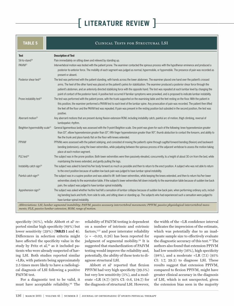
TABLE 5 Clinical Tests for Structural LSI
Abbreviations: LSI, lumbar segmental instability; PAIVM, passive accessory intervertebral movements; PPIVM, passive physiological intervertebral move-ments; PLE, passive lumbar extension; ROM, range of motion.
Test Description of Test
Sit-to-stand34 Pain immediately on sitting down and relieved by standing up.
PAIVM23 Intervertebral motion was tested with the patient prone. The examiner contacted the spinous process with the hypothenar eminence and produced a
posterior-to-anterior force. The mobility of each segment was judged as normal, hypermobile, or hypomobile. The presence of pain was recorded as
present or absent.
Posterior shear test23 The test was performed with the patient standing, with hands across the lower abdomen. The examiner placed one hand over the patient’s crossed
arms. The heel of the other hand was placed on the patient’s pelvis for stabilization. The examiner produced a posterior shear force through the
patient’s abdomen, and an anteriorly directed stabilizing force with the opposite hand. The test was repeated at each lumbar level by changing the
point of contact of the posterior hand. A positive test occurred if familiar symptoms were provoked, and is proposed to indicate lumbar instability.
Prone instability test23 The test was performed with the patient prone, with the trunk supported on the examining table and the feet resting on the floor. With the patient in
this position, the examiner performed a PAIVM test to each level of the lumbar spine. Any provocation of pain was recorded. The patient then lifted
the feet off the floor and the PAIVM test was repeated. If pain was present in the resting position but subsided in the second position, the test was
positive.
Aberrant motion23 Any aberrant motions that are present during flexion-extension ROM, including instability catch, painful arc of motion, thigh climbing, reversal of
lumbopelvic rhythm.
Beighton hypermobility scale23 General ligamentous laxity was assessed with the 9-point Beighton scale. One point was given for each of the following: knee hyperextension greater
than 10°, elbow hyperextension greater than 10°, fifth finger hyperextension greater than 90°, thumb abduction to contact the forearm, and ability to
flex the trunk and place hands flat on the floor with knees extended.
PPIVM1 PPIVMs were assessed with the patient sidelying, and consisted of moving the patient’s spine through sagittal forward-bending (flexion) and backward-
bending (extension), using the lower extremities, while palpating between the spinous process of the adjacent vertebrae to assess the motion taking
place at each motion segment.
PLE test30 The subject was in the prone position. Both lower extremities were then passively elevated, concurrently, to a height of about 30 cm from the bed, while
maintaining the knees extended, and gently pulling the legs.
Instability catch sign30 The subject was asked to bend his/her body forward as much as possible and then to return to the erect position. A subject who was not able to return
to the erect position because of sudden low back pain was judged to have lumbar spinal instability.
Painful catch sign30 The subject was in a supine position and was asked to lift both lower extremities, while keeping the knees extended, and then to return his/her lower
extremities slowly to the examination table. If the subject’s lower extremities fell down instantly to the examination table because of sudden low back
pain, the subject was judged to have lumbar spinal instability.
Apprehension sign30 The subject was asked whether he/she had felt a sensation of lumbar collapse because of sudden low back pain, when performing ordinary acts, includ-
ing bending back and forth, from side to side, and sitting down or standing up. The subjects who had experienced such a sensation were judged to
have lumbar spinal instability.
41-03 Alqarni.indd 136 2/24/2011 4:35:13 PM

journal of orthopaedic & sports physical therapy | volume 41 | number 3 | march 2011 | 137
of both active and passive testing proce-dures for LSI.
The posterior shear test was original-ly described by Delitto et al14 as a test to diagnose LSI. The results of this review demonstrated relatively poor sensitiv-ity (57%), specificity (48%), and, conse-quently, a small +LR (1.1) for this test,23 which has also been shown to have poor intrarater (κ = 0.27)23 and interrater (κ = 0.22) reliability.28 These results indicate that the posterior shear test has limited overall diagnostic ability to diagnose LSI.
The prone instability test33 demon-strated low to moderate sensitivity (61%) and specificity (57%), and a low +LR (1.4),23 which suggests that the test has limited ability to accurately diagnose structural LSI. Moderate intrarater re-liability (κ = 0.69)23 and the previously reported “almost perfect” interrater re-liability (κ = 0.87) for this test are sug-gested to support this finding.28
The Beighton hypermobility scale4 has been previously shown to have a positive correlation with structural LSI.23 This test had low sensitivity (36%) and high specificity (86%), and a relatively small +LR (2.5). The high intrarater reliabil-ity (ICC = 0.72) and specificity values in the retrieved study,23 combined with previously reported high reliability coef-ficients (ICC = 0.79),28 suggest that this test may be of some clinical use to rule in patients with a positive diagnosis of LSI. However, the low +LR, which in clini-cal interpretation is considered to be of greater diagnostic value than sensitivity and specificity values alone, makes this unlikely.
Maigne et al34 examined the ability of the sit-to-stand test to diagnose struc-tural LSI. The study compared patients whose LBP occurred immediately upon sitting down and was relieved on stand-ing up, with a control group whose LBP did not show this pattern. The sit-to-stand test had a specificity of 100% and a sensitivity of 31% within this patient population. However, recruitment bias might explain the high specificity, as only patients who previously reported a
positive sit-to-stand test were selected for the study group. This article had the lowest QUADAS score, and the authors reported that the test result might vary, depending on time of day that the test was conducted, the type of seat employed, and the patients’ symptom levels before the test.46 The authors also reported that these symptoms were observed in only 1 of 70 patients presenting with CLBP, which is much lower than the expected incidence of structural LSI in the CLBP population.49,59 Due to these factors, ad-ditional research is needed to address identified study limitations and to deter-mine the test’s true diagnostic ability and clinical utility.
The PLE test is a relatively new meth-od for examining structural LSI origi-nally reported by Kasai et al.30 The test requires the patient to lie prone, while the clinician lifts both lower extremities into extension to a height of approxi-mately 30 cm, while providing some traction to the lower extremities. During this maneuver, a positive test is based on an increase in pain that disappears on return to the starting position. In an at-tempt to diagnose structural LSI, Kasai et al30 used the PLE test to examine 122 patients with mixed lumbar pathology (TABLE 4). The PLE test was standardized and performed twice, at an interval of 2 to 4 weeks, by 2 independent orthopae-dists. They reported no disparate test results between the 2 sessions, which suggests substantial test-retest reliabil-ity. The sensitivity, specificity, and +LR of the PLE test were reported as 84%, 90%, and 8.8 (95% CI: 4.5, 17.3), respectively, indicating that the PLE test is a poten-tially effective clinical test to diagnose structural LSI. However, the prevalence rate for structural LSI in this study was relatively high (31%), which might be due to the study’s elderly population sample, with high rates of spinal degeneration, stenosis, spondylolisthesis, and concur-rent LSI. Therefore, although these re-sults are promising, further investigation of this test should be undertaken in other patient populations, across different age
groups, and with different assessors, to further evaluate its reliability and accu-racy to diagnose structural LSI.
Kasai et al30 also investigated a range of active movement signs/tests to diag-nose structural LSI (TABLE 1). The instabil-ity catch sign, painful catch sign, and the apprehension sign all had relatively low sensitivity and high specificity, resulting in very small +LRs. These results sug-gest that these tests would more likely produce high false negative rates if used to diagnose structural LSI in research and clinical practice (TABLE 1). Similarly, Fritz et al23 reported that a selection of aberrant motion test procedures dem-onstrated the same pattern of diagnostic accuracy and poor intrarater reliability (κ = –0.07), confirming the limited value of these signs/tests to diagnose structural LSI.
Clinical tests, such as those described in this review (TABLE 5), are not the only measures reported in the literature that are suggested to aid the diagnosis of structural LSI. Patient history of asso-ciated signs and symptoms suggestive of LSI has also been reported. Kasai et al31 interviewed 368 patients with lum-bar degenerative disease, of which 88 patients had structural LSI identified by imaging (translation greater than or equal to 5 mm). The results showed that pain on standing up and rolling over had the highest sensitivity (58% and 55%, respectively) and specificity (88% and 93%, respectively) for LSI, and a report of morning pain, with morning being the most painful time of day, had a sensitiv-ity of 74% and specificity of 80%. Symp-toms exacerbated by worsening weather had 65% sensitivity and 94% specificity. These results highlight the possibility that symptoms exacerbated by specific movements and the timing of symptoms could assist clinicians in diagnosing structural LSI.
The clinical implications of a positive diagnosis of structural LSI are inevitably related to either surgical or conservative management of this condition. Lumbar spinal fusion is usually suggested for
41-03 Alqarni.indd 137 2/24/2011 4:35:14 PM

138 | march 2011 | volume 41 | number 3 | journal of orthopaedic & sports physical therapy
[ literature review ]patients with severe symptoms and ra-diographic evidence of hypermobility (greater than 4 mm of vertebral transla-tion), who do not respond to conservative treatment.53 More commonly, conserva-tive treatment is indicated; however, there are few studies that have specifi-cally addressed the conservative man-agement of radiologically determined structural instability.38,39,43 Reported con-servative management has included brac-es or corsets14,54 and patient education to avoid overloading the passive stabilizing structures of the spine at end range.21,22 However, the mainstay of conservative management for instability-related lum-bar spine pain has focused on exercises to improve neuromuscular control of the spine.9 Various strengthening programs targeting specific groups of muscles have been reported in the literature36,37,42,51; however, no one specific program has demonstrated superiority over another.9,21 Further research is needed to identify the most effective strategies for patients with identified instability classifications.21 Conversely, the clinical implications of negative test findings for structural LSI are that alternative diagnoses need to be made and, in patients whose symptomol-ogy remains suggestive of lumbar insta-bility, a potential diagnosis of functional LSI could be considered.
As with most diagnostic studies for lumbar pathology, limitations exist that may affect the validity of the results. Firstly, the literature that has reported the accuracy of clinical tests to diagnose structural LSI is limited, and only 4 of the articles retrieved met this literature review’s inclusion criteria. Secondly, all 4 articles1,23,30,34 used flexion-extension radiographs to identify abnormal ver-tebral translational motion to diagnose structural LSI.1,27 While flexion-extension radiographs have historically been con-sidered the radiological reference test of choice for LSI, they have been sug-gested to be complicated by false positive rates and have significant variation in asymptomatic persons.27,45 Additionally, differences in patient positioning might
account for the 10% to 15% variation in observed vertebral displacement.11
The reported cutoff values for ver-tebral translatory motion employed to diagnose the presence of structural LSI also remain somewhat contentious and vary between 3 to 5 mm in the litera-ture.17,18,27,32,52 The majority of studies in this review employed cutoff values for translational motion greater than 4.5 to 5.0 mm, with Abbott et al1 using transla-tion beyond 2 SDs from the mean of an asymptomatic population as the cutoff value. The effects of differing translation-al cutoff values on the diagnostic ability of clinical tests to diagnose structural LSI are not currently known.
It has also been proposed that aber-rant motion and dysfunction from struc-tural LSI exist not only at end range but during midrange spinal movements, which these tests might not identify.35 Flexion-extension radiographs simply assess vertebral displacement statically at end range,32,52 which, theoretically, would only detect the function of the pas-sive stabilizing subsystem.22 This might have significant limitations in detecting dysfunction from structural LSI that oc-curs within the neutral zone (midrange spinal motion).44,48
Digital video fluoroscopy has also been utilized to identify normal and abnormal lumbar motion in vivo.3,56,64,65 This type of imaging has recently demonstrated an ability to identify movement abnormali-ties in patients with suspected functional LSI2; however, further research is needed to confirm and substantiate these find-ings in a population with structural LSI. Despite these recognized limitations, flexion-extension radiographs remain, at present, the most common criterion ref-erence standard to diagnose structural LSI.47
A possible limitation of the methods used in this study was that only 1 reviewer searched the literature to identify articles for inclusion. Another factor that may limit the generalizability of the results of LSI diagnostic studies is the heterogene-ity and sample size of the cohort under
investigation. Study sample sizes identi-fied in this review ranged from 4234 to 1381 patients (TABLE 4). These relatively low samples might have affected the in-ternal validity and diagnostic accuracy of the results, and it has been suggested that studies of this nature should, theoretical-ly, contain over 600 participants to have meaningful diagnostic value.20
Additionally, quality assessment of retrieved articles is considered to be an essential component of most systematic reviews.13 The QUADAS tool was used to assess the quality of articles in this study; however, this and other well-utilized tools are suggested to have a number of associated limitations. These include the possibility that even well-conducted studies may score poorly if the methods and results of the study are not reported in sufficient detail,62 and that most qual-ity assessment tools used in diagnostic studies do not include items that assess statistical power, which can subsequently affect a study’s validity.
Almost all of the clinical tests inves-tigated in this systematic review, due to their high specificity, demonstrated the ability to diagnose patients with LSI, when the tests were positive. However, the trade-off for the majority of tests was the low sensitivity, which means that these tests may not be able to rule out people who test negative for structural LSI. Both PAIVM and PPIVM appear to have modest ability to diagnose structural LSI. The PLE test, however, had both the highest +LR (8.8) and lowest –LR (0.2) of the tests investigated, demonstrating a moderate but important role for both ruling in and ruling out structural LSI. These results suggest that the PLE test may be useful in clinical practice to diag-nose structural LSI.
It is, however, important to note that the reliance on a single test in isolation is not usually recommended in muscu-loskeletal and orthopaedic clinical prac-tice, and it is likely that a combination of valid and reliable tests, and the inclusion of patient-specific signs and symptoms, including historical elements, might fur-
41-03 Alqarni.indd 138 2/24/2011 4:35:15 PM

journal of orthopaedic & sports physical therapy | volume 41 | number 3 | march 2011 | 139
ther assist clinicians in diagnosing and managing patients with LSI. Further in-vestigation of these combined diagnostic factors is required in future studies that include larger patient populations, differ-ing age ranges, and different assessors, to ensure the validity and diagnostic accu-racy of the tests.
CONCLUSION
This is the first systematic re-view that has been conducted to identify the accuracy of clinical tests
for diagnosing structural LSI. A total of 11 clinical tests were identified from the literature that met the study inclusion criteria. The reviewed articles were con-sidered to be of sufficient quality to as-certain the diagnostic value of each test evaluated. The majority of tests had high specificity but low sensitivity. The PLE test was found to have the highest com-bined sensitivity and specificity, as well as the highest +LR, suggesting that in the absence of, or as an adjunct to, radiologi-cal imaging this test might be of use in musculoskeletal and orthopaedic clinical practice to diagnose structural LSI. How-ever, additional research of the diagnostic accuracy of the PLE test across a range of patient populations with different asses-sors is recommended to further evaluate its validity to diagnose structural LSI. t
ACKNOWLEDGEMENTS: We gratefully acknowl-edge the assistance of Masahiro Takemura for translation of the Japanese articles.
REFERENCES
1. Abbott JH, McCane B, Herbison P, Moginie G, Chapple C, Hogarty T. Lumbar segmental instability: a criterion-related validity study of manual therapy assessment. BMC Mus-culoskelet Disord. 2005;6:56. http://dx.doi.org/10.1186/1471-2474-6-56
2. Ahmadi A, Maroufi N, Behtash H, Zekavat H, Parnianpour M. Kinematic analysis of dynamic lumbar motion in patients with lumbar segmen-tal instability using digital videofluoroscopy. Eur Spine J. 2009;18:1677-1685. http://dx.doi.org/10.1007/s00586-009-1147-x
3. Auerbach JD, Wills BP, McIntosh TC, Balderston RA. Evaluation of spinal kinematics following lumbar total disc replacement and circumfer-ential fusion using in vivo fluoroscopy. Spine (Phila Pa 1976). 2007;32:527-536. http://dx.doi.org/10.1097/01.brs.0000256915.90236.17
4. Beighton P, Horan F. Orthopaedic aspects of the Ehlers-Danlos syndrome. J Bone Joint Surg Br. 1969;51:444-453.
5. Binkley J, Stratford PW, Gill C. Interrater reliabil-ity of lumbar accessory motion mobility testing. Phys Ther. 1995;75:786-792; discussion 793-785.
6. Bram J, Zanetti M, Min K, Hodler J. MR abnor-malities of the intervertebral disks and adjacent bone marrow as predictors of segmental instability of the lumbar spine. Acta Radiol. 1998;39:18-23.
7. Brown MD, Holmes DC, Heiner AD. Measurement of cadaver lumbar spine motion segment stiff-ness. Spine (Phila Pa 1976). 2002;27:918-922.
8. Childs JD, Fritz JM, Piva SR, Erhard RE. Clinical decision making in the identification of patients likely to benefit from spinal manipulation: a traditional versus an evidence-based approach. J Orthop Sports Phys Ther. 2003;33:259-272.
9. Cleland JA, Schulte C, Durall C. The role of ther-apeutic exercise in treating instability-related lumbar spine pain: a systematic review. J Back Musculoskelet. 2002;16:105-115.
10. Cook C, Brismee JM, Sizer PS, Jr. Subjective and objective descriptors of clinical lumbar spine instability: a Delphi study. Man Ther. 2006;11:11-21. http://dx.doi.org/10.1016/j.math.2005.01.002
11. Danielson B, Frennered K, Irstam L. Roent-genologic assessment of spondylolisthesis. I. A study of measurement variations. Acta Radiol. 1988;29:345-351.
12. Davidson M. The interpretation of diagnostic test: a primer for physiotherapists. Aust J Phys-iother. 2002;48:227-232.
13. Deeks JJ. Systematic reviews in health care: systematic reviews of evaluations of diagnostic and screening tests. BMJ. 2001;323:157-162.
14. Delitto A, Erhard RE, Bowling RW. A treatment-based classification approach to low back syndrome: identifying and staging patients for conservative treatment. Phys Ther. 1995;75:470-485; discussion 485-479.
15. Denegar CR, Fraser M. How useful are physi-cal examination procedures? Understanding and applying likelihood ratios. J Athl Train. 2006;41:201-206.
16. Di Fabio RP, Boissonnault W. Physical therapy and health-related outcomes for patients with common orthopaedic diagnoses. J Orthop Sports Phys Ther. 1998;27:219-230.
17. Dupuis PR, Yong-Hing K, Cassidy JD, Kirkaldy-Willis WH. Radiologic diagnosis of degenerative lumbar spinal instability. Spine (Phila Pa 1976). 1985;10:262-276.
18. Dvorak J, Panjabi MM, Chang DG, Theiler R, Grob D. Functional radiographic diagnosis of the lumbar spine. Flexion-extension and lateral bending. Spine (Phila Pa 1976). 1991;16:562-571.
19. Farfan HF, Gracovetsky S. The nature of instabil-ity. Spine (Phila Pa 1976). 1984;9:714-719.
20. Flahault A, Cadilhac M, Thomas G. Sample size calculation should be performed for design accuracy in diagnostic test studies. J Clin Epidemiol. 2005;58:859-862. http://dx.doi.org/10.1016/j.jclinepi.2004.12.009
21. Fritz JM, Cleland JA, Childs JD. Subgrouping patients with low back pain: evolution of a clas-sification approach to physical therapy. J Orthop Sports Phys Ther. 2007;37:290-302.
22. Fritz JM, Erhard RE, Hagen BF. Segmental instability of the lumbar spine. Phys Ther. 1998;78:889-896.
23. Fritz JM, Piva SR, Childs JD. Accuracy of the clinical examination to predict radiographic instability of the lumbar spine. Eur Spine J. 2005;14:743-750. http://dx.doi.org/10.1007/s00586-004-0803-4
24. Greenhalgh T. How to read a paper. Papers that report diagnostic or screening tests. BMJ. 1997;315:540-543.
25. Hall H, McIntosh G, Boyle C. Effectiveness of a low back pain classification system. Spine J. 2009;9:648-657. http://dx.doi.org/10.1016/j.spinee.2009.04.017
26. Haneline M, Cooperstein R. Weighing the reli-ability and validity of clinical tests. J Amer Chi-ropr Assos. 2006;43:19-22.
27. Hayes MA, Howard TC, Gruel CR, Kopta JA. Roentgenographic evaluation of lumbar spine flexion-extension in asymptomatic individuals. Spine (Phila Pa 1976). 1989;14:327-331.
28. Hicks GE, Fritz JM, Delitto A, Mishock J. Inter-rater reliability of clinical examination measures for identification of lumbar segmental instability. Arch Phys Med Rehabil. 2003;84:1858-1864.
29. Jaeschke R, Guyatt GH, Sackett DL. Users’ guides to the medical literature. III. How to use an article about a diagnostic test. B. What are the results and will they help me in caring for my patients? The Evidence-Based Medicine Working Group. JAMA. 1994;271:703-707.
30. Kasai Y, Morishita K, Kawakita E, Kondo T, Uchida A. A new evaluation method for lumbar spinal instability: passive lumbar extension test. Phys Ther. 2006;86:1661-1667. http://dx.doi.org/10.2522/ptj.20050281
31. Kasai Y, Morishita K, Takegami K, Uchida A. Clinical symptoms of patient with lumbar spinal instability [in Japanese]. Clin Orthop Surg. 2003;38:463-467.
32. Knutsson F. The instability associated with disk degeneration in the lumbar spine. Acta Radiol. 1944;25:593-609.
33. Magee DJ. Orthopedic Physical Assessment. 3rd ed. Philadelphia, PA: W.B. Sauders Company; 1997.
34. Maigne JY, Lapeyre E, Morvan G, Chatellier G. Pain immediately upon sitting down and relieved by standing up is often associated with radiologic lumbar instability or marked anterior loss of disc space. Spine (Phila Pa 1976). 2003;28:1327-1334. http://dx.doi.org/10.1097/01.BRS.0000065569.76853.E9
41-03 Alqarni.indd 139 2/24/2011 4:35:16 PM
140 | march 2011 | volume 41 | number 3 | journal of orthopaedic & sports physical therapy
[ literature review ]
@ MORE INFORMATIONWWW.JOSPT.ORG
35. Maitland GD. Vertebral Manipulation. 5th ed. London, UK: Butterworth & Co Ltd; 1986.
36. McGill SM. Low back stability: from formal description to issues for performance and reha-bilitation. Exerc Sport Sci Rev. 2001;29:26-31.
37. McGill SM, Grenier S, Kavcic N, Cholewicki J. Coordination of muscle activity to assure stabil-ity of the lumbar spine. J Electromyogr Kinesiol. 2003;13:353-359.
38. Moller H, Hedlund R. Surgery versus conserva-tive management in adult isthmic spondylolis-thesis--a prospective randomized study: part 1. Spine (Phila Pa 1976). 2000;25:1711-1715.
39. Nelson BW, O’Reilly E, Miller M, Hogan M, Wegner JA, Kelly C. The clinical effects of in-tensive, specific exercise on chronic low back pain: a controlled study of 895 consecutive patients with 1-year follow up. Orthopedics. 1995;18:971-981.
40. Nizard RS, Wybier M, Laredo JD. Radiologic assessment of lumbar intervertebral instability and degenerative spondylolisthesis. Radiol Clin North Am. 2001;39:55-71, v-vi.
41. Ogon M, Bender BR, Hooper DM, et al. A dynamic approach to spinal instability. Part I: Sensitization of intersegmental motion profiles to motion direction and load condition by insta-bility. Spine (Phila Pa 1976). 1997;22:2841-2858.
42. O’Sullivan PB. Lumbar segmental ‘instability’: clinical presentation and specific stabilizing exercise management. Man Ther. 2000;5:2-12. http://dx.doi.org/10.1054/math.1999.0213
43. O’Sullivan PB, Twomey L, Allison GT. Altered abdominal muscle recruitment in patients with chronic back pain following a specific exer-cise intervention. J Orthop Sports Phys Ther. 1998;27:114-124.
44. Panjabi MM. The stabilizing system of the spine. Part II. Neutral zone and instability hypothesis. J Spinal Disord. 1992;5:390-396; discussion 397.
45. Panjabi MM, Lydon C, Vasavada A, Grob D, Crisco JJ, 3rd, Dvorak J. On the understanding of clinical instability. Spine (Phila Pa 1976). 1994;19:2642-2650.
46. Paris SV. Physical signs of instability. Spine (Phila Pa 1976). 1985;10:277-279.
47. Pitkanen MT, Manninen HI, Lindgren KA, Sih-vonen TA, Airaksinen O, Soimakallio S. Segmen-tal lumbar spine instability at flexion-extension radiography can be predicted by conventional radiography. Clin Radiol. 2002;57:632-639. http://dx.doi.org/10.1053/crad.2001.0899
48. Pope MH, Frymoyer JW, Krag MH. Diagnosing instability. Clin Orthop Relat Res. 1992;60-67.
49. Pope MH, Panjabi M. Biomechanical defini-tions of spinal instability. Spine (Phila Pa 1976). 1985;10:255-256.
50. Posner I, White AA, 3rd, Edwards WT, Hayes WC. A biomechanical analysis of the clinical stability of the lumbar and lumbosacral spine. Spine (Phila Pa 1976). 1982;7:374-389.
51. Richardson CA, Jull GA. Muscle control-pain control. What exercises would you pre-scribe? Man Ther. 1995;1:2-10. http://dx.doi.org/10.1054/math.1995.0243
52. Shaffer WO, Spratt KF, Weinstein J, Lehmann TR, Goel V. 1990 Volvo Award in clinical sci-ences. The consistency and accuracy of roent-genograms for measuring sagittal translation in the lumbar vertebral motion segment. An experimental model. Spine (Phila Pa 1976). 1990;15:741-750.
53. Sonntag VK, Marciano FF. Is fusion indicated for lumbar spinal disorders? Spine (Phila Pa 1976). 1995;20:138S-142S.
54. Spratt KF, Weinstein JN, Lehmann TR, Woody J, Sayre H. Efficacy of flexion and extension treat-ments incorporating braces for low-back pain patients with retrodisplacement, spondylolisthe-sis, or normal sagittal translation. Spine (Phila Pa 1976). 1993;18:1839-1849.
55. Taylor J, O’Sullivan P. Lumbar ‘segmental’ in-stability: pathology, diagnosis and conservative management. In: Twomey L, Taylor J, eds. Physi-cal Therapy of the Low Back. Philadelphia, PA: WB Saunders; 2000:201-247.
56. Teyhen DS, Flynn TW, Childs JD, et al. Fluoroscopic video to identify aberrant lumbar motion. Spine (Phila Pa 1976). 2007;32:E220-229. http://dx.doi.org/10.1097/01.brs.0000259206.38946.cb
57. Wadsworth C, DiFabio R, Johnson D. The spine.
In: Wadsworth C, ed. Manual Examination and Treatment of the Spine and Extremities. Balti-more, MD: Williams and Wilkins; 1988:70-71.
58. Wall BF, Kendall GM, Edwards AA, Bouffler S, Muirhead CR, Meara JR. What are the risks from medical X-rays and other low dose radiation? Br J Radiol. 2006;79:285-294. http://dx.doi.org/10.1259/bjr/55733882
59. Weiler PJ, King GJ, Gertzbein SD. Analysis of sagittal plane instability of the lumbar spine in vivo. Spine (Phila Pa 1976). 1990;15:1300-1306.
60. White AA, Panjabi MM. Clinical Biomechanics of the Spine. 2nd ed. Philadelphia, PA: J.B. Lip-pincott Company; 1990.
61. Whiting P, Harbord R, Kleijnen J. No role for quality scores in systematic reviews of diagnostic accuracy studies. BMC Med Res Methodol. 2005;5:19. http://dx.doi.org/10.1186/1471-2288-5-19
62. Whiting P, Rutjes AW, Reitsma JB, Bossuyt PM, Kleijnen J. The development of QUADAS: a tool for the quality assessment of studies of diag-nostic accuracy included in systematic reviews. BMC Med Res Methodol. 2003;3:25. http://dx.doi.org/10.1186/1471-2288-3-25
63. Williams CM, Maher CG, Hancock MJ, et al. Low back pain and best practice care: A survey of general practice physicians. Arch Intern Med. 170:271-277. http://dx.doi.org/10.1001/archinternmed.2009.507
64. Wong KW, Luk KD, Leong JC, Wong SF, Wong KK. Continuous dynamic spinal motion analysis. Spine (Phila Pa 1976). 2006;31:414-419. http://dx.doi.org/10.1097/01.brs.0000199955.87517.82
65. Zheng Y, Nixon MS, Allen R. Lumbar spine visualisation based on kinematic analysis from videofluoroscopic imaging. Med Eng Phys. 2003;25:171-179.
FIND Author Instructions & Tools on the Journal’s Website
JOSPT’s instructions to authors are available at www.jospt.org by clicking “AUTHOR TOOLS & INSTRUCTIONS” in the upper right-hand column of the home page, or by visiting “INFORMATION FOR AUTHORS”, located in the site’s navigation bar in the left-hand column. The Journal’s editors have assembled a list of useful tools and links for authors as well as reviewers.
41-03 Alqarni.indd 140 2/24/2011 4:35:17 PM