Embed Size (px)
Citation preview

Loyola University ChicagoLoyola eCommons
Master's Theses Theses and Dissertations
1979
Clinical Evaluation of the Accuracy of InterocclusalRecording MaterialsLaDeane Fattore-BrunoLoyola University Chicago
This Thesis is brought to you for free and open access by the Theses and Dissertations at Loyola eCommons. It has been accepted for inclusion inMaster's Theses by an authorized administrator of Loyola eCommons. For more information, please contact [email protected].
This work is licensed under a Creative Commons Attribution-Noncommercial-No Derivative Works 3.0 License.Copyright © 1979 LaDeane Fattore-Bruno
Recommended CitationFattore-Bruno, LaDeane, "Clinical Evaluation of the Accuracy of Interocclusal Recording Materials" (1979). Master's Theses. Paper3026.http://ecommons.luc.edu/luc_theses/3026
CLINICAL EVALUATION OF THE ACCURACY
OF INTEROCCLUSAL RECORDING MATERIALS
By
LaDeane Fattore-Bruno, B.S., D.D.S.
A Thesis Submitted to the Faculty of the Graduate School
of Loyola University in Partial Fulfillment of
the Requirements for the Degree of
Master of Science
April, 1979

ACKNOWLEDGEMENTS
The author wishes to acknowledge the following people:
The Junior and Senior dental students and staff who volunteered
their time for this study.
Doctor Timothy Hart for his willing assistance in the statistical
analysis of this research.
My husband, Doctor James Bruno, for his moral support.
The members of my committee, Doctor William F.P. Malone, Doctor
Boleslaw Mazur, and Doctor James L. Sandrik.
Special gratitude goes to Doctor Malone for his instruction, gui
dance, and patience.
ii

VITAE
LaDeane Fattore-Bruno was born in Chicago, Illinois, on March 8,
1950. She attended Willow Springs School and Argo Community High
School in Argo-Summit, Illinois.
She received a Bachelor of Science degree in June, 1973, from
Loyola University, Chicago, Illinois.
A Doctor of Dental Surgery degree was conferred upon her by
Loyola University Dental School, 1977.
In July, 1977, she entered graduate school at Loyola University
to pursue a specialty in Fixed Prosthodontics and a Master of Science
degree in Oral Biology.
From June 1978, to May, 1979, she was a Clinical Instructor in
the Undergraduate Department of Fixed Prosthodontics at Loyola Uni
versity School of Dentistry.
iii

TABLE OF CONTENTS
CHAPTER PAGE
I
II
III
IV
ACKNOWLEDGEMENTS. . . • . . . . . . . . . . . . . . . . . . . . . . ii
VITAE. • • . . . . . . . • • . . • . . • . . . . . . • . . . . . . . . • . . . iii
TABLE OF CONTENTS . . . . . . . . . . . . . . . . . . • . . . . . . i v
LIST OF TABLES ........••...•.......•••... · vi
LIST OF ILLUSTRATIONS. . . . . . . . . . . . • . . • . . . • . vii
CONTENTS OF THE APPENDIX ......•••...•..... viii
INTRODUCTION .......•..............•..•... • 1
REVIEW OF THE LITERATURE ..............•.•. 5
Centric Relation.......................... 5 Centric Relation and Vertical Dimension... 6 How Reproducible is Centric Relation?..... 8 Causes of Error in Recording Centric Relation.................................. 10 Centric Occlusion......................... 12 Centric Occlusion in Swallowing and Mastication............................... 14 Centric Occlusion and Rehabilitation...... 16 Characteristics of an Ideal Interocclusal Bite Medium............................... 18 Waxes..................................... 19 ZOE....................................... 20 Potential Sources of Error in Recording Centric Position.......................... 22 Polyether as an Interocclusal Record...... 25 Summary of the Review of Literature....... 27
METHODS AND MATERIALS ...........•.•.•..... 30
EXPERIMENTAL RESULTS ........••......•....• 41
iv

CHAPTER
v
PAGE
DISCUSSION ...........•.....•......... 54
Limitation of Study............ . . . . . . 58 Comparison of Results to Literature and Its Clinical Implications. • . . . . . . • . . . . . . • . . . . . . . . . 60 Clinical Significance of Study....... 61 Future Studies....................... 62
BIBLIOGRAPHY. • . . • . . • . . • . . . . . . • . . . . . . . 64
APPENDIX............................. 67
v

LIST OF TABLES
TABLE PAGE
1 Means and Standard Deviations of Four Interocc1usa1 Records............... 41
2 F Ratios ................................ . 50
Vi

LIST OF ILLUSTRATIONS
FIGURE PAGE
1 Zinc Oxide and Eugenol Paste with Coe Tray Carrier ••...•.••.••..•...•.....•.•. 35
2 Reinforced Wax and Pink Baseplate Wax ...... . 36
3 Coe Bite Registration Trays •••....•......•• 37
4 Polyether Interocclusal Recording Medium .••. 38
5 Buhnergraph Mounted on Whip Mix Articulator. 39
6 Recording Position of Mandibular Cast ...•... 39
7 Patient Record ..••..••....••••..••.••.•••... 40
A 55
B Declanation ..••.•....•••...••.•.•.......•... 56
c Rotation ...••••••••••.•••..•••...•.•...•.••• 57
D 45
E ............................................ 45
F 46
G ............................................ 46
H 48
vii

CONTENTS OF THE APPENDIX
Rotation and Declanation in Degrees and the X and Y Movement of the Center of
PAGE
the Articulator Hinge Axis in Millimeters ....• 68
Angles Formed by Intersection of Lines CO-CO' and the Frankfort Horizontal Plane •...• 73
Measurement of Line CO-C0 1 •••••••••••••••••••• 78
viii

CHAPTER I
INTRODUCTION
When dentists establish a patient's occlusion by restorative
procedures, they develop :an occlusal relationship congruent with the
concept of an "ideal" or "optimal condyle-fossa" relationship. How
ever, a great deal of controversy exists over the location of this
"ideal" condyle position and how to attain it.
Even if there were general agreement to an "optimum condyle
position," dentists would still be faced with the problem of recording
and transferring it to an articulator. This critical transfer, which
can be one of the weakest links in a painstaking technique, is fre
quently accomplished with some type of interocclusal record.
Wax has achieved wide acceptance for this precise transfer.
However, its inherent nature constituted an opportunity for error
(Nagle, 1959). Dental waxes can be hard, soft, thick or thin, heated
or chilled throughout its bulk without uniformity. (Berman, 1960).
A wax record is subjected to being scraped, blunted, distorted, and
compressed. Complete closure into waxes was not achievable under
pressures comparable to those of a clinical setting (Millstein, Clark,
Kronman, 1973).
No wax permitted the complete seating of casts (Shanahan and
Leff, 1960). Waxes incorrectly registered the incisal and occlusal
forms of teeth (Tylman, 1978). Wax caused patients to close into
1

2
undesirable patterns. Wax had the tendency to move teeth into ab
normal positions.
Zinc oxide and eugenol paste was felt by some researchers and
clinicians to be the material of choice for interocclusal records
(Berman, 1960; Sindledecker, 1978; Tylman, 1978). The material had
true fluidity, offered no resistance to closure, set to a hard,
noncompressible consistency and was sharp and easily read (Berman,
1960). Casts were articulated accurately without fear of distortion
or compression of the record unequally in the vertical dimension
(Berman, 1960). ZOE very accurately reproduced the incisal or
occlusal form of teeth. It remained rigid with little or no dimen
sional change after setting. It can be easily reassembled if the
record was broken or damaged in any way.
However, ZOE cannot be modified, corrected, or verified with
comparative ease. This material, once set, was irreversible. Only
minor changes can be accomplished. Final accuracy must be assumed
without verification. This assumption was very often incorrect
(Wirth and Aplin, 1971).
ZOE had other difficulties. It had a relatively long setting
time in which registration errors were introduced. The details of
the teeth on the record reproduced surpassed the detail on many
casts. ZOE had the tendency to fracture or stick to the teeth
(Wirth and Aplin, 1971). This lead to unseen distortion of the
record upon its removal from the teeth or through its trimming to

3
allow casts to seat completely.
Ramitec, a new polyether interocclusal recording material, is
proving to be as useful as its impression material counterpart.
This elasmmeric compound was made from polyethers terminated with
amino groups which are cross-linked with strong acids such as
aromatic sulfonic acid esthers.
This cross-linked rubber was reported to have high dimensional
accuracy after polymerization and storage (JADA, 1977). When com-
pared to other elastomeres, it was reported to possess the highest
recovery after deformation and the least dimensional change following
removal from the mouth.
Polyether Chemical Formula:
0
" CH3-cH2-,H-0-C-CH2-0-
/' CH2
CH2
The purpose of this study was to determine the accuracy of four
materials commonly used for recording interocclusal relationships.
Thirty-one subjects age twenty-five to thirty with a full complement
of teeth were selected to participate. There were seventeen females
and fourteen males used in this study.
Maxillary casts of each patient were mounted on an arcon articu-
lator with a face bow. Mandibular casts were mounted in maximum
intercuspal position. A Buhnergraph with bilateral recording devices

4
attached to the condylar assembly of the articulator was used to
record and evaluate the accuracy of the four materials. Any measure
ment which deviated from the pre-established centric occlusion
position in a vertical, antero-posterior and declanation-rotational
direction was recorded. The resultant numerical deviations were
subjected to a statistical analysis of variance.

CHAPTER II
REVIEW OF THE LITERATURE
CENTRIC RELATION
It has been considered essential to certain clinicians and authors
to distinguish between the terminal hinge and the habitual automatic
opening and closing movement. Beyron (1969) stated the terminal hinge
movement was the posterior opening and closing movement carried out
with the mandible retruded. He believed the position after terminal
hinge closure was the retruded contact position or centric jaw relation.
The joint structures and their ligaments together with passive
muscle resistance were believed by many investigators to be the deter
miners of centric jaw relation (Sieber, 1965; Arstadt, 1954; Zola,
1963; Boucher and Jacoby, 1961; Brotman, 1960).
The retruded contact position was purported to be reproducible
and may be recorded with a predictable percentage of accuracy. It
was believed to be the position where the condyles can be stationarily
seated in both joints simultaneously. Many investigators thought cen
tric relation was an excellent reference position. They believed it
can accurately evaluate maxillomandibular relationships (Beyron, 1969;
Granger, 1963; Stuart and Stallard, 1960).
However, centric relation was surrounded by controversy starting
with the very definition of it. Sears (1960) stated that in 1950
when the Committee on Principles, Concepts, and Practices of the
Academy of Denture Prosthetics tried to formulate statements dealing
5

6
with centric jaw relation, they found they all had different inter
pretations of the definition in the Academy's Glossary of Prosthodontic
Terms. In that same year Sicher wrote, "Where, then, do we stand
today in the controversy over the jaw relations in centric position?
I think it would be best to reserve judgment and to consider 'centric'
or 'ideal' or 'harmonious' mandibular position as the definition of a
problem rather than of a solution." Sicher was prophetic. Current
dental literature still showed a lack of agreement in the use of terms
for jaw relations and associated concepts.
CENTRIC RELATION AND VERTICAL DIMENSION
Granger (1963) stated a centric interocclusal record involved
the use of a material in the mouth upon which the patient would bite
with his own muscular force, while he maintained the hinge axis in its
posterior terminal position. The proprioceptive mechanism necessitated
a bite made at an increased vertical dimension with a minimum of clo
sing pressure. The "bite" had to be made with a pure rotary closure.
It was then mounted on the hinge axis. Here the cast was closed to
the correct vertical relation to the maxilla.
Cohen (1960) and Brotman (1960) also agreed centric relation
could be taken at an increased vertical dimension. According to
Brotman, all true hinge-axis records, no matter how thick, fit pro
perly between two casts if the mounting was accurate and if the axis
was located properly. Vertical dimension could be altered as much
as 3 or 4mm between the mounting of the casts.

7
According to Coulourites (1955), however, the correct centric
relation depended upon the correct vertical dimension. John Ulrich
(1959) did a study with 3 subjects where he asked them to hold the
mandible back while opening maximally. Two of the individuals could
not suppress the forward movements of the condyles. The third indi
vidual did carry out an opening movement but to only half of its
extent without any significant forward glide of the condyles.
Shanahan and Leff (1966) advised against the change of vertical
dimension of occlusion on a hinge articulator because it was physio
logically inaccurate. Thick wax interocclusal records were used to
mount casts on the instrument because deflective occlusal contacts
were believed to direct the mandible from the correct centric rela
tion.
Centric relation has been defined as the most retruded position
of the condyles from which free lateral movements can be made at a
given vertical dimension. Standard and Lepley (1955) found nothing
wrong with this definition provided dentists accepted the concept
that with every change in vertical dimension, there was a different
centric relation. They proposed what they believed was a better
definition: "The balanced position of the condyles from which free
lateral movements can be made when the mandible is in physiologic
rest position."

8
HOW REPRODUCIBLE IS CENTRIC RELATION?
Wood (1968) didn't believe maxillomandibular relations were fixed
mechanical entities in the living skull. He believed they were only
as stable as the living tissues of everchanging patients. Centric
relation did not necessarily represent the most physiologic relation
of the tissues involved. Wood believed a prosthodontist could not be
dogmatic about the registration. If he seriously worked with a patient,
the results obtained were their mutual best effort. Different den
tists could possibly select different "centers" but the possibility of
a usable and practical center would not be nullified. The right and
left centers of rotation were to be considered the day's recording.
In another week, the points of pure rotation may have moved.
Wood (1968) stated the experienced dentist may have used his
special technique and personal magic to minimize the many hazards of
recording centric relation but if the patient's mandible did not
function there, his effort failed.
Schireson (1963) also did not believe in the immutability of
centric relation. Maxillomandibular relations changed bringing with
them changes in the occlusal relations, force patterns, and habit
patterns of the teeth. He cited tests with templates in the form of
maxillomandibular acrylic resin immobilizing splints where centric
relation changed appreciably within a few weeks. These tests were
made with the use of the most posterior clinical position of the
mandible as the point of reference.

9
According to Brotman (1960) it wasn't clear whether or not the
hinge axis position was the proper maxillomandibular relationship. The
arch relationship didn't always exist in mouths with natural, healthy
dentitions. Yet, for many years, fixed occlusal restorations were
built with the mandible in centric relation and the restorations worked
satisfactorily. He lamely concluded the hinge axis is a physiologically
acceptable maxillomandibular relationship.
Moyers (1956) discussed centric relation reproducibility in terms
of growth and the neuromuscular complex. During growth of the cranio
facial complex of bones, centric relation must change, for the mandible
grows at a faster rate downward and forward than do the maxillae.
Centric relation was not the same when the patient was tense and tired
as when he was freshened and relaxed. It was different when he was
afraid than when quiet and at ease. Since centric relation is a neu
rological concept, Moyers felt the reflexes controlling centric
relation must be learned and be capable of learning. Both the pos
tural position (centric occlusion) and centric relation became
relatively more stable with age, but the concept of a fixed and
immutable centric relation was contrary to all that is known of
neuromuscular physiology.
Moyers (1956) pointed out that neuromuscular reflexes were
unique to the patient, not the dentist. The position of the mandible
of any patient was determined by the patient's neuromuscular mecha
nisms and not by the dentist's "paraphenalia". Centric relation was

not determined by the dentist's wishes. Jaw relations were regis
tered by the patient. Moyers said any successful technique worked,
not because of the ingenuity of the engineering involved, but because
of adherence to physiologic guidelines.
According to Moyers there was a lack of convincing evidence the
most retruded position of the condyles from which lateral movements
may be made coincides in all patients with centric relation, except
by definition. His experiments have shown that 76% of the subjects
observed demonstrated muscle imbalance and straining when the jaws
were closed with the condyles in their most retruded positions. His
data had been obtained from some 1,000 observations taken electro
myographically. In edentulous patients, only 32% had shown muscle
relaxation and balance when the condyles were in their most retruded
position. Moyers' data supported well the laminographic work of
Rickets. In Moyers' opinion, the greater the occlusal disharmony,
the more likely centric relation is to be found somewhere other than
in the most retruded position, simply because greater joint mobility
developed with eccentric occlusions.
CAUSES OF ERROR IN RECORDING CENTRIC RELATION
Kingery (1959) grouped sources of error into two categories:
technical and those of patient origin.
TECHNICAL CAUSES OF ERROR included poorly adapted registration
bases, carelessness in assembling the record on an occluding frame,
displacement of the recording bases by the dentist in attempting
10

to force the mandible into its terminal position, excessive closing
pressure by the patient, and the use of too-resistant recording
medium.
ERRORS OF PATIENT ORIGIN included those caused by tensions,
habits, moods of the patient and the influence of the dentist's
attitude.
11
G. Newell Wood (1968) said the location of an individual patient's
mandibular hinge position was a human effort requiring both cooperation
and coordination between the dentist and his patient. The muscles
that move the mandible must be completely relaxed. The patient or
the patient with the dentist's gentle guidance, may retrude the
mandible and_ arc the jaw in a true hinge position. However, the
range of pure arcing without translation was limited to approximately
15°. According to Wood, if the dentist-patient team coordinated and
if the operator persevered within the narrow arcing range, the
centric relation position could be found.
An accurate jaw position required a motor act which was the
result of proprioceptor impulses originating anywhere in the masti
catory mechanism, or even from outside of the mechanism. Where
periodontal proprioception was lost (as when teeth are lost), motor
activity may be influenced even more by touch, pressure, and other
painful stimuli arising from closure on the denture base (Berman,
1960).
Guichet (1977) pointed out occlusal contact patterns programmed

adaptive muscle responses. In the same manner, any medium placed
between the teeth generated proprioceptive signals and adaptive
12
muscle responses. Attempts by the dentist to manipulate the patient's
condyles into centric relation may be strongly hindered when the
patient's musculature reacted in a protective reflex action.
Granger (1963) stated there were many techniques for obtaining
a centric relation record, equally good in the hands of various den
tists. However, a full occlusal restoration made with a correct
centric relation record was rarely seen. Therefore, a technique was
not of much value without an understanding of what was required. The
purpose of a centric relation record was not merely to record a re
truded relation of the mandible. The most important part of this
record was to elevate the condyles out of their rest positions and
up to their functioning fulcral positions.
CENTRIC OCCLUSION
The habitual, automatic opening and closing movement-rapid
closure from open position was carried out within the total envelope
of motion. Thus it had been considered an intraborder movement.
Repeated habitual closing movements vary slightly, but the last
part of the movement into the occlusal position was remarkably stable.
Regulation was accomplished by past and present muscle memories. The
position after closure was the intercuspal position or maximum inter
cuspation of opposing teeth. This position was commonly called
centric occlusion (Beyron, 1969).

13
In a smoothly functioning system the habitual, automatic opening
and closing movement was performed with well-synchronized muscular
action. The intercuspal position attained after such a closure was
a stable contact position. However, the automatic closure can be
altered by various interferences in occlusion. This position was
then considered unreliable for evaluation of the maxillomandibular
relationships (Beyron, 1969).
Beyron (1964) examined adolescent and adult primitive people,
Australian aborigines, with practically complete dental arches and
morphologically acceptable occlusion. In all his studies, most of
the subjects were found to have an anteroposterior distance between
the retruded contact position and the intercuspal position. As a
rule, it was in only 10 percent of the subjects that the intercuspal
position coincided with the retruded position of the mandible. On
an average, the distance between the two positions was found to be
about lmm (with a variation of 0 to 2mm). Similar values of about
the same magnitude have also been reported for children. From these
morphologic studies it may be concluded maximum intercuspation nor
mally occurred anterior to the retruded contact position at a varying
but short distance.
It is often stated maximum intercuspation should coincide with
the retruded contact position. The reproducibility of the retruded
contact position and the accuracy with which it can be obtained was
however, no proof it was the physiologically optimal maxillomandibular

relation. Maximum intercuspation was a maxillomandibular relation
with a small range anterior to the retruded contact position that
must be considered physiologically normal (Beyron, 1969).
Posselt (1958) had shown in his study the habitual closing po
sition to be from 1.0 to 1.4mm anterior to the most retruded or axis
position.
CENTRIC OCCLUSION IN SWALLOWING AND MASTICATION
In 1953 Jankelson, Hoffman and Hendron did a cineflourographic
study on mastication. They found centric occlusion to be the only
tooth contact of any significance occuring during stomatognathic
function. Evidence of eccentric tooth balance during eating was not
found.
14
In 1961 Jerome Schweitzer performed some research studies on
masticatory function in man. He found the posterior borderline or
hinge closure did not seem to be reached in functional chewing in the
sagittal plane. In power closure both condyles should have been in
the hinge position when the mandible reached the level of inter
cuspation. In some theories of occlusion the mandible followed the
posterior border path during power closure. This occurred infre
quently in Schweitzer's research.
In June, 1967, at the Tufts Berkshire Conference, Pameijer,
Glickman and Roeber reported the following: "Centric relation does
not seem to be an important functional position in swallowing or
chewing. Of 477 chewing contacts studied in the sagittal plane,

15
only 7 occurred with the mandible in habitual occlusion or anterior
to it. The so-called 'habitual occlusion' is really the working
occlusion since it is the position in which tooth contact occurs most
often in chewing and swallowing." They also found, as Schweitzer did
in 1961, only 4 of 30 swallowing contacts made with the transmitter
registering in the sagittal plane occurred in centric relation.
In a subsequent study they reported by guided closure and by
instruction, it was possible to register tooth contacts in the most
retruded position of the mandible. However, this position was used
relatively infrequently in eating and swallowing. Most contacts
during chewing occurred in habitual occlusion. The few tooth con
tacts during chewing posterior to habitual occlusion were recorded
both during chewing and swallowing. In most instances the teeth
contacted in habitual occlusion during swallowing or a retrusive
glide occurred from an anterior position into habitual occlusion.
Besides the findings of Schweitzer and Glickman, there were
others who have demonstrated power closure of the mandible did not
occur in the most retruded position of the condyle. Among them were
Higley and Logan ( 1960), Jankelson (1973), and Shanahan and Leff
(1966).
There was the implication that if centric occlusion did not
coincide with centric relation, the patient had disharmonies and was
therefore, predisposed to periodontal disease. From the studies
mentioned and those of Posselt (1962), Weinberg (1964), Ricketts (1950)

16
and Frisch (1966), the indication was that, in most patients examined,
centric occlusion did not coincide with centric relation. Therefore,
the close relationship between occlusal disharmonies, occlusal trauma,
and periodontal disease was, at the very least, open to serious doubt.
CENTRIC OCCLUSION AND REHABILITATION
Douglas Lyon (1960) stated centric relation was the most important
concern in occlusal rehabilitation and, if a satisfactory, tolerated
and functional centric occlusion was present, there was no need to de
stroy the relationship. The dentist would then have to resort to some
arbitrary method to re-establish this relationship. Unsuccessful
results in occlusal rehabilitation had not been caused by the use
of, or failure to use a specific instrument, hinge axis, recording,
or any other registration. Faulty diagnosis was generally the
cause of unsuccessful results. Many dentists failed to take into
consideration some occlusions were best left alone, others should be
treated conservatively, and others gave best results when duplicated
in a harmonious functional occlusion.
Strohaver (1972) stated when an occlusion must be reconstructed
on an articulator without benefit of centric occlusion, some other
repeatable reference relationship was necessary. Centric relation
was considered to be the only other repeatable reference relationship
with which to coordinate the occlusion. Although it has been stated
no other phase of dentistry was so important as a clear understanding
of centric relation, this relationship continued to mean different

17
things to different people. Definitions of centric relation were so
numerous and varied this term was practically useless unless it was
accompanied by the user's definition.
Brecker (1959) in a classic article stated many natural denti
tions requiring rehabilitation possessed a satisfactory, tolerant and
functional centric occlusion. This position was the most important
concern of occlusal rehabilitation. There was no need to destroy the
position and then resort to the arbitrary method employed for complete
dentures to restore the relationship. Once the tolera·ted and cor
rect vertical dimension of occlusion was lost or destroyed, it is
almost impossible to record the correct one again. The sooner the
treatment of occlusion in dentitions was separated from that used in
complete dentures, the more conservative and the more successful will
be the rehabilitation.
METHODS USED TO RECORD CENTRIC JAW POSITION
When dentists attempted to re-establish a patient's occlusion
by restorative procedures, they must develop a new occlusal relation
ship coinciding with their concept of an "ideal" or "optimal" condyle
fossa relationship. However, much controversy existed over the
location of this "ideal" condyle position and how to establish it
(Lundeen, 1974).
Even if there were general agreement about the definition of
centric relation, dentists would still be faced with the problem of
recording and transferring it to an articulator (Lundeen, 1974;

18
Strohaver, 1972). This critical transfer, which can be one of the
weakest links in a painstaking technique, was frequently accomplished
with some type of interocclusal record. Attaining an accurate centric
relation record had been called the most important single step in the
construction of any prosthesis. Despite the importance attributed
to this procedure, there was a wide divergence of opinion concerning
the methods and materials to be used for the recording (Strohaver,
1972).
CHARACTERISTICS OF AN IDEAL INTEROCCLUSAL BITE MEDIUM
The general requirements of an ideal material used in the regis
tration of interocclusal records are: 1) Material which did not
displace the teeth during intercuspation; 2) Exhibited little or no
dimensional change upon setting; 3) Accurately recorded the occlusal
and incisal surfaces of the teeth; 4) Remained rigid after setting;
5) Minimal resistance form so it did not affect the normal closing
pattern of the mandible or caused abnormal movement of the teeth
during closure (Pipia, 1978; Berman, 1960). Silverman (1957)
stated an interocclusal record should offer no resistance to closure,
have fluidity, and permit the masticatory mechanism to operate free
from strain. An accurate centric relation can be obtained only with
minimal closing force. Any attempt to make a record with anything
more than minimal closing force generally leads to an incorrect
centric relation. 6) Material had the capacity and ease of veri
fication; 7) Ease of use and modification; 8) Material caused no

19
adverse effects on tissues involved in the recording (Wirth and
Aplin, 1971).
For a high degree of accuracy, all interocclusal records should
be checked several times in the patient's mouth. More than one
record may be desired or even necessary (Kingery, 1959; Brotman, 1960;
Sindledecker, 1978; Tylman, 1978).
WAXES
Restorative procedures require the transfer of tooth and jaw
relationships to some form of an articulator. Wax had achieved wide
acceptance for this precise transfer despite the fact its inherent
nature constituted an opportunity for error (Nagle, 1959). While
wax was a versatile material, it's far from the perfect medium for
the registration of the critical interocclusal record (Berman, 1960).
Berman's study (1960) showed dental waxes could be hard, soft,
thick or thin, heated or chilled throughout its bulk without uni
formity. Once the record was made, it was subject to being scraped,
blunted, distorted and compressed. The softest wax required a
weight load of 102 grams for full penetration, while the hardest
wax required 357 grams.
Shanahan and Leff (1960) studied baseplate and impression wax.
They found neither wax made a satisfactory interocclusal record for
mounting casts on an articulator. As interocclusal bite registration
media these waxes rendered only approximate accuracy. Neither wax
permitted the complete seating of the casts.

20
Millstein, Clark, and Kronman (1973) studied the accuracy of two
brands of baseplate wax by varying thickness, initial heating tempera
ture, and closing pressures. The wax records were subjected to vary
ing storage environments, time intervals, and seating pressures. A
factorial experimental design for the investigation of the main and
interactive effects of these variables was used. The important
findings were: 1) Complete closure into the waxes was not achievable
under pressures comparable to those of a clinical setting; 2) Storage
of wax records in water produced the greatest change in dimension
while air cooling produced the least; 3) Considerable vertical and
rotational changes occurred when the test model was replaced in a
previously formed wax registration; and 4) Exact reproduction of the
original wax recordings was never achieved.
Tylman (1978) stated waxes have distinct disadvantages when used
as interocclusal recording material. They do not accurately repro
duce the incisal and occlusal forms of the teeth. Wax tended to
spread laterally as the teeth close into the material, thus incor
rectly registering the occlusal or incisal form. Waxes had consider
able dimensional change caused by any fluctuation in temperature.
Further, the texture and nature of the wax material tended to cause
a patient to close in an undesirable pattern. Wax also had the
tendency to move the teeth into abnormal positions.
ZOE
Wirth and Aplin (1971) classified interocclusal records into

two categories: Chemical setting (ZOE paste, Nogenol, plaster,
Ramitec, etc.) or Thermoplastic waxes.
21
Berman (1960) felt that plaster, although it could readily flow,
was not a great chemical setting interocclusal record. It had to be
mixed thinly, lacked adherence, and broke easily. This breakage
often caused vital parts of the record to be lost.
Berman (1960) felt ZOE paste was the material of choice for an
interocclusal record. The material mixes to a 1) true fluidity,
2) offers no resistance to closure, 3) adheresto its carrier, 4) sets
to a hard, noncompressible consistency, 5) is sharp and easily read,
and 6) articulation of casts can be accomplished accurately without
fear of distortion or compression of the record unequally in the
vertical dimension.
Tylman (1978) stated the zinc oxide and eugenol washes came
closest to meeting all the requirements of an ideal material to be
used for interocclusal records~ It accurately reproduced the
incisal and occlusal form of teeth. It remained rigid with little or
no dimensional change after setting. It is a material that can be
easily reassembled if the interocclusal record is broken or damaged
in any way.
Strohaver (1972) did a study comparing the accuracy of articu
lator mountings utilizing various interocclusal records. He found
zinc oxide and eugenol paste with a "Lucia jig" produced the least
variable group of articulator mountings made with interocclusal

22
records.
Wirth and Aplin (1971), however, had some reservations about the
properties of the so-called "chemical-setting" records. They felt
wax could be modified, changed, corrected, and verified with compara
tive ease. Materials like ZOE paste and plaster, once set, were
irreversible. They could not be modified. Consequently, once the
record was made and removed from the mouth, only minor changes, such
as trimming, were permitted. Final accuracy had to be assumed with
out verification. Too often this assumption was incorrect and the
need to make it represented a serious deficiency.
Wirth and Aplin (1971) mentioned additional difficulties with
such materials: 1) The lengthy setting time in which registration
errors can be introduced; 2) The tendency to fracture or stick to
the teeth; 3) The details of the teeth on the record reproduced may
surpass the detail on many casts. This can lead to unseen distortion
of the record upon its removal from the teeth or through its trimming
to allow casts to set completely.
POTENTIAL SOURCES OF ERROR IN RECORDING CENTRIC POSITION
According to Strohaver (1972) the biologic variability of the
anatomic hinge cannot be overcome resulting in many potential sources
of error in recording and transferring centric position to an articu
lator. It was clinically impractical to construct clutches, locate
the hinge axis, make multiple interocclusal records, use a fully
adjustable articulator, and split - cast mounting procedure for every

23
patient. More arbitrary techniques, in the hands of careful dentists,
produced clinical results within the adaptive range of most patients.
Osborne (1969) stated when only a small number of artificial teeth
were required, the hand articulation method was indicated. When large
numbers of teeth have to be replaced, it was necessary to use some
form of adjustable articulator. No articulator, however, can or will
reproduce mandibular movements with 100 percent accuracy. Consequently,
no articulator more complex than a Hanau H or Dentatus need be em
ployed. Such instruments reproduced the general pattern of movement
of the patient, but final adjustment of occlusion must always be
carried out in the mouth.
In his study on articulator mountings using interocclusal
records, Strohaver (1972) discovered the least variable of ALL
methods for mounting the mandibular cast was the hand articulation
technique using centric occlusion.
However, Carl Boucher (1972) took issue with Strohaver's study.
He said the reason hand articulation turned out to be the most
accurately repeatable technique was due to the variable of jaw
separation. Centric relation was accurate only at the rest vertical
dimension. Centric relation was accurate only at the rest vertical
dimension. Boucher felt the vertical dimension variable was well
controlled with hand articulation but not with the interocclusal
recording media techniques. Boucher did state, however, that even
though centric relation was the most constant jaw relation, it is

difficult to record with repeatable accuracy.
Clinical variables, which were more difficult to control than
technique variables, were those which had to do with physical defor
mation of the anatomic parts being related. There were two rather
significant variables to be included in this category (Douglass,
1975).
The first was the physical deformation of the mandible during
eccentric or opening movements. The second was the physical dis
placement of teeth under an occlusal load (Douglass, 1975).
24
In order to significantly control mandibular distortion, a
technique must register arch form at or near the vertical dimension
of occlusion (Douglass, 1975). DeMarco and Paine (1974) found
distortion to be quite negligible up to 28 percent of maximal
opening. Techniques requiring impressions made at a vertical opening
of the jaws, such as the opposing full-arch techniques, were subject
to error if the mandible was beyond 28 percent of maximum opening.
This was also true, although not to as significant a degree, for
those techniques utilizing quadrant casts made with the jaws in an
open position (DeMarco and Paine, 1974).
In terms of establishing centric occlusion, the second variable
was probably more significant than the first. Goto (1971) found
shifting of the teeth occurred when the dentition assumed a maximal
interdigitation. Therefore, casts made of teeth resting in un
strained periodontal membranes cannot be placed in maximal inter-

25
digitation. The centric occlusion seen in the mounted full-arch casts
was not the same as the centric occlusion the patient was able to
attain. Cast restorations made to this incorrect centric occlusion
may, therefore, feel "high" at the clinical try-in.
The physical displacement of teeth in occlusion was also a sig
nificant factor in techniques using paste materials for registration
to mount quadrant casts (Douglass, 1975). The discrepancy occurred when
the cast was made of teeth "at rest", while the "bite registration"
was made of teeth in maximum interdigitation. Invariably, an error
will be introduced when the quadrant cast was placed in the registration
paste for mounting purposes (Douglass, 1975).
POLYETHER AS AN INTEROCCLUSAL RECORD
Polyether was patented in 1969. Polyether was an elastomere.
This elastomeric compound was made from polyethers terminated with
amino groups crosslinked with such strong acids as aromatic sulfonic
acid esthers. The crosslinked rubber was reported to have high
dimensional accuracy after polymerization and storage (Wilson, 1977).
Docking (1970) stated polyether material possessed the highest
recovery after deformation when compared to other elastomeres. It
also had the least dimensional change following removal from the
mouth.
The polyether system is a paste-paste system. Base paste contains
an unsaturated polyether with imine end groups, a plasticizer, and a
filler. The reactor paste contains an aromatic sulphonate, a

26
plasticizer, and an inorganic filler. The setting reaction of a
polyether is a cationic polymerization reaction. It involves cross-
linking of imine groups (Combe, 1975).
Ramitec is a new interocclusal recording medium made of polyether.
As yet, little is known about it. Research, however, is forthcoming.
Chemical formula of 0
Polyether:
II CH 0
I 3 II CH -CH -CH-0-C-CH -0-
3 2 l 2
cH(~H2
-~-0-CH2-c-O-~H-CH2-cH3
CH3 /N, CH2 CH2

27
SUMMARY OF THE REVIEW OF LITERATURE
(Excluding Centric Occlusion - Centric Relation Controversy)
AUTHOR ~1ATERIAL
Berman (1960)
Wirth and Aplin (1971)
Berman (1960)
Shanahan and Leff (1960)
dental waxes
baseplate wax and impression wax
Millstein, Clark base plate wax and Kronman (1973)
STATEMENT
Ideal Interocclusal Bite Medium:
1) doesn't displace teeth 2) little dimensional change
upon setting 3) remains rigid after setting 4) minimal resistance to closure
5) capacity and ease of verification
6) ease of use and modification 7) causes no adverse effects on
tissues
Found softest wax required 102 grams for full penetration; hardest required 357 grams.
Found neither wax permitted complete seating of casts; rendered only approximate accuracy.
Found:
1) Complete closure into the waxes was not achievable under pressures comparable to a clinical setting.
2) Storage in. water produced the greatest change in dimension.
3) Considerable vertical and rotational changes occurred when cast placed into wax registration.
4) Exact reproduction of the original wax recordings was never achieved.

AUTHOR
Ty1man (1978)
Berman (1960)
Tylman (1978)
Wirth and Aplin (1971)
MATERIAL
waxes
ZOE
ZOE
ZOE
STATEMENT
Stated:
1) Waxes don't accurately reproduce the incisal and occlusal forms of teeth,
2) Wax spreads laterally in closure,
28
3) Considerable dimensional change caused by fluctuation in temperature,
4) Causes patient to close into undesirable patterns.
Stated:
1) Material mixed to a true fluidity,
2) Offered no resistance to closure,
3) Adhered to its carrier, 4) Set to a hard, noncompressible
consistency, 5) Sharp and easily read, 6) Articulation of casts accom
plished without distortion or compression of the record unequally in the vertical dimension
Stated: ZOE
1) Accurately reproduced incisal and occlusal forms of teeth,
2) Remains rigid with little or no dimensional change after setting,
3) Material can be easily reassembled if broken.
Mentioned difficulties with ZOE:
1) lengthy setting time, 2) tendency to fracture and
stick to teeth, 3) details on record may surpass
detail on casts.

AUTHOR
Osborne (1969)
Strohaver (1972)
Douglass (1975)
DeHarco and Paine (1974)
Goto (1971)
Docking (1970)
Wilson (1977)
Combe (1975)
MATERIAL
Polyether
29
STATEMENT
Stated: Hand Articulation indicated when only small number of teeth must be replaced.
Stated: Least variable of all methods for mounting casts is hand articulation.
To control flexion of mandible, impression technique must register vertical dimension at or near the vertical dimension of occlusion.
Found distortion of mandible negligible up to 28% of maximal opening.
Found shifting of teeth occurred when dentition assumed maximal interdigitation. Impressions were made with mandible in static position.
Actual centric occlusion of patient thus not attained.
Stated: Polyether possessed highest recovery after deformation when compared to other elastomeres.
Stated: Polyether had high dimensional accuracy after polymerization and storage.
Described setting reaction of polyether: A cationic polymerization involving crosslinking of imine groups.

CHAPTER III
METHODS AND MATERIALS
Thirty-one patients for this study were selected from amongst the
dental school population of Loyola University. The following criteria
were used for selection: 1) little restorative treatment, 2) presence
of all teeth (with the exception of third molars or first bicuspids),
and 3) adequate posterior and anterior occlusal stops.
Maxillary and mandibular alginate impressions were taken of each
patient. These were poured in stone and mounted on a Whip-Mix
articulator utilizing the face bow. The mandibular cast was mounted
to the maxillary in centric occlusion using the hand articulation
technique.
Interocclusal records were taken of each patient with four kinds
of media: 1) two thicknesses of pink baseplate wax*, 2) reinforced
wax,** 3) zinc oxide and eugenol paste*** and a COE frame carrier
(Figure 1 and 4), a new polyether**** interocclusal recording medium.
The material was utilized with and without a carrier.
The baseplate wax was cut in half and placed in aplastic bowl
filled with very warm water for two minutes. It was then removed,
*Hygienic Company **Lactona Company, Philadelphia.
***Kerr Bite Registration Paste, Sybron/Kerr, Romulus, Michigan. ****Premier Dental Products Company, Norristown, PA.
30

folded to two thicknesses, and placed into the patient's mouth. It
was removed and trimmed anterior-posteriorly so the anterior teeth
would not be included in the record. It was also trimmed laterally
to prevent the wax from impinging upon the inner surfaces of the
cheeks. The wax was placed back onto the patient's mandibular
posterior teeth bilaterally. The patient was instructed to close
into centric occlusion. Cool water was directed on the wax for one
31
minute with the teeth still occluding. The wax was carefully removed,
air dried, wrapped in paper toweling, and stored in a cool place.
The reinforced wax (Figure 2) was placed in a warm water bath
for one minute. It was placed onto the patient's mandibular teeth
to determine if the wax needed trimming in the retromolar pad area.
It was then removed and placed in the warm water bath for two minutes.
The wax was placed back onto the patient's mandibular teeth. The
patient was instructed to close into centric occlusion. Cool water
was directed on the wax for one minute with the teeth still occluding.
The wax was carefully removed, air dried, wrapped in paper toweling,
and stored in a cool place.
A Coe frame (See Figure 3) was placed into the patient's mouth
and adjusted for the proper width. If the frame was too long antero
posteriorly, the frame was trimmed. The investigator placed the
Coe frame into the patient's mouth to avoid impingement of the
plastic rim upon the teeth or soft tissues. The patient was asked
to close into centric occlusion. He was asked if he was biting on

32
the plastic part of the frame. If the plastic rim was impinging upon
the teeth or soft tissues, the patient's response was immediate.
Zinc oxide and eugenol paste (See Figure 1) was then thinly
placed on both sides of the frame. The carrier was then placed onto
the patient's mandibular teeth. The patient was asked to close into
centric occlusion. The paste was allowed to set for three minutes.
The record was removed from the mouth, air dried, wrapped in paper
toweling, and placed in a cool place.
The patient's teeth were then debrided of any remaining ZOE
paste. Next, the polyether record (See Figure 4) utilizing a Coe
frame was taken. Again, the frame was adjusted and placed in the
mouth several times to give the investigator and patient a chance to
practice taking the record.
The polyether accelerator and catalyst were placed in two one
inch lengths upon a mixing pad. The material was mixed to a uniform
consistency and thinly placed upon both sides of the frame. This
carrier was then placed onto the mandibular teeth. The patient was
asked to close into centric occlusion. The polyether was allowed to
set for five minutes, the manufacturer's suggested setting time.
The interocclusal record was removed, air dried, wrapped in paper
toweling, and placed in a cool place.
Next, the polyether was loaded into a syringe provided by the
manufacturer (See Figure 4). It was simply injected onto the
occlusals of the mandibular bicuspids and molars. The patient was

33
asked to close. The material was allowed to set for five minutes.
It was removed, air dried, wrapped in paper toweling, and stored with
the other records.
RECORDING THE POSITION OF THE MAXILLARY CAST UTILIZING EACH OF THE INTEROCCLUSAL RECORDS
Centric occlusion was used to mount the casts to minimize the
variables involved in this clinical study and to avoid the centric
relation controversy. The hand articulated casts mounted in centric
occlusion were used as the standard by which the interocclusal
records were measured. Proper selection of patients and Strohaver's
study on articulatormountings made the researcher confident that hand
articulation was a proper standard. Hand articulation is reproduc-
ible. Reproducibility is an absolute necessity for a standard in
this study.
The ZOE paste registration and the two polyether interocclusal
records were carefully trimmed with a Bard Parker blade. Excess
bulk around the buccal, lingual, and interproximal areas of the
intaglios of the teeth were trimmed away to prevent improper seating
of the maxillary cast to the mandibular.
The condyle heads were removed from the Whip Mix articulator
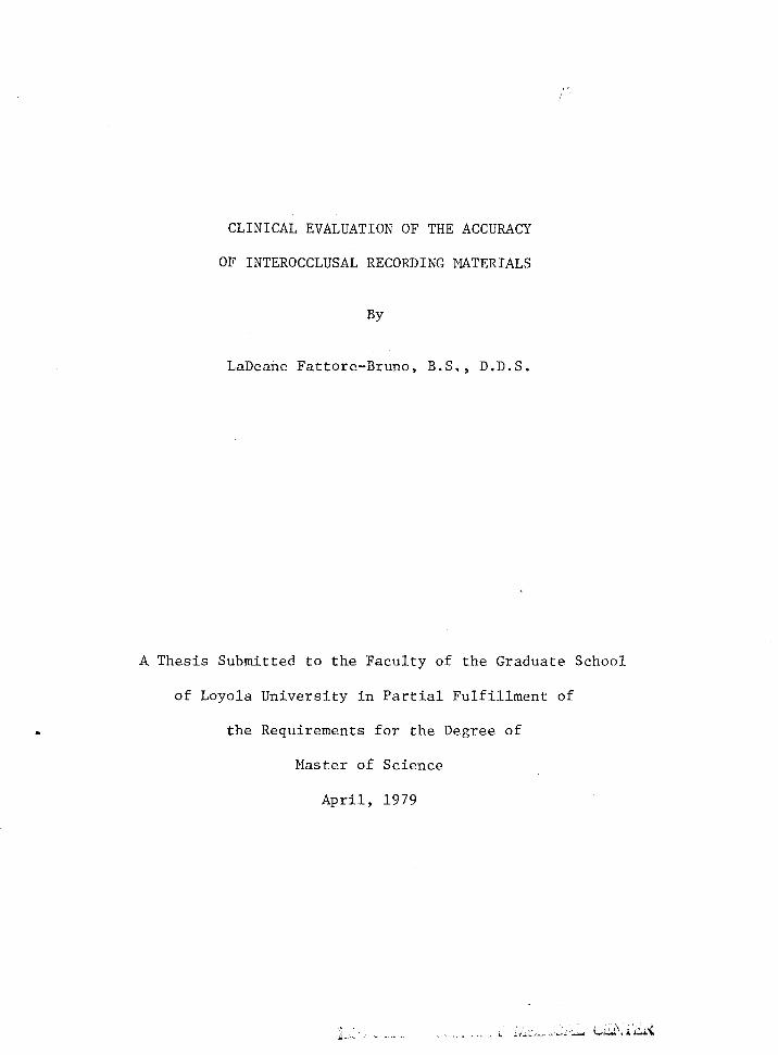
and a Buhnergraph was screwed on in place of them (See Figure 5).
Graph paper attached to cardboard was placed on the condylar housing
using double stick tape. The cardboard reinforced the graph paper
to prevent distortion while making the recordings. The double stick

tape kept the graph paper firmly attached to the housing. Stamp pad
inks of different colors were used to make the recordings (See
Figure 6).
First, the position of the maxillary cast when hand articulated
with the mandibular cast was determined. The casts were placed in
centric occlusion. Black ink was placed on the Buhnergraph styli.
With the casts carefully supported, the positions of the tips of the
pointer rods were marked on the graph paper. The ink was cleaned
34
off the styli and a different color ink was placed on them. An
interocclusal record was placed on the mandibular cast. The maxillary
cast was set into it. The casts were carefully supported in the
imprints of the record. Again the positions of the tips of the
styli rods were marked on the graph paper. This procedure was re
peated for each interocclusal record (See Figure 7).
The distance between the position of the hand articulated casts
and the casts mounted into the interocclusal record was then measured.
The measurements were made on a Gaertner traveling microscope
micrometer.

35
Figure 1

36
Figure 2

37
Figure 3

38
Figure 4
39
Figure 5
Figure 6

40
Figure 7
Ho\ND \HICUL.HION - 3L . .\CK
LT
POLYETHER ;r rHou·r COli: rRAY-GilEEN
lBC Q){IDE .\ND EUG.,;NQL P sr~-BLUi>
~.:.I. !'0 iC:>O o.U- PU'lPLE
;.Ji 0·(1·
,.,. 101 ,. -< T ,,.
PI. K AX-3 O.o/N
" ct·•• 7"
t•" .,..

CHAPTER IV
EXPERIMENTAL RESULTS
Table 1
Means and Standard Deviations of Four
Interocclusal Records
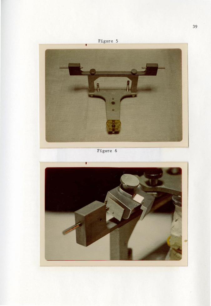
Pink Wax
X-V ector Y-Vector Rotation Declanation
Reinforced Wax
X-V ector Y-Vector Rotation Declanation
Polyether With Coe Tray
X-V ector Y-Vector Rotation Declanation
Polyether Without Coe Tray
X-V ector Y-Vector Rotation Declanation
Means
.0102
.2180
.0375 -.0870
-.0203 .1650
-.0298 .0830
-.0099 .0953 .0154
-.0500
-.0066 .0325
-.0230 -.0069
41
Standard Deviations
.0760
.0797
.5037
.6718
.0581
.0755
.3950
.3254
.0333
.0586
.1813
.2392
.0171
.0218
.1152
.0954

Means Standard Deviations
ZOE
X-V ector Y-Vector Rotation Declanation
-.0116 .1067 .0178 .0174
.0290
.0542
.2325
.2327
The right and left poles of the articulator hinge axis have been
condensed to four factors of displacement:
1) X-Vector - X is the displacement anterior-posteriorly of the center of the hinge axis
2) Y-Vector - Y is the displacement vertically of the center point of the hinge axis
3) Rotation - the degree of angular displacement in the horizontal plane
4) Declanation - the angular displacement in the sagittal plane as measured from the center of the hinge axis
The plane of the graph paper attached to the condylar housing
is tangent to the arc defined by the articulator hinge axis. A
vector originated from a point representing the original centric
occlusion position (CO). The vector terminated at a point represent-
ing the new CO position. The investigator thus measured a vector in
space and a projection in the given plane (horizontal or vertical).
The mandibular cast was the moving member not the maxillary
cast. The upper cast is fixed due to facebow utilization. The
graph paper is also part of the fixed upper cast. The maxillary cast
is fixed. The lower cast is mounted to it. It is important to
understand this concept. The maxillary cast cannot be thought of as
42

43
a floating member. To do so is to misinterpret how the standard was
obtained. In the clinical lab procedures, the standard was always
obtained by mounting the mandibular cast to the fixed maxillary. When
interocclusal records are placed between the casts, the investigator
isagain mounting the lower cast to the upper. The Buhnergraph,
mandibular cast, and lower component of the Whip Mix articulator were
in effect moving about the fixed maxillary cast as determined by the
facebow.
THE MOVEMENTS MADE BY THE MANDIBULAR CAST WERE REFERENCED TO THE CENTER OF THE HINGE AXIS OF THE ARTICULATOR.
ANY WAY THE MANDIBULAR CAST MOVED, THE CENTER OF THE ARTICULATOR HINGE AXIS ALSO MOVED.
THE MOVEMENTS MADE BY THE MANDIBULAR CASTS ARE VERTICAL, HORIZONTAL, AND LATERAL. LATERAL MOVEMENT, AS MENTIONED, COULD NOT BE MEASURED.
From the data the investigator analyzed: 1) Bodily movement of
the center of the articulator hinge axis in space; 2) Angular dis-
placement of the hinge axis around its center in two planes - in
the horizontal plane (or rotation) and in the vertical plane (or
declanation).
In this study the investigator measured the X and Y displacement
of the two poles (left and right) of the hinge axis and rotation and
declanation of the hinge axis around its center. These measurements
were used for the statistical analysis.
An interocclusal record when placed between the teeth can cause
both vertical and horizontal movement - not a pure movement. It is

44
necessary to divide this movement into its component parts - X andY.
The distance from the centric occlusion position (CO) to the new
position as determined by the interocclusal record (CO') was known.
The angle 9 formed by the line CO-CO' with the horizontal line deter-
mined by the Frankfurt Horizontal plane was determined through use of
a protractor. Because the condylar housings were set at 30°, 30° had
I • to be added to the angle formed by the co-co llne and the horizontal
base line in order to establish the angle 9. (See Figure D).
Through use of trigonometry, the X-component was determined to be:
..l X=<COS 9 (Hypotenuse)
..)
Y=SIN 9 (Hypotenuse) (See Figure E)
Rotation was the next factor of displacement considered. To
determine rotation, geometric assumptions were made:
1) WHEN THE MANDIBULAR CAST CHANGED POSITIONS, THE POINTERS MARKED THE INTERSECTION OF THE GRAPH PAPER WITH THE MANDIBULAR AXIS
2) GIVEN A LINE (LINE A), AND ANOTHER LINE (LINE B), NOT PARALLEL TO LINE A, AND AN INTERSECT ANGLE 9
8, ,..B II
- --8 ,, -- -- 8 --- ::.. ..,-" -- _::. B" ' I
' ...- - ..-' --- _,.... ~ .... - 1)-- - -- -- e -- ....--...- ..- -- ,..-- ----ALL':LINES PARALLEL TO LINE B HAVE SAME INTERSECT ANGLE.
3) CONSTRUCT A LINE PARALLEL TO BUHNERGRAPH STYLI THAT INTERSECTS ONE OF THE GRAPH PAPERS AT THE POINT OF THE MAXILLARY HINGE AXIS. CALL LINE M'. (See Figure F).
The maxillary cast axis is equal to the diameter of the Whip Mix
upper member from the outer surface of the right condylar housing to

FIGURE ~
FRANKFORT HORIZONTAL PLANE-._ .......... ___ _
FIGURE E
ACTUAL ANGLE FROM HORIZONTAL:
" 9=cX+30
CO' CO=ORIGINAL CENTRIC OCCLUSION POSI-TION
CO'=NEW CENTRIC OCCLUSION POSITION AS DETERMINED BY THE INTEROCCLUSAL RECORD
Y-C MPONENT
co
ENLARGEMENT OF CONDYLAR POINTS ON GRAPH PAPER
-l XY DISPLACEMENT (DISTANCE, DIRECTION)
X COMPONENT=COS 9 (HYPOTENUSE) 8=ANGLE MEASURED WITH PROTRACTOR HYPOTENUSE=DISTANCE MEASURED FROM CO ESTABLISHED BY HAND ARTICULATION TO CO' ESTABLISHED BY INTEROCCLUSAL _!)ECORD Y=SIN 9 (HYPOTENUSE)

STYLUS
~
FIGURE F
M =MANDIBULAR AXIS AFTER BITE REGISTRATION
M'=LINE CONSTRUCTED PARALLEL TO M
GRAPH PAPER
STYLI POINT TO LINE REPRESENTING MAXILLARY AXIS
46
GRAPH PAPER
MAXILLARY AXIS=MANDIBULAR AXIS AT CO ESTABLISHED BY HAND ARTICULATION
~ ~
VT=VR-VL
FIGURE G
GRAPH PAPER
GRAPH PAPER
-.l. -l VT=VR-VL
) STYLU~ < VT
ORIGINAL POSI~ -----F-------~------------------------~~~~ Maxillary cast TION OF POINTERS DIAMETER OF WHIP MIX axis
VT=X component, X component of pole
ATN=arc tangent or inverse tangent
KNOWN: KNOWN: KNOWN:
w DIAMETER OF WHIP MIX=D (ADJACENT SIDE)
~ ~
VT=VR-VL=XCO~FONENT l-X COMPONENT L TAN 9= OPP~SITE SIDE/ADJACENT SIDE TAN 9=VT/D THEREFORE 9=ATN (QfPOSITE SIDE)/ (ADJ. SIDE)
=ATN (VT /D""') 9 = ROTATION

47
the outer surface of the left condylar housing plus 2mm. Each graph
paper was lmm in thickness due to the cardboard mounts. Two mm was
thus added to the diameter of the Whip Mix. ....... _.l. -I. VT is equal to VR-VL.
~ .....)
VR is known. VR=X component right. This was previously measured. ~ -1 VL=X component left. This was measured. Thus rotation = TAN cn/R)
~
or 1/TAN (H/R). (See Figure G).
Declanation was determined the same was as rotation except
..... -I. ..,.) ~
VT=VR-VL where VR=Y component right. VL=Y component left. Declana-
-1..... ~ tion =TAN (V/R) or 1/TAN (V/R).
Thus the angular displacement in a given plane (Horizontal =
ROTATION, Vertical = DECLANATION) is therefore the inverse tangent
of the rotation displacement vector divided by the diameter of the
instrument.
-' Rotation and declanation have been established. However, X and
~
Y are displacement components determined for both the left and right
sides. Data cannot be effectively analyzed unless the X and Y poles
are condensed in terms of the movement of the center of the articu-
later hinge axis.
paper left pole
(See Figure H).
center of
articulator hinge
axis
graph paper
right pole

FIGURE H
LINE THROUGH CENTER OF ARTICULATOR HINGE AXIS
sV::r CONDENSATION OF 2 POLES IN X PLANE M,
Mo .---1
48
ROT VJ..
RSV ROT VR
SV::rLSV::rRSV BECAUSE M0 IS PARALLEL TO M2
--" __.). ---l -1
ROTL • HVL + LSV ROTR =a HVl~ + RSV
ROTL •-ROTR
--' LSV ____.. --1.
) HVL + . - (HVR + RSV
( _,. -· (HVR
.....J. HVL + sv ) . - + sv )
--l. _.. ~ --l HVL + sv . - HVR -sv
sV + SV+ HVL = -"HVR __,. --" ---l
2 SV • -HVR - HVL -.1 ----J. ----1
SV • -~ ( HVR + HVL )
lsVI·I~ ( HVR + HVL ) I
MOVEMENT OF CENTER OF ARTICULATOR
HINGE AXIS IS IN DIRECTION ( POSITIVE OR
NEGATIVE) OF LARGEST COHPONENT VECTOR

-l HVL=X component left. This is known. _ __., HVR=X component right. This is known.
-l. SV=the movement of the center of the articulator hinge axis as
determined by the X components of both the left and right sides.
Baseline=maxillary cast
M0=line formed by mandibular member when interocclusal record is placed between casts.
M1 and M2 are lines drawn parallel to Mo. Because Ro and M2 are parallel, SV=LSV=RSV
49
lsvl = ll/2 (ERlR+InlL)I or the movement of the center of the articulator hinge axis is equal to 1/2 the movement of the right and left X components.
For the determination of the movement of the center of the articu-
lator hinge axis in terms of the right and left Y components, the
same geometrical explanation as Figure D was used. However, the right
---l. ----1. and left Y components were substituted for HVL and HVR. Thus Y= ~ --1.. __. --l
(LV+RV)/2 where LV=left vertical component and RV=right vertical
component.
A statistical analysis (F RATIO) was used to analyze the data
for the four components of movement for each of the materials studied.
(See Table 2).
For the comparison of all 5 treatments, the F ratio of 38.525
for the Y-vector was statistically significant. The waxes, with
their great resistance to closure, were the probable cause of this
vertical displacement.
Rotation, declanation, and the anteroposterior components of

Table 2
F RATIOS
Statistical Significance *
~ ROTATION
Reject Null Hypothesis if F>2.44 at .05 level.
Five Treatments Compared 2.065 38.525* .261
Reject Null Hypothesis if F>4.17 at .05 level.
Zoe and Polyether With Coe Tray
Pink Wax and Polyether Without Coe Tray
Pink Wax and Reinforced Wax
Reinforced Wax and Polyether Without Coe Tray
Polyether With Coe Tray and Polyether Without Coe Tray
Reinforced Wax and Polyether With Coe Tray
Zoe and Polyether Without Coe Tray
.068
1.648
3.83
1. 396
.367
.728
.837
.719
1.648
6.876*
87.234*
34.437*
17.452*
45.968*
-3 2 X 10
.409
. 328
.01
.744
.306
1.102
Reject Null Hypothesis if F~3.15 at .05 level.
Reinforced Wax, Pink Wax, and Polyether Without Coe Tray
ZOE, Polyether With and Without Coe Tray
2.552 65.423* .299
23.09*
50
DECLANATION
1.118
1. 389
.425
1.878
2.124
.897
3.757
.297
1.264

51
movement were all insignificant. Centric occlusion was probably
responsible for these results. With centric occlusion a patient has
definite stops which his neuromuscular mechanisms are "programmed" to
locate. Patients can find their intercuspal position. They may not
be able to close into it completely due to an intervening material,
but they "know" where the position is located. With centric relation
we have a different situation. Declanation and the X-vector could be
significantly affected when the mandible is "de-programmed."
The next analysis of variance comparison was ZOE and polyether
with a Coe Tray. Both materials utilized a carrier. The investigator
was interested in finding out how the materials compared to each
other (Coe Tray carrier variable cancelled out).
The results for all 4 components of displacement were as ex
pected. The F ratios for all 4 components of displacement were insig
nificant. ZOE and polyether have,little if any resistance to closure.
The materials are fluid-like. Closure into centric occlusion is
unimpeded.
The next analysis of variance run was pink wax and polyether
without a Coe Tray. All 4 F ratios were insignificant. The results
were not as expected. Polyether is a material with little resistance.
Pink wax has a great deal of resistance to closure.
Pink wax and reinforced wax were compared. The two waxes were
significantly different in the vertical plane. Anteroposterior
deviation, although statistically insignificant, was more pronounced
than with other materials.

52
Reinforced wax and polyether without a Coe Tray were next examined.
The two materials were significantly different in the vertical displace
ment analysis. This was as expected. Reinforced wax has a great deal
of resistance to closure.
Polyether with Coe Tray and polyether without Coe Tray were com
pared. For this analysis all results were as expected. However, the
Y-vector F ratio was a little surprising (F=34.437). Rotation, de
clanation, and anteroposterior displacement were expected to be
negligible. Polyether has no resistance to closure. To be so statis
tically significant in the vertical plane was due to the carrier.
Reinforced wax and polyether with Coe Tray were next examined.
The results indicated reinforced wax caused a significant amount of
displacement of the center of the articulator hinge axis in the
vertical plane. This again was caused by its resistance to closure.
Declanation was insignificant, but the F ratio of 3.757 indicated
reinforced wax was capable of causing a deviation in the sagittal
plane.
ZOE and polyether without a Coe Tray were compared. The F ratio
was statistically significant in the Y plane. Interference from the
tray can be assumed to be the responsible factor since ZOE and poly
ether have no resistance to closure.
Reinforced wax, pink wax, and polyether without Coe Tray were
compared. The results indicated waxes have resistance to closure in
the vertical plane. The anteroposterior displacement was insignificant
but an F ratio of 2.552 indicated waxes can cause anteroposterior

53
deviation of the center of the articulator hinge axis.
ZOE, polyether with and without a Coe Tray were compared only in
the vertical dimension (Y-vector). The F ratio was 23.09 for this
analysis. The Coe Tray carrier was probably responsible for the
statistically significant F ratio for the Y-vector.

CHAPTER V
DISCUSSION
This study is attempting to examine and measure a three-dimen
sional displacement of the center of the Whip Mix articulator hinge
axis in two dimensions. This is a geometrical projection in 2-D of
a 3-D clinical situation (def. Projection= N-1 representation of an
N dimensional object).
The Buhnergraph can measure only anteroposterior and vertical
components of displacement of the articulator hinge axis. It cannot
measure pure lateral components of movements. That is, a sideshift
invoked by the material cannot be detected by this recording device.
For this study, the lateral component of movement does not
negatively affect the validity of the Buhnergraph recordings. In
fact, this shortcoming of the device favors the interocclusal records
used in this study. The resistance of waxes are notorious for
causing displacement of the maxillomandibular relationship. However,
if a wax displaces the mandible laterally, this malposition will not
be recorded. The material will in fact appear to be more clinically
useful than it actually is (Figure A).
ASSUMPTIONS MADE IN THIS STUDY:
1. The investigator is studying a representation of a patient's
hinge axis as determined by the Whip Mix face-bow.
54

55
2. In the experimental design of this study, the investigator is
assuming the hinge axis is related to the mandibular member of the
articulator in a proper centric occlusion without an intervening mate-
rial (Hand Articulation= Standard).
3. Interocclusal recording media have the potential to cause
errors in both clinical patient procedures and cast mounting procedures.
No matter what the mechanism behind the alteration of the center of
the hinge axis of the articulator, the sum total of these errors will
manifest themselves as two dimensional displacements of the poles of
the articulator hinge axis.
4. The Null Hypothesis states: Interocclusal recording media do
not alter the position of the center of the articulator hinge axis in
space.
In analyzing the data obtained in this study, the anteroposterior
component of movement, X, and the vertical component, Y, must be con-
sidered separately. Declanation and Rotation are a part of the X and
Y movement. A pure vertical and pure anteroposterior movement
probably rarely exists when an interocclusal media is placed between
the teeth. Declanation is the angular displacement .in the sagittal
plane as measured from the center of the hinge axis. Rotation is
the degree of angular displacement in the horizontal plane.
FIGURE A
STYLUS -~ ~ ~
POSITION POSITION ONE TWO

56 FIGURE B
DECLANATION

1 57
FIGURE C
ROTATION - As Seen From Above
---J
---- - --:x_--

1 I
58
LIMITATIONS OF STUDY:
EXPERIMENTAL DESIGN
1. Reiterating a point previously discussed, this study is based
on a two-dimensional projection of a three-dimensional entity in
space.
2. The lateral component of movement, although not negatively
affecting the study, cannot be measured with a Buhnergraph. Poor
interocclusal recording media can appear to have more clinical use-
fulness than actually exists.
The investigator attempted to control the following variables.
However, the clinical judgment required is a variable itself.
CLINICAL TECHNIQUE
3. Improper impression technique can minimize the usefulness as
a standard of the hand articulated casts in centric occlusion. Taking
alginate impressions at a significantly open vertical dimension will
give a centric occlusion different from that which the patient can
attain. The interocclusal records are taken with the teeth in maximum
intercuspation. These records will not appear as satisfactory as they
might be when compared to this standard. Flexion of the mandible is
the cause of this problem.
4. If the patient does not completely close into the inter-
occlusal recording material until his teeth come together in centric
occlusion, the record reproduced will be incorrect. The true nature
of the interocclusal material will not be recorded. Patients often

59
do not know whether they have closed all the way through a material.
MOUNTING PROCEDURES
5. The improper mounting of the mandibular cast to the maxillary
cast in centric occlusion could invalidate the standard for that
particular patient. If the investigator does not carefully note wear
facets, anterior and posterior stops, the plane of occlusion of all
teeth, malposed teeth, etc. an improper hand-articulated mounting in
centric occlusion will result.
6. Failure to properly trim interocclusal records could result
in improper seating of the maxillary cast into the recording media.
Incorrect recordings will result.
COLLECTION OF DATA
7. Articulating the maxillary and mandibular casts and then
marking the graph paper grid with the Buhnergra.ph styli is both
arduous and critical. If the maxillary cast is not properly and
carefully held into the record, the upper member of the articulator
will declanate slightly. The wrong position will then be recorded.
8. The Gaertner Traveling Microscope Micrometer is another
possible source of error in this study. If the micrometer is not
calibrated at properly designated intervals, "play" in the vernier
will result in inaccurate recordings.

60
COM?ARISON OF RESULTS TO LITERATURE AND ITS CLINICAL IMPLICATIONS:
Zinc oxide and eugenol was compared with polyether utilizing a
Coe tray. The rotation analysis of variance was negligible. Declana
tion, anteroposterior, and vertical analysis of variance were insig
nificant. It can be concluded from this study the two materials
utilizing a carrier behave equally well.
Berman (1960) and Tylman (1978) stated ZOE was the material of
choice for an interocclusal record. They said the material mixes to
a true fluidity and offers no resistance to closure. This study did
not indicate ZOE was the material of choice for an interocclusal
record. It did, however, bear out the claims Berman and Tylman made
for the fluidity and lack of resistance to closure of ZOE.
Reinforced wax and polyether without a Coe tray were compared.
The F ratio for the Y-vector was statistically significant. It can
be concluded reinforced wax, due to its resistance to closure,
dimensional instability, and deformation-prone nature is an unreliable
interocclusal record.
Reinforced wax and polyether with a tray were compared. The F
ratio for the vertical dispacement vector was statistically signifi
cant. Declanation came very close to significance. Again, it can be
concluded the inherently poor nature of reinforced wax caused the
deviations indicated.
Reinforced wax, ?ink wax, and polyether without a tray were
compared. Again the Y-vector had a statistically significant F ratio.

Waxes are poor interocclusal records. This study confirms the
work of Berman (1960), Shanahan and Leff (1960), and Millstein,
Clark, and Kronman (1973). It supports the claims of Nagle (1959),
Silverman (1957), and Tylman (1978).
CLINICAL SIGNIFICANCE OF STUDY
For the dental clinician interocclusal recording waxes are best
left alone. Compared to other available recording media, they are
decidedly inferior.
61
Zinc oxide and eugenol paste is a very good interocclusal record
ing material. Polyether with a carrier is equally good. However,
there are some disadvantages with both of these materials.
ZOE cracks and sticks to the teeth. Vital parts of it can be
lost through this breakage. Once the record has been used to mount
casts, it can no longer be used again. Wirth and Aplin noted these
disadvantages in 1971.
ZOE must be trimmed. The flash around the intaglios of the
teeth, unless cut back, can prevent the accurate seating of casts.
This is especially true of polyether.
There is a "spring" to this elastomere. There is also a decided
amount of friction in seating a stone model into it. If polyether is
not trimmed and carefully seated into casts, it can be worse than any
material examined in this study.
As a matter of fact, if this investigator had not trimmed ZOE
and Ramitec in the 3 treatments out of 5 presented in this study,

there is a good possibility the Null Hypothesis could have been
proved.
Polyether and ZOE must be used thinly. This is important for
ZOE and critical for polyether. Too much polyether around the
intaglios of the teeth will significantly prop open a pair of casts.
The chemical setting recording media studied are not especially
easy to use, if any are. They are relatively convenient to use
clinically but they prove to be more difficult to utilize in the
clinical lab procedures. This is extremely important for the dental
clinician to realize. Unfortunately, only the laboratory technician
seems to understand this well. He uses HAND ARTICULATION.
The results of this study indicated polyether without a Coe tray
was the best interocclusal recording material studied. The carrier
appeared to interfere with proper centric jaw closure. The investi
gator was careful to keep the plastic frame part of the carrier away
from the occluding surfaces of the teeth. However, the frame often
touched the inner surfaces of the cheeks and other soft tissues.
This might have set up a negative neuromuscular response in the
patient resulting in a less than optimum centric oc.clusion.
FUTURE STUDIES
To improve future studies using a Buhnergraph, a third point
should be devised to record lateral displacement. A hinged needle
attached to the Buhnergraph might be a solution. Interocclusal
materials with and without resistance could then be more accurately
62

compared to each other. Their clinical usefulness could be more
comprehensively evaluated.
63
Clinical research involves many variables---some that can and
some that can't be controlled. The sheer difficulty in designing a
scientifically valid clinical study points out clearly the importance
of this type of research. Dental laboratory research is necessary
and important but clinical evaluation must follow these types of
studies.
Clinical studies of dental materials are sorely lacking. How
good is dental research, then, if two very important factors, the
patient and operator variables, are left out? No matter how many
claims are made for a product, if it does not work in the hands of
the clinician, if it isn't compatible with the patient, if it
possesses properties causing difficulty in clinical and laboratory
procedures, it isn't a product desirable for the dental profession.
It does not work.

BIBLIOGRAPHY
Arstadt, T. "The Capsular Ligaments of the Temperomandibular Joint and Retrusion Facets of the Dentition in Relationship to Mandibular Movements,u Akademisk Forlag, Oslo, 1954.
Berman, M. "Accurate Interocclusal Records," J Pros Dent., Vol. 10, 1960.
Beyron, H.L. "Occlusal Relations and Mastication in Australian Aborigines," A.C.T.A. Odont. Scand., Vol. 22, 1964, 597-678.
Beyron, H.L. "Optimal Occlusion," Dent. Clinics of North America, Vol. 13, No. 3, July, 1969.
Brecker, S.C. "Conservative Occlusal Rehabilitation," J Pros Dent., Vol. 9, No. 6, Nov-Dec., 1959, 1002.
Boucher, Carl. "Through the Eyes of the Editor,u J Pros Dent., Vol. 28, No. 4, October, 1972, 354.
Boucher, L.J. and Jacoby, J. "Posterior Border Novements of the Human Mandible," J Pros Dent., Vol. 11, 1961, 836-841.
64
Brotman, Don. "Contemporary Concepts of Articulation," J Pros Dent., Vol. 10, March-April, 1960.
Brotman, Don. "Hinge Axis. Part I. The Transverse Hinge Axis," J Pros Dent., Vol. 10, May-June, 1960, 440.
Cohen, R. "The Hinge Axis and Its Practical Application in the Determination of Centric Relation, u J Pros Dent., Vol. 10, March-April, 1960.
Cohn, L.A. "The Factors of Dental Occlusion Pertinent to the Restorative and Prosthetic Problem," J Pros Dent., Vol. 9, 1959, 256-277.
Combe, E.C. Notes on Dental Materials. 2nd Ed., ChurchillLivingston, 1975.
Coulourites, Alex. "Freeway Space," J Pros Dent., Vol. S', March, 1955.
De Marco and Paine. "Mandibular Dimensional Change," J Pros Dent., 1974, 428-485.

65
Docking, A.R. "Modern Materials in Dental Practice," Aust. Dent. J., Vol. 15, 1970, 303-309.
Douglass, Gordon. "The Cast Restoration-Why is it High?" J Pros Dent., Vol. 34, 1975, 491-495.
Frisch, J., Bhaskar, S.N., and Bales, D. "Clinical Study of Normal Occlusion (unpublished), 1966.
Goto. "An Experimental Study on the Physiologic Mobility of a Tooth," Shikwa Gakuho, Vol. 71, 1971, 1415-1444.
Granger, E. "The Principles of Obtaining Occlusion in Occlusal Rehabilitation," J Pros Dent., Vol. 13,1963.
Guichet, N. "Biologic Laws Governing Functions of Muscles that Move the Mandible. Part IV. Degree of Jaw Separation and Potential for Maximum Jaw Separation, J Pros Dent., Vol. 38, No. 3, September, 1977, 305-306.
Hoel, Paul. Elementary Statistics, Third Edition, New York, London, Sydney, Toronto: John Wiley and Sons, Inc., 1971.
Jankelson, Hoffman, and Hendron. "The Physiology of the Stomatognathic System" J.A.D.A., No. 46, 1953.
Kingery, R. "Maxillomandibular Relationship of Centric Relation," J Pros Dent., Vol. 9, Nov-Dec., 1959, 924-925.
Long, J.H. "Location of the Terminal Hinge Axis by Intraoral Means," J Pros Dent., Vol. 23, 1970.
Lyon, Douglas M. "The Establishment of Occlusion," Dental Clinics of North America, Nov. 1960.
Millstein, Clark, and Kronman. "Accuracy of Wax Interocclusal Registrations," J Pros Dent., Vol. 29, 1973.
Moyers, R. "Some Physiologic Considerations of Centric and Other Jaw Relations," J Pros Dent., Vol. No. 6, 1956.
Nagle, R.J. "Personal Communication to Martin Henry Berman," July 6, 1959.
Osborne, J. "Occlusion and Prosthetic Dentistry," Dental Clinics of North America, Vol. 13, No. 3, July, 1969, 623.
Pameijer, J., Glickman, I., and Roeber, F.W. "Intraoral Occlusal Telemetry. II Registration of Tooth Contacts in Chewing and Swallowing," J Pros Dent., Vol. 19, 1960, 151.

66
Posselt, U. Physiology of Occlusion and Rehabilitation. Philadelphia: The F.A. Davis Co., 1962.
Posselt, U. "Range of Movements of the Mandible," J.A.D.A., Vol. 56, 1958, 10-13.
Posselt, U. "An Analyzer for Mandibular Positions," J Pros Dent., Vol. 7, 1957, 368-374.
Ricketts, R.M. "Variations of the Temperomandibular Joint as Revealed by Cephalometric Laminography," Am J Orthodont., Vol. 36, 1950, 877.
Schireson, S. "Grinding Teeth for Masticatory Efficiency and Gingival Health," J Pros Dent., Vol. 13, Mar-April, 1963, 344.
Schweitzer, J. "Dental Occlusion: A Pragmatic Approach," Dental Clinics of North America, Vol. 13, No. 3, July, 1969, 706-707.
Silverman, S.I. "Management of the Trial Denture Base," Dental Clinics of North America, March, 1957, 231-243.
Sicher, H.L. "The Biologic Significance of Hinge Axis Determination," J Pros Dent., Vol. 6, 1956, 620.
Sicher, H.L. Oral Anatomy, 4th Ed., St. Louis: The C.V. Mosby Co., 1965, 505-510.
Sindledecker, L. "Effect of Different Centric Relation Checkbites on Condylar Position," Masters Dissertation, University of Michigan, 1978.
Tylman, S. "Methods of Recording Interrocclusal Relationships," Tylman's Theory and Practice of Fixed Prosthodontics, 7th Ed., St. Louis: C.V. Mosby Co., 1978.

APPENDIX
67

68
ROTATION AND DECLANATION IN DEGREES AND THE X AND Y MOVEMENT OF THE CENTER OF THE ARTICULATOR HINGE AXIS IN MILLIMETERS
_. --" X y ROTATION DECLANATION
Pink Wax
Patient:
1 .0966 .28974 .2378 .3106 2 .08796 .13694 .2653 .1046 3 -.02835 .1824 -.0692 -.4786 4 . 09778 .29444 .2305 .2809 5 . 07189 .15631 .2141 . 6724 6 .04204 .14511 -.5765 .0243 7 -.06254 .25518 .0871 -1.4693 8 -.04466 .25627 -.8985 .4204 9 -.1505 .37955 .4711 -.1.2122
10 -.01201 .15805 -.5665 -.1358 11 -.13437 .15579 -.105 .1748 12 -.02939 .13972 .5697 -.2552 13 -2.8x1o-4 .17823 -.2323 .0896 14 .01567 .20213 -.2297 .2754 15 -.03318 .22306 -.6648 -.8272 16 -.1109 .14287 1. 2635 -.6452 17 -.02398 .16897 .3861 -.3424 18 -.13373 .15945 .165 .141 19 .10557 .14631 -.4745 .2882 20 .05335 .18004 -.0787 .536 21 .01785 .28576 -.2629 .8907 22 .03038 .38196 -.5862 .2321 23 -6.9x1o-4 .1266 -.5586 -.2083 24 .03849 .38365 -.1817 -1.6136 25 6.43x10 .20248 .3658 .3667 26 .0582 .16164 .3499 .1089 27 .0281 .18236 8.6x10⁻³ -.4783 28 . 07287 .17009 .0356 .1887 29 8.46x10 .33821 .5667 -1.1913 30 .17103 .31808 1.209 1. 3676 31 .07907 .25601 .2209 -.3201
69
~ -" X y .ROTATION DECLANATION
Reinforced Wax
Patient:
1 .06296 .12912 .4146 -.5485 "l -.07622 . 06713 .4027 -.1454 L
3 5.L~x1c-3 .20562 .2702 .2958 4 .06301 .12908 .4149 -.548 5 -.01591 .20704 -.2443 .0366 6 .03656 .15006 -.0881 .033 7 -.01037 .20215 -.3613 -. 0472 8 -.0783 .15179 .1996 .0675 9 -.09987 .13656 -.2221 -.0348
10 . Ol!327 .20695 -.1.1278 .0682 11 .06828 .42397 -.2731 .0949 12 -.0403 .15912 .1196 -.4408 13 -. 07296 .20616 -.0571 -.0272 14 -.07288 .15931 .117 .0357 15 -.14233 .18125 .5027 .0685 16 -.07741 .10499 -.0929 .1381 17 -.05113 .09142 -.0672 -3.2x1o-3 18 .03861 .14698 . 0118 -.1729 19 -.03789 .06981 -.4791 -.0559 20 -·.04572 .08258 -.4751 .3978 21 -.06447 • 07702 .3065 -.0337 22 3.16x1o-3 .16871 .1108 .2551 23 -.05578 .26009 -.2808 .4751 24 -.10415 .15905 -.1333 .4004 25 -.01058 .08794 .0157 .4501 26 -.034.26 .19234 .0853 .337lf 27 .0949 .24032 .9756 .2265 28 .02991 .1043 .197 .6868 29 -2. L•xlo-3 .12424 -.1751 -.24.74 30 -1. 52x1o-3 .31362 -.5733 .9355 31 .01888 .17502 -.418.6 -.1265

70
-1 .... X y ROTATION DECLANATION
Po1yether With Coe Trav
Patient:
1 .05725 .09343 .0863 -.0533 2 .0374 .09033 -.0548 .0517 3 -3 .07454 .076 -.0796 -6.08x10 4 .06057 .09906 .0887 -.0609 5 -.0317 .09677 -.3161 .0852 6 -6.45xlo-3 .25309 -.0817 -.5773 7 -.04307 .25817 .5713 -.9309 8 -.01602 .07599 -.1863 .0848 9 -.05539 .09097 .0716 -.0479
10 -.02251 .03844 -.2587 .0162 11 -.0778 • 0965!+ .1409 -.0526 12 -.0196 .07219 -.164 .0759 13 3.16x1o-3 .0915 .05 -.0652 14 9x1o-4 .09835 .1966 --.1464 15 0.02231 .10239 -. 0172 .0367 16 -.0253 .08457 .0337 .0961 17 2.4xlo-4 .031 .1675 -.2041 18 -1.4x1o-4 .05381 -.2038 -2.3x1o-3 19 4.5x1o-3 -.20761 .2349 -6. t+x1o-3 20 .05358 .09228 .2509 -.2231 21 -7.27x1o-3 .03029 -.Ol:-79 .1995 22 -.02635 .03205 -.0599 .1223 23 8.95x1o-3 .20904 .116 .2882 24 6. 91x1o-3 .07446 -.1603 -.3352 25 -.01172 .10199 .0667 -.0685 26 .01876 .10383 .1879 .3676 27 -.01791 .07032 -.181 -.1368 28 -.04455 .05171 -1. 7x1o-3 5.lo-3
29 -.06237 .06584 -.1Lf26 -.0992 30 -5.64x1o-3 .04492 .0916 .0683 31 -.05447 .06325 -.0769 .0426

71
..... ~
X y ROTATION DECLANATION
Po1yether Without Coe Tray
Patient:
1 0 0 0 0 2 .02024 .05833 .0831 .1135 3 9.42xlo-3 .0181 -.062 -.1192 4 3.67xlo-3 .01981 • 02!~2 .1305 5 0 0 () 0 6 -.02367 .02673 .0358 .0384 7 1. 99;~1o- 3 .03607 -.2429 -.1022 8 -.0289 .05109 .1662 .0579 9 -.01667 . 01105 -.0896 .0592
10 0 0 0 0 11 -. OLI46 .03741 -.0133 -.0762 12 -.01341 .02693 -.0189 -. 0372 13 4.86xlo-3 . 032if9 -.032 -. 214 14 -2.33x1o-3 .02218 .0154 -.1461 15 -2.4xlo-4 .02254 -.0914 -.0487 16 -.01679 . 02234 -.099 .0816 17 -7.21x1o-3 .02661 -1. 7x1o-3 .056 13 -8.42x1o-3 .01137 .0554 -.0749 19 -.01253 .02789 .0906 1. 2xlo-3 20 9.87xl0-3 .06132 -.3372 . 015/o~ 21 5.25x1o-3 .05997 -.1807 -.1516 22 -9.97x1o-3 .04701 .0679 -.0574 23 .01767 .09618 -.1351 -.097 24 .0317 .04092 -.1269 .0256 25 -.03359 .05009 -.1112 .1055 26 -.0335 .0296 .0692 . 017 L1 27 -4. Olxlo-· 3 .02275 .0264 -.11.~98
28 -. 02097 .06612 .2605 ,l{pJ5 29 -3.81x1o-3 .02405 -.0251 .158ll 30 -3.74x1o-3 .04925 .0218 -':l -6.3x10 ~
31 -9.01x1o-3 9.87x1o-3 -.062 .065

I
' 72
...... J
X y ROTATION DECLANATION
ZOE
Patient:
1 .04706 .10448 7.9x10⁻³ -6.5x1o-3 2 • 01119 .06221 -.0347 .1481 3 -.03152 .07017 .3249 .0461 4 .05959 .03262 -.017 -.0682 5 .04746 .08792 -.0426 .1244 6 -.01664 .15396 -.1257 .0928 7 -.01312 .1218 -.0353 -.0333 8 -.01551 .10906 .1909 -.0869 9 -.0248 .10733 .1918 -.0548
10 6.2x1o-4 .10953 -.23 .2194 11 -.01528 .13854 .3352 -.0665 12 -.06663 . 09729 .1327 -.0651 13 -.02712 .17122 -.0631 .3984 14 -. 02977 .25162 .5017 -.6253 15 -.03829 .14912 .3212 -.1935 16 5.32x1o-3 .13431 -.1016 .0681 17 -.01648 . 24713 .1695 -.2327 18 -.018 .12494 -.1351 -.126 19 .01474 • 06134 .2455 .2141 20 -.02909 .16587 -.6476 .6283 21 -.05147 .08268 -.0571 .2676 22 -.04215_3 . 08871 -.223 -.1091 23 2.87x10 .10578 -. 2377 .0922 24 1.16x1o-3 .04897 -.2146 .0432 25 -.05735 . 07782 .1736 -.3049 26 1.88x1o-3 . 09711 .1922 .0513 27 -.0259 .03698 .1705 -.2436 28 .01405 .04595 .0925 .~026
29 -5.58x1o-3 .04944 .0836 -.1568 30 -. 02211 .03209 -.2154 .2113 31 -.01746 .04085 -.2006 5.3x1o-3

ANGLES FORMED BY INTERSECTION OF LINES CO-CO' AND THE FRANKFORT HORIZONTAL PLANE
*9=Angles enumerated + 30
Patient:
1 2 3 4 5 6 7 8 9
10 11 12 13 14 15 16 17 18 19 20 21 22 23 24 25 26 27 28 29 30 31
Pink lvax Right Condyle Left Condyle
Angle Angle
76 68.5 94 76 54 47.5 99 64.5
111.5 67.5
132.5 123
78 72.5 79
141.5 110.5 139
30 56.5 69 71 62 84
108.5 88 84 64.5 98.5 96.5 81.5
68.5 50
109.5 68.5 68
107 147 119.5 112 125.5 129.5
60.5 100.5 94.5
144 29 73.5
121 80 81 93 98
132 85.5 76.5 58 75 68.5 59 56 61.5
73

Patient:
1 2 3 4 5 6 7 8 9
10 11 12 13 14 15 16 17 18 19 20 21 22 23 24 25 26 27 28 29 30 31
74
Reinforced Wax Right Condyle Left Condyle
Angle Angle
90 147 102.5
90 84 71 78
127.5 115
42.5 75
104.5 107 120.5 142 127 114
78 66 40
143.5 96 94
130.5 123.5 103.5 104.5
0 81.5 63.5 67
~20 108.5
79.5 20
104 81.5
108.5 106.5 135.5 120.5
86.5 103.5 112 108.5 109 126 124
71.5 151 129.5 104
84.5 106.5 119.5
93 95 48.5 74
108.5 101 106

75
Pol·iether With Coe Tray
Right Condyle Left Condyle Angle A'1g1e
Patient:
1 66.5 50.5 2 61 73.5 3 101.5 85 4 66.5 50.5 5 79 126 6 89 96.5 7 108 69.5 8 79 116.5 9 124 118
10 65 146.5 11 133.5 122.5 12 85 118 13 92.5 82.5 14 103.5 68 15 101.5 103 16 113.5 101.5 17 112.5 0 18 62.5 118 19 98.5 79 20 83 32.5 21 0 103.5 22 142 125 23 93 84 24 76 126.5 25 101 91 26 101.5 73.5 27 84 132.5 28 131 130.5 29 117 149 30 119.5 81.5 31 127 133.5

76
Pol.Y§!ther lJithout
Coe Tray Right Condyle Left Condyle
Angle Angle
Patient:
1 0 79.5 2 79.5 66.5 3 62.5 0 4 0 79.5 5 0 0 6 148.5 125.5 7 53 149.5 3 142 93.5 9 146 146.5
10 0 0 11 131 151 12 115.5 135 13 81.5 0 14 96 0 15 65.5 133 16 100 132.5 17 111 102 18 126.5 0 19 133.5 87.5 20 44 123 21 68.5 121 22 110 89.5 23 71 92 24 36 74.5 25 122.5 130 26 148.5 125.5 27 100 0 28 143.5 78 29 0 99 30 98 90.5 31 0 133

77
ZOE Right Condyle Left Condyle
Angle Angle
Patient:
1 66.5 65 2 67.5 86 3 142 77 4 66.5 65 5 52 69 6 89 102 7 93.5 99 8 110 82 9 115 87.5
10 65 103.5 11 114 74.5 12 129 118 13 99 99 14 107 73.5 15 116 85 16 80.5 94 17 98.5 87.5 18 89 110 19 128 61 20 45.5 116 21 135.5 116 22 94.5 136.5 23 67 105.5 24 51.5 119.5 25 124 134.5 26 107 73.5 27 125 0 28 0 73 29 104 74.5 30 0 130.5 31 72 139

78
MEASUREMENT OF LINE CO-CO'
CO=Hand Articulation
CO'=Position with interocclusal record between casts
ZOE Pink Wax Reinforced Wax Patient Right Left Right Left Right Left
1 .l150 .l142 .2500 .3621 .2124 .1340 2 .0432 .0849 .1301 .1995 .1638 .0475 3 .1026 .0792 .2557 .l164 .1646 .2548 4 .1559 .1349 .2595 .3623 .2123 .1341 5 .0876 .l144 .0670 .2787 .2026 .2191 6 .1399 .1718 .1918 .1556 .1534 .1568 7 .1271 .l182 .4843 .0588 .2140 .2056 8 .1301 .0968 .2132 .3678 .1784 .1690 9 .1276 .0991 .6058 .2108 .1565 .1873
10 .0841 .1469 .1934 .1688 .2910 .2522 ll .1627 .1333 .1753 .2366 .4240 .4392 12 .1379 .0990 .2128 .l160 .2335 .l173 13 .l121 .2346 .1683 .1951 .2199 .2179 14 .3624 .1634 .1681 .2447 .1786 .1737 15 .1986 .0202 . 3552 .1658 .2775 .2027 16 .1257 .1450 .3869 .0926 .1052 .1557 17 .2856 .2120 .2359 .1220 .1006 .1097 18 .1441 .1126 .2104 .2l10 .1771 .1273 19 .0366 .1073 .2051 .1930 .0857 .1265 20 .0988 .2907 .l183 .2647 .0345 .1853 21 .0600 .1372 .1612 .4216 .1381 .0741 22 .1056 .1048 .3667 .4213 .1307 .2084 23 .0997 .1243 .1792 .1278 .1884 .2542 24 .0542 .0638 .6322 .1390 .1292 .2526 25 .1497 .0442 .1548 .2655 .0235 .1565 26 .0934 .1094 .1452 .2101 .1488 .2445 27 .0903 .0000 .2564 .1136 .2127 .3668 28 .0000 .0961 .1567 .2136 .0000 .2170 29 .0755 .0266 .5249 .1835 .1636 .0914 30 .0106 .0844 .llll .6342 .1917 .0462 31 .0421 0635 .3080 .2360 .2l10 .1621
All figures are in centimeters

79
Patient: P With Coe Tray P Without Coe Tray
1 .1107 .1106 .0000 .0403 2 .0943 .1024 .0418 .0824 3 .0884 .0627 .0408 .0000 4 .1181 .1164 .0000 .0403 5 .0854 .1356 .0000 .0000 6 .1181 .1164 .0000 .0403 7 .4201 .1247 .0646 .0405 8 .0643 .0993 .0687 .0600 9 .1185 .0948 .0037 .0363
10 .0397 .0741 .0000 .0000 11 .1441 .1050 .0649 .0533 12 .0609 .0948 .0361 .0301 13 .1015 .0823 .0657 .0000 14 .1240 .0821 .0446 .0000 15 .0988 .1108 .0329 .0207 16 .0763 .1012 .0471 .0101 17 .0671 .0252 .0194 .0359 18 .0667 .0662 .0283 .0000 19 .2109 .2105 .0382 .0281 20 .1271 .1087 .0849 .0759 21 .0000 .0623 .0892 .0431 22 .0219 .0318 .0593 .0383 23 .1655 .2542 .1173 .0815 24 .1292 .0293 .0630 .0465 25 .1145 .0916 .0404 .0863 26 .0490 .1665 .0516 .0396 27· .0916 .0672 .0462 .0000 28 .0675 .0690 .0753 .0894 29 .0908 .0986 .0000 .0487 30 .0397 .0559 .0507 .0483 31 • 0711 .0961 .0004 .0270
All figures are in centimeters.

80
APPROVAL SHEET
The thesis submitted by LaDeane Fattore-Bruno has been read and
approved by:
Doctor William F. Malone
Doctor Boleslaw Mazur
Doctor James L. Sandrik
The final copies have been examined by the director of the thesis
committee and the signature which appears below verifies the fact that
any necessary changes have been incorporated, and that the thesis is
now given final approval with reference to content, form and mechanical
accuracy.
The thesis is therefore accepted in partial fulfillment of the
requirements for the Degree of Master of Science.
DATE I SIGNATURE OF ADVISOR