Embed Size (px)
Citation preview
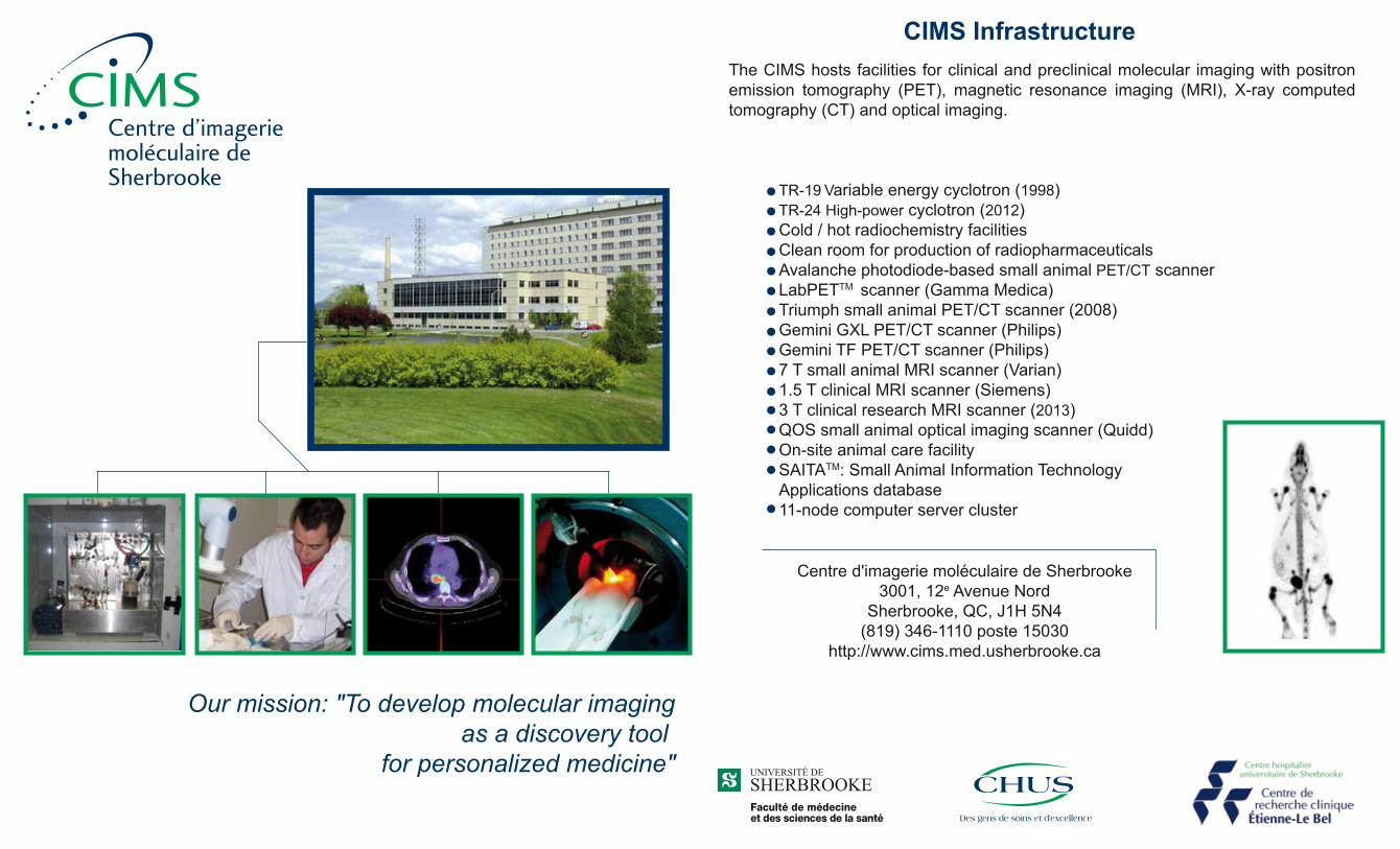
The CIMS hosts facilities for clinical and preclinical molecular imaging with positron emission tomography (PET), magnetic resonance imaging (MRI), X-ray computed tomography (CT) and optical imaging.
TR-19 Variable energy cyclotron (1998) TR-24 High-power cyclotron (2012) Cold / hot radiochemistry facilities Clean room for production of radiopharmaceuticals Avalanche photodiode-based small animal PET/CT scanner LabPETTM scanner (Gamma Medica) Triumph small animal PET/CT scanner (2008) Gemini GXL PET/CT scanner (Philips) Gemini TF PET/CT scanner (Philips) 7 T small animal MRI scanner (Varian) 1.5 T clinical MRI scanner (Siemens) 3 T clinical research MRI scanner (2013) QOS small animal optical imaging scanner (Quidd) On-site animal care facility SAITATM: Small Animal Information Technology Applications database 11-node computer server cluster
Centre d'imagerie moléculaire de Sherbrooke3001, 12e Avenue Nord
Sherbrooke, QC, J1H 5N4(819) 346-1110 poste 15030
http://www.cims.med.usherbrooke.ca
CIMS Infrastructure
Our mission: "To develop molecular imagingas a discovery tool
for personalized medicine"
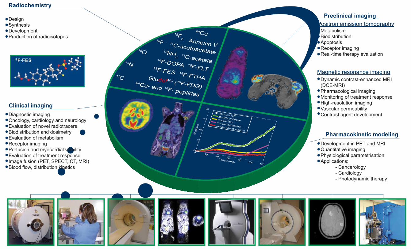
Radiochemistry
Preclinical imagingPositron emission tomography
Magnetic resonance imaging
Clinical imaging
Pharmacokinetic modeling
DesignSynthesisDevelopmentProduction of radioisotopes
Diagnostic imagingOncology, cardiology and neurologyEvaluation of novel radiotracersBiodistribution and dosimetry Evaluation of metabolism Receptor imagingPerfusion and myocardial viabilityEvaluation of treatment responseImage fusion (PET, SPECT, CT, MRI)Blood flow, distribution kinetics
Development in PET and MRIQuantitative imagingPhysiological parametrisationApplications: - Cancerology - Cardiology - Photodynamic therapy
Metabolism BiodistributionApoptosis Receptor imaging Real-time therapy evaluation
Dynamic contrast-enhanced MRI (DCE-MRI)Pharmacological imagingMonitoring of treatment responseHigh-resolution imagingVascular permeabilityContrast agent development
18F-FES