Embed Size (px)
Citation preview
Editorial
Chromaffin cells at the beginning of the 21st
century
Survival of a man, as well as of a beast, critically
depends on the rapid release of two related hormones,
adrenaline and noradrenaline, which provide for a
general mobilization of vital resources (Garcia et al.
2006). These hormones are synthesized and liberated
from post-ganglionic sympathetic neurones and from a
set of neuro-endocrine cells (which form the inner part
of the adrenal gland) generally known as ‘chromaffin
cells’. In addition, adrenal chromaffin cells release a
wide variety of peptides (e.g. opioids, or VIP), purines
(ATP, ADP, AP4A) and proteins that can act as
prohormones (DBH, granins).
The story of chromaffin (i.e. demonstrating affinity to
chromium salts) cells began a little more than 100 years
ago in Prague, when Alfred Kohn produced the first
detailed description of these cells (Kohn 1898, 1902,
1903): he identified these cells as secretory and discov-
ered their close relation to the neurones in the sympa-
thetic ganglia. The structure of the adrenal gland
received considerable attention from morphologists
(notably from R.E. Coupland) when microscopy tech-
niques became widely available. Dıaz-Florez et al. in
this issue contribute to update this subject with their
paper, which is accompanied by a set of images that
illustrate the structure and function of these cells.
Development of tissue culture techniques in the 1970s
extended earlier classical studies performed on perfused
adrenals in situ or in vitro and allowed for extensive
usage of biochemical, electrophysiological, electro-
chemical, fluorescence microscopy and molecular biol-
ogy techniques. In 1976, Lloyd Green and Arthur
Tischler created the PC12 cells from a rat phaeochro-
mocytoma; subsequently these cells were used for
important studies of cell differentiation and enzyme
activity as well as secretion. In this issue, Westerink and
Ewing have revisited the contribution of PC12 cells to
our knowledge of secretory pathways.
Almost 40 years ago Douglas (1968), using data
obtained from perfused cat adrenals, promulgated the
theory of stimulus-secretion coupling, which high-
lighted Ca2+ ions as signals coupling cellular excitation
with initiation of secretion. Since then, chromaffin cells
became one of the most popular and widely used
cellular models for investigating the molecular mecha-
nisms of secretion. Here, De Diego et al. present a
comprehensive description of stimulus-secretion cou-
pling and the role of neurotransmitter receptors and ion
channels. Further, the specific role of nicotinic cholino-
receptors in stimulus-secretion coupling is overviewed
by Sala et al., whereas the role of muscarinic receptors
is covered by Olivos and Artalejo. Additional amplifi-
cation of initial depolarization caused by activation of
nicotinic receptors is achieved by opening of voltage-
gated sodium channels; their contribution to the regu-
lated secretion is discussed by Wada et al. The central
role in stimulation-secretion coupling belongs to Ca2+
ions passing through voltage-gated Ca2+ channels.
Marcantoni et al. and Fox et al. overview the biophys-
ical and functional role of the various Ca2+ channel
types expressed by chromaffin cells in normal condi-
tions as well as under stress, while the current under-
standing of Ca2+ handling by cell organelles is presented
by Garcıa-Sancho and Verkhratsky.
The principal functional organelle of chromaffin cells
is represented by secretory vesicles, also known as
chromaffin granules. Their transport from the trans-
Golgi apparatus to the plasma membrane is conducted
by cytosolic proteins; these are described by Trifaro
et al. The main intravesicular proteins are chromogra-
nins, which are involved in many cellular functions such
as granule sorting, buffering of soluble components and
providing a source of bioactive peptides as overviewed
by Montero-Hadjadje et al. The molecular physiology
of exocytosis is a topic of the paper by Shuzo Sugita.
Further, the actual exocytotic process visualized by
evanescent wave techniques (total internal reflection
fluorescent microscopy – TIRFM) allows trailing of the
single vesicle in the narrow submembrane area, as
described by Holz and Axelrod. Electrophysiology and
electrochemistry applied to the study of secretion were
first assayed in chromaffin cells and then expanded to
other cells; however, chromaffin cells are still consid-
ered one of the favourite models to study the exocytosis/
endocytosis processes by capacitance recordings, ampe-
rometry and patch-amperometry, as presented by Bor-
ges et al.
Maintenance of regulated exocytosis is inseparable
from membrane retrieval accomplished through endo-
cytosis, which also can be followed by TIRFM; these
investigations are overviewed by Barg and Machado.
Finally, adrenomedullary tissues are implicated in
various forms of pathology and the relevant diseases
are summarized by Fung et al.
The spread of chromaffin cells as universal models
was very much determined by their common experi-
mental availability and by the large number of labora-
tory techniques, which have been developed to study
Acta Physiol 2008, 192, 143–144
� 2008 The AuthorsJournal compilation � 2008 Scandinavian Physiological Society, doi: 10.1111/j.1748-1716.2007.01819.x 143
them. Although this special issue of Acta Physiologica
cannot comprise all the aspects of the seminal research
carried out with chromaffin cells since William Doug-
las’s time, we have tried to present the reader with a
comprehensive view on the chromaffin cell physiology
and pathophysiology. This special issue represents a
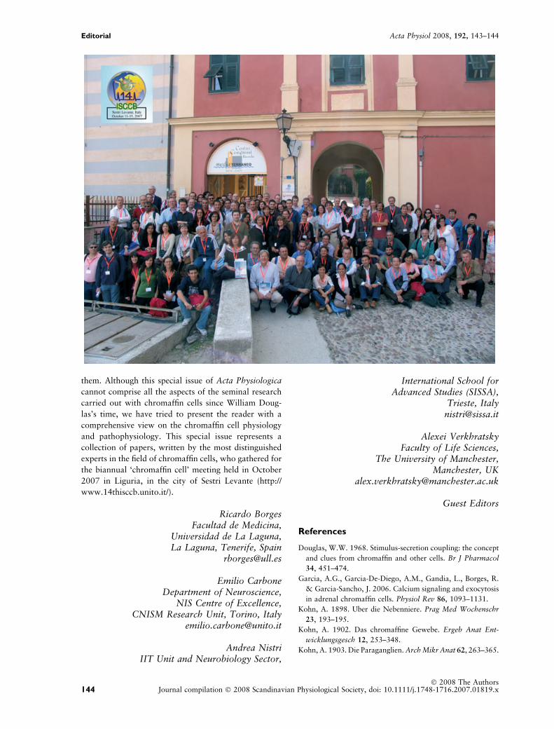
collection of papers, written by the most distinguished
experts in the field of chromaffin cells, who gathered for
the biannual ‘chromaffin cell’ meeting held in October
2007 in Liguria, in the city of Sestri Levante (http://
www.14thisccb.unito.it/).
Ricardo BorgesFacultad de Medicina,
Universidad de La Laguna,La Laguna, Tenerife, Spain
Emilio CarboneDepartment of Neuroscience,
NIS Centre of Excellence,CNISM Research Unit, Torino, Italy
Andrea NistriIIT Unit and Neurobiology Sector,
International School forAdvanced Studies (SISSA),
Trieste, [email protected]
Alexei VerkhratskyFaculty of Life Sciences,
The University of Manchester,Manchester, UK
Guest Editors
References
Douglas, W.W. 1968. Stimulus-secretion coupling: the concept
and clues from chromaffin and other cells. Br J Pharmacol
34, 451–474.
Garcia, A.G., Garcia-De-Diego, A.M., Gandia, L., Borges, R.
& Garcia-Sancho, J. 2006. Calcium signaling and exocytosis
in adrenal chromaffin cells. Physiol Rev 86, 1093–1131.
Kohn, A. 1898. Uber die Nebenniere. Prag Med Wochenschr
23, 193–195.
Kohn, A. 1902. Das chromaffine Gewebe. Ergeb Anat Ent-
wicklungsgesch 12, 253–348.
Kohn, A. 1903. Die Paraganglien. Arch Mikr Anat 62, 263–365.
144� 2008 The Authors
Journal compilation � 2008 Scandinavian Physiological Society, doi: 10.1111/j.1748-1716.2007.01819.x
Editorial Acta Physiol 2008, 192, 143–144