Embed Size (px)
Citation preview

CHARACTERISTICS OF EPIGLOTTIC INVERSION IN CHILDREN WITH CEREBRAL PALSY
by
Abigail Cryan
B.A. Biological Sciences, Carnegie Mellon University, 2013
Submitted to the Graduate Faculty of
School of Health and Rehabilitation Sciences in partial fulfillment
of the requirements for the degree of
Master of Science, Speech Language Pathology
University of Pittsburgh
2016

ii
UNIVERSITY OF PITTSBURGH
SCHOOL OF HEALTH AND REHABILITATION SCIENCES
This thesis was presented
by
Abigail Cryan
It was defended on
April 21, 2016
and approved by
Thesis Advisor: James L.Coyle, Ph.D., CCC-SLP; BCS-S, Associate Professor
Roxann Diez Gross, Ph.D., CCC-SLP, Adjunct Assistant Professor
Katherine Verdolini Abbott, Ph.D., CCC-SLP, Professor

iii
Copyright © by Abigail Cryan
2016

iv
This single cohort, retrospective, descriptive study, was conducted because of anecdotal, clinical
observations that most children with cerebral palsy exhibit limited epiglottic inversion during
swallowing. Puree bolus swallows from previously recorded Videofluoroscopic Swallowing
Studies (VFSS) of children with cerebral palsy were analyzed with image processing software to
compare the degree of the epiglottic angle before the swallow to the epiglottic angle at maximum
rotation, and the competence of airway protection using the Penetration-Aspiration Scale. Data
were analyzed to describe correlation between angle of epiglottic inversion and entry of material
into the laryngeal vestibule. Descriptive statistics were used to describe means and standard
deviation of epiglottic inversion, penetration-aspiration score, and the age and sex of the
participants. A comparison was made between angle of epiglottic inversion in children with
cerebral palsy and healthy norms. Findings of this study included significantly limited epiglottic
inversion in children with cerebral palsy. A correlation was not demonstrated between epiglottic
inversion and penetration aspiration scores.
CHARACTERISTICS OF EPIGLOTTIC INVERSION IN CHILDREN WITH
CEREBRAL PALSY
Abigail Cryan, M.S.
University of Pittsburgh, 2016

v
TABLE OF CONTENTS
PREFACE ................................................................................................................................. VIII
1.0 INTRODUCTION ........................................................................................................ 1
1.1 SPECIFIC AIMS AND HYPOTHESES ........................................................... 6
2.0 STUDY DESIGN AND METHODS ........................................................................... 8
2.1 STUDY DESIGN ................................................................................................. 8
2.2 RESEARCH METHODS .................................................................................... 8
2.3 DEPENDENT VARIABLES ............................................................................ 11
2.4 MEASUREMENTS ........................................................................................... 12
2.5 STATISTICAL ANALYSIS ............................................................................. 19
3.0 RESULTS ................................................................................................................... 21
4.0 DISCUSSION ............................................................................................................. 24
4.1 LIMITATIONS AND SUGGESTIONS FOR FUTURE RESEARCH ........ 27
4.2 CLINICAL IMPLICATIONS .......................................................................... 28
BIBLIOGRAPHY ....................................................................................................................... 30
vi
LIST OF TABLES
Table 1: Characteristics of Study Participants .............................................................................. 11
Table 2: Summary of Percent Agreement - Epiglottic Rotation ................................................... 18
Table 3: Percent Exact Agreement - PA Scoring .......................................................................... 18
Table 4: Intraclass Correlation Results – Epiglottic Rotation ...................................................... 19
Table 5: Intraclass Correlation Results - PA Scoring ................................................................... 19
Table 6: Results of Epiglottic Inversion and Penetration Aspiration Score. ................................ 21
vii
LIST OF FIGURES
Figure 1: Mechanics of Epiglottic Inversion .................................................................................. 5
Figure 2: Landmarks Used to Measure Epiglottic Inversion ........................................................ 14
Figure 3: Measurement of Angles................................................................................................. 16
Figure 4: Relationship between Epiglottic Inversion and PA Score (p>.05) ................................ 23
Figure 5: Relationship between Epiglottic Inversion and PA score without outliers (p>.05) ...... 23
Figure 6: Suprahyoid muscles ....................................................................................................... 26

viii
PREFACE
Acknowledgments: This thesis was my first experience creating and implementing a research
project. I would like to acknowledge and thank the people who helped me through this process. I
could not have completed this project without the guidance, instruction, and support of my thesis
advisor, Dr. James Coyle. I would also like to thank Dr. Roxann Diez Gross for her advice,
discussion, a space to work, and teaching me how to use the lab equipment. Thank you to my co-
investigator, Marybeth Trapani-Hanasewych for your clinical observations, working through
measurement training with me, and being my reliability checker. I would like to thank Dr.
Katherine Verdolini-Abbott for being part of my committee. Thank you to Ronit Gisser, who
helped me through the Children’s Institute IRB process. I would also like to thank Dr. Lauren
Terhorst for her statistical counselling and helpful suggestions. Finally, thank you to Atsuko
Kurosu for helping with reliability measurements.

1
1.0 INTRODUCTION
Children with cerebral palsy (CP) are predisposed to dysphagia. The consequences of dysphagia
include malnutrition, which can lead to delayed development and a higher incidence of infection
(Kim, 2013). Few studies have characterized the kinematics of swallowing in this population.
Efforts to more clearly understand swallowing kinematics in children with CP will ultimately
benefit clinicians evaluating Videofluoroscopic Swallowing Studies (VFSS). Findings may guide
clinical interpretation and information provided to families.
Cerebral Palsy: Cerebral palsy is a general diagnosis given to individuals who experience a
nonprogressive motor disorder caused by damage in the brain of the fetus or infant. Cerebral
palsy may affect sensation, perception, cognition, behavior, and communication (Schiariti,
2015). It is the most common physical disability occurring in early childhood. Cerebral palsy
results from damage to the central nervous system (CNS). The damage can occur from in utero
to 2 years of age with up to 80% of cases occurring in utero and 10% of cases occurring through
asphxia during birth. After birth, cerebral palsy is most often caused by “central nervous system
infection, trauma, strokes, and severe hypoxic events” (Erasmus, 2011). Cerebral palsy presents
differently depending on the location and extent of the lesion. Though the lesion does not change
with development, the presentation of the impairment may change depending on the ability of
the CNS to adapt. Therefore, classifications are used to further differentiate presentations. These
classifications are: spasticity, athetosis, rigidity, hypotonia, dystonia, or mixed. However, most

2
cases of cerebral palsy are classified by a non-specific code (Koman, 2004). Cerebral palsy is a
highly variable and common disability.
Swallow Physiology: Swallow physiology can be described in five stages: oral preparatory,
oral transit, stage transition, pharyngeal, and esophageal. During the oral preparatory stage
salivation begins digestion, mastication breaks the bolus into an edible size and texture, and the
bolus is positioned and contained. In the oral transit stage, propulsion of the bolus and
velopharyngeal closure begin as the bolus leaves the oral cavity. During stage transition,
hyolaryngeal excursion begins which starts the movement of the epiglottis and moves the airway
out of the bolus path while applying traction force on the upper esophageal sphincter (UES).
During this stage, the bolus enters the pharynx. In the pharyngeal stage, lingual propulsion and
pharyngeal constriction push the bolus into the pharynx. Hyolaryngeal excursion continues to
reposition the epiglottis in the horizontal position, the larynx is pulled toward the hyoid, and the
UES is opened while the entry to the larynx is closed. The bolus and tongue muscles pull the
epiglottis over the larynx, and finally, the bolus tail enters the UES (Coyle, 2014). Entry of the
bolus into the UES begins the esophageal stage. Swallowing is a complex process. If any parts of
this process are disturbed, dysphagia, or difficulty swallowing, may occur.
Dysphagia: Dysphagia is defined as a swallowing abnormality. Dysphagia can lead to “poor
nutrition or dehydration, risk of aspiration which can lead to pneumonia and chronic lung
disease, and less enjoyment of eating or drinking” (ASHA, 2016). Aspiration occurs as a result
of the impaired interactions of many factors. These impaired interactions are caused by
oropharyngeal dysphagia (OPD). OPD is a condition that disables normal transfer of the bolus
safely from the mouth through the pharynx to the esophagus. OPD may be explained by many

3
factors. Those specific to neurological damage (which is the cause of cerebral palsy) include
damage to the cranial nerves in the brainstem swallowing center, cortical and subcortical areas of
the brain, or motor neurons. Common complaints from patients with oropharyngeal dysphagia
including, food “sticking”, coughing and choking, weight loss, and lung infections (Rommel,
2015). Besides diet changes, OPD can be treated by a variety of compensatory strategies and
exercises which may or may not be possible for a child with cerebral palsy depending on their
neuromuscular or cognitive abilities.
Swallowing and Feeding Problems in Children with Cerebral Palsy: Swallowing and
feeding disorders specific to children with cerebral palsy typically include delayed pharyngeal
swallow initiation and reduced pharyngeal motility. Children with cerebral palsy also have a
difference in “salivary parameters” which suggests an autonomic impairment (Ferreira, 2011).
An autonomic impairment may affect digestion and ability to absorb nutrients. These difficulties
with digestion are likely the cause of feeding aversions, which are also common in this
population.
Dysphagia has previously been characterized in children with cerebral palsy. Kim et al.
(2013) described findings from Videofluoroscopic Swallowing Studies (VFSS) on 29 children
with cerebral palsy. In the severe cerebral palsy group, reduced lip closure, inadequate bolus
formation, residue in the oral cavity, delayed triggering of pharyngeal swallow, reduced
laryngeal elevation, coating on the pharyngeal wall, delayed pharyngeal transit, multiple
swallows, and aspiration were significantly more common with increasing cerebral palsy
severity. This study also showed that dysphagia is closely related to severity of cerebral palsy,
with as many as 50% of children with severe cerebral palsy aspirating, mostly silently. The

4
authors therefore recommended early dysphagia evaluations for children with cerebral palsy,
even without overt signs of aspiration. Though many characteristics of the swallow of children
with cerebral palsy were investigated in this article, movement of the epiglottis was not
characterized.
Mechanisms of Epiglottic Inversion: The epiglottis is a piece of cartilage at the base of the
tongue which inverts over the airway during the swallow. It cannot invert on its own, but is
moved as a result of a series of mechanisms which make up a healthy swallow. Mechanisms of
epiglottic inversion were previously characterized, and therefore allow for a hypothesis to be
made about its movement for children with cerebral palsy. Vandaele et al. (1995) determined
which structures and mechanisms are responsible for epiglottic inversion. This mechanism was
characterized through dissection of 20 adult cadavers and 5 VFS studies. The authors described
how laryngeal elevation and anterior hyoid movement exerts traction to bring the epiglottis into a
position below the horizontal. Two epiglottic movements were also characterized. In the first, the
epiglottis is moved into a somewhat horizontal position by tongue movement. The second
movement brings the epiglottis below the horizontal when the bolus passes through the pharynx,
which is the result of biomechanical forces generated by movement of the hyoid bone and
thyroid cartilage. Though this study described epiglottic inversion in adults, there are no
available description of this mechanisms in children. When the epiglottis does not invert, it is
indicative of impairments in other aspects of the swallow. A major contributor to epiglottic
inversion is hyolaryngeal excursion, which is the central movement that protects the airway
during the swallow. Because of the characteristics of swallowing in CP discussed by Kim et al.
(2013), specifically delayed triggering of pharyngeal swallow, reduced bolus formation, and
reduced laryngeal elevation, it is plausible that epiglottic inversion would be decreased in

5
children with cerebral palsy. These characteristics are a reflection of both reduced tongue
movement and reduced HLE. Further, because limited epiglottic inversion is indicative of
impaired swallowing dynamics, the airway is likely more vulnerable to laryngeal penetration or
aspiration in this population.
Figure 1: Mechanics of Epiglottic Inversion In these frames taken from the Vandaele article, the epiglottis is at rest in frame “a”. The first epiglottic movement in seen in frame “e” in which tongue movement bring the epiglottis into the somewhat horizontal position. The second epiglottic movement is seen in frame “f” in which the epiglottis in brought below the horizontal by movement of the hyoid bone and thyroid cartilage.
Kinematic Measures: In this study, kinematic measures were performed to measure total
rotation of epiglottic inversion. In order to measure epiglottic inversion, measures were aligned
to movement of spinal landmarks. Angles were calculated from the base of the vallecula to the
tip of the epiglottis. This allowed for an exact calculation of how far the epiglottis
rotated/inverted for each subject. This method is based on a previous study measuring hyoid
displacement. Steele et al. (2011) used an X,Y coordinate system in which the Y-axis was
aligned to with the spine (C2 to C4 vertebra) and the X-axis intersected perpendicular to the
spine. Structural movement tracing was used to calculate the displacement of the hyoid and

6
arytenoid cartilages relative to the spine. In the current study, the method also incorporated
movement alignment with the spine and use of anatomical landmarks to measure movement.
However, in the current study the anatomical landmarks are the base of the valleculae and the tip
of the epiglottis rather than the arytenoid cartilages and hyoid.
To our knowledge, there has not been a previous study characterizing the epiglottic
inversion in any children. Epiglottic inversion of aging adults has been characterized by Kang et
al. (2010). In the youngest group for this study (≤45 years old) the smallest angle of epiglottic
inversion was 89 degrees in a healthy adult. This number will therefore be used in the hypothesis
as the smallest epiglottic angle which could occur in healthy individuals.
Clinically, limited epiglottic inversion of children with cerebral palsy has been anecdotally
observed, however characterizing it in a systematic fashion will provide clinicians and
researchers with information about the types of issues children with cerebral palsy may have
with swallowing.
1.1 SPECIFIC AIMS AND HYPOTHESES
The aims of this research were to describe the epiglottic inversion of a small group of dysphagic
children with cerebral palsy and to determine whether the degree of epiglottic inversion is
predictive of swallowed material entering the airway (penetration-aspiration scale score) and the
severity of airway compromise. It was hypothesized that epiglottic inversion would be limited to
less than 89 degrees for most of the children with cerebral palsy. It was also hypothesized that

7
fewer degrees of inversion would correlate with higher (worse) scores of the penetration
aspiration scale (PAS).

8
2.0 STUDY DESIGN AND METHODS
2.1 STUDY DESIGN
This is a descriptive, retrospective, single cohort study that made objective frame-by-frame
measurements of epiglottic inversion during swallowing from previously completed
videofluoroscopic studies. Epiglottic inversion data was compared to published values from
prior research of epiglottic inversion. This comparision sample was obtained from the population
most closely age-matched to the cohort of children with cerebral palsy. This study also included
a correlational analysis between degree of epiglottic inversion and penetration-aspiration score.
2.2 RESEARCH METHODS
2.2.1 Ethical Considerations
Videos analyzed were collected in previous clinical VFS studies. Swallowing studies were
completed with the patients in lateral view under videofluoroscopy and recorded on a Kay
Pentax Swallowing Work Station. Studies were conducted using continuous fluoroscopy and a
frame rate of 30 frames/second. Studies were performed by a speech language pathologist at a
specialty rehabilitation hospital. These studies were taken before this study began for clinical
purposes and were therefore not collected with a standardized approach. Investigators had no

9
control over methods used in these studies. Limitation of fluoroscopy time was ensured by the
radiologist conducting each examination to account for the fact that children are more
radiosensitive than adults. We obtained consents from the guardians of participants to use the
pre-recorded videorecorded swallow study data for this study. Participants were also given the
opportunity to provide assent, but all were unable to provide assent because of severe
communication disorders. This study was approved by the Institutional Review Boards of both
the University of Pittsburgh and the Children’s Institute of Pittsburgh.
2.2.2 Inclusion Criteria
Eligible participants were children who (1) were diagnosed with cerebral palsy; (2) had
swallowed at least one puree bolus during the videofluoroscopic examination; (3) were under the
age of 18 at the time of the study. Signed consent forms from the guardians of the participants
were also required for inclusion. Puree bolus was selected because it was presumed this
consistency would be presented at similar volumes for each swallow, as puree is spoon fed.
Puree consistencies in the VFSS included applesauce as well as general puree consistencies
brought to the evaluation by the family of the participants.
2.2.3 Exclusion Criteria
Ineligible participants (1) were 18 or older at the time of the study; (2) did not swallow a puree
bolus during the VFS study; (3) did not have a diagnosis of cerebral palsy. If consent forms from
the guardians of the participants were not returned, these participants were not included.

10
2.2.4 Recruitment
Prior to IRB review, twenty-eight potential participants were identified who had both a diagnosis
of cerebral palsy and a puree swallow during their VFSS. At this point, age was not a limiting
factor in this study. However, following IRB review, it was determined that participants over the
age of 18 would need separate consent forms sent to them if they were “decisionally able” or
“not decisionally able”. In our study, we did not have access to records which would allow us to
determine this, so the participants were limited to less than 18 years old. Potential participants
were then limited to a total of 18. Two of these participants were either deceased or moved
without change of address provided so recruitment letters and consent forms were sent to 16
possible participants who underwent VFSS evaluations between the dates of 9/14/2007 and
8/28/2015. Recruitment letters were sent from the participants’ speech-language pathologist, who
had access to their addresses and medical records. The recruitment letter explained the study to
the parents and also provided an explanation as to how consent or assent could be obtained. The
consent form was written to the participants with the intention that it could be read to the
participants by their parents if this was cognitively appropriate. An assent form was also attached
which provided a space for the participant to sign if possible, circle a “yes” or “smiley face” if
possible, or a space for the parent to describe why assent was not possible. A total of 5 letters
were returned with consent to participate in this study.
2.2.5 Participants
Five eligible participants with cerebral palsy meeting all inclusion criteria were identified who
had a VFSS between the dates of 9/5/2008 and 7/8/2014. These eligible participants consisted of
3 females, aged 3 and 10 years old and 2 males, aged 5 and 7 years old. A fifth eligible

11
participant (5 year old female) who provided consent was not included in the study because the
quality of the video was not adequate for measurement. This was caused by constant head
movements of the participant. Consequently, a total of 4 participants’ VFS studies were analyzed
(2 male, 2 female). Average age of participants in this pool was 6.25 years old with a standard
deviation of 2.99 years. Participants were referred for evaluation for various reasons including
swallowing difficulty, determining safety of eating, and poor weight gain. Studies were
completed at a pediatric specialty rehabilitation hospital. Participant characteristics are described
in Table 1. Diagnostic information such as etiology and type of cerebral palsy was not available.
Table 1: Characteristics of Study Participants
Participant Age Sex Number of swallow studies
Number measurable swallows
1 5 M 1 1 2 10 F 1 1 3 3 F 2 10 4 7 M 1 4
2.2.6 Instrumentation
Kinematic analyses of all measures were performed using ImageJ (National Institutes of Health)
image processing software on a Windows 7 personal computer.
2.3 DEPENDENT VARIABLES
The dependent variables were epiglottic rotation and PA scores.
2.3.1 Pre-study judgment training
Initial training: The PI was trained by the mentor who is an expert in the kinematic analysis of
videofluoroscopic swallow data, including epiglottic rotation and penetration-aspiration scale
12
scores. During training and before measuring any data from the present study, inter-rater
reliability of PA scores was established using a set of VFSS videos used to train laboratory
judges. Intra-rater reliability of epiglottic rotation measurements was also established using a set
of VFSS studies used to train laboratory judges. Intraclass correlation coefficient (ICC) was
computed to determine the degree of inter-rater reliability of PA scoring between the expert
judge (the thesis advisor) and PI for PA scores using the ICC tool from the website of the
Department of Obstetrics and Gynaecology of the Chinese University of Hong Kong. This tool
calculates the ICC using the number of raters, number of cases, and a matrix for data. An ICC
score of 0.872 was measured for PA scoring. Intra-rater reliability for measuring epiglottic
rotation was also established by the principal investigator using non-study data. An ICC score of
0.9139 was measured. Following use of this tool, all data from both judges were entered into
SPSS Version 23 to confirm the tool’s accuracy. The results were identical, therefore the ICC
tool was used for the remainder of reliability testing. Percent agreement within 1 PA point was
used for PA scoring while percent agreement within 25 degrees was used for measuring
epiglottic rotation (Tables 2 and 3).
2.4 MEASUREMENTS
All measurable puree swallows in each video were used. Both studies were measured for the
patient who was evaluated by VFS studies on two separate dates. Because these studies were
taken for clinical purposes, consistencies were not presented to each patient in a uniform manner.
Puree was selected under the presumption it would be presented in a more consistent volume
from study to study because it is spoon fed. Puree was also selected because it is a common
13
consistency included in VFS studies. Every puree swallow for each participant was considered,
however many could not be used because of movements of the participants or unclear anatomical
landmarks. A total of 16 puree bolus swallows were analyzed for this study across all
participants. Videos had been recorded at 30 frames per second.
2.4.1 Measurement of dependent variables: Epiglottic rotation
The video frame showing the epiglottis at rest before the onset of epiglottic inversion, and the
video frame demonstrating maximal epiglottic inversion, were selected for plotting the epiglottic
landmarks using ImageJ. The frame preceding the first rotational motion of the epiglottis that
led to the pharyngeal swallow was selected as the “at rest” frame for each swallow, and the
frame in which maximal epiglottic inversion/rotation had occurred was defined as the “maximal
epiglottic inversion” frame.
Four anatomic landmarks on each frame were identified (Figure 2): First, both the base
of the vallecula and the tip of the epiglottis were plotted. Second, the anterior inferior corner of
the C2 vertebra and the anterior inferior corner of the C4 vertebra were plotted. These two
vertebral points formed a stationary participant-referenced spinal axis anchor, which was used to
add or subtract gross rotational motion of the entire head that may artificially increase or
decrease the measured epiglottic rotation (See figure 3 for example). ImageJ uses a Cartesian
coordinate system to indicate degrees of rotation. The maximal angle of rotation was subtracted
from the epiglottic angle “at rest” to determine the amount of epiglottic rotation. These two
measurements were recorded both at rest and at maximum epiglottic inversion.

14
2.4.2 Measurement of dependent variables: Airway compromise
For each swallow, the 8-point Penetration Aspiration Scale (Rosenbek et al., 1996) was used to
rate the depth of, and patient response to, laryngeal penetration and aspiration.
Figure 2: Landmarks Used to Measure Epiglottic Inversion
2.4.3 Data analysis
Epiglottic and spinal axis displacement measures from ImageJ were entered into a spreadsheet.
Gross epiglottic rotation was calculated by subtracting the degrees of rotation present at maximal
epiglottic rotation from the degrees of rotation present at rest. Head movement was then
accounted for by subtracting (if clockwise) or adding (if counterclockwise) the degrees of spinal
axis rotation from the degrees of epiglottic rotation to determine the angle of epiglottic rotation.
See Figure 2 for example frames. In frames 1 and 3, the epiglottic angle is plotted. In frames 2
and 4, the spinal axis is plotted. Epiglottic rotation appears limited between frames 1 and 3, and
spinal axis rotation between frames 2 and 4 increased apparent epiglottic rotation by rotating the
entire head counterclockwise.

15
2.4.4 Comparison to norms
The measurements from this study were compared to the only available norms for epiglottic
inversion. Kang et al. (2010) investigated the influence of aging on movement of the hyoid bone
and epiglottis in healthy adults. The youngest group in this study was less than 45 years old and
the maximal angle of the epiglottis was measured using only liquid boluses. For data analysis,
the average rotation of the epiglottis in the youngest group was used, 102.4 degrees. This average
rotation was used for a one-sample t-test comparing this 1 average to all epiglottic inversion data
from this study.
2.4.5 Establishment of judgment reliability.
Epiglottic rotation and PA score measurements were re-rated 2 times by both PI and the co-
investigator (3 ratings total) for all swallows and the average of these measurements was used in
the data analysis.

16
Figure 3: Measurement of Angles
The lines in each image correspond to the following: 1) angle of the epiglottis at rest 2) angle of
the spinal y-axis at rest 3) angle of the epiglottis at maximum epiglottic inversion 4) angle of the
spinal y-axis at maximum epiglottic inversion. The angle of the spine moved counterclockwise
between images 2 and 4. This forward head movement was accounted for by adding the
difference between the spinal angles in images 2 and 4 angle to epiglottic rotation values.

17
2.4.6 Measurement of Reliability:
Intra-rater reliability: The PI independently rated the dependent variable penetration-aspiration
scores, and measured the two epiglottic dependent variables (epiglottic angle at rest, epiglottic
angle at maximum rotation) for each swallow (Figure 2). This PI then randomly re-rated a
randomly selected 50% of the analyzed swallows (8 swallows total) at least 2 weeks after initial
measurement in order to determine intra-rater reliability. The intraclass correlation coefficient
(ICC) determined test-retest reliability between the judge’s first and second judgments for both
dependent variables. ICC for intra-rater reliability of measurements of epiglottic inversion was
calculated to be 0.6182. Fleiss (1986) defines ICC of between 0.40-0.75 “fair to good”. This
decrease from the ICC for training can be explained by the decreased definition for pediatric
anatomy, and the movements of the participants. ICC for intra-rater reliability of the PA score
was 0.7918.
Because each measure of epiglottic rotation used for the results is an average of 3
measurements, this allowed for two types of intra-rater ICC ratings. When determining
correlation between the 3 measured rotations used to determine an average as the final
measurement, the ICC result was 0.7875.
Inter-rater reliability: Inter-rater reliability was measured by having a second judge (a
trained speech language pathologist) independently rate penetration-aspiration scores and
epiglottic angles of the same randomly selected set of 50% of the analyzed swallows. ICC
analysis for epiglottic inversion of 8 swallows was calculated at 0.5222. This fair to good
correlation could be explained by a difference in training between the PI and second judge as
well as the factors listed above.
18
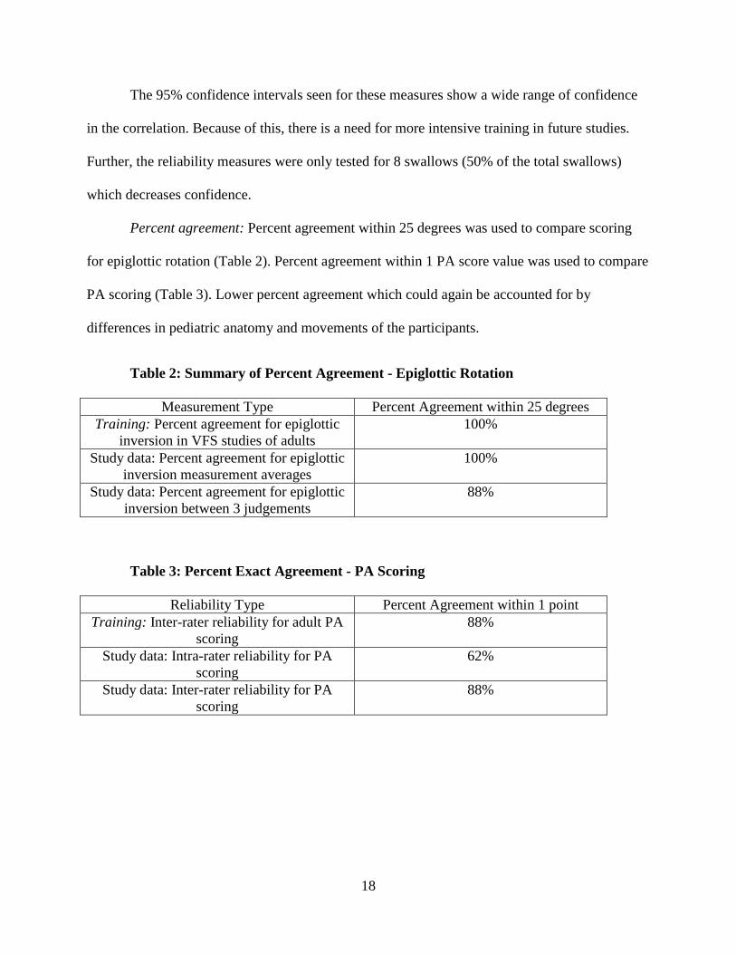
The 95% confidence intervals seen for these measures show a wide range of confidence
in the correlation. Because of this, there is a need for more intensive training in future studies.
Further, the reliability measures were only tested for 8 swallows (50% of the total swallows)
which decreases confidence.
Percent agreement: Percent agreement within 25 degrees was used to compare scoring
for epiglottic rotation (Table 2). Percent agreement within 1 PA score value was used to compare
PA scoring (Table 3). Lower percent agreement which could again be accounted for by
differences in pediatric anatomy and movements of the participants.
Table 2: Summary of Percent Agreement - Epiglottic Rotation
Measurement Type Percent Agreement within 25 degrees Training: Percent agreement for epiglottic
inversion in VFS studies of adults 100%
Study data: Percent agreement for epiglottic inversion measurement averages
100%
Study data: Percent agreement for epiglottic inversion between 3 judgements
88%
Table 3: Percent Exact Agreement - PA Scoring
Reliability Type Percent Agreement within 1 point Training: Inter-rater reliability for adult PA
scoring 88%
Study data: Intra-rater reliability for PA scoring
62%
Study data: Inter-rater reliability for PA scoring
88%

19
Table 4: Intraclass Correlation Results – Epiglottic Rotation
Reliability Type Intraclass Correlation Coefficient
95% Confidence Interval
Training: Intra-rater reliability for epiglottic inversion in VFS studies of adults
0.914 0.472-0.981
Study data: Intra-rater reliability for epiglottic inversion measurement averages
0.618 -0.72-0.911
Study data: Intra-rater reliability for 3 epiglottic inversion measurements used for
averages
0.788 0.585-0.912
Study data: Inter-rater reliability for epiglottic inversion
0.686 -0.372-0.935
Table 5: Intraclass Correlation Results - PA Scoring
Reliability Type Intraclass Correlation Coefficient
95% Confidence Interval
Training: Inter-rater reliability for adult PA scoring
0.872 0.755-0.915
Study data: Intra-rater reliability for PA scoring
0.792 0.296-0.954
Study data: Inter-rater reliability for PA scoring
-0.098 -7.180-0.794
2.5 STATISTICAL ANALYSIS
Descriptive data includes age and sex of participants (Table 1). Means and standard deviations of
epiglottic rotation, PA score, age, and sex were calculated. Inferential statistics was deployed but
were weakened by the small sample size and serial dependency of the data. The correlation
between rotation and penetration-aspiration scores was determined using a Pearson correlation
coefficient on SPSS Version 23 software and summarized visually using a scatterplot. A one-
sample t-test was performed using SPSS Version 23 software to compare the mean rotation

20
values from this study to the mean from a study of normal adults (Kang, 2010). No reference
data for healthy children is currently available for comparison.

21
3.0 RESULTS
Table 6: Results of Epiglottic Inversion and Penetration Aspiration Score.
Swallow ID Total Epiglottic Rotation (degrees) PA Score
1_1 28.77 2
2_1 50.04 1
4_1 48.48 8
4_2 57.41 4
4_3 17.71 8
4_4 34.12 4
4_5 14.84 1
5_1 47.99 1
5_2 72.93 1
5_3 55.62 1
5_4 70.53 8
5_5 53.83 1
6_1 42.36 1
6_2 2.04 1
6_3 17.32 1
6_4 9.06 1

22
The results of the VFS studies showed that epiglottic rotation in children with cerebral palsy is
limited. Consistent with the hypothesis that epiglottic inversion would be limited to less than 89
degrees, the highest angle of epiglottic inversion recorded was 72.93, 16.07 degrees less than the
lowest rotation in a group of healthy adults under 45 years old. Forty-four percent (7/16) of the
measurable swallows exhibited less than 35 degrees of rotation. The average rotation for
children with cerebral palsy was 38.94 degrees, significantly less than the normal adult rotation
average of 102.4 degrees. The standard deviation was higher for children with cerebral palsy at
21.9 degrees while the standard deviation was 13.4 degrees for normal adults under 45 years old.
However, this may be artifact of our small sample size. The one-sample t-test showed epiglottic
rotation in children with cerebral palsy was statistically significantly lower than the normal adult
population (<45 years old), t(15) = -11.011, p = .000.
Average PA score was 2.75 with a standard deviation of 2.79. However, visual inspection
of the plotted PA scores and epiglottic rotation angles (Figure 3) did not demonstrate any
correlation between the two, which does not support our hypothesis. In fact, there is a slight
increase in penetration aspiration score with increased epiglottic inversion (Figure 4) though this
may have been due to the small amount of data points. This slight correlation is also seen
following removal of outliers (Figure 5). A Pearson correlation also showed no significant
correlation (R=0.186, p =0.492). Aspiration occurred with epiglottic inversion angles of 17.71,
48.48, and 70.53 degrees. From these data, it cannot be concluded that there is a correlation
between epiglottic inversion and penetration aspiration scores for children with cerebral palsy.

23
Figure 4: Relationship between Epiglottic Inversion and PA Score (p>.05)
Figure 5: Relationship between Epiglottic Inversion and PA score without outliers (p>.05)

24
4.0 DISCUSSION
This study investigates a new method of analyzing the kinematics of swallowing. In this study,
rotation of the epiglottis was measured in order to obtain more information about swallowing
kinematics in an at-risk population. It is a new method which compensates for head movement.
Head movement is a symptom of motor disorders which may may occur in populations beyond
those with cerebral palsy, such as patients with Parkinson’s Disease. We were able to obtain data
from these novel measures with fair to high reliability for most measures.
The results of our study indicated that epiglottic inversion during swallowing in children
with cerebral palsy is less than that seen in healthy young adults. Although this may be an
artifact of differences in aerodigestive tract anatomy between children and adults, this
preliminary study provides the first measurement of epiglottic rotation of persons in this
population, and useful data regarding swallow kinematics in children with cerebral palsy who are
disproportionately affected by dysphagia. Given that research into swallowing physiology using
x-ray imaging in healthy human children is considered ethically undesirable, our comparison to
young adults represents the only plausible comparison available. Studies comparing these same
dependent variables between groups of children with cerebral palsy and may shed more light on
this question.
The hypothesis that epiglottic inversion would be limited for children with cerebral palsy
was supported by our investigation of this small sample, as all epiglottic inversion was less than

25
89 degrees. Nearly half of the swallows analyzed failed to produce more than 35 degrees
rotation. The limited epiglottic inversion for children with cerebral palsy is likely explained by
the decreased HLE and tongue movement previously studied in this population. Because the
epiglottis is a piece of cartilage, it is moved by other muscles in the swallowing mechanism such
as tongue muscles and the muscles involved in HLE. If HLE or tongue movement do not occur,
the epiglottis will not be inverted and will not contribute to closure of the supraglottic passage.
Mechanisms of spasticity and athetosis occurring in cerebral palsy likely explain this change in
swallowing kinematics. However, because of our small sample, they warrant further
investigation with stratification of participants by type of motor impairment.
The hypothesis that fewer degrees of inversion would correlate with higher (worse)
scores of the PAS was not found to be true in this study. Surprisingly, there was not a strong
correlation between reduced epiglottic inversion and airway penetration and aspiration. This is
counterintuitive because complete epiglottic inversion is a component of airway closure.
However, this result may have been caused by a small sample size that produced a wide range of
PA scores over a small number of swallows, and predominance of swallows in our data set from
one participant. Naturally, a larger sample would have provided a more valid sampling of this
population’s swallow kinematics. Another explanation as to why this hypothesis was not
supported is that closure occurs from the epiglottic petiole to the arytenoids, as opposed to
epiglottic inversion for protection (Logeman, 1992). This may represent a compensatory
adaptation that ensures airway protection, which is necessary for physiologic survival. Other
explanations may include that epiglottic inversion is not essential for airway protection (Medda,
2002), or that in children, anatomical orientation of structures lowers airway protection
dependency on epiglottis coverage of the laryngeal vestibule.
26
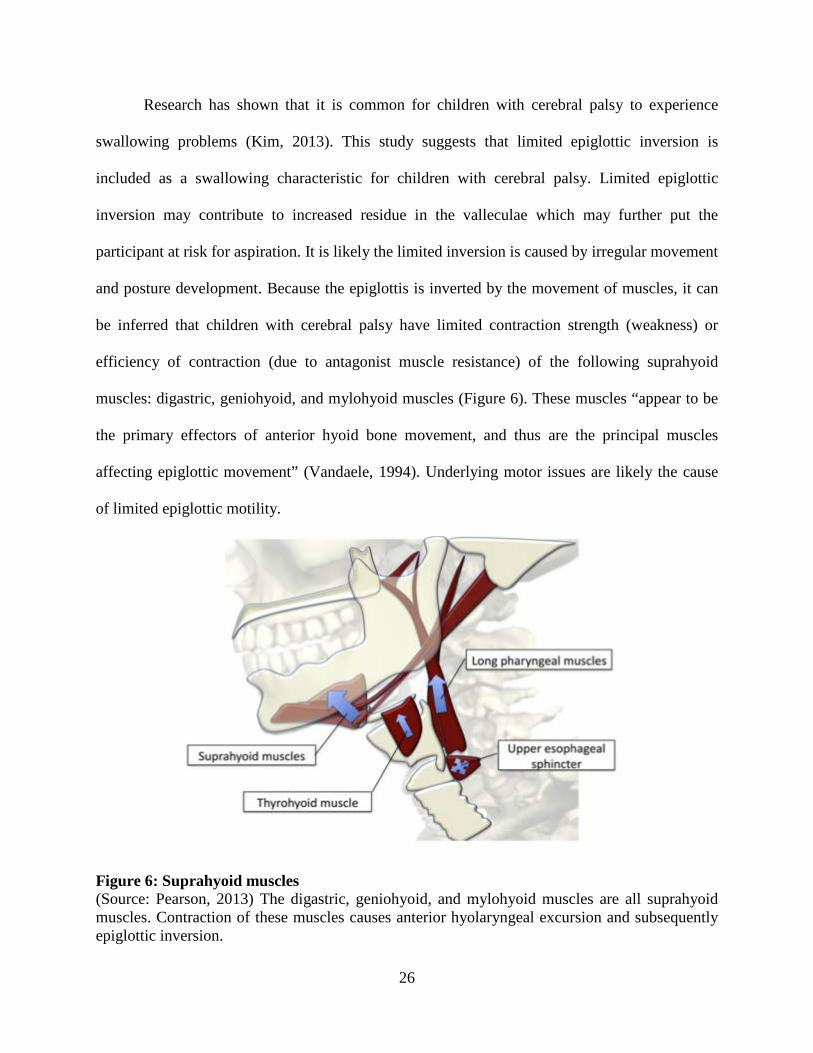
Research has shown that it is common for children with cerebral palsy to experience
swallowing problems (Kim, 2013). This study suggests that limited epiglottic inversion is
included as a swallowing characteristic for children with cerebral palsy. Limited epiglottic
inversion may contribute to increased residue in the valleculae which may further put the
participant at risk for aspiration. It is likely the limited inversion is caused by irregular movement
and posture development. Because the epiglottis is inverted by the movement of muscles, it can
be inferred that children with cerebral palsy have limited contraction strength (weakness) or
efficiency of contraction (due to antagonist muscle resistance) of the following suprahyoid
muscles: digastric, geniohyoid, and mylohyoid muscles (Figure 6). These muscles “appear to be
the primary effectors of anterior hyoid bone movement, and thus are the principal muscles
affecting epiglottic movement” (Vandaele, 1994). Underlying motor issues are likely the cause
of limited epiglottic motility.
Figure 6: Suprahyoid muscles (Source: Pearson, 2013) The digastric, geniohyoid, and mylohyoid muscles are all suprahyoid muscles. Contraction of these muscles causes anterior hyolaryngeal excursion and subsequently epiglottic inversion.
27
4.1 LIMITATIONS AND SUGGESTIONS FOR FUTURE RESEARCH
There were several limitations in our study’s design. The small sample size decreases
generalizability. In terms of reliability, there is a strong need for increased training in
future studies, as demonstrated by the wide range of confidence intervals. Also, the type and
severity of cerebral palsy were unknown to the principal investigator, who only had access to
the VFSS, age, and sex of each participant. This additional information would have been
useful because cerebral palsy is such a varied disorder. By noting the subtype and severity,
as well as the predominating form of motor impairment, more useful conclusions might
have been drawn which could point to both the etiology and clinical significance for this
diverse population. Children with cerebral palsy have also demonstrated variability in
mobility across settings (Tieman, 2007). This variability could account for the differences
seen between the 2 dates of VFS studies of one participant. For future research, variability of
swallowing at different days and times may be of interest.
It would also have been helpful to have knowledge of the norms for epiglottic
movement in typical children instead of adults. However as discussed earlier, there are
ethical concerns about exposing healthy children to x-rays for research purposes. Knowledge
of norms for the pediatric population would have helped draw appropriate conclusions about
the extent of the limited inversion. Without this data, it is not possible to be confident that
limited epiglottic inversion is not seen in the general population in pediatrics.
We also did not know the exact bolus volumes that were administered by clinicians
during the VFS studies. The epiglottis is partially inverted by the size of the bolus. If the bolus is
not consistent it cannot be clearly determined that an increase or decrease in epiglottic inversion

28
is not caused by a difference in bolus size. For future research, a consistent bolus volume would
remove that possibility.
Further, comparison data for norms should also include equivalent bolus consistencies.
The adult epiglottic rotation was measured with a liquid bolus while this study measured with a
puree bolus. As mentioned above, epiglottic inversion is affected by the type and size of the
bolus. In future studies, comparisons should be made across the same bolus consistency and size.
Lastly, the number of measureable swallows for each participant should be consistent. In
this study, the majority of measured swallows were from one participant. This caused a skew in
the data that decreased generalizability. By analyzing an equal number of measurable swallows,
the confidence in conclusions would increase.
For future research, more control of variables could be obtained by a prospective rather
than retrospective study. The volume of the bolus should be controlled as well as the number of
measurable swallows per participants. This consistency in measurements would increase
confidence that the measured difference in epiglottic inversion are caused by the motor
impairment instead of other factors such as skew in the data, or bolus consistency or volume.
4.2 CLINICAL IMPLICATIONS
The results shed light on some of the possible biomechanical explanations for misdirection of
swallowed material during swallowing in children with cerebral palsy, who have elevated
prevalence of pneumonia. Aspiration while swallowing can be a major contributor to pneumonia
pathogenesis. Because aspiration is prevalent in this population, careful evaluation of the
aerodigestive kinematics rather than global manifestations of cerebral palsy are warranted.

29
The results of this study increase awareness into the motoric and swallowing related
impairments of children with cerebral palsy. This information can be shared with clinicians,
patients, and their families and discussed when interpreting VFS studies. Through motion
analysis, researchers are able to begin to differentiate neuronal damage and hypothesize
treatment strategies. Limited epiglottic inversion indicates underlying muscle impairment. In
this population, these findings are important because they increase the awareness of the types of
issues children with cerebral palsy may have with swallowing and may lead to future research to
lower the risk this impairment poses.

30
BIBLIOGRAPHY
Coyle, J. L. (2012). Biomechanical Analysis. In R. D. Newman & J. M. Nightingale (Eds.), Videofluoroscopy: A Multidisciplinary Team Approach (pp. 107-122). San Diego: Plural.
Coyle, J. (2014). Swallow physiology in a nutshell [class handout]. Department of Communication Science and Disorders, University of Pittsburgh, Pittsburgh, PA.
Erasmus, C.E., Hulst, K., Rotteveel, J.J., Willemsen, M. A. A. P., & Jongerius, P.H. (2011). Clinical practice. European Journal of Pediatrics, 171(3), 409-414. doi: 10.1007/s00431-011-1570-yFerreira, 2011 cite
Fleiss JL. The Design and Analysis of Clinical Experiments. New York: John Wiley & Sons; 1986.
Intraclass correlation. (2014). Retrieved fromhttps://department.obg.cuhk.edu.hk/researchsupport/IntraClass_correlation.asp
Kang, B., Oh, B., Kim, I., Chung, S., Kim, S., & Han, T. (2010). Influence of Aging on Movement of the Hyoid Bone and Epiglottis during Normal Swallowing: A Motion Analysis. Gerontology, 474-482.
Koman, L. A., Smith, B.P., & Shilt, J.S. (2004). Cerebral palsy. The Lancet, 363(9421), 1619-1631. doi: http://dx.doi.org/10.1016/S0140-6736(04)16207-7
Kim, J., Han, Z., Song, D., Oh, H., & Chung, M. (2013). Characteristics of Dysphagia in Children with Cerebral Palsy, Related to Gross Motor Function. American Journal of Physical Medicine & Rehabilitation, 912-919.
Logemann, J. A., Kahrilas, P. J., Cheng, J. O. A. N., Pauloski, B. R., Gibbons, P. J., Rademaker, A. W., & Lin, S. H. E. Z. H. A. N. G. (1992). Closure mechanisms of laryngeal vestibuleduring swallow. American Journal of Physiology-Gastrointestinal and Liver Physiology,262(2), G338-G344.
Medda, B. K., Kern, M., Ren, J., Xie, P., Ulualp, S. O., Lang, I. M., & Shaker, R. (2003). Relative contribution of various airway protective mechanisms to prevention of aspiration during swallowing. American journal of physiology. Gastrointestinal and liver physiology, 284(6), G933-9.
31
Pearson, W. G., Hindson, D. F., Langmore, S. E., & Zumwalt, A. C. (2013). Evaluating Swallowing Muscles Essential for Hyolaryngeal Elevation by Using Muscle Functional Magnetic Resonance Imaging. International Journal of Radiation Oncology, Biology, Physics, 85(3), 735–740. http://doi.org/10.1016/j.ijrobp.2012.07.2370
Rasband, W.S. (2014). ImageJ [Computer Software]. Bethesda, Maryland, USA: U. S. National Institutes of Health, http://imagej.nih.gov/ij/.
Rommel, N., & Hamdy, S. (2016). Oropharyngeal dysphagia: manifestations and diagnosis. Nat Rev Gastroenterol Hepatol, 13(1), 49-59. doi: 10.1038/nrgastro.2015.199Rosenbek, JC, Robbins, J, Roecker EV, Coyle, JL, & Woods, JL. (1996) A Penetration-Aspiration Scale. Dysphagia 11:93-98.
Schiariti, V., & Mâsse, L.C. (2015). Relevant Areas of Functioning in Children With Cerebral Palsy Based on the International Classification of Functioning, Disability and Health Coding System: A Clinical Perspective. Journal of Child Neurology, 30(2), 216-222. doi: 10.1177/0883073814533005 Cite SPSS
Steele, C., Bailey, G., Chau, T., Molfenter, S., Oshalla, M., Waito, A., & Zoratto, D. (2011). The relationship between hyoid and laryngeal displacement and swallowing impairment. Clin Otolaryngo.
Swallowing Disorders (Dysphagia) in Adults. (n.d.). Retrieved April 06, 2016, from http://www.asha.org/public/speech/swallowing/Swallowing-Disorders-in-Adults/
Tieman, B., Palisano, R. J., Gracely, E. J., & Rosenbaum, P. L. (2007). Variability in mobility of children with cerebral palsy. Pediatric Physical Therapy, 19(3), 180-187.
VanDaele, D., Perlman, A., & Cassell, M. (1995). Intrinsic fibre architecture and attachments of the human epiglottis and their contributions to the mechanism of deglutition. Journal of Anatomy, 186.