Embed Size (px)
Citation preview

Embryology of the larynx:
Larynx is derived from branchial arch system
Anatomy of the larynx:
It extends from tip of epiglottis C3, to lower edge of cricoid C6.
It is if formed of cartilaginous framework, connected by ligaments and
muscles, and lined by mucous membrane.
Laryngeal cartilages:
A- Single cartilages
1- Thyroid 2- Cricoid 3- Epiglottis
1-thyroid notch 1-facet for arytenoid 1-posterior surface
2-superior horn 2-facet for inf.thyroid horn 2-sup.thyroid horn
3-thyroid lamina 3-posterior lamina 3-thyroid
4-inferior horn 4-cricoid ring 4-arytenoid
5-laryngeal prominence(adam apple) 5-upper border 5-inf.thyroid horn
6-narrow anterior arch 6-post.part of cricoid
7-lower border
B- Paired cartilage
1- Arytenoid 2-Corniculate 3-Cuneiform
1-thyroid cartilage
2-cricothyroid ligament
3-cricoid cartilage
4-thyrohyoid membrane
5-sup.thyroid horn
6-inf.thyroid horn

Laryngeal ligaments & membranes
1- Crico thyroid membrane. 2-Thyrohyoid membrane.
3-Crico tracheal ligament. 4-Thyro epiglottic ligament.
5-Hyo epiglottic ligament
6- Two intrinsic ligaments: form a broad sheet beneath lining
mucosa:
a- Superiorly: quadrangular ligament between lateral border of
epiglottis & arytenoid.
b- Inferiorly: conus elasticus, from arch of cricoid to vocal ligament
1-thyrohyoid membrane 1-epiglottos 1-laryngeal inlet
2-cricothyroid ligament 2-aryepiglottic fold 2-aryepiglottic fold
3-hyoid bone 3-cuneiform 3-epiglottis
4-lateral part of thyrohyoid memb. 4-corniculate 4-quadrangular
ligament
5-thyroid cartilage 5- quadrangular ligament 5-vocal fold
6-lat.part of cricothyroid lig. 6-vestibular ligament 6-glottis
7-cricoid cartilage 7-vocal ligament 7-lat.part of
cricothyroid lig
8-cricotracheal ligament 8-cricothyroid ligament
9-first tracheal ring
Laryngeal folds:
1- Vocal folds: covering the vocal ligament that is attached on both
sides from the vocal process of arytenoid to middle of inner surface
of thyroid angle.
2- Ventricular folds (false vocal cords): above vocal folds
3- Ary epiglottic folds: between epiglottis and arytenoids.
4- Glosso epiglottic folds two lat & one median.
1-thyro-epiglottic ligament 2-epiglottis

3-yyo-epiglottic ligament 4-hyoid bone
5-pre epiglottic space 6-thyrohyoid membrane
7-thyriod cartilage
Laryngeal inlet:
Oblique, directed slightly posteriorly, bounded by: epiglottis, ary
epiglottic folds, and arytenoids
Cavity of the larynx: 3 areas
1- Glottis space: between V.F extend to 1 cm below
2- Supraglottis: above V.F(from floor of ventricle) extend to tip of
epiglottis
3- Subglottis: from below glottis to lower border of cricoid
Laryngeal muscles:
A- Intrinsic muscles: responsible for vocal cord mobility & tension
1- Abductor: Posterior crico arytenoid (posticus)
2- Adductors: Lateral crico arytenoid - Inter arytenoid
- Thyro arytenoid & vocalis (V.F bulk) - Crico thyroid
muscle
3- Tensors: Crico thyroid muscle.
Vocalis (internal part of thyro arytenoid).
B- Extrinsic muscles
1- Depressors: sternohyoid, sterns thyroid, omohyoid.
2- Elevators: mylohyoid, geniohyoid, digastric, hyoglossus.
3- Inferior constrictor of the pharynx.

1-origin of PCA from back of cricoid lamina 1-origin of LCA from upper border of arytenoid
2-insertion into muscular process of arytenoid 2-insertion into front of muscular process of
arytenoid
Laryngeal spaces:
- Pre epiglottic space - Paraglottic space
Laryngeal mucosa
Larynx is lined by pseudo stratified columner ciliated epithelium, with
goblet cells (respiratory epithelium)
Except: True vocal cords
Tip of epiglottis
Ary epiglottic fold
Nerve supply
1- Motor: all muscles by RLN, except cricothyroid by external branch of superior laryngeal nerve.
2- Sensory: Above V.F: internal branch of superior laryngeal nerve
Below V.F: RLN
Blood supply: Arteries & veins are closely related to nerves
a- Sup. Laryngeal A&V: branches of sup thyroid A&V, join internal branch of sup. laryngeal nerve to pierce thyrohyoid membrane.
b- Inferior laryngeal A&V: branches from inferior thyroid A&V: accompany RLN
Lymphatic drainage:
- Supraglottis (rich): to UDCLN
- Glottis : no lymphatics in Rienk’s space
- Subglottis : pretracheal (delphian), LDCLN, supraclavicular, sup. mediastinal
Physiology (Functions of the larynx):
A: Air way protection: from aspiration by:

1- Closure of 3 sphincters: ary epiglottic fold, ventricular fold & true V.F.
2- Laryngeal elevation during swallow.
3- Reflex cough.
4- Reflex inhibition of respiration during swallow.
B: phonation
C: Respiration: adjustment of glottic aperture helps to regulate acid base balance
D: Fixation of chest during straining
Laryngeal Symptoms & Examination
Laryngeal symptoms:
1- Change of voice (hoarseness)
2- Dyspnea: difficulty in breathing.
3- Stridor: noisy respiration.
4- Pain: local or referred.
5- Cough, expectoration, haemptysis
6- Dysphagia
7- 7- Chocking
8- 8- Swelling
Examination:
A- External Examination
inspection: position & movement, contour, swellings, and scars.
palpation: tenderness, crepitus, crepitation, swellings, laryngeal click
Side to side movement of larynx over the vertebral column, normally a click is felt, if lost (+ve moure’s sign) … mass between larynx & vertebral column e.g. post cricoid carcinoma.
B- Indirect laryngoscopy
C- Flexible fiberoptic laryngoscopy
- When I.L is difficult, it is introduced via the nose.
- Photographic documentation.
D- Direct laryngoscopy under GA:
By rigid endoscope
Operating microscope can be used: Microlaryngoscopy(MLS)
N.B.: - Any lesion affect laryngeal patency: stridor
- Any lesion affect vocal fold: hoarseness
Laryngeal investigations:
1- Direct laryngoscopy ± biopsy 2-Radiology (X ray.CT,MRI)
Congenital diseases
1- Congenital web
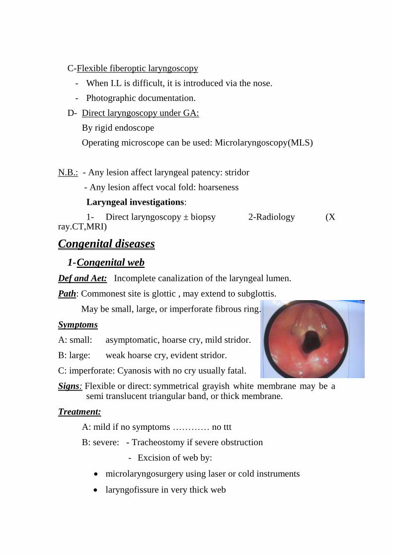
Def and Aet: Incomplete canalization of the laryngeal lumen.
Path: Commonest site is glottic , may extend to subglottis.
May be small, large, or imperforate fibrous ring.
Symptoms
A: small: asymptomatic, hoarse cry, mild stridor.
B: large: weak hoarse cry, evident stridor.
C: imperforate: Cyanosis with no cry usually fatal.
Signs: Flexible or direct: symmetrical grayish white membrane may be a semi translucent triangular band, or thick membrane.
Treatment:
A: mild if no symptoms ………… no ttt
B: severe: - Tracheostomy if severe obstruction
- Excision of web by:
microlaryngosurgery using laser or cold instruments
laryngofissure in very thick web
C: imperforate web: Urgent tracheostomy or rigid bronchoscope to rupture the web
2- Laryngomalacia (congenital laryngeal strider)
Def: stridor due to weak flaccid larynx.
Aet: cartilaginous framework of larynx is abnormally soft & gets collapsed during inspiration especially supraglottic structures.
Incid: Most common congenital anomaly.
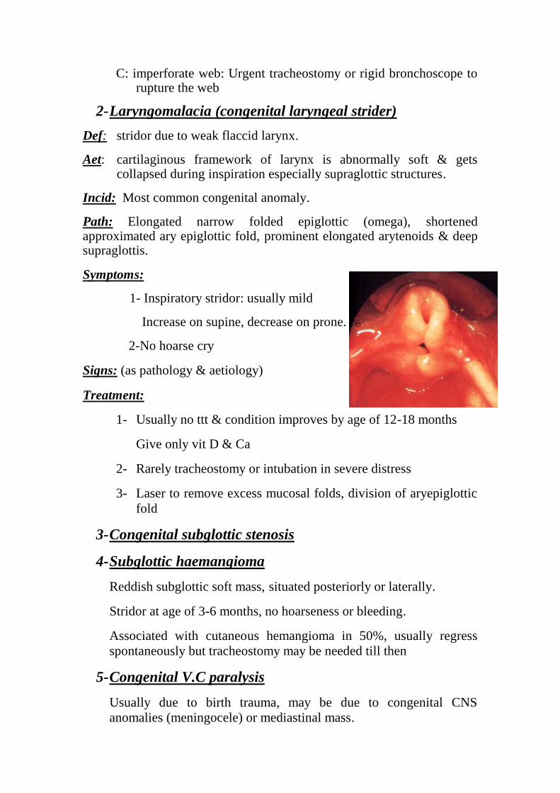
Path: Elongated narrow folded epiglottic (omega), shortened approximated ary epiglottic fold, prominent elongated arytenoids & deep supraglottis.
Symptoms:
1- Inspiratory stridor: usually mild
Increase on supine, decrease on prone.
2-No hoarse cry
Signs: (as pathology & aetiology)
Treatment:
1- Usually no ttt & condition improves by age of 12-18 months
Give only vit D & Ca
2- Rarely tracheostomy or intubation in severe distress
3- Laser to remove excess mucosal folds, division of aryepiglottic
fold
3- Congenital subglottic stenosis
4- Subglottic haemangioma
Reddish subglottic soft mass, situated posteriorly or laterally.
Stridor at age of 3-6 months, no hoarseness or bleeding.
Associated with cutaneous hemangioma in 50%, usually regress
spontaneously but tracheostomy may be needed till then
5- Congenital V.C paralysis
Usually due to birth trauma, may be due to congenital CNS
anomalies (meningocele) or mediastinal mass.

6- Laryngo tracheo-oesophageal cleft
Abnormal communication between larynx & trachea anteriorly and
hypopharynx & oesophagus posteriorly…….. stridor,chocking &
aspiration……….pneumonia
7- Congenital cysts
Laryngeal (saccular) cyst
Mucous filled cysts,arise from ventricle, false folds or aryepiglottic
fold.
Asymptomatic if large: hoarseness& stridor.
Ttt: endoscopic excision or marsupialisation.
Laryngocele:
Special type of congenital cyst develops from laryngeal saccule
(anterior portion of the ventricle between false & true cords).
May be internal, external, combined.
Symptoms: hoarseness, stridor & neck swelling.
Signs: bulge of the ventricle.
Soft, expansile, compressible mass, on lateral neck
Invest: X ray air filled sac CT :diagnostic
ttt excision endoscopic: laser or MILS or external
Laryngeal trauma
Types
I- Mechanical
1- Sharp (penetrating): stab, gunshot, cut throat.
2- Blunt: blow, strangulation, motor car accident.
3- Inhaled F.B
4- Surgical :rough endoscopy, high tracheostomy
5- Intubation injury: rough intubation, prolonged intubation
II- Chemical Corrosive ingestion (potash)
Inhaled irritant gases

III- Physical: Irradiation injury
Symptoms:
History of trauma Dyspnea & stridor Hoarseness of voice
Severe local pain Dysphagia Haemptysis
External swelling Hge, shock (hypovolaemic or neurogenic)
Signs:
1- External:
Inspection: Swelling due to oedema, haematoma, surgical emphysema
Deformity as depressed cartilage
External wound
Palpation: Localized tenderness & crepitus
Surgical emphysema
Deformity
2- By flexible or direct laryngoscopy
Mucosa: laceration Submucosa: oedema or haematoma
V.F: paralysis or avulsion Cartilage: displaced
Joints: dislocated Arytenoids: intubation granuloma
investigations: - D.L - C.T
treatment:
Air way secure by tracheostomy or intubation
Shock management
Drugs antibiotics Steroids
Open reduction: suture laceration & fractured cartilage + stent
F.B
In the larynx
Rare, mostly goes to trachea, sharp or large FB may be impacted
Sudden severe respiratory obstruction
Ttt: Heimlich maneuver: sudden abdominal compression
Turn child upside down & slap back
Removal by DL
Tracheostomy or cricothyrotomy
Inhaled FB
incid: children, mentally retarded
Types: vegetable: seeds, beans non vegetable: pins, buttons.
endogenous: vomitus, blood
Sites: rare in larynx (passes to esophagus or bronchus) in bronchi usually right main bronchus (wider & in line with the trachea).
Clinical picture:
A- initial stage sudden severe cough, cyanosis, dyspnea and sometimes haemptysis, this stage may pass unnoticed
B- Latent stage: mild cough & expectroation - localized wheeze
C- Manifest stage:
I- Mechanical obstruction
Complete (collapse) Partial (emphysema)
- Dyspnea, cough, expectoration - Dyspnea, cough, expectoration
- mediastinum shift to same side - To opposite side
- Dullness on percussion - Hyper resonance
- No air entry - Localized wheeze
II- Inflammatory changes
Bronchitis, bronchopneumonia, lung abscess (more in organic F.B)
D- Large F.B: Impacted in larynx may cause suffocation
Investigations: 1- D.L & bronchoscopy 2- Xray neck & chest
Ttt: 1-Urgent tracheostomy in large impacted F.B
2- Removal by direct laryngoscopy or bronchoscopy
Laryngeal stenosis

Def: Fibrotic narrowing of endolarynx leading to respiratory distress
Aet: A-Congenital: Congenital laryngeal atresia, congenital laryngeal web
congenital subglottic stenosis
B-Trauma (see before)
C-Chromic inflammations (granulomas): Scleroma, T.B, $, leprosy
D-Laryngeal neoplasms: chondroma, fibroma, carcinoma
Incid: most common is subglottic (& most difficult to treat)
Path: 1- supra glottic 2- glottic (ant, post, complete). 3- Subglottic
Clinical picture:
- Asymptomatic Stridor on exertion
- Biphasic stridor & dyspnea Exam shows the stenosis
Investigations: 1-DL & bronchoscopy
2-C.T to show length & degree of stenosis
3-Pulmonary functions for follow up
Treatment:
A) Tracheostomy in severe cases
B) Endoscopic procedure: Laser excision, repeated dilatation
C) External: laryngoplasty: split cricoid, insert costal graf & put Montgomery tube
Resection & end to end anastomosis.
Laryngitis I- Acute
(A) Non specific in adults, in children, epiglottistis & laryngo tracheobronclitis
(B) Specific: Diphtheria
II- Chronic
(A) Non specific
hypertrophic Diffuse
Localized: nodule , polyp, and leukoplakia

Atrophic
(B) Specific (granulomas): scleroma, TB, S, leprosy, & mycosis
I- Acute Laryngitis
(A) Acute non specific laryngitis
(1) In adults
Def.: acute catarrhal inflammation of laryngeal m.m.
Aet.: follow upper respiratory tract infection
Viral: influenza, rhinovirus Bacteria: Strept. pneumonia, strept., straph.
Predisposing factors:
- Voice abuse. - Smoking.
- Post nasal discharge as in sinusitis. - Dust & fumes.
Incid.: see above Path: congestion and edema
Symptoms:
General: fever (occasionally), headache, malaise, and fatigue.
Local: - hoarseness.
- Discomfort & pain on phonation.
- Dry cough + yellow sputum.
Signs: Diffuse symmetrical congestion & oedema, + mucoid secretions.
Treatment:
1- Voice rest. 2- Fluids.
3- Humidificatoin :steam, tincture benzoini.
4- Systemic antibiotics. 5- Mucolytics.
(2) Acute non specific laryngitis in children
Def.: as before.
Aet.: as before.
Path: Difference between adult & child larynx.
1- Anatomical factors.
- Lumen is relatively smaller.
- Loose submucosal tissue.
- Funnel shaped lumen.
- More abundant lymphatic supply in mucosa.
More oedema, minimal edema cause marked obstruction.

2- Immature immune system.
3- Immature nervous regulation = spasm.
Symptoms:
General: as usual.
Local: - Hoarseness. - Dry cough. - Stridor, dyspnea.
Signs: Diffuse congestion and oedema. + mucoid secretion.
Subglottic oedema
D.D.: causes of stridor in children
Treatment: as adult +
- Hospitalization, oxygen.
- Steroids to decrease oedema.
- Careful observation of respiratory obstruction if severe distress → intubation, or rarely tracheostomy (avoided in children unless no other way).
(3) Acute Epiglottitis
Def.: Acute non specific laryngitis affecting mainly epiglottis.
Aet.: Haemophilus influenza usually.
Incid.: more in infants & children.
Symptoms:
General: high fever, malaise, headache, and anorexia.
Local: - Severe odynophagia, drippling of saliva.
- Muffled voice (hot potato). -Rapidly progressive stridor.
Signs:
- Oedema of supraglottis. - Epiglottis appears as red swollen mass.
- Pharyngeal oedema & congestion. - Enlarged UDCLN.
Ttt: as before.
(4) Acute laryngo tracheobronchitis
Def.: Acute respiratory infection spreading to entire respiratory system.
Clinical picture: In adult: as laryngitis + cough, expectoration.
In children: as laryngitis + cough, expectoration.

Treatment: as laryngitis.
(B) Laryngeal diphtheria.
Symptoms:
General: Toxaemia
Local: - hoarseness, stridor, Dyspnea, cough.
Signs: grayish, white, dirty membrane.
Treatment: as usual. + Tracheostomy.
II- Chronic Laryngitis.
(a) Chronic non specific laryngitis.
(1) Hypertrophic
(a) Chronic diffuse hypertrophic laryngitis.
Aet.: - Repeated acute laryngitis predisposing factors ----
Symptoms:
- Hoarseness of voice - Cough & expectoration.
- Hemming (desire for frequent throat clearing).
Signs: Congested thickened V.F
in oedematous type (reinke oedema) :pale polypoidal VF.
ttt: 1- Remove predisposing factor. 2- Like acute but no antibiotics.
3- VF stripping: MLS or laser in resistant cases
(b) Chronic localized hypertrophic laryngitis.
(1) Vocal cord nodules (2) Vocal cord polyp.
Aet.: - Voice abuse (singer). - Voice abuse.
- reflux
Incid.: More in females, children. - More in males.
Path: Epithelial hyperplasia of - Localized subepithelial
Free edge of V.F. oedema, or vascular eng-
Fresh: soft& red orgement, followed by fibrosis.
mature:hard&white
Symptoms: hoarseness, weakness - Hoarseness.

-Rarely stridor if large.
- Chocking.
Signs: Bilateral. - Unilateral
Small tiny nodules. - Small or large
Sessile -Usually pendunculated.
White or pink. - Grey, white or red.
At free margin of V.F. - From undersurface or V.F.
Junction of ant 1/3 & post 2/3. - Site as nodule.
Treatment: Voice rest, avoid misuse. - Removal by MLS
Speech therapy. - Laser.
+ removal by MLS - Speech therapy.
Laser. - Voice rest, avoid misuse.
(3) Leukoplakia.
Def.: white patch on laryngeal mucosa.
Aet.: Irritation.
Pathology: Epithelial hyper plasia & hyper keratinization.
Symptoms: persistent hoarseness.
Signs: white patch on V.F.
May appear as diffuse, villous, or verrucous.
ttt: Excision by MLS or Laser, follow up as it is pre cancerous.
(2) Atrophic.
Aet: dusty atmosphere - Industrial fumes - Post irradiation
therapy

Incid: more in women
Symptoms: hoarseness, offensive breath &dyspnea(crusts)
Signs: dry pale atrophic mucosa, & crusts
Ttt: Avoid predisposing factors, moisture, menthol inhalation,
laryngoscopy to remove crusts
(B) Chronic specific laryngitis (Granuloma).
(1) Laryngoscleroma.
Def.: chronic specific inflammation affecting upper respiratory tract.
Aet.: gram – ve bacilli; klebsiella rhinoscleromatis.
Incid.: endemic in Egypt. Usually 2ry to nasal scleroma may be 1ry.
Usually subglottic region.
Path: see nose.
Symptoms: - stridor & dyspnea.
- Hoarseness may be present.
- Cough & expectoration.
Signs: pale pinkish smooth swelling on both side of subglottis covered by greenish crusts fibrosis & subglottic stenosis.
Investigations:
- DL & biopsy. - CT for length & degree of stenosis.
ttt:
1- Medical as in rhinoscleroma. 2-Voice rest & humidification.
3-Tracheostomy, if severe distress.
4- Excision of the web by Laser.
External: graft & stent
Resection anastomosis
(2) T.B laryngitis.
Aet: 2ry to pulmonary T.B. Path.: affect post part of larynx.
Symptoms:
I- General: 1- T.B toxemia: night fever, night sweat, loss of weight, loss of
appetite.

2- Pulmonary T. B cough, expectoration, haemptysis.
II- Local:
1- Hoarseness: progressive, phonosthesia( weak voice)
2- Stridor & dyspnea.
3- Pain & referred otalgia.
4- Odynophagia (marked).
Signs:
I- External: Tenderness due to perichondritis.
II- by I.L or flexible:
1- T.B granulations on arytenoids.
2- T.B ulcer: thin undermined edge, yellow caseous floor.
3- Impaired V.F mobility.
Investigations:
1- DL & biopsy. 2- Chest X ray for T.B. 3- Tuberculin test: good
negative.
Treatment:
1- Tracheostomy in severe distress.
2- Anti tuberculous drugs e.g. rifampicin, PASA, streptomycin & pyrazinamide.
3- Local anesthetic spray before meals.
(3) Syphilis of the larynx.
Aet.: Treponema pallidum (spirochete). Incid.: Very rare.
Path.: 1ry chancre. 2ry mucous patches. 3ry gumma (commonest).
Symptoms: - Hoarseness.
-Stridor.
- Cough & discomfort (no pain).
Signs: $ affects ant. part of larynx
Gumma of epiglottis appears swollen & ulcerated.
Diffuse symmetrical infiltration without ulcerated.
Investigations:
1- DL & biopsy. 2- Radiology. 3- Serology for $.
Ttt: 1- Tracheostomy if needed.
2- Penicillin.
(4) Lupus
Aet.: attenuated T.B. Incid.: mostly 2ry to nasal lupus.
Path.: apple jelly nodule.
Ulceration on one side.
Fibrosis, notched epiglottis.
Symptoms: vague discomfort.
Signs: see pathology.
Investigation: DL & biopsy. - X ray chest.
ttt: like T.B + vit. D.
(5) Leprosy
- Lepromatous or tuberculoid types.
- Nodules ulceration fibrosis.
- Ttt: Dapson + rifampicin.
(6) Fungal infection (mycosis).
a) candidiasis (moniliasis)
- Caused by candida albicans, whitish grey fibrinous membrane.
- Ttt: Remove the underlying cause,topical nystatine or miconazole.
b) Aspergillosis.
Perichondritis
Def: inflammation of the perichondrium of laryngeal cartilage
Aet: trauma…………
Infection: TB, S, typhoid
Advanced carcinoma
Path perichondritis……subperichondrial abscess… cartilage necrosis & stenosis
Symptoms: fever, malaise, pain increase on swallowing, hoarseness, & stridor
Signs: marked tenderness, enlarged laryngeal framework

Laryngeal mucosa is red, edematous, &covered with pus
Ttt: hospitalization, high dose of antibiotics, steroids
Incision of abscess, remove necrosed cartilage.
Tracheostomy or intubation. - Laryngectomy in advanced cases.
Laryngeal oedema
Aet: trauma………..
Infection: laryngitis, perichomdritis, spread of infection from quinsy, ludwig
Angioneurotic oedema: allergic (foods, inhalants& drugs) or hereditary
Non inflammatory oedema: heart renal or liver failure, myxodema .
Symptoms: hoarseness, stridor & dyspnea
Ttt: Rest, oxygen, ttt of the cause
Tracheostomy or intubation in sever cases
Antihistaminics, ,and hydrocortisone
Tumors of the larynx
I- Benign
A) Epithelial Papilloma.Adenoma.
B) Mesenchymal Haemangioma, Chondroma, Fibroma.
Papilloma (commonest benign)
(A) Single papilloma (B) Multiple papillomatosis
Adult papolloma recurrent papillomatosis.
Juvenile multiple papillomatosis
Aet.: - True papilloma - Viral HPV
Autoimmune
Hormonal imbalance
Incid.: - Adult 30-50 y - Children .
Male: female 2/1. Equal sex.
Path.: - Commonest site is free - V.F, any where in larynx extend
edge of V.F, may to trachea, bronchi,

affect other sites.
- Vascular C.T core covered by - Similar.
hyperplastic st. sq. epithelium.
Symptoms:
- hoarseness - Stridor.
+ stridor if large. Hoarseness.
Signs: - single, white or pink. - Multiple white or pink.
warty like growth sessile or warty, sessile affecting any
pedunculated variable size. portion of the larynx.
ttt: Surgical excision by: MLS using cold instruments or laser
MLS using cold instruments or laser
laryngofissure.
Tracheostomy usually needed
(try to avoid……spread).
Interferon.
Malignancy: - 3% - no
Recurrence: - rare - very common
II- Malignant (Cancer larynx)
Def.: malignant tumor of the larynx.
Aet Pre disposing factors tobacco: smoking the most important.

alcohol ingestion supra glottic
carcinoma.
irradiation.
Precancerous lesions: Leukoplakia Adult papilloma.
Incid: Old over 40 with peak in 60. Male to female 8:1(now
less).
Common neoplasm, 1% of all malignancies.
Path.:
(A) Site: glottic 70% Supraglottic 25% Subglottic
5%
(B) G.P: Malignant ulcer. Fungating mass.
Infiltrating nodule.
(C) M.P: commonest is squamous cell carcinoma.
(D) Spread:
1- Direct
Glottic Supraglottic subglottic
2- Lymphatic spread.
a) Glottic: very rare, as there is no lymphatics in Rinek’s space.
b) Supraglottic: common & early due to rich lymphatics UDCLN.
c) Subglottic: common may be bilateral pre laryngeal, pre tracheal, paratracheal middle & LDCLN.
3- Blood spread rare & late : lungs, liver, bone, brain.
(E) T.N.M classification
(a) T for primary tumor
Tis Carcynoma in situ.
T1 One region (in glottic T1a : one cord, T1b : two cords).
T2 Two regions.
T3 Fixed cord.
T4 Extra laryngeal spread.
(b) N: LN metastasis.
No.: No palpable LN.
N1: Single, 3 cm or less, ipsilateral.
N2a: Single, 3-6, ipsilateral.
N2b: Multiple, 3-6 ,ipsilateral.
N2c: Bilateral < 6cm.
N3: more than 6 cm.
(c) M Distant metastasis.
M0: No clinical or radiological evidence of metastasis.
M1: Present clinical or radiological evidence of metastasis.
Symptoms:
1- Hoarseness of voice - progressive. -early in glottic cancer.
2- Dyspnea & stridor due to air way obstruction.
3- Discomfort in the throat.
Late symptoms:
4- Pain, referred otalgia along Arnold branch of X.
5- Cough & irritation.
6- Neck swelling: LN metastasis or direct tumor infiltration.
7-Dysphagia, haemptysis. 8-Foetid breath, cachexia.
Signs:
I General: teeth for sepsis & oral hygiene. Chest : distant metastasis.
II- Local: (Larynx)
(1) External (Neck):
- L.N, describe. - Swelling, broadening.
- Tenderness. - Thyroid.

(2) by I.L flexible or D.L examine:
Tumor: - hyper keratotic warty or papillary growth.
- Malignant ulcer.
- Raised nodule.
V.F mobility: - Freely mobile.
- Fixed: deep muscle invasion.
- Limited: weight of tumor or moderate invasion.
Extension: to hypopharynx, trachea or tongue.
NB: some areas are difficult to be examined: ventricle, subglottis, posterior surface of epiglottis.
Investigations:
1- CT or MRI neck most important radiological diagnosis for accurate staging.
2- DL & biopsy. Site, extent, biopsy.
3- Metastatic work up.(chest X ray, abdominal sonar & bone scan)
4- Routine preoperative investigations.
Treatment:
TIS: surgery: V.F stripping by MLS.or Laser , followed by regular follow up
Radiotherapy (not preferred).
T1, T2: surgery: Partial laryngectomy aiming at complete tumor excision with preservation of phonation, normal breathing °lutition
Endoscopic Laser excision.
Radiotherapy.
T3: Total laryngectomy + post operative radiotherapy
Also in subglottic tumors & recurrent cases after conservative surgery or radiation
- Large T2 or small T3: subtotal laryngectomy (cricohyoidoepiglottopexy).
T4: - Total laryngectomy + post operative radiotherapy.
- Trail of chemo radio therapy.

Management of cervical metastasis.
If palpable LN RND.
If no palpable LN Glottic (nothing)
Supra glottic prophylactic neck dissection, or
radiotherapy.
Sub glottic prophylactic neck dissection, or
radiotherapy
Indications of total laryngectomy:
1- T3, T4 glottic, supraglottic.
2- All subglottic & transglottic.
3- Recurrence or failure after conservative surgery.
4- Recurrence or failure after radiotherapy.
5- Contraindication for conservative or radiotherapy.
6- Certain histological types.
Contra indications of total laryngectomy:
1- Poor general condition. 2-Patient refusal.
3-Distant metastasis. 4-Involvement of unresectable structures.
Disadvantages of total laryngectomy.
1- Loss of voice. 2-Inability to increase intra thoracic
pressure.
3-Permanent tracheostomy. 4-Loss of nasal functions.
2- Limitation of activities.
Rehabilitation after surgery:
1- After partial laryngectomy: voice therapy.
2- After total laryngectomy: Oesophageal speech.

Tracheo oesophageal puncture.
Electronic larynx.
Palliative ttt
Indicated in: unresectable tumors, surgery refusal, distant metastasis &
poor general condition
1- Tracheostomy. 2- Ryle or gustrostomy for feeding.
3- Palliative laser, radiotherapy, chemotherapy.
4- Pain killers. 5- Antibiotics.
Prognosis: early cancer has good prognosis.
Glottic cancer has best cure rate up to 90% due to early
presentation (hoarseness) &absent lymphatic spread.
Vocal Cord Paralysis
a- Unilateral v.c paralysis
Aet: Vagal or RLN injury.
(A) Peripheral
1- Congenital Hydrocephalus
2- Surgical trauma 20%.
- Thyroidectomy - Radical neck dissection.
- Pharyngeal pouch - Esophageal surgery.
- Cardio vascular surgery.
3- Non surgical trauma 20%.
- Neck trauma e.g. stangulation, open injuries.
- Cricothyroid joint dislocation.
- Fracture skull base.
4- Inflammatory 5%.
- Apical pulmonary T.B. - Meningitis,
osteomyelitis.
5- Peripheral neuritis

- Infections: herpes, influenza, and diphtheria. - DM
- Chemicals: alcohol, lead. -Ascending polyneuritis
6- Neoplastic 25%.
- Thyroid malignancy. - Hypopharyngeal
malignancy.
- Bronchogenic carcinoma. - Oesophageal
carcinoma.
- Malignant L.N.
7- Miscellaneous
- Myasthenia gravis. - Rheumatoid arthritis,
SLE.
8- Idiopathic: unknown cause usually viral in aetiology.
(B) Central causes.( Bulbar):
- Head trauma.
- Thrombosis, Hge, embolism.
- Encephalitis, polio, diphtheria. -
- Medullary tumors.
Vocal cord positions
1- Median: both cords in midline.
2- Paramedian : glottis 3-5 mm.
3- Cadaveric: glottis 7 mm.
4- Slight abduction: glottis 14 mm.
5- Full abduction: 18 mm.
Explanation of vocal cord position:
(A) Semon’s law
In progressive RLN injury, abductor paralysis occurs 1st v.c in
median or paramedian position, then adductor paralysis cadaveric position.
(B) Adductors are more powerful than abductors so, when RLN is injured, adductors takes upper hand.
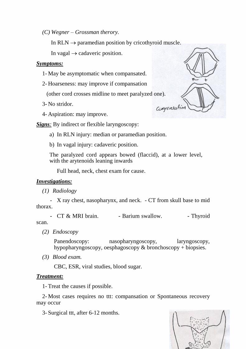
(C) Wegner – Grossman therory.
In RLN paramedian position by cricothyroid muscle.
In vagal cadaveric position.
Symptoms:
1- May be asymptomatic when compansated.
2- Hoarseness: may improve if compansation
(other cord crosses midline to meet paralyzed one).
3- No stridor.
4- Aspiration: may improve.
Signs: By indirect or flexible laryngoscopy:
a) In RLN injury: median or paramedian position.
b) In vagal injury: cadaveric position.
The paralyzed cord appears bowed (flaccid), at a lower level, with the arytenoids leaning inwards
Full head, neck, chest exam for cause.
Investigations:
(1) Radiology
- X ray chest, nasopharynx, and neck. - CT from skull base to mid thorax.
- CT & MRI brain. - Barium swallow. - Thyroid scan.
(2) Endoscopy
Panendoscopy: nasopharyngoscopy, laryngoscopy, hypopharyngoscopy, oesphagoscopy & bronchoscopy + biopsies.
(3) Blood exam.
CBC, ESR, viral studies, blood sugar.
Treatment:
1- Treat the causes if possible.
2- Most cases requires no ttt: compansation or Spontaneous recovery may occur
3- Surgical ttt, after 6-12 months.

Indicated in: persistent hoarseness or aspiration.
Operation performed (medialization)
a) Teflon or Collagen injection.
b) Throplasty: external medialization.
c) Nerve muscle pedicle reinnervation.
b- Bilateral Vocal Cord Paralysis
a- (Bilateral Abductor Paralysis)
Aet.: injury to both RLN (peripheral).
1- Surgical trauma. Thyroidectomy, esophageal surgery.
2- Peripheral neuritis.
3- Neoplastic: cancer thyroid.
Symptoms:
1- Good voice but tries easily.
2- Stridor may be severe, increase by exertion & infection.
Signs:
1- By indirect or flexible laryngoscopy: Vocal cords are in median or paramedian position.
2- Head, neck, chest exam for cause.
Investigations: same
Treatment:
1- If sever stridor Tracheostomy.
2- In established cases:laryngeal widening procedure ,3-6 months later.
(a) Endoscopic arytenoidectomy with posterior cordectomy using MLS or laser.
(b) Woodman’s operation. External operation to fix aryternoid laterally.
(c) Reinnervation procedure.
(d) Tracheostomy with speaking valve.
b- Bilateral adductor paralysis
Presents with aphonia & aspiration (fatal)
Ttt: tracheostomy with speaking valve
Laryngeal closure
Total laryngectomy
Tetany neonatorum & laryngismus stridulus
Def: active laryngeal spasm Aet: Ca deficiency
Incid: young under nourished rickety children, commonly with adenoids
Clinical picture: stridor suddenly at night, cyanosis, may be carpo-pedal spasm
Ttt: A: during the attack: slapping child back, traction on the tongue
B: in between attacks: adenoidectomy, Ca
Laryngeal symptoms
Stridor
Def.: noisy respiration indicating partial airway obstruction in larynx &/or trachea.
Types: a. inspiratory laryngeal: at or above glottis.
b. Biphasic: subglottic or tracheal.
c. Expiratory: bronchial.
Causes:
I- Congenital:
- Congenital web - laryngo malacia.
- Subglottic stenosis - subglottic haemangioma.
- Congenital cyst - congenital bil. cord palsy.
II- Traumatic:
Mechanical: sharp, blunt, surgical, intubation, F.B.
Physical: radiotherapy, thermal.
Chemical: corrosives.
III- Inflammatory:
- Acute non-specific laryngitis, in children -Acute epiglottitis.
- Acute laryngo tracheo brochitis in children. -Laryngeal diphtheria.
- Chronic specific laryngitis (granulomas)
IV- Neoplastic
Benign: multiple papillomatosis. Subglottic chondroma.
Malignant: carcinoma.
V- Miscellaneous
Neurological: bil abductor paralysis.
Laryngeal spasm e.g. tetany
Tumor like conditions: Laryngeal cysts, laryngocele, amyloidosis.
Laryngeal oedema.
VI- Tracheal causes
Retrosternal goiter, mediastinal LN, thymus hyperplasia,& deep neck infection
Clinical picture:
1- Stridor 2-Dyspnea &
tachypnea.
3-Irritability, restlessness, fatigue, sweating. 4-Working ale nasi.
5-Suprasternal, supraclavicular & intercostal retraction.
6-Working accessory muscles.
7- Tachycardia. 8-Congested neck veins during
expiration
9-Late: cyanosis, bradycardia Finally: coma & cardiac
arrest.
+ Hoarseness of voice. NB: Stridor in children.
Hoarseness of voice
Def.: change in quality & timbre of voice, so it becomes rough, harsh &
low pitch. caused by incomplete coaptation, tension or vibration.
Causes of hoarseness.
I- Congenital: web.
II- Traumatic: Mechanical, physical, chemical + voice abuse.
III- Inflammatory: all except subglottic scleroma.
IV- Neoplastic: Benign & malignant.
V- Miscellaneous: Unilateral cord paralysis
Crico arytenoid arthritis.
General weakness: myasthenia or convalescence
Lack of mucus secretion: atropine
Hysterical.

NB: Stridor without hoarseness.
Hoarseness without stridor.
Laryngeal operations
Tracheostomy
Creation of surgical opening, in anterior tracheal wall.
Indications:
I- Upper respiratory tract obstruction.
a- Congenital b- Traumatic
c- Inflammatory d- Neoplastic
e- Miscellaneous
f- Extra laryngeal causes (injury, infection, oedema, tumors)
Injury: maxillofacial. Infection: retropharyngeal
abscess.
Edema: tongue & neck edema Tumors: oral cavity, tongue,
pharynx.
II- Lower respiratory tract obstruction
1- Secretory obstruction.
Value:
1- Frequent accurate aspiration of secretion.
2- Elimination of dead space.
3- Prevent aspiration by cuffed tube.
4- Avoids complications of prolonged intubation.
Causes:
A) Central
1- Head injury: contusion, laceration. 2-Drug intoxication
3- Uraemia, ketoacidosis. 4-Brain tumors & abscess.
5-Stroke: Hge, embolism, thrombosis

B) Peripheral
1)Respiratory muscle paralysis. 2) Chest injury e.g. ribs fracture.
2- Respiratory failure:
- COPD - Neurological disorders e.g. polio, myasthenia.
III- Elective
A) Before laryngeal surgery.
B) Before major operations in head e.g. angiofibroma, maxillectomy,
Types:
Site Advantages Disadvantages
High 1st, 2
nd rings Easy
Rapid
Emergency
Cricoid injury
Low 5th
, 6th
Papillomatosis
Subglottic Cancer
Subglottic Stenosis
Difficult
Pleural injury.
Innominate v. injury.
Slips easily.
Mid 3rd
, 4th
behind isthmus
Avoid previous disadvantages.
Technique:
1- Anesthesia: Usually local infiltration.
General: in elective cases, better in children.
No anesthesia in real emergency.(better do
cricothyroidotomy).
2- Position: Supine with extended neck.
If patient distressed: sitting or semisitting.
3- Incision: Vertical: rapid, lower border of thyroid….manubrium

Transverse: cosmetic,between lower border of cricoid and
supra sternal notch
4- Incise fat, fascia, separate pretracheal muscles.
5- Dissect, incise & transfix isthmus.
6- Open the trachea as a flap.
7- Put a suitable tube.
8- Adequate haemostasis.
9- Close the wound not too tight, fix tube to skin.
Types of Tracheostomy tubes:
1- Metal of silastic. 2-Cuffed or not.
3-Tubes with inner & outer layers. 4-Tubes with expiratory
value.
Complications:
1- A nesthetic complications local or general.
2- Apnea
- When operation done under L.A
- Due to rapid wash out of co2 which is stimulus for respiratory
center.
- ttt: close the opening for a short time, allow patient to breath 95%
O2 in 5% co2 , or assisted ventilation.
3- Bleeding
a) Primary ant. jugular v. Thyroid gland. Innominate v.
b) Reactionary slipped ligature, from previously collapsed v.(open the wound & ligate the vessel)
c) Secondary due to infection.(antibiotics & fresh blood)
4- Pneumothorax.
Due to: pleural injury. Manifested by: dyspnea air entry, X ray.

ttt: intercostal tube connected to underwater seal.
5- Pneumomediastinum.
Due to: Excessive inferior dissection.
If mild: resolve spontaneously. If severe: acute heart failure.
6- Crustation.
No filtration of inspired air. Decreased mucociliary clearance.
7- Delayed complications:
a) Subglottic stenosis: due to cricoid injury.
b) Tracheal stenosis: due to erosion by tube or infection.
c) Difficult extubation.
d) Tracheo oesophageal fistula.
e) Tracheo cutaneous fistula.
8- Emphysema (surgical). Air accumulation under skin.
Due to: - Improperly fitting tube. -Excessive lateral neck dissection.
ttt: Remove a skin suture Insert a more fitting tube.
9- Embolism (air embolism) :due to injury of large neck vein.
ttt: Pour saline into wound. Compression of opened vein.
Elevate foot of bed. Blood transfusion.
10- Injury
-Thyroid gland Hge. - Apex of pleura pneumothorax.
- Cricoid cartilage subglottic stenosis.
-Posterior tracheal wall Tracheo oesophageal fistula.
ttt: Ryle feeding, surgical repair.
-Big vessels Hge. - RLN V.F paralysis.
11- Infection
Wound infection. Chest infection.
12- Tube complications:
a) Slipped tube:
Due to low tracheostomy, wide stoma, short neck, or short tube.
ttt reposition.

b) Blocked tube by dried secretions.
ttt Frequent suction. Cleaning with NaHco3.
Post Operative Care.
1- Patient lies in semi sitting position.
2- Observation for vital signs.
3- Observation for bleeding.
4- Observation for respiratory distress.
Known by recurrence of stridor, absence of air current, absence of mirror dimness, patient can speak without closing the tube.
5- Humidification by steam inhalation.
6- Antibiotics.
7- Mucolytics.
8- Care of tube:
a) Frequent suction, NaHco3 to dissolve mucous.
b) Regular removal of inner tube for cleaning.
9- Extubation: tube is closed with cork for daytime, then day & night and then removed.
Cricothyroidotomy(laryngotomy)
A surgical opening made into the circothyroid membrance.
Indications:
It is done only in real emergency.
If facilities or experience for tracheostomy are not available.
Technique:
A transverse stab is made with any available knife or even scissors.
A special combined laryngostomy tube and introducer (if available) is used, however, if it is not present any tube is inserted e.g. a ball point pen barrel.
Total laryngectomy (see before)
Laryngofissure (Median thyrotomy):
Opening the larynx in the mid-line through an external incision by splitting the thyroid cartilage.
Indications:

1- Post-traumatic laryngeal stenosis 2- Impacted F.B.
3-Post-inflammatory laryngeal stenosis e.g. scleroma. 4-Large benign tumors
2- Malignant tumors: T1 glottic carcinoma.
Partial laryngectomy
A: partial vertical (for glottic carcinoma)
Types: Vertical hemilaryngectomy: one cord
Frontolateral hemilaryngectomy: extends to ant. Commissure, up to third of opposite cord
Frontal partial laryngectomy: ant. Commissure, or third of one or both cords
B: Partial horizontal: (for supraglottic tumors)
Limited to supraglottis with free mobile cord
C: Cricohyoidopexy or cricohyiodoepiglottopexy:a special type of partial
laryngectomy, leaving only one or two arytenoids.
Direct laryngoscopy
This is direct visualization of the larynx under general anesthesia.
Indications:
Diagnostic
1- Examination of the larynx in infants and children.
2- Examination of the larynx in adults with difficult indirect
laryngoscopy (mirror examination) is difficult.
3- To asses extent & site of a lesion, as for staging of malignant tumors.
4- To take a biopsy.
Therapeutic
1- Removal of a foreign body.
2-Microlaryngosocpic sugery (MLS) e.g. to remove a polyp, nodules, or small localized tumors under magnification.
Phoniatrics
Phoniatrics: medical branch that deals with disorders of language, speech & voice

Language: the communication of meanings by means of symbols, it has 4 modalities comprehension, speaking, reading, & writing.
Speech: articulation of different speech sounds, controlled via: pyramidal tract, extra pyramidal, cranial nuclei (5, 7, 10&&12) & cerebellum.
Voice: physical act of sound production by means of VF with the exhaled air stream
A: language disorders
1-Delayed language development (DLD)
Causes: brain damage (mental retardation, cerebral palsy)
Sensory deprivation (hearing or visual)
Psychiatric disturbance
Assessment: investigations for the cause, investigations for language level
Management: correction of auditory, visual, nervous, or endocrinal problems
General language stimulation
2-Aphasia
Def: acquired impairment of language processes.
Causes: stroke, cranial trauma, cerebral tumors, degenerative diseases (Alzheimer)), metabolic, & infections
Assessment: neurological examination, radiological investigations
Psychological & language assessment
Ttt:of the cause & language stimulation
B: speech disorders
1-stuttering
Def: intraphoneme disruption resulting in part-sound, part-word repetition
Aet: obscure, may be due to organic or a learnt behavior
Clinical picture: may be associated with muscular activity, vocal abnormalities, breath disturbance
Ttt: speech therapy, tranquilizers or muscle relaxants
2-velopharyngeal incompetence
Def: velum does not close oropharyngeal ithmus properly
aet: (causes of nasal regurge)
Clinically: hypernasality, nasal regurge, ear & psychological
problems
Diagnosis: history, endoscopy, radiology & speech analysis
DD: hyponasality
Ttt: correct cleft palate, pharyngeal flap & speech therapy
3-dyslalia
Def: persistent defective speech sounds
aet: tongue tie, dental problem, hearing loss or functional
Ttt: of the cause & speech therapy
C: voice disorders
1-Organic: congenital, traumatic, inflammatory, neoplastic or
neurological.
2-Minimal associated pathology: VF polyp, cyst, nodules, or edema.
3-Functional: no organic cause, excessive muscular force or faulty
respiration.