Embed Size (px)
Citation preview

1
CHAPTER IX – SURGICAL PATHOLOGY OF PERICARDIUM -- Horea FEIER 9. 1. Anatomy: The pericardium is the fibro-serous sac that surrounds the heart, venae cavae, the terminal portion of the pulmonary veins, pulmonary arteries and aorta to the origin of brachiocephalic arterial trunk. It is made up of two layers: The outer layer consists of a fibrous conjunctive tissue. It is composed mainly of collagen and elastic fibers, acting as a resistance. The internal layer consists of a single layer of mesothelial cells. The portion which covers the heart is called the visceral pericardium (epicardium), and its portion in contact with the fibrous pericardium is called the parietal pericardium. This layer has a secretory role, the mesothelian cells forming a serous membrane, the source of pericardial fluid. Between the two layers there is normally a small amount of pericardial fluid (15-30 ml), having a lubrication role. The pericardium has motor innervation by parasympathetic nerve fibers originating from the vagus nerve, recurrent larynx and esophageal plexus and the sympathetic fibers originating in the superior thoracic stellate ganglion, which are distributed by aortic, cardiac and diaphragmatic nerve plexus. Sensitive pericardial innervation is transmitted through the phrenic nerve fibers that enter the spinal cord at the C3-C5 level. Arterial vascularization of the pericardium is provided by pericardo-phrenic arteries (right and left), a branch of internal mammary artery. It is fixed to the chest and surrounding structures by pericardo-phrenic, sternopericardial and pericardo-vertebral ligaments. Physiology: The pericardium performs many physiological functions. Protection: a mechanical barrier against extension of infectious or malignant processes. Limiting heart distension: pericardium helps to limit acute cardiac distension. This protective effect disappears during chronic volume overload due to increased compliance and progressive pericardial distension. Ventricular interdependence: pericardium has an important role in the transmission of increased right ventricular pressure on left ventricle by ventricular septal deviation to the left. The result is an inversion of normal septal curvature and decreased left ventricular telediastolic volume. This effect is diminished if the pericardium is absent and it is emphasized in cardiac tamponade. Distribution of intrathoracic pressure on the heart: during inspiration, pericardium is extended, leading to lower intrapericardial pressure and thus increase of transmural pressure gradient in the right cavities. This leads to increased right cardiac filling and lowering blood pressure due to increased pulmonary transit time. This effect disappears in calcareous constrictive pericarditis.
2
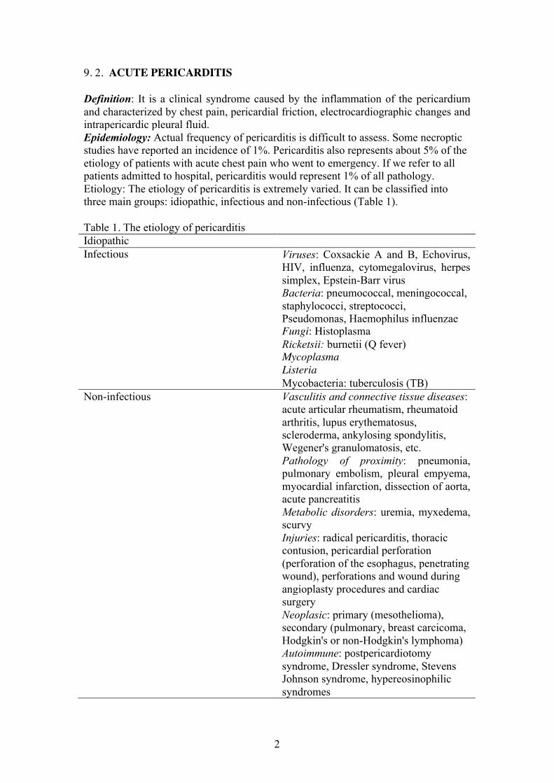
9. 2. ACUTE PERICARDITIS Definition: It is a clinical syndrome caused by the inflammation of the pericardium and characterized by chest pain, pericardial friction, electrocardiographic changes and intrapericardic pleural fluid. Epidemiology: Actual frequency of pericarditis is difficult to assess. Some necroptic studies have reported an incidence of 1%. Pericarditis also represents about 5% of the etiology of patients with acute chest pain who went to emergency. If we refer to all patients admitted to hospital, pericarditis would represent 1% of all pathology. Etiology: The etiology of pericarditis is extremely varied. It can be classified into three main groups: idiopathic, infectious and non-infectious (Table 1). Table 1. The etiology of pericarditis Idiopathic Infectious Viruses: Coxsackie A and B, Echovirus,
HIV, influenza, cytomegalovirus, herpes simplex, Epstein-Barr virus
Bacteria: pneumococcal, meningococcal, staphylococci, streptococci, Pseudomonas, Haemophilus influenzae
Fungi: Histoplasma Ricketsii: burnetii (Q fever) Mycoplasma Listeria Mycobacteria: tuberculosis (TB) Non-infectious Vasculitis and connective tissue diseases:
acute articular rheumatism, rheumatoid arthritis, lupus erythematosus, scleroderma, ankylosing spondylitis, Wegener's granulomatosis, etc.
Pathology of proximity: pneumonia, pulmonary embolism, pleural empyema, myocardial infarction, dissection of aorta, acute pancreatitis
Metabolic disorders: uremia, myxedema, scurvy
Injuries: radical pericarditis, thoracic contusion, pericardial perforation (perforation of the esophagus, penetrating wound), perforations and wound during angioplasty procedures and cardiac surgery
Neoplasic: primary (mesothelioma), secondary (pulmonary, breast carcicoma, Hodgkin's or non-Hodgkin's lymphoma)
Autoimmune: postpericardiotomy syndrome, Dressler syndrome, Stevens Johnson syndrome, hypereosinophilic syndromes

3
Most pericarditis are idiopathic, despite the remarkable progress of diagnostic laboratory techniques - between 30 and 50% by some authors. Currently, the most common etiology is viral, viruses representing up to 70% of all infectious pericarditis. Among the subtypes of the virus, Coxsackie B and influenza viruses are most frequently involved. Viral pericarditis with cytomegalovirus, Epstein-Barr virus or herpes simplex may occur in immunodepressive patients.This also occurs in patients with HIV: most pericarditis occur in these patients by mycobacterial superinfection (tuberculosis) or the development of mediastinal lymphomas. Viral pericarditis may occur in association with viral myocarditis with the same etiology. Many idiopathic pericarditis are actually viral.
Bacterial pericarditis occur through the extension of a proximity infection (lungs, pleura) to the pericardial area and represent 5% of pericarditis. Tuberculous pericarditis represent 4% of pericarditis in developed countries and up to 70% in African countries. In Romania the etiology is still important. Among non-infectious etiologies, neoplasic etiology and uremic pericarditis are the most common. Pericarditis may occur after surgery (postpericardotomy syndrome) or interventional cardiac procedures or they may accompany acute myocardial infarction either acute or remote (Dressler syndrome). Finally, radic etiology is an important cause of chronic constrictive pericarditis. Classification:
By its occurrence: acute or chronic By the presence of pleural fluid: exudative or not, acute pericarditis without pleural fluid
Etiology: All the factors presented in Table I cause in the first phase “dry” inflammation of the pericardium. Morpho-pathology: From a macroscopic point of view, the pericardium is thickened edematiated and reddish. False membranes and adhesions can occur between the pericardium and myocardium and lead to constriction if they are fibrous. Microscopically we find an inflammatory infiltrate with polymorphonuclear, vasodilation and deposition of fibrin in extracellular matrix. Clinically: It is characteristic to associate chest pain, pericardial friction and typical electrocardiographic changes. Chest pain is alive, placed retrosternally or precordially. It may irradiate to the supraclavicular pit, trapezius muscle and shoulder. This irradiation is explained by the fact that the sensitive pericardial afferences are transmitted through the phrenic nerves (C3-C5) and are projected at dermatoma. It may be emphasised by inspiration, swallowing and supine position and diminished by the front-leaning position.
Pericardial friction is characteristic of acute "dry" pericarditis. It appears as a harsh, rough, systolic-diastolic blow. Electrocardiographic manifestations appear as disorders of repolarization phase , which evolves in 4 stages (Table 2). ECG manifestations in the acute phase, are a consistent elevation, ie without image in the mirror, the ST segment, making the differential diagnosis of acute myocardial infarction, followed by the return of ST segment to isoelectric line in a few days. T wave evolves in 4 stages (Table 2) from positive
4
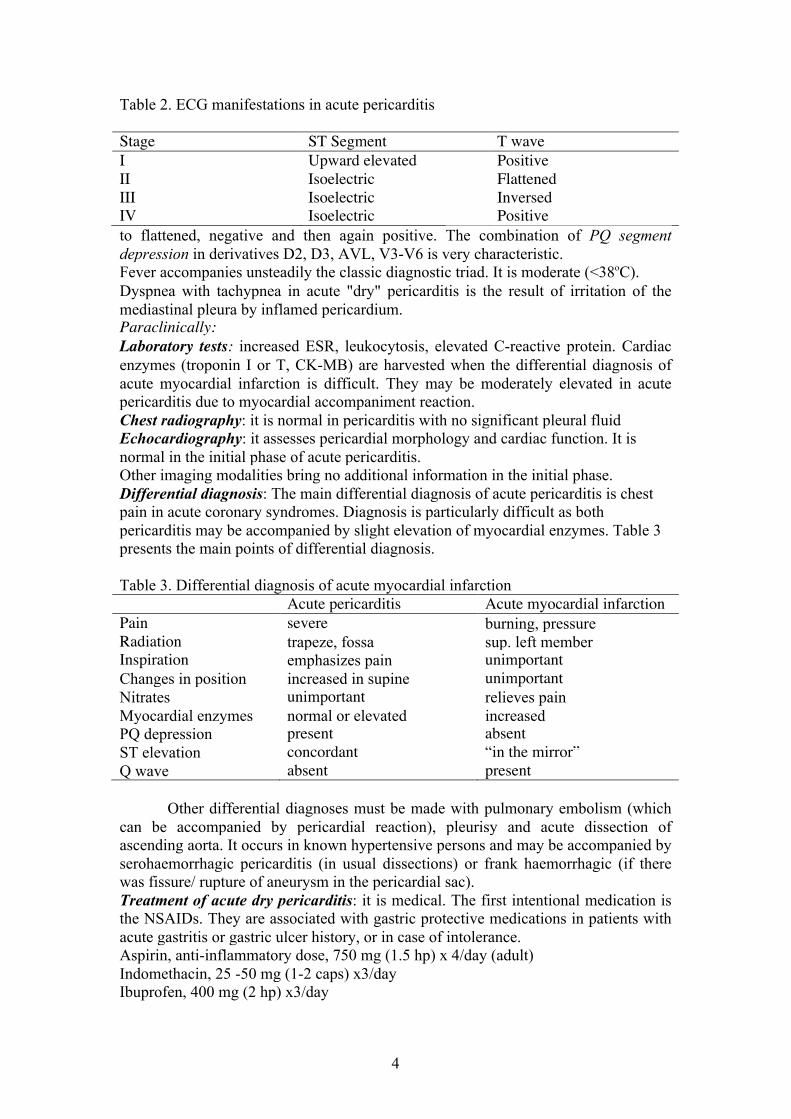
Table 2. ECG manifestations in acute pericarditis Stage ST Segment T wave I Upward elevated Positive II Isoelectric Flattened III Isoelectric Inversed IV Isoelectric Positive to flattened, negative and then again positive. The combination of PQ segment depression in derivatives D2, D3, AVL, V3-V6 is very characteristic. Fever accompanies unsteadily the classic diagnostic triad. It is moderate (<38oC). Dyspnea with tachypnea in acute "dry" pericarditis is the result of irritation of the mediastinal pleura by inflamed pericardium. Paraclinically: Laboratory tests: increased ESR, leukocytosis, elevated C-reactive protein. Cardiac enzymes (troponin I or T, CK-MB) are harvested when the differential diagnosis of acute myocardial infarction is difficult. They may be moderately elevated in acute pericarditis due to myocardial accompaniment reaction. Chest radiography: it is normal in pericarditis with no significant pleural fluid Echocardiography: it assesses pericardial morphology and cardiac function. It is normal in the initial phase of acute pericarditis. Other imaging modalities bring no additional information in the initial phase. Differential diagnosis: The main differential diagnosis of acute pericarditis is chest pain in acute coronary syndromes. Diagnosis is particularly difficult as both pericarditis may be accompanied by slight elevation of myocardial enzymes. Table 3 presents the main points of differential diagnosis. Table 3. Differential diagnosis of acute myocardial infarction Acute pericarditis Acute myocardial infarction Pain severe burning, pressure Radiation trapeze, fossa sup. left member Inspiration emphasizes pain unimportant Changes in position increased in supine unimportant Nitrates unimportant relieves pain Myocardial enzymes normal or elevated increased PQ depression present absent ST elevation concordant “in the mirror” Q wave absent present Other differential diagnoses must be made with pulmonary embolism (which can be accompanied by pericardial reaction), pleurisy and acute dissection of ascending aorta. It occurs in known hypertensive persons and may be accompanied by serohaemorrhagic pericarditis (in usual dissections) or frank haemorrhagic (if there was fissure/ rupture of aneurysm in the pericardial sac). Treatment of acute dry pericarditis: it is medical. The first intentional medication is the NSAIDs. They are associated with gastric protective medications in patients with acute gastritis or gastric ulcer history, or in case of intolerance. Aspirin, anti-inflammatory dose, 750 mg (1.5 hp) x 4/day (adult) Indomethacin, 25 -50 mg (1-2 caps) x3/day Ibuprofen, 400 mg (2 hp) x3/day
5
Corticosteroid medication is reserved for cases that did not respond to classical treatment. Evolution of acute pericarditis without pleural fluid: It is mostly benign. Complications of acute pericarditis are the development of a pleural fluid, or evolution to chronic state, with successive reccurences and the emergence of pericardial constriction. Acute Exudative Pericarditis Definition: They are acute pericarditis complicated by the appearance of a pleural pericardial fluid. Clinical symptoms depend on the accumulated amount of fluid. Morpho-pathology: The pericardial biopsy fragments show the aspects of inflammation enumerated above. In case of specific (neoplastic or tuberculous) pericarditis, the etiologic diagnosis of the fluid may be made. Its macroscopic appearance is often sero-citrine, especially in pure inflammatory or viral forms. Flakes of fibrin may occur, caused by protein clotting, where a protein concentration> 2 gr/100 ml (positive Rivalta test). If the macroscopic appearance of the liquid is sero- sanguinolent, etiology is most often tuberculous or neoplastic, but "idiopathic" pericarditis can occur with a similar aspect. Bacterial pericarditis is relatively easily identified by the presence of purulent, viscous, yellowish pleural fluid. Chylopericardium is characterized by lactescent intrapericardial fluid due to increased fat content of thoracic lymph. Finally, pure sanguinolent fluids are found in penetrating intracardiac wounds, iatrogenic coronary artery perforations during cardiac catheterization of the electrophysiological or post-operative examinations, postinfarct rupture of ventricle, rupture of atrioventricular junction after mitral prosthesis or acute dissection of ruptured aorta in the pericardium. Microscopic aspect of the liquid can give information about its etiology. Cytology can detect the presence of malignant cells in neoplastic fluids. In the inflammatory ones, it is usually non-specific. The direct microscopic examination, after Gram and Ziehl-Nielsen staining, detects presence of tuberculous bacilli. Culture of the liquid on specific media identifies the bacteria in purulent fluids. Biochemical examination, with the concentration of protein, glucose, the LDH, allows differentiation between transudate and pericardial effusion, and diagnosis of tuberculous pericarditis by increased concentration of adenine deaminase. Newer techniques of PCR and immunohistochemistry allow, respectively, the etiological diagnosis of viral pericarditis or autoimmune pericarditis. Clinically: clinical signs of exudative pericarditis depend on the amount of accumulated fluid. Pain is present from the “dry” phase of the pericarditis. Dyspnoea is increasing due to distension caused by pericardial liquid, in addition to the irritation of the phrenic and mediastinal pleura. Electrocardiographic signs are the same (changes in ST segment that evolve in 4 stages and are consistent in all derivatives) and the microvoltage aspect (maximum amplitude less than 5 mm) and the electric alternance (change of QRS axis due to movement of swinging heart in abundant fluids-"swinging heart") Pericardial friction, patognomonic sign of acute pericarditis disappears with the emergence of a liquid. Heart sounds become deafening once the pleural fluid was

6
formed. Subfebrilities are inconstantly found. Laboratory: Laboratory examinations: ESR's and C-reactive protein are constantly high and hyperleukocytosis is frequently encountered. Table 4 specifies laboratory tests to be conducted to clarify the etiologic diagnosis of pleural fluid. Table 4. Laboratory tests in pericarditis Etiology Test Viral Viral serodiagnosis Connective tissue diseases Antinuclear antibodies, rheumatoid factor Articular rheumatism ASO Tuberculosis Tuberculin IDR Myxedema T3, T4, TSH Neoplastic Tumor markers (carcinoembryonic
antigen) Chest radiography: it shows cardiac shadow increase,if the quantity of liquid is > 250 ml. It is characteristic that the shadow is stationary with heart movements in radioscopy (Fig. 1) Fig.1
Echocardiography: It is the most important imaging test. This test diagnoses exudative pericarditis, it measures the size of anterior and posterior pericardial decollation, cardiac systolic function, presence of fibrin flakes or blood clots and

7
pretamponade or tamponade diagnosis with diastolic collapse of right cavities, their severe compression and expansion of inferior vena cava. It is generally considered that anterior or posterior decollation, greater than 2 cm, means a large amount of liquid. Cardiac catheterization: it is not a routine practice in patients with pericardial fluid, unless coronary disease is suspected or if the pericardial fluid is considered to be due to acute aortic dissection. CT Scan : it is not a routine procedure as preoperative exploration in exudative pericarditis unless acute aortic dissection is suspected. Postoperatively, if exploration of pericardial fluid and biopsy piece suggests a neoplastic disease, CT scan is needed to determine the starting point and the tumor extension. Nuclear magnetic resonance - the same indications as the CT Scan. Differential diagnosis of acute exudative pericarditis is made, on the one hand, with the conditions responsible for acute chest pain, similar to acute "dry" pericarditis (acute coronary syndromes, pleurisy, aortic dissection) plus those responsible for acute dyspnea: pulmonary embolism, acute pulmonary edema, respiratory disease, etc. Treatment of acute exudative pericarditis is medical, interventional or surgical. Medical treatment: it is identical to the treatment of acute pericarditis with no pericardial fluid. Nonsteroidal anti-inflammatory drugs (NSAIDs) are medications of first choice. Their posology is the same as in cases of acute pericarditis without pericardial fluid. Corticosteroids are used in cases not responding to treatment with NSAIDs. Interventional treatment: pericardocentesis is performed for diagnostis and curative purposes, in patients with> 2 cm of anterior pericardial fluid. Puncture technique is subxiphoid after Seldinger technique.The left xiphocostal angle is punctured at an angle of 30-45%, orienting the needle to the left shoulder. Once entered into the pericardial area, a quantity of 40-50 ml of liquid is withdrawn to be sent for laboratory tests. With a guide introduced into the puncture needle, a drainage catheter is placed in the pericardial area, which will connect to an active drainage system. To reduce the risk of complications of this technique (right ventricular puncture, puncture of a coronary artery), the puncture needle can be connected to an ECG electrode or a pressure chamber. In the ideal situation, the puncture is guided by ultrasounds. The role of the surgery is to drain the pericardial fluid diagnostically and curatively in its acute phase. Subxiphoid drainage is performed on the pericardium through a median incision in the right xiphoid appendix. A section of pericardium is resected and sent for morphopathological examination. The fluid is completely evacuated and 50 ml are sent for laboratory tests. It is the most used technique. Pericardial window is the procedure of wide communication ("window") between the pericardial and pleural area by resecting a section of the pericardium and mediastinal pleura. Thus, if the pericardial effusion recovers, it will be evacuated in the pleural area and from there slowly reabsorbed. The indication of this technique is pericarditis at greater risk of relapse, the presence of subjacent chronic diseases (neoplastic, uremic, connective tissue pericarditis, etc.). It is made by right or left anterolateral thoracotomy, with a pleural area procedure and resection of a rectangular section of pleura and pericardium.

8
Surgical drainage through median sternotomy. It is recommended in hemorrhagic frank effusions (to detect the source of bleeding and achieve hemostasis), postoperative purulent or tuberculous effusions. In the latter etiologies complete evacuation of adhesions is necessary to prevent formation of constrictive pericarditis and installation of a lavage-drainage system. Thoracoscopic pericardial drainage is an elegant and minimally invasive method of draining the pericardial fluids. It consists in the introduction of a thoracoscope and two instruments (aspirator, cautery or clip) through three thoracic incisions. It has the advantage of direct visualization of the pericardial space, with lysis of formed adhesions, complete evacuation of clots, the collection of bioptic fragments, etc.. Its disadvantage is the need for a specific material. Surgical drainage through robotic techniques is similar to the thoracoscopic drainage but it is performed using a surgical robot. It is extremely expensive. Complications of acute exudative pericarditis are the occurrence of cardiac tamponade, the relapse of the exudate and formation of constrictive pericarditis. 9. 3. Cardiac tamponade Definition: Cardiac tamponade is a syndrome of extrinsic compression of the heart (mainly the right cavities) and clinically manifested by Beck's triad (hypotension, increased central venous pressure, faint heart sounds), paradoxical pulse, and dyspnea. Cardiac tamponade is a medical-surgical emergency, evolving to death in all untreated cases. Physiopathology: Cardiac tamponade occurs in exudative pericarditis if accumulation of intrapericardial fluid was rapid, or in chronic pericardial fluids if they exceeded the dilating reserve of the pericardial sac. Both situations show increased intrapericardial pressure, which becomes positive (normally it is -2 mm Hg) and compression of cardiac structures. The most affected cavities by pressure growth are right cavities, working in a lower pressure regime. Two phases of evolution are described: in the first phase the intrapericardial pressure is lower than the ventricular filling pressure. Cardiac output is maintained, with increased telediastolic ventricular volume and a compensatory tachycardia. If intrapericardial pressure continues to increase, it becomes higher than ventricular telediastolic pressure and ventricular filling is severely affected. In the case of the left ventricle,it is also caused by inversion of physiological curvature of the interventricular septum. The result is severe decrease of cardiac output. Clinically: Clinical consequences of this situation are the following: Increased ventricular interdependence: During inspiration venous return to the right heart increases. Due to increased intrapericardial pressure, high right ventricular filling will lead to reversal of normal curvature of ventricular septum and decreased left ventricular telediastolic volume. The hemodynamic effect is the emergence of Kussmaul's paradoxical pulse, which is defined by an inspiration decrease of systolic pressure with > 10 mm Hg. Hypotension due to impaired cardiac filling (when the intrapericardial pressure becomes greater than the right atrium one) and the reversal of the physiological curvature of the septum. Increase of jugular pressure and central venous pressure. Clinically, jugulars are turgid, X wave is increased on the venous jugular curve (pronounced filling of the

9
right atrium during ventricular systole) and Y wave disappears (poor right ventricular filling in diastole, due to external compression with hypodiastolic phenomena). Reactive tachycardia, a fast mechanism which tends to compensate for reduced cardiac output. Severe dyspnoea by phrenic nerve irritation. There are electrocardiographic signs of acute exudative pericarditis (microvoltage, concordant ST elevation) with electric alternation increase (changes of electrical axis of QRS complex), by a balance move of the heart in the intrapericardial fluid ("swinging heart"). Paraclinically: there are the same aspects as in acute exudative pericarditis. Due to the emergency, making the whole battery of laboratory tests is often impossible. The only two exams to be performed are thoracic radiography and cardiac echocardiography. Chest radiography in anterior-posterior incidence shows an increase in cardiac silhouette (immobile in radioscopy) and reverse of middle left arc curvature. Transthoracic echocardiography shows diastolic collapse of right cavities and their extrinsic compression (in the apical incidence 4 chambers), reverse of the physiological curvature of the interventricular septum, dilation of inferior vena cava. It assesses cardiac function and calculates, in M-mode or 2D, the distance of anterior and posterior transonic space. It is the most important exam to be performed under clinical suspicion of pericardial tamponade, the only certain diagnosis in an emergency. (Figure 2.3) The treatment of cardiac tamponade is only interventional or surgical and the medical treatment only serves to stabilize the patient. The medical treatment is performed through an effective filling of the patient, administration of oxygen by mask, low / moderate dose of inotropes (dobutamine) which do not increase peripheral resistance. Fig.2
10
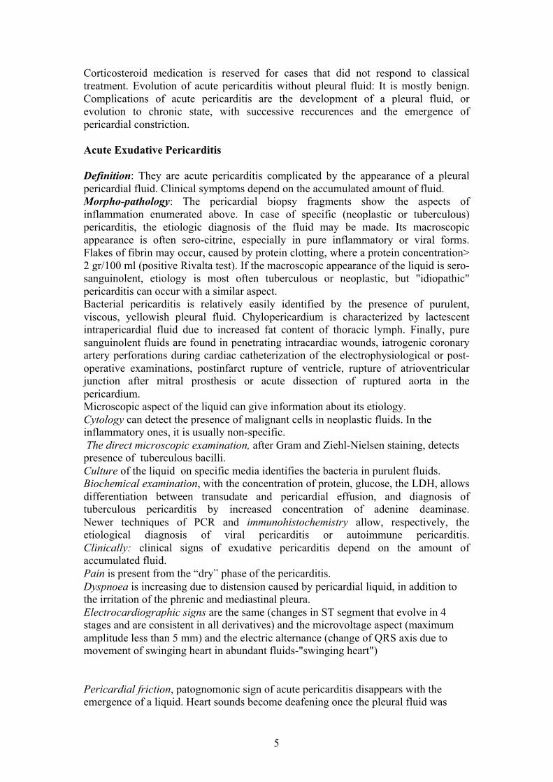
Right ventricular collapse in cardiac tamponade. Transthoracic echographic image in the parasternal incidence long axis. Fig.3
The collapse of the right atrium (RA) in cardiac tamponade. The extracardiac transonic image marked (p) is the pericardial fluid. Pericardiocentesis is elected in an absolute emergency, whether it is a pericardial fluid without fibrin clots or floaters, and if there is no evidence to suggest a chronic etiology. It is made by the technique described in exudative forms. Surgical drainage is chosen if we are dealing with a postoperative pericardial fluid, if there are clots or fibrin at ultrasound, if we are dealing with a tuberculous, uremic or malignant etiology. It is also preferable in extremely abundant fluids. Techniques are the same as in exudative forms, with a preference for subxifoidian surgical drainage, which is performed quickly in an emergency situation, with hemodynamic instability. A median sternotomy procedure is necessary if the pericardial fluid contains clots or pus. 9. 4. Chronic Constrictive Pericarditis Definition: The chronic pericarditis is a syndrome caused by excessive thickening of the pericardium, which is inextensible, fibrous or chalk, and is manifested clinically by poor ventricular filling (hypodiastole). Etiology: The main etiology of constrictive pericarditis is tuberculous, but it can occur after irradiation (radical pericarditis), postsurgical or chronic fungal pericarditis (Histoplasmosis). Morphopathology: the pericardial sac is much thickened and it may present isolated or extensive areas of calcification (Panzerherz). There are marked adhesions between the visceral and parietal pericardium. From a microscopic point of view we have a chronic inflammatory exudate with lymphocytes, fibrosis and calcification.
11
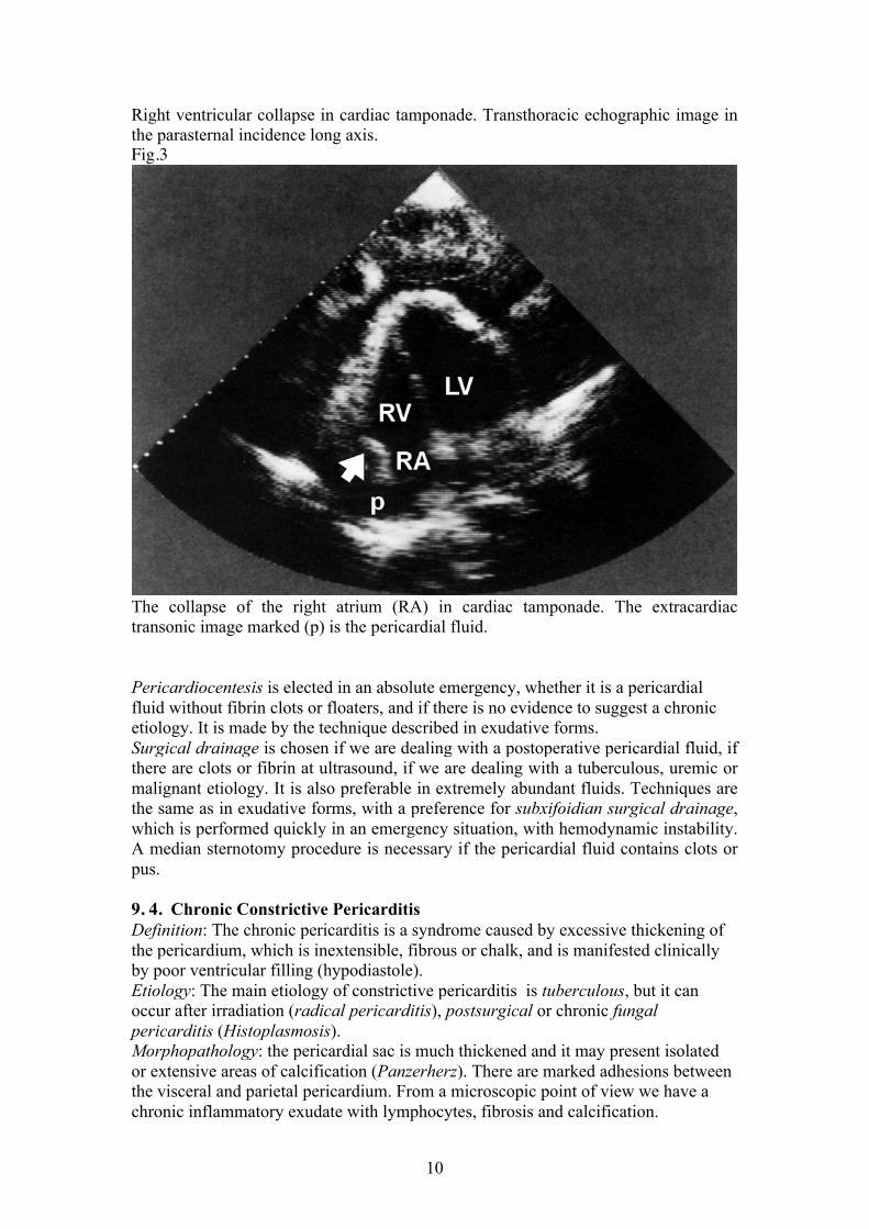
Physiopathology: The presence of the inextensible rigid pericardial sac, leading to alteration of ventricular filling mechanism. Thus, after opening the atrio-ventricular valves there is a rapid ventricular filling, followed by abrupt filling stop when the ventricular telediastolic volume reaches the limit imposed by pericardial sac. This aspect is reflected in the ventricular pressure curve as a sharp decrease in pressure during protodiastole followed by a plateau phase in late diastole ("dip- plateau "). The jugular pressure curve, unlike the case of cardiac tamponade,in which the rapid ventricular filling is impaired, shows an increase of x and y curves. Due to the rigid and inextensible character of the pericardium, intrathoracic pressure is no longer transmitted in the intrapericardial area. This leads to Kussmaul sign: increased central venous pressure in deep inspiration, opposite to the normal situation. Compensatory response of the heart, before a fixed diastolic filling, is a reactive tachycardia. If the situation becomes chronic, hydrosaline retention occurs due to central venous stasis on the one hand, and secondly because of the inhibition of atrial natriuretic factor secretion. The myocardium may also atrophy, with damage of myocytes and their replacement with collagen. Clinically: Jugular turgidity Hepatomegaly, and other signs of liver dysfunction in advanced forms, such as ascites, jaundice, palmar erythema, angioma “spider” Lower limb edema Dyspnoea Dispnee Paraclinically: Fig. 4
Laboratory tests. They are not pathognomonic. They show hydrosaline retention, liver function alteration, hypoalbuminemia. ESR-forms can be increased in chronic inflammatory forms and IDR with positive tuberculin in tuberculous pericarditis. Thoracic radiography may be diagnosed in the lateral and anterior posterior incidence if pericardium is calcified, with "eggshell" image .(Fig. 4)

12
Thoracic radiography in anterior posterior and lateral incidences of a patient with calcific chronic constrictive pericarditis. ECG is nonspecific. “Mitral” P wave and atrial fibrillation occur, due to chronic atrial dilation. Echocardiography. M-mode and 2D show thickened pericardium, dilation of inferior vena cava, and in Doppler mode characteristic changes, synchronous with respiration, occur. Thus, in inspiration transtricuspidian flow is increased and the transmitral flow decreased and in expiration, the changes are reversed. These changes occur due to inthoracic pressure drop in inspiration, blocked capillary pressure and left atrial pressure. The effect will be a poor left ventricular filling in diastole, reversing the physiological curvature of the interventricular septum. Left ventricular function may be altered in patients with constrictive pericarditis by atrophy and fibrosis, with a decrease of ejection fraction. Computertomography (CT scan) and magnetic resonance imaging (MRI) have a higher sensitivity than echography to detect calcifications and pericardial thickening (Fig. 4) Fig.5
Thoracic CT scan. The thickened and calcified pericardium is to be noticed. Right and left cardiac catheterization is made mandatory in constrictive pericarditis. Simultaneous catheterization of both ventricles reveals telediastolic pressure equalization,the characteristic aspect "dip- plateau " or "square root" of the ventricular curve (Fig.6), increase of right atrial pressure in deep inspiration (Kussmaul sign). Coronary angiography may rarely reveal external compression of the coronary arteries.

13
Fig.6
Differential diagnosis in constrictive pericarditis is to be made, first with restrictive cardiomyopathy, and also with valvular pathology of the right heart (tricuspidian stenosis or insufficiency) myxoma of the right atrium, hypertrophic cardiomyopathy, superior cava syndrome, nephrotic syndrome. The most difficult diagnosis is in restrictive cardiomyopathy (Table 5). Table 5. Differential diagnosis with restrictive cardiomyopathy Parameter Constrictive pericarditis Restrictive cardiomyopathy Anamnesis Surgical history, irradiation
system diseases, tuberculosis,neoplasia
Insignificant
Valvular insufficiencies absent IM �i IT AP systolic pressure < 40 mm Hg > 40 mm Hg Diastolic pressure balance
Frequent Rarely
VIS curvature reversal in inspiration
Yes No
Thoracic radiography Visible calcifications 25-30%
No
CT / MRI Pericardial thickening / calcification
No
Myocardial biopsy Normal Infiltrative diseases Treatment of constrictive pericarditis. Medical treatment has a limited symptomatic role of reducing venous stasis and edema. Loop and antialdosteronic diuretics are used. If atrial fibrillation occurs, it should be converted because it lowers 25% of ventricular filling. Sinus tachycardia should be respected as it is reactive to a fixed stroke volume. Surgery is the only cure. It is achieved by a subtotal interphrenic pericardiectomy through median sternotomy. Thickened or calcified pericardium is resected from the right cavities and pulmonary artery trunk as they are the main affected ones by constriction. Resection extends to the phrenic nerves, which represent lateral edge of excision. Intervention may be difficult in some cases in which the visceral
14
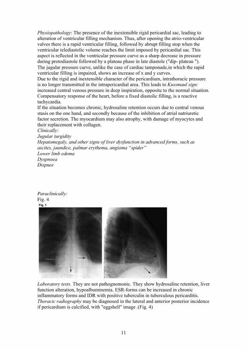
pericardium is closely adhering to the heart area. This intervention leads to a significant decrease in central venous pressure with increasing ventricular filling and cardiac output. Perioperative complications of pericardiectomy are the chamber injuries in case of strong adhesions, lesions of phrenic nerve or low cardiac output syndrome. This can occur in a lasting pericardial contraction , with consequent atrophy of myocardial fibers and their replacement by collagen fibers with impaired left ventricular systolic function after decompression. Constrictive exudative chronic pericarditis Definition: It is a rare form of chronic pericarditis, which associates the presence of a pericardial fluid with thickened visceral pericardium. Some consider this pathology a form of transfer from exudative recurrent pericarditis to chronic constrictive pericarditis. Physiopathology: Constriction of the right cavities is made predominantly by the large amount of liquid. Evacuation of fluid will only partially lead to protodiastole to improve diastolic filling, which is then limited by the restriction performed by inextensible visceral pericardium. Diagnosis of this clinical entity is difficult, especially if the visceral pericardium is not calcified, and is made by highlighting the thickened viceral pericardium associated with intrapericardial fluid during M mode echography. New imaging methods such as computed tomography or magnetic resonance imaging also allow the diagnosis of this form of pericarditis (Figure 7). Fig.7
Exudative constrictive pericarditis. Thickened pericardium and pericardial fluid in small quantity. Evolution of constrictive exudative pericarditis may be made to partial resorption of fluid and shifting to pure constrictive pericarditis. The treatment is surgical, through median sternotomy, and it consists in the evacuation of liquid and subtotal resection of the pericardium.

15