Embed Size (px)
Citation preview

Change of bones lending from
24th October till time of autopsies
Monday and Tuesday closed
Wednesday 9-14
Thursday 9-16
Friday 9-14

Arthrology arthros (joint), logos (science)

Arthrology – study of joints (articulatio)
1) Synarthrosis (continuous/fibrous joint)– continuous connection of
bones by connective tissue (fibrous tissue, cartilage, bone)
2) Diarthrosis (Synovial joint) – movable connection of bones by
contact of articular surfaces covered by articular cartilage and with
additional features
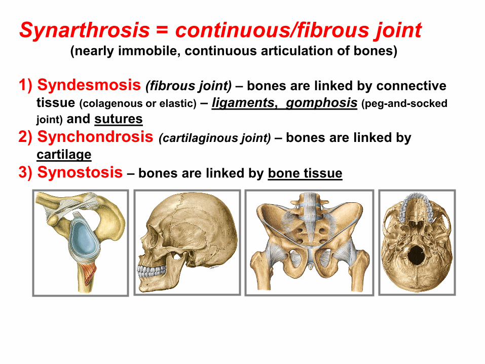
Synarthrosis = continuous/fibrous joint (nearly immobile, continuous articulation of bones)
1) Syndesmosis (fibrous joint) – bones are linked by connective
tissue (colagenous or elastic) – ligaments, gomphosis (peg-and-socked
joint) and sutures
2) Synchondrosis (cartilaginous joint) – bones are linked by
cartilage
3) Synostosis – bones are linked by bone tissue

I. Synarthrosis (continuous/fibrous joint)
1. syndesmosis 2. synchondrosis 3. synostosis
II. Diarthrosis (synovial joint)

Ad 1. Syndesmosis (fibrous joint)
1. Ligaments 2. Membranes 3. Gomphosis (peg-and-socked joint)
4. Sutures

4. Sutures
Sutura serrata
Sutura squamosa (Squamous suture)
Sutura plana

hyalinní chrupavka vazivová chrupavka
2. Synchondrosis (cartilaginous joint)
Discus intervertebralis (intervertebral disc)
Symphysis pubica
3. Synostosis Os sacrum
Os coccygis
Synarthrosis (continuation)

II. Articulatio=synovial joint= Diarthrosis movable connection of two or more bones by touch of contact
articular surfaces covered by articular cartilage and with auxiliary
facilities

General features of a synovial joint
(diarthrosis)
Facies articulares (articular surfaces) (fossa articularis, caput articulare)
Capsula articularis (joint capsule) = (stratum fibrosum and stratum
synoviale) – fibrous and synovial layers)
Cavitas articularis – capillary space filled by synovia (synovial fluid)
Synovia (Synovial fluid) – nourishes the articular cartilage, increases
adhesion and decreases friction of contact surfaces (lubricant)
Synovial plicae or synovial villi
Rete articulare (Articular network)

Auxiliary facilities of joints a) labrum articulare – fibrocartilaginous ring – broadening of a shallow
articular fossa by a strip of fibrous tissue and/or cartilage
b) disci and menisci articulares – plates of cartilage - serves as elastic pad,
discs divide the articular cavity into two parts, menisci only partly
c) ligamenta are present in the most joints as ligamenta capsularia,
extracapsularia or intracapsularia (capsular, extracapsular or intracapsular
ligaments)
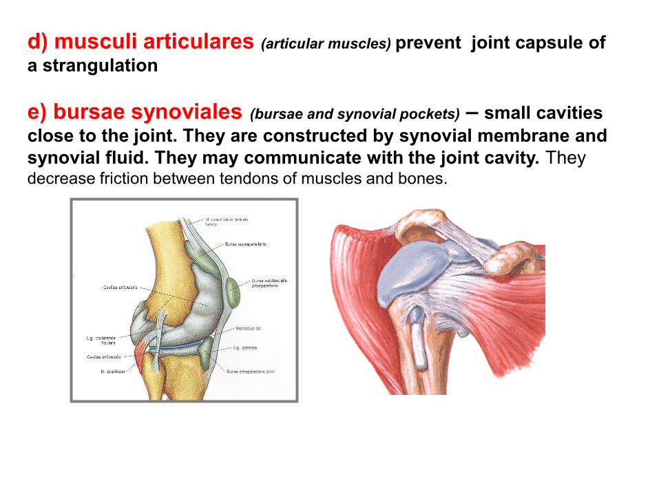
d) musculi articulares (articular muscles) prevent joint capsule of
a strangulation
e) bursae synoviales (bursae and synovial pockets) – small cavities
close to the joint. They are constructed by synovial membrane and
synovial fluid. They may communicate with the joint cavity. They decrease friction between tendons of muscles and bones.

Division of joints
A. According to a number of bone in contact:
Articulatio simplex Simple joint
Articulatio composita Compound joint
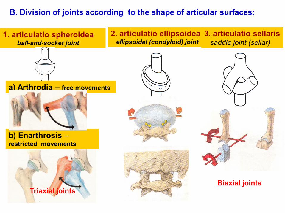
B. Division of joints according to the shape of articular surfaces:
1. articulatio spheroidea ball-and-socket joint
a) Arthrodia – free movements
b) Enarthrosis – restricted movements
2. articulatio ellipsoidea ellipsoidal (condyloid) joint
3. articulatio sellaris saddle joint (sellar)
Biaxial joints Triaxial joints

4. Articulatio cylindroidea cylindrical joint
4b) ginglymus
5) Articulatio trochlearis hinge (trochlear) joint
(with ledge-shaped elevation)
Monoaxial joints
4a) Articulatio trochoidea
pivot joint (trochoid)

6. Amfiartrosis Joint with minimal movements
5. Articulatio plana Joint with sliding movements
Middle position of the joint – position in which a joint capsule is evenly
and maximum relaxed (the joint is the least loaded).

Movements in joints
flexion extension
abduction adduction
supination pronation
I.
II.
III.
Special arthrology
Connection on the skull
1. Craniovertebral connection
2. Skull syndesmoses and synchondroses
3. Temporomandibular joint
4. Connection of the hyoid bone (os hyoideum)

1. Connections of the skull A. Synarthrosis (1. syndesmosis, 2. synchondrosis, 3. synostosis)
B. Diarthrosis=synovial joint (articulatio temporomandibularis).
suturae ligamentum stylohyoideum
ligamentum stylomandibulare
I.1. Syndesmosis cranii
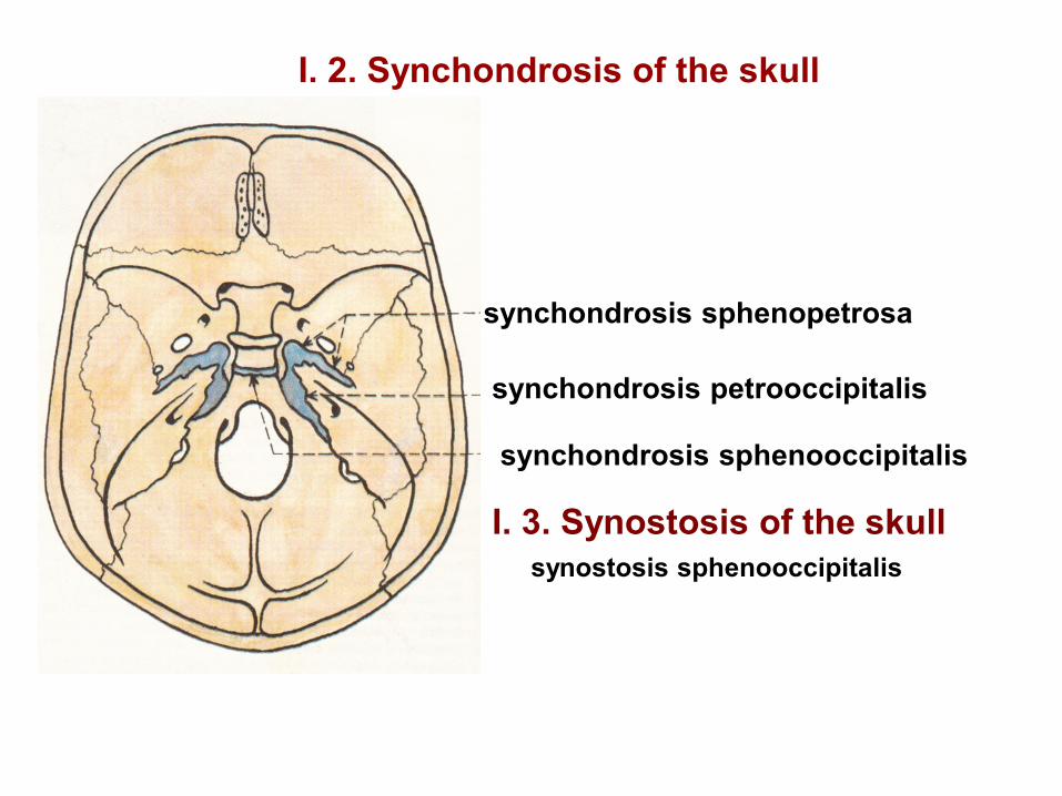
I. 2. Synchondrosis of the skull
synchondrosis sphenopetrosa
synchondrosis petrooccipitalis
synchondrosis sphenooccipitalis
I. 3. Synostosis of the skull
synostosis sphenooccipitalis
Description (characterization) of synovial joints
1. Name of a joint
2. Articular surfaces
3. Articular capsule
4. Auxiliary facilities
5. Type of joint
6. Movements

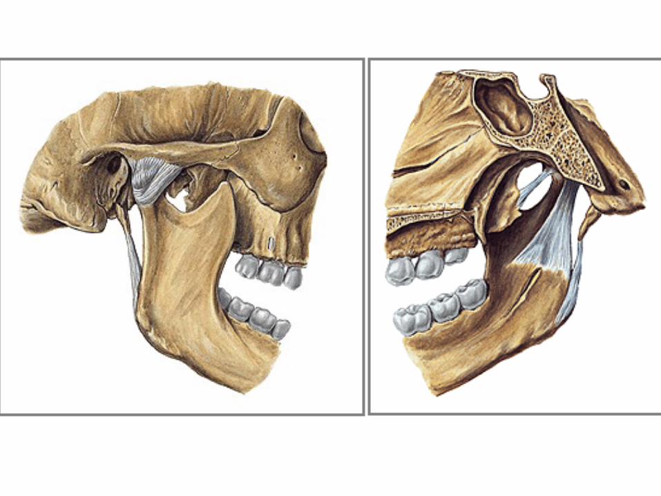
Articulatio temporomandibularis
Temporomandibular joint
Articular surfaces: caput mandibulae with fossa mandibularis and tuberculum
articulare of the temporal bone
Articular capsule: is attached to the margins of contact articular surfaces, ventrally and
dorsally is lax, its medial part is tense
Auxiliary facilities: discus articularis – with a thin center and thicker margins. It divides
joint cavity into the upper pars discotemporalis and lower pars discomandibularis. Lig.
laterale.
Close to the joint are located lig. sphenomandibulare and lig. stylomandibulare
Type of joint: composite and paired joint (Gynglimus)
Movements: complicated (rotary, sliding, chewing movements)
¨

Movements: opening (mandibular depression ) and closing mouth
(elevation), rotary and sliding movements and chewing movements -
mandibular protraction (movement of mandible ventrally) and retraction
movement of mandible dorsally)

I. Craniovertebral joints 1. Articulatio atlantooccipitalis – (atlanto-occipital joint)
articular surfaces:……………….
articular capsule: is attached to the margins of the articular surfaces
auxiliary facilities: membrana atlantooccipitalis anterior and posterior
(anterior and posterior atlanto-occipital membranes)
membrana tectoria (tectorial membrane)
type of joint: art. ellipsoidea (ellipsoidal joint)
movements: flexion and extension of the head and its minimal lateral motion

2a. Articulatio atlantoaxialis mediana
facies articularis anterior dentis and fovea dentis
facies articularis posterior dentis
and lig. transversum atlantis
2b. Articulatio atlantoaxialis lateralis
facies articulares inferiores atlantis
processus articulares superiores axis
2. Articulatio atlantoaxialis – a compound joint
Articular surfaces:

Articulatio atlantoaxialis mediana et lateralis
Articular capsule: is common for both 1) and 2) joints
and is attached to the margins of contact articular
surfaces
Auxiliary facilities: lig. apicis dentis, ligg. alaria, lig.
transversum atlantis, lig. cruciforme atlantis = lig.
transversum atlantis and fasciculi longitudinales
(longitudinal bands)
membrana atlantooccipitalis anterior and posterior
(Anterior and posterior atlantooccipital membranes) ,
membrana tectoria
Type of joint: functionally – the mechanical unit. Atlas
rotates around dens axis in about 60o

Juncturae columnae vertebralis (Junctions of the spine)
There are both synarthrosis (fibrous joints) and diarthrosis (synovial joints)
on the spine.
Connections of adjacent vertebrae 1. between vertebral bodies – by disci intervertebrales (23)
2. between vertebral arches by ligg. flava
3. between vertebral processes by ligg. intertransversaria, interspinalia and
supraspinale (lig. nuchae)
4. Synovial joints (diarthrosis) – articulationes intervertebrales (intervertebral joints)

Anulus fibrosus
Nucleus
pulposus
= jelly-like nucleus
– remnant of chorda
dorsalis
=spherical focus for
movement
1. Connection between vertebral bodies
(intervertebral disc)
23 discs, form about ¼ of the spine length
discs act as shock absorbers

Compression of disc (nucleus pulposus distributes the pressure)

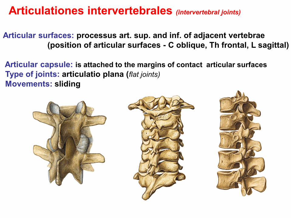
Articulationes intervertebrales (intervertebral joints)
Articular surfaces: processus art. sup. and inf. of adjacent vertebrae
(position of articular surfaces - C oblique, Th frontal, L sagittal)
Articular capsule: is attached to the margins of contact articular surfaces
Type of joints: articulatio plana (flat joints)
Movements: sliding

Common connections for all vertebrae (tie vertebral column)
Lig. longitudinale anterius - anterior longitudinal ligament
Lig. longitudinale posterius – posterior longitudinal lig. continues cranially as
membrana tectoria
caudal continuation of both – ligg. sacrococcygea

Long ligaments of spine Ligamentum supraspinale (ligamentum nuchae)
Ligamentum longitudinale anterius
Ligamentum longitudinale posterius (membrana tectoria)

Shape and curvature of spine Spine has cervical and lumbar lordosis
(C4-5, L3-4)
and thoracic and sacral kyphosis (Th6-7)

Movements of the spine
Cervical spine – anteflexion, retroflexion, lateral flexion, rotation
Thoracic spine – rotation
Lumbar spine – anteflexion, retroflexion, lateral flexion

II. Curvature of the spine in frontal plane – skoliosis
Physiological skoliosis Th3-5

Connections of the thorax I) Juncturae thoracis (connections of thoracic cage)
A. Articulationes costovertebrales (costovertebral joints)
1. Articulationes capitis costae (joints of the rib head)
Articular surfaces: facies articularis capitis costae and foveae costales
of the thoracic vertebrae bodies
Articular capsule: is attached to the margins of contact articular surfaces.
Auxiliary facilities: lig. capitis costae radiatum
lig. capitis costae intraarticulare 2. – 10. ribs
Movements: – around axis paralel to the collum costae (neck of rib)
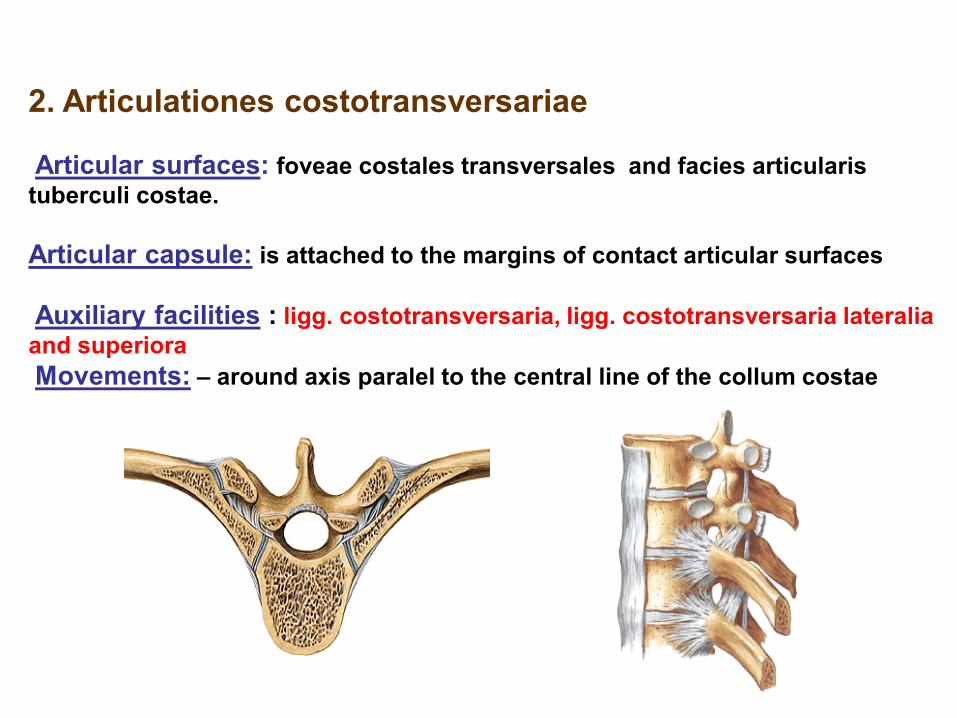
2. Articulationes costotransversariae
Articular surfaces: foveae costales transversales and facies articularis
tuberculi costae.
Articular capsule: is attached to the margins of contact articular surfaces
Auxiliary facilities : ligg. costotransversaria, ligg. costotransversaria lateralia
and superiora
Movements: – around axis paralel to the central line of the collum costae

B. Juncturae sternocostales (sternocostal articulations)
1. Synchondrosis sternocostalis - connection of the incisura costalis sterni (sternal
costal notch and cartilage of the 1. and often the 6th and 7th ribs.
2. Articulationes sternocostales (sternocostal synovial joints) between 2.–5. ribs and
sternum.
Auxiliary facilities: ligg. sternocostalia radiata form membrana sterni externa
and interna
C. Connection of 5th – 9th ribs
1. Articulationes interchondrales (interchondral joints)
Articular connection between cartilagines costales 5.–9th with short articular
capsule.
2. Membranae intercostales – membranae intercostales externae - between
costal cartilages close to the sternum. Membranae intercostales internae are
located near to the spine.

II. Shape of thorax (chest) a) Ventral wall – sternum, costal cartilages and ribs b) Lateral wall – anguli costae
c) Dorsal wall – vertebrae thoracicae and bone parts of the ribs
d) Entrance (apertura thoracis superior) is limited by the 1. thoracic
vertebra, the 1. rib and cranial margin of sternum
e) Exit (apertura thoracis inferior) is limited by 12. thoracic vertebra, 12.
and 11. ribs and arcus costarum.
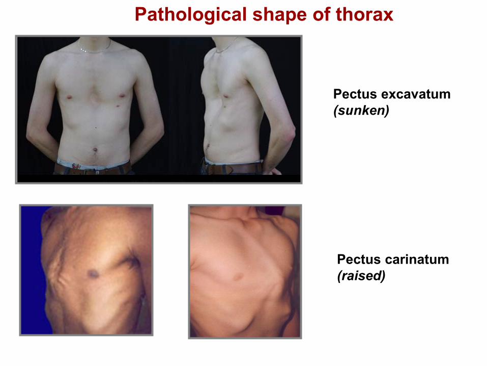
Pectus excavatum
(sunken)
Pectus carinatum
(raised)
Pathological shape of thorax

III. Movements of thorax (chest)
in costovertebral joints - rotation along longitudinal axis - runs through
collum costae
increasing volume of the chest - inspiration (inspire)
decreasing volume of the chest - expiration (expire)

Upper limb connections (juncturae ossium extremitatis superioris)
1) Articulationes cinguli membri superioris
Connections of shoulder girdle
2) Articulationes membri superioris liberi
Connections of the free part of the upper limb

Articulationes cinguli membri superioris (Connections of shoulder girdle)
Articulatio sternoclavicularis (sternoclavicular joint)
Articulatio acromioclavicularis (acromioclavicular joint)
Syndesmosis scapulae (ligaments of scapula)
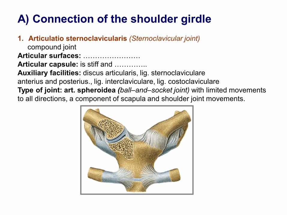
A) Connection of the shoulder girdle
1. Articulatio sternoclavicularis (Sternoclavicular joint)
compound joint
Articular surfaces: ……………………
Articular capsule: is stiff and …………..
Auxiliary facilities: discus articularis, lig. sternoclaviculare
anterius and posterius., lig. interclaviculare, lig. costoclaviculare
Type of joint: art. spheroidea (ball–and–socket joint) with limited movements
to all directions, a component of scapula and shoulder joint movements.

2. Articulatio acromioclavicularis (Acromioclavicular joint) (usually a compound joint)
Articular surfaces : ………………..
Articular capsule : is attached to…………..
Auxiliary facilities : often is present discus articularis, lig. acromioclaviculare,
lig. coracoclaviculare
Type of joint: spheroid or plane (ball–and–socket/plane joint) with limited
movements - a component of scapula and shoulder joint movements.

3. Syndesmosis (Ligaments) of the scapula Lig. transversum scapulae - superius and inferius (Superior and inferior transversal
scapular ligament)
Lig. coracoacromiale (coracoacromial ligament) - together with both bone processus
forms fornix humeri. Abduction/elevation of the upper limb is always associated with movements of
scapula!
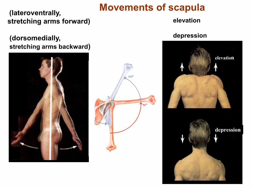
Movements of scapula (lateroventrally,
stretching arms forward)
(dorsomedially,
stretching arms backward)
elevation
depression
elevation
depression

B) Articulationes membri superioris liberi Connections of the free part of the upper limb
1. Articulatio humeri (Shoulder joint) Articular surfaces:………………….
Articular capsule: on the medial side of humerus runs more distally. Vagina synovialis
intertubercularis.
Auxiliary facilities: labrum glenoidale, ligg. glenohumeralia, lig. coracohumerale.
Articular capsule is reinforced by tendons of muscles (m. subscapularis, m.
supraspinatus, m. infraspinatus, m. teres minor called rotator cuff).
Type of joint: arthrodia (ball-and-socket), three degrees of movements freedom).
Middle position of the joint - flexion and abduction at about 40-45o, synovial bursae


Articulatio humeri
Type of joint: spheroidea – ball and socket (arthrodia)
Movements: triaxial joint, to all directiones
flexion extension
abduction
adduction
pronation
supination
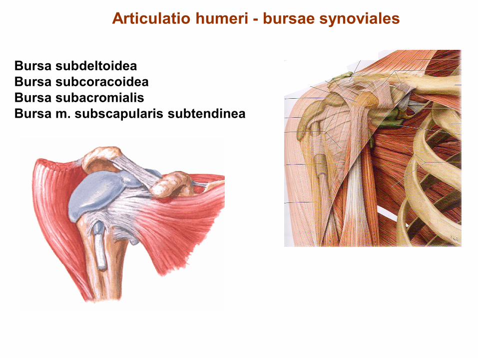
Articulatio humeri - bursae synoviales
Bursa subdeltoidea
Bursa subcoracoidea
Bursa subacromialis
Bursa m. subscapularis subtendinea

Articulatio cubiti (elbow joint)
Articulatio composita:
1. Articulatio humeroradialis
2. Articulatio humeroulnaris
3. Articulatio radioulnaris proximalis

– 2. Articulario cubiti (Elbow joint) compound joint
Articulatio humeroradialis (humeroradial joint)
Articulatio humeroulnaris (humeroulnar joint)
Articulatio radioulnaris proximalis (radioulnar proximal joint)
Articular surfaces :……………………………..
Articular capsule : both epicondyli of humerus are free, all fossae
of humerus are located intracapsularly, on the radius runs to the
collum radii – recessus sacciformis.
Auxiliary facilities: ligg. collateralia rad. and uln., anulare radii
Type of joints:
Movements: flexion and extension, rotation (inner-pronation) and
external rotation (supination).
Subcutaneous and subtendinous olecranon bursa.


Articulatio cubiti - movements
flexion and extension
(hyperextension over 180o)
pronation and supination of forearm
flexion
extension
pronation
supination

Used pictures come from:
Moore, K. L. (1992): Clinical oriented anatomy. Third edition.
Williams&Wilkins, A Waverly Company.
Gilroy, A. M. et all. (2009): Atlas of Anatomy. Thieme New York, Stuttgart.
Putz, R. (2008):
Atlas of Human Anatomy Sobotta. Elsevier Books.
Platzer, W., Kahle, W., Leonhardt H. (1992):
Locomotor system. Georg Thieme Verlag, Stuttgart,
New York, 4th edition.
Čihák, R. (1987): Anatomie 1. Avicenum, Zdravotnické nakladatelství.