Embed Size (px)
Citation preview
C AD / C AM SYSTEMS | INSTRUMENTS | HYGIENE SYSTEMS | TRE ATMENT CENTERS | IMAGING SYSTEMS
C A D / C A M C A M E r A S . M A D E t o i n S p i r E
CEREC OMNICAM AND CEREC BLUECAM.THE fIRST CHOICE IN EVERY CASE.
CEREC omnicam: sCanning simpliCity.
Unrivalled handling, powder-free scanning and precise 3D impressions in natural color. Taking CEREC Omnicam optical impressions is easy, intuitive and ergonomic.
The natural color appearance will not only impress your patients.
powder-free digital scanning in full color

CErEC omnicam
powder-free scanningCEREC Omnicam is optimized for powder-free scanning of natural tooth structures and gingiva. Simply place the camera over the relevant area and the scan starts automatically. The elimination of the powder coating means that the scanning process is easier to learn. full-arch and half-arch scans can now be performed more conveniently and quickly than ever before.
Unrivalled handling benefitsperfect accessCEREC Omnicam fits perfectly into the user’s hand. Thanks to the slim-line design and the compact camera tip, scanning the posterior teeth presents no problems. The rounded outer contours ensure that it is easy to rotate. Regardless of whether the patient is sitting upright or reclining, you can scan the upper and lower jaws ergonomically without having to adjust your natural working posture.
Seamless scanning processThe user simply moves the camera head closely over the teeth in a single flowing movement. The data is generated successively into a 3D model. The seamless scanning process delivers an impressive depth of field. What’s more, you can interrupt and resume the scan at any time.
precise 3D scans in natural colorIt is impressive to see the 3D model displayed in full color on the monitor. The various surfaces are shown in their natural shades. This direct and realistic feedback helps you navigate your way around the oral cavity and enables you to distinguish between amalgam, gold or composite. Clinically it provides a clear differentiation between gingiva and the preparation margin. Effective communication with patients CEREC Omnicam offers a further decisive benefit. In the patient coun-seling mode you can record video clips and present these directly to the patient. The lifelike visualization of the teeth and gingiva helps to convince the patient of the need for treatment. He or she will more readily understand and accept your therapy proposals.
Scanning simplicity – the details.
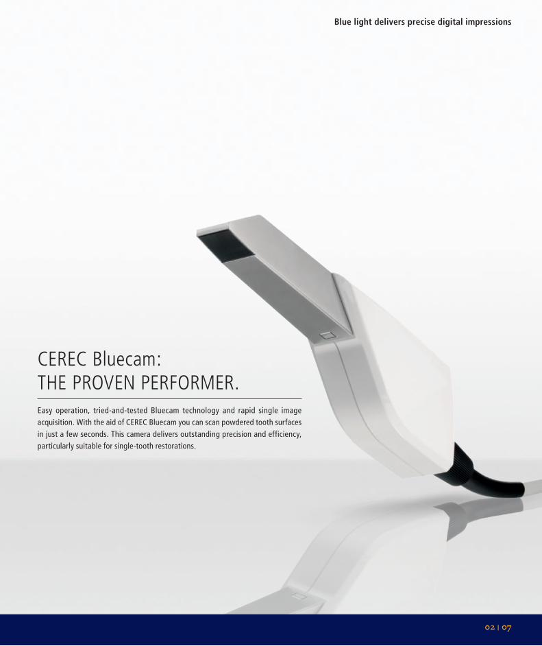
CEREC bluecam: thE pRovEn pERfoRmER.Easy operation, tried-and-tested Bluecam technology and rapid single image acquisition. With the aid of CEREC Bluecam you can scan powdered tooth surfaces in just a few seconds. This camera delivers outstanding precision and efficiency, particularly suitable for single-tooth restorations.
02 | 07
Blue light delivers precise digital impressions
CErEC Bluecam
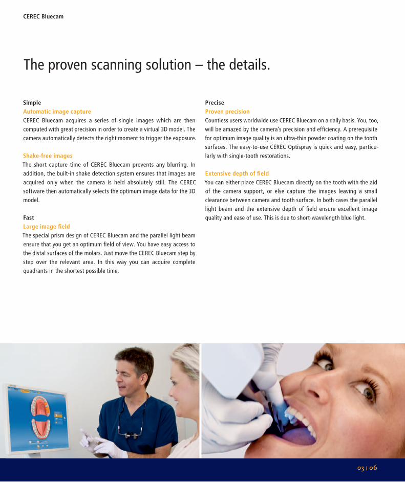
SimpleAutomatic image captureCEREC Bluecam acquires a series of single images which are then computed with great precision in order to create a virtual 3D model. The camera automatically detects the right moment to trigger the exposure.
Shake-free imagesThe short capture time of CEREC Bluecam prevents any blurring. In addition, the built-in shake detection system ensures that images are acquired only when the camera is held absolutely still. The CEREC software then automatically selects the optimum image data for the 3D model.
FastLarge image fieldThe special prism design of CEREC Bluecam and the parallel light beam ensure that you get an optimum field of view. You have easy access to the distal surfaces of the molars. Just move the CEREC Bluecam step by step over the relevant area. In this way you can acquire complete quadrants in the shortest possible time.
preciseproven precisionCountless users worldwide use CEREC Bluecam on a daily basis. You, too, will be amazed by the camera’s precision and efficiency. A prerequisite for optimum image quality is an ultra-thin powder coating on the tooth surfaces. The easy-to-use CEREC Optispray is quick and easy, particu-larly with single-tooth restorations.
Extensive depth of fieldYou can either place CEREC Bluecam directly on the tooth with the aid of the camera support, or else capture the images leaving a small clearance between camera and tooth surface. In both cases the parallel light beam and the extensive depth of field ensure excellent image quality and ease of use. This is due to short-wavelength blue light.
The proven scanning solution – the details.
03 | 06
Acquiring digital impressions with the CErEC omnicam and the CErEC Bluecam
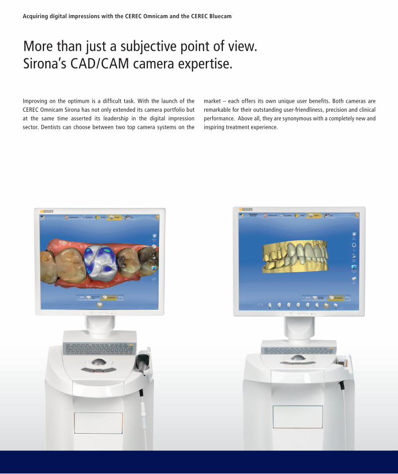
More than just a subjective point of view. Sirona’s CAD/CAM camera expertise.
Improving on the optimum is a difficult task. With the launch of the CEREC Omnicam Sirona has not only extended its camera portfolio but at the same time asserted its leadership in the digital impression sector. Dentists can choose between two top camera systems on the
market – each offers its own unique user benefits. Both cameras are remarkable for their outstanding user-friendliness, precision and clinical performance. Above all, they are synonymous with a completely new and inspiring treatment experience.
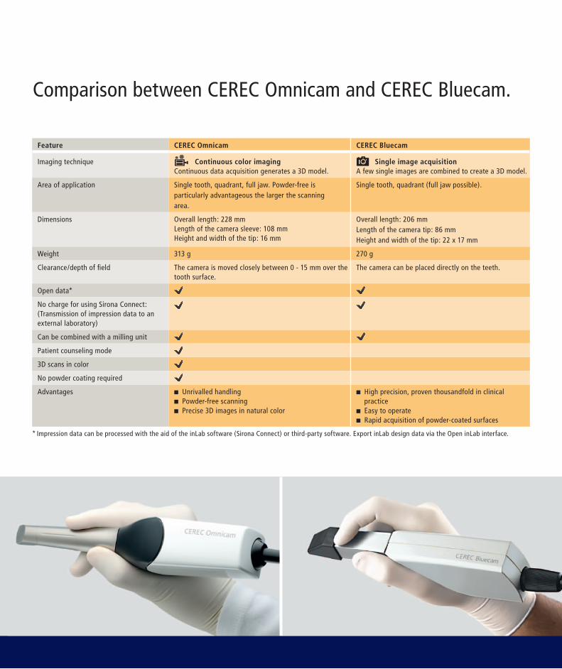
Comparison between CEREC Omnicam and CEREC Bluecam.
Feature CErEC omnicam CErEC Bluecam
Imaging technique Continuous color imagingContinuous data acquisition generates a 3D model.
Single image acquisitionA few single images are combined to create a 3D model.
Area of application Single tooth, quadrant, full jaw. Powder-free is particularly advantageous the larger the scanning area.
Single tooth, quadrant (full jaw possible).
Dimensions Overall length: 228 mmLength of the camera sleeve: 108 mmHeight and width of the tip: 16 mm
Overall length: 206 mmLength of the camera tip: 86 mmHeight and width of the tip: 22 x 17 mm
Weight 313 g 270 g
Clearance/depth of field The camera is moved closely between 0 - 15 mm over the tooth surface.
The camera can be placed directly on the teeth.
Open data*
No charge for using Sirona Connect:(Transmission of impression data to an external laboratory)
Can be combined with a milling unit
Patient counseling mode
3D scans in color
No powder coating required
Advantages n Unrivalled handlingn Powder-free scanningn Precise 3D images in natural color
n High precision, proven thousandfold in clinical practicen Easy to operaten Rapid acquisition of powder-coated surfaces
* Impression data can be processed with the aid of the inLab software (Sirona Connect) or third-party software. Export inLab design data via the Open inLab interface.
Subj
ect t
o te
chni
cal c
hang
es a
nd e
rror
s in
the
text
, Ord
er N
o. A
9110
0-M
43-B
610_
7600
, Prin
ted
in G
erm
any,
Disp
o-N
o. 0
4601
, JP1
2-01
38 W
S 07
126.
V0
CAD /CAM SYSTEMS | INSTRUMENTS | HYGIENE SYSTEMS | TREATMENT CENTERS | IMAGING SYSTEMS
Sirona – unique worldwide systems expertise in dental equipment productsSirona develops and manufactures a comprehensive range of dental equipment, including CAD/CAM Systems for dental practices (CEREC) and laboratories (inLab), Instruments and Hygiene Systems, Treatment Centers and Imaging Systems. Sirona manufactures high technology products that guarantee ease of use and a high return on investment – for the good of your practice and for the benefit of your patients. In this way, you can approach every challenge you face with confidence. Enjoy every day. With Sirona.
Sirona Dental Systems · E-mail: [email protected] · sirona.com
Be part of our dental community!sirona.com/social
See the new CEREC Omnicam in action www.sirona.com/Omnicam