Embed Size (px)
Citation preview
CEREBRAL BLOOD
CIRCULATION
Khaleel Alyahya, PhD, MEd King Saud University School of Medicine @khaleelya
OBJECTIVES
At the end of the lecture, students should be able to:
o List the cerebral arteries.
o Describe the cerebral arterial supply regarding the origin,
distribution and branches.
o Describe the arterial Circle of Willis .
o Describe the cerebral venous drainage and its termination.
o Describe arterial & venous vascular disorders and their clinical
manifestations.
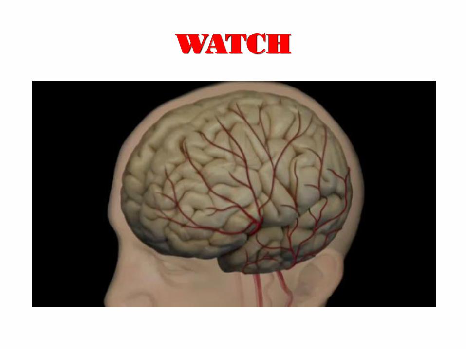
WATCH
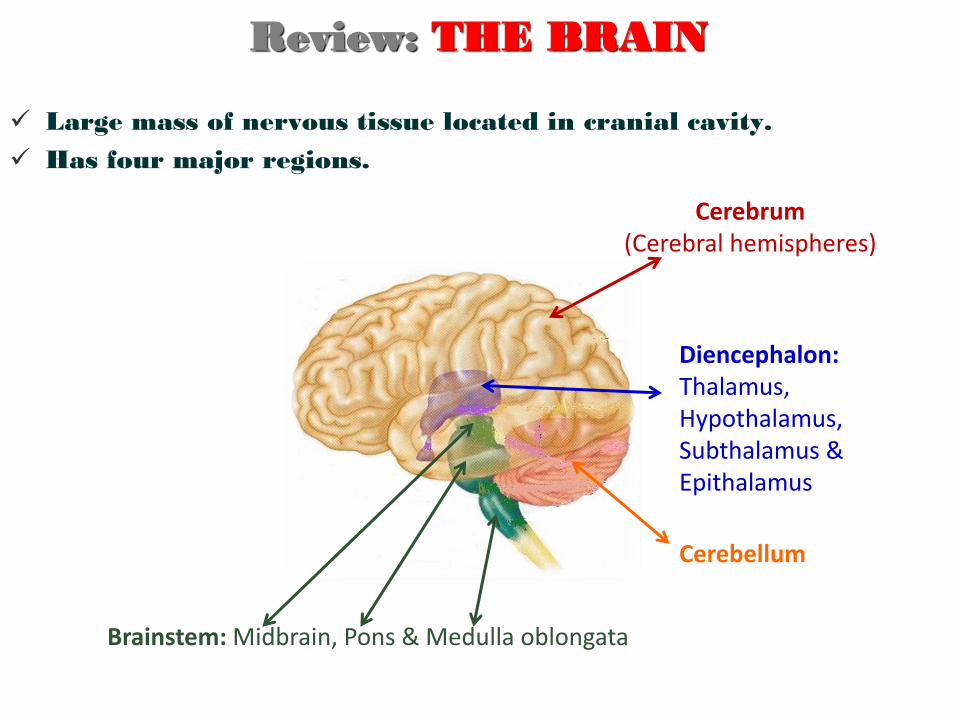
Large mass of nervous tissue located in cranial cavity.
Has four major regions.
Cerebrum (Cerebral hemispheres)
Cerebellum
Brainstem: Midbrain, Pons & Medulla oblongata
Diencephalon: Thalamus, Hypothalamus, Subthalamus & Epithalamus
Review: THE BRAIN
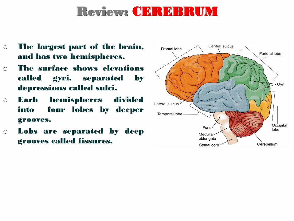
Review: CEREBRUM
o The largest part of the brain,
and has two hemispheres.
o The surface shows elevations
called gyri, separated by
depressions called sulci.
o Each hemispheres divided
into four lobes by deeper
grooves.
o Lobs are separated by deep
grooves called fissures.
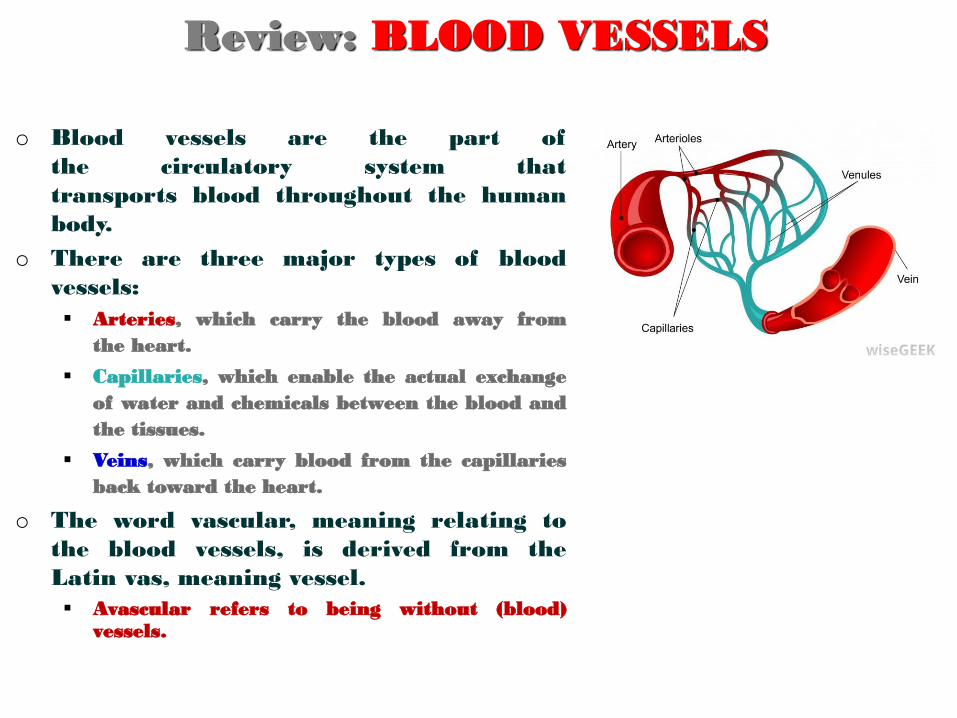
o Blood vessels are the part of
the circulatory system that
transports blood throughout the human
body.
o There are three major types of blood
vessels:
Arteries, which carry the blood away from
the heart.
Capillaries, which enable the actual exchange
of water and chemicals between the blood and
the tissues.
Veins, which carry blood from the capillaries
back toward the heart.
o The word vascular, meaning relating to
the blood vessels, is derived from the
Latin vas, meaning vessel.
Avascular refers to being without (blood) vessels.
Review: BLOOD VESSELS
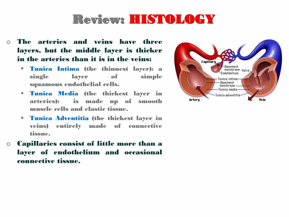
o The arteries and veins have three
layers, but the middle layer is thicker
in the arteries than it is in the veins:
Tunica Intima (the thinnest layer): a
single layer of simple
squamous endothelial cells.
Tunica Media (the thickest layer in
arteries): is made up of smooth
muscle cells and elastic tissue.
Tunica Adventitia (the thickest layer in
veins) entirely made of connective
tissue.
o Capillaries consist of little more than a
layer of endothelium and occasional
connective tissue.
Review: HISTOLOGY
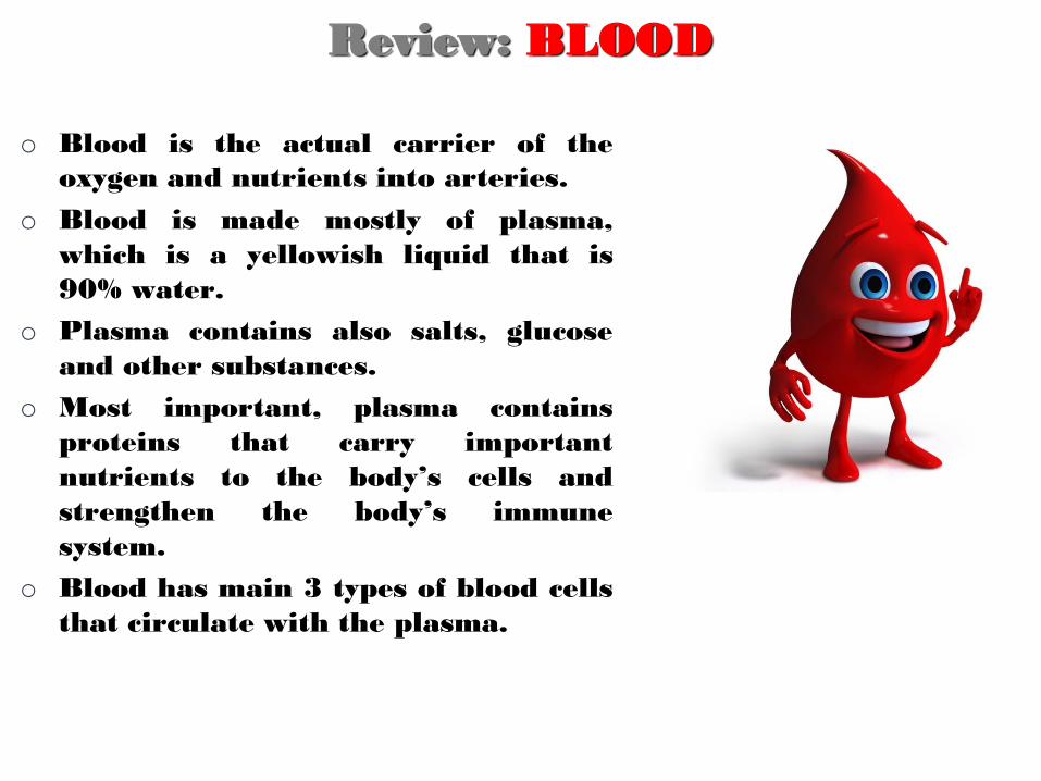
o Blood is the actual carrier of the
oxygen and nutrients into arteries.
o Blood is made mostly of plasma,
which is a yellowish liquid that is
90% water.
o Plasma contains also salts, glucose
and other substances.
o Most important, plasma contains
proteins that carry important
nutrients to the body’s cells and
strengthen the body’s immune
system.
o Blood has main 3 types of blood cells
that circulate with the plasma.
Review: BLOOD
CEREBRAL CIRCULATION
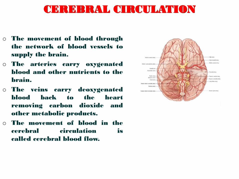
o The movement of blood through
the network of blood vessels to
supply the brain.
o The arteries carry oxygenated
blood and other nutrients to the
brain.
o The veins carry deoxygenated
blood back to the heart
removing carbon dioxide and
other metabolic products.
o The movement of blood in the
cerebral circulation is
called cerebral blood flow.
CEREBRAL ARTERIAL SUPPLY
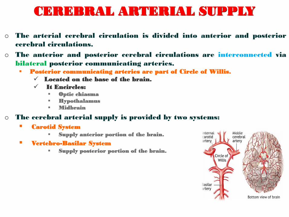
o The arterial cerebral circulation is divided into anterior and posterior
cerebral circulations.
o The anterior and posterior cerebral circulations are interconnected via
bilateral posterior communicating arteries. Posterior communicating arteries are part of Circle of Willis.
Located on the base of the brain.
It Encircles: Optic chiasma
Hypothalamus
Midbrain
o The cerebral arterial supply is provided by two systems:
Carotid System
Supply anterior portion of the brain.
Vertebro-Basilar System
Supply posterior portion of the brain.
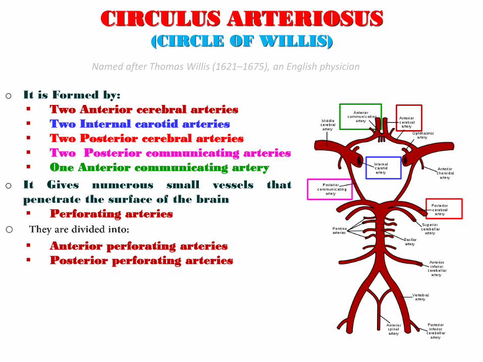
CIRCULUS ARTERIOSUS (CIRCLE OF WILLIS)
o It is Formed by:
Two Anterior cerebral arteries
Two Internal carotid arteries
Two Posterior cerebral arteries
Two Posterior communicating arteries
One Anterior communicating artery
o It Gives numerous small vessels that
penetrate the surface of the brain
Perforating arteries
o
Anterior perforating arteries
Posterior perforating arteries
Named after Thomas Willis (1621–1675), an English physician
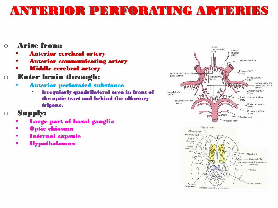
ANTERIOR PERFORATING ARTERIES
o Arise from: Anterior cerebral artery
Anterior communicating artery
Middle cerebral artery
o Enter brain through: Anterior perforated substance
• irregularly quadrilateral area in front of
the optic tract and behind the olfactory
trigone.
o Supply: Large part of basal ganglia
Optic chiasma
Internal capsule
Hypothalamus
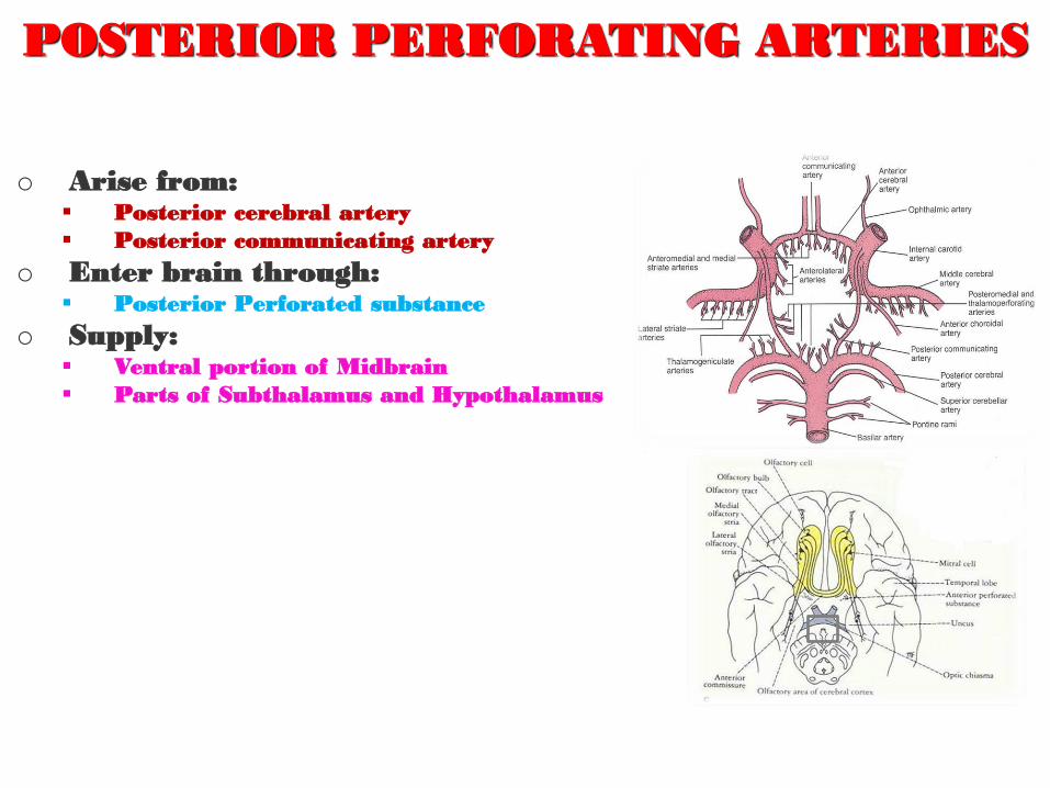
POSTERIOR PERFORATING ARTERIES
o Arise from: Posterior cerebral artery
Posterior communicating artery
o Enter brain through: Posterior Perforated substance
o Supply: Ventral portion of Midbrain
Parts of Subthalamus and Hypothalamus
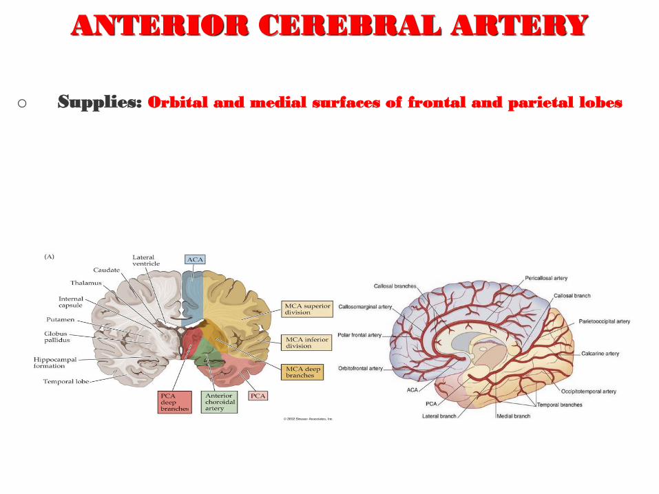
ANTERIOR CEREBRAL ARTERY
o Supplies: Orbital and medial surfaces of frontal and parietal lobes
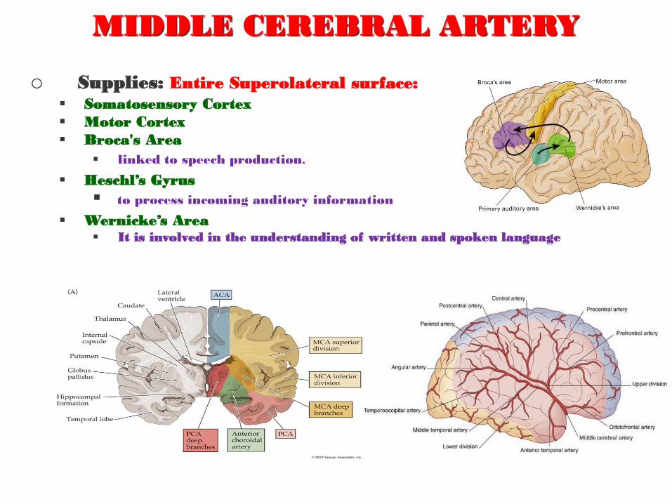
MIDDLE CEREBRAL ARTERY
o Supplies: Entire Superolateral surface:
Somatosensory Cortex
Motor Cortex
Broca's Area
linked to speech production
Heschl’s Gyrus
to process incoming auditory information
Wernicke’s Area It is involved in the understanding of written and spoken language
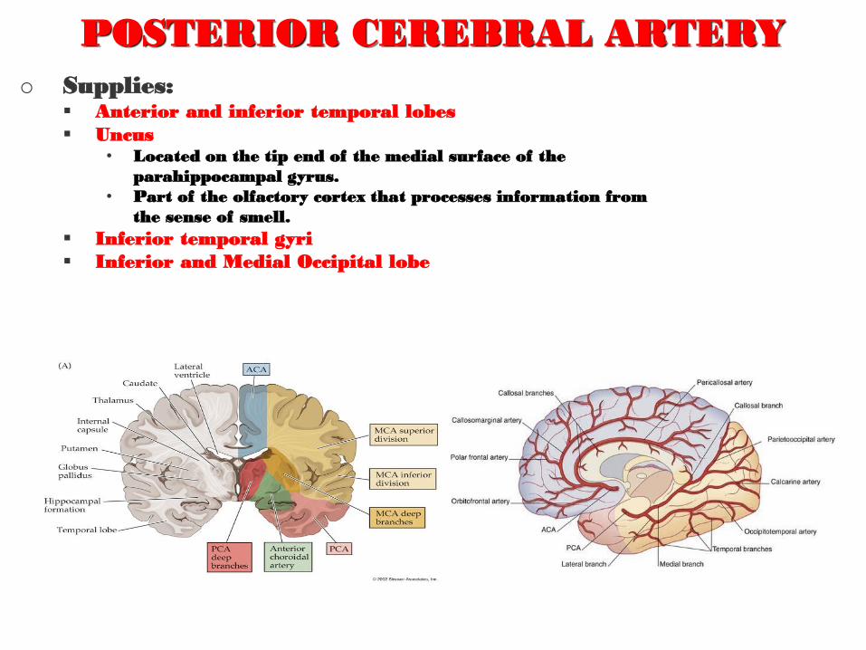
POSTERIOR CEREBRAL ARTERY
o Supplies: Anterior and inferior temporal lobes
Uncus • Located on the tip end of the medial surface of the
parahippocampal gyrus.
• Part of the olfactory cortex that processes information from
the sense of smell.
Inferior temporal gyri
Inferior and Medial Occipital lobe
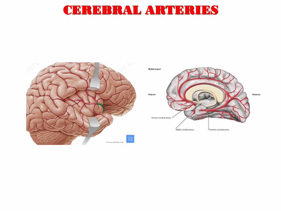
CEREBRAL ARTERIES
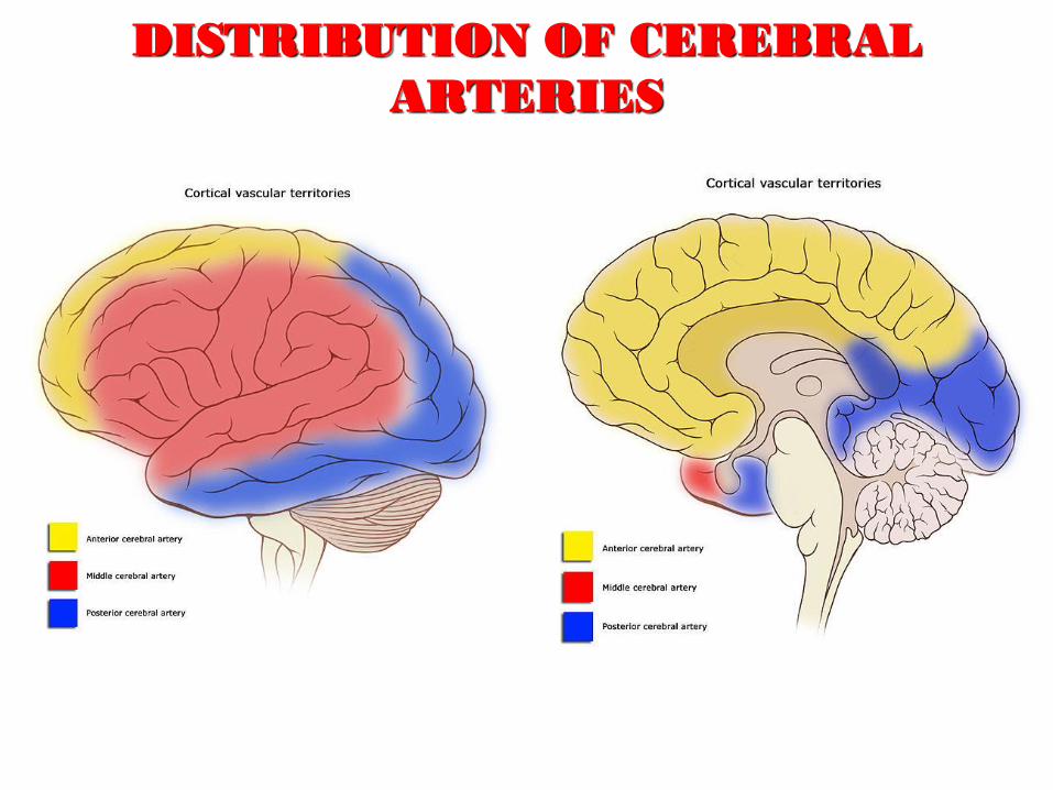
DISTRIBUTION OF CEREBRAL
ARTERIES
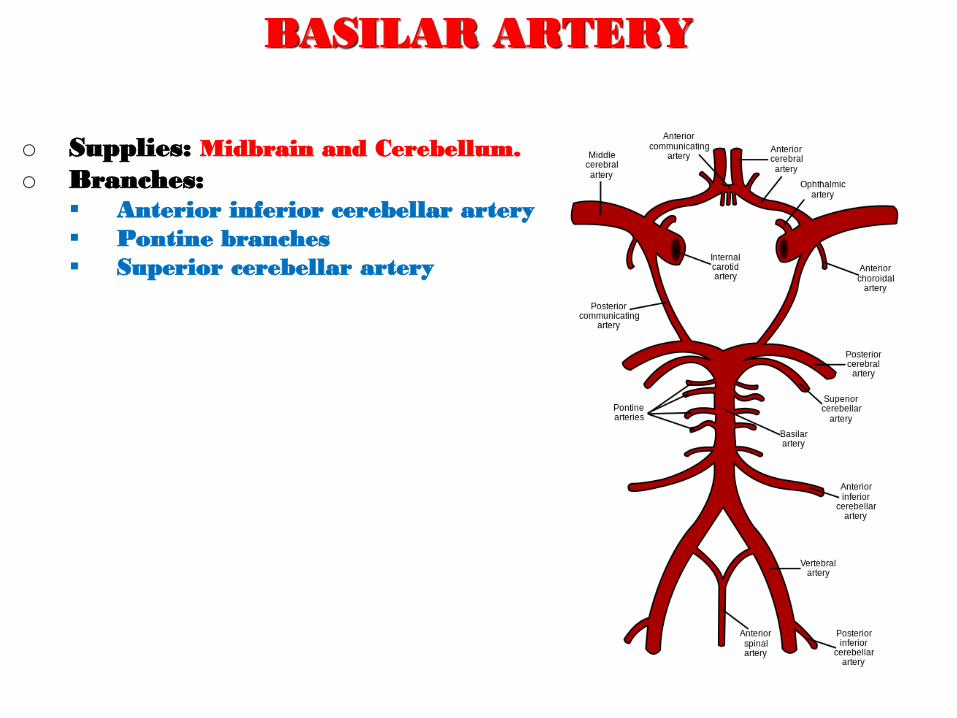
BASILAR ARTERY
o Supplies: Midbrain and Cerebellum.
o Branches: Anterior inferior cerebellar artery
Pontine branches
Superior cerebellar artery
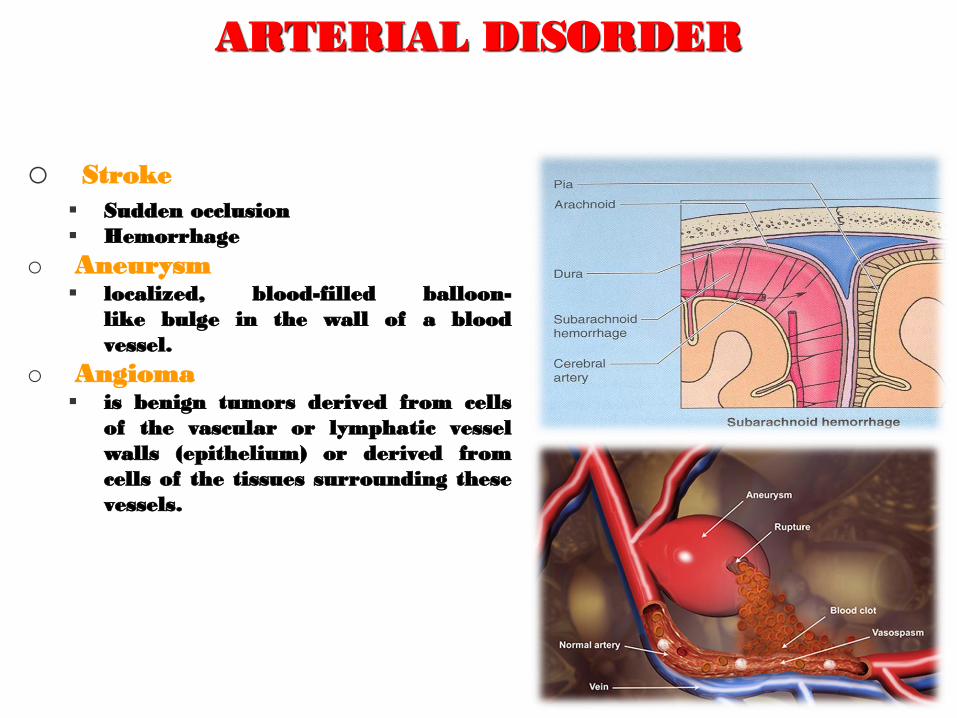
ARTERIAL DISORDER
o Stroke
Sudden occlusion
Hemorrhage
o Aneurysm localized, blood-filled balloon-
like bulge in the wall of a blood
vessel.
o Angioma is benign tumors derived from cells
of the vascular or lymphatic vessel
walls (epithelium) or derived from
cells of the tissues surrounding these
vessels.
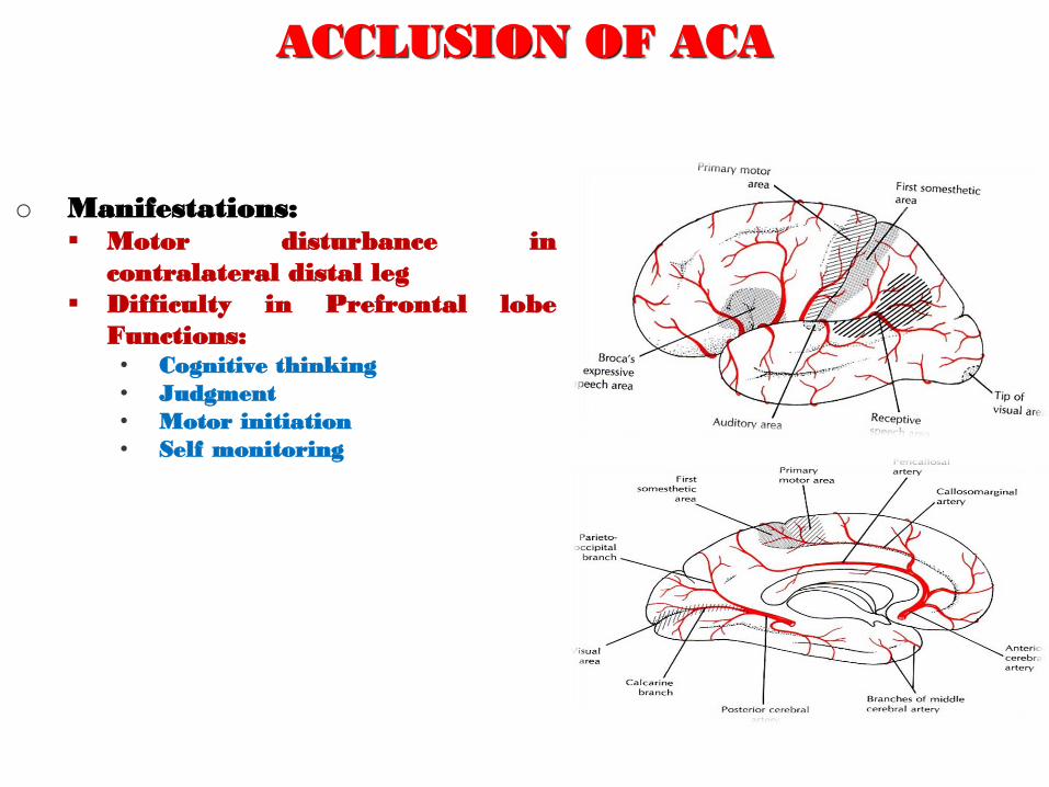
ACCLUSION OF ACA
o Manifestations: Motor disturbance in
contralateral distal leg
Difficulty in Prefrontal lobe
Functions: • Cognitive thinking
• Judgment
• Motor initiation
• Self monitoring
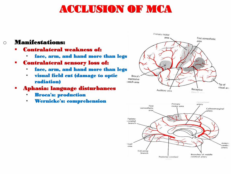
ACCLUSION OF MCA
o Manifestations: Contralateral weakness of:
• face, arm, and hand more than legs
Contralateral sensory loss of: • face, arm, and hand more than legs
• visual field cut (damage to optic
radiation)
Aphasia: language disturbances • Broca's: production
• Wernicke's: comprehension
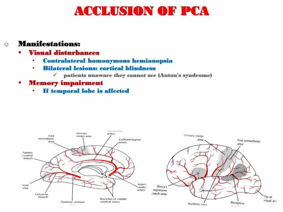
ACCLUSION OF PCA
o Manifestations: Visual disturbances
• Contralateral homonymous hemianopsia
• Bilateral lesions: cortical blindness patients unaware they cannot see (Anton's syndrome)
Memory impairment • If temporal lobe is affected
HOW WE ARE DOING ..?
o Which statement(s) of the following is NOT Wrong?
o Anterior cerebral arteries supply Broca's and Wernicke’s Area..!!
o Occlusion of MCA causes difficulty in Prefrontal lobe’s functions..!!
o Middle cerebral arteries are part of Willis Circle..!!
o Aneurysm is benign tumors derived from cells of
the vascular or lymphatic vessel walls..!!
o Posterior cerebral arteries supply anterior and inferior temporal
lobes..!!
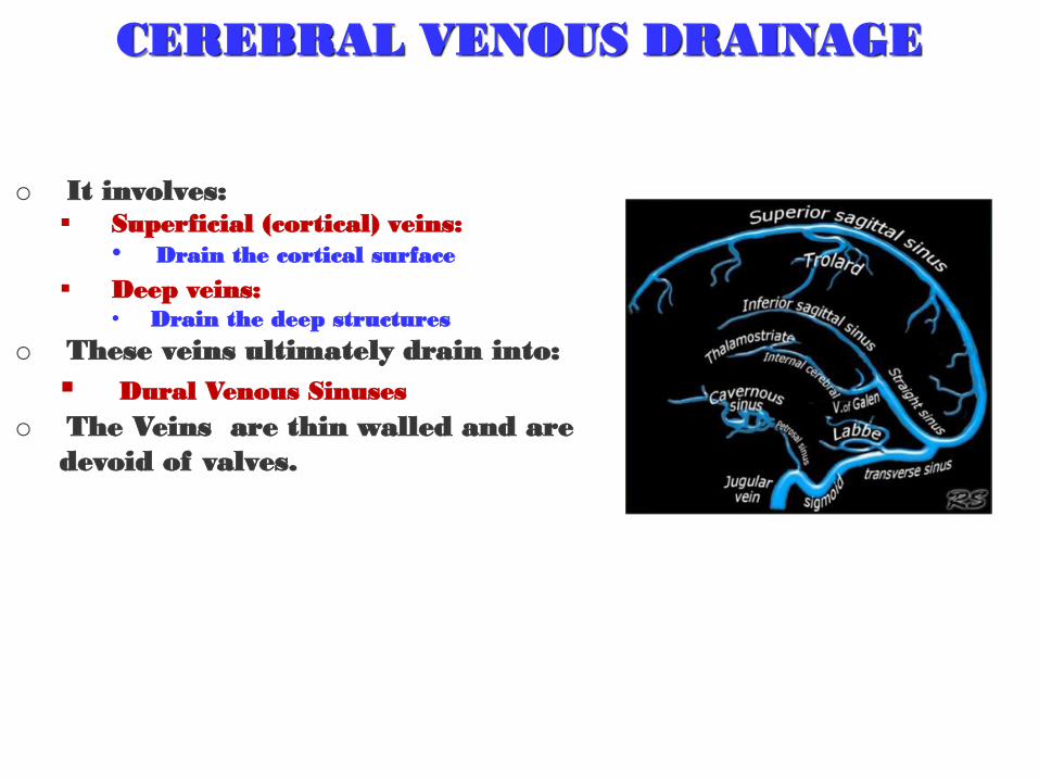
CEREBRAL VENOUS DRAINAGE
o It involves: Superficial (cortical) veins:
• Drain the cortical surface
Deep veins: • Drain the deep structures
o These veins ultimately drain into:
Dural Venous Sinuses
o The Veins are thin walled and are
devoid of valves.
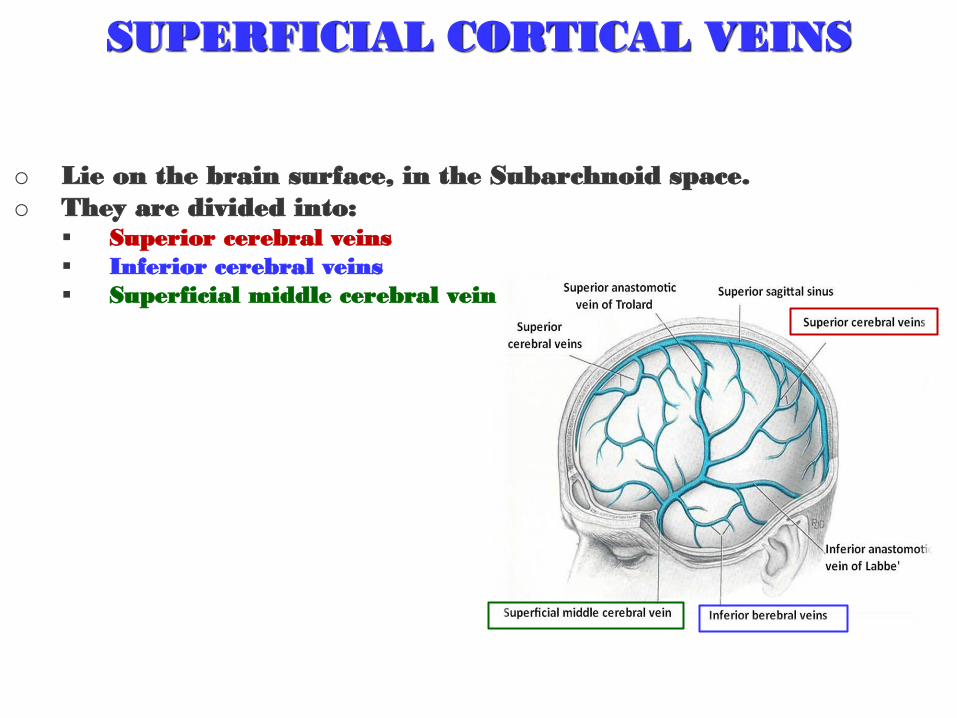
SUPERFICIAL CORTICAL VEINS
o Lie on the brain surface, in the Subarchnoid space.
o They are divided into: Superior cerebral veins
Inferior cerebral veins
Superficial middle cerebral vein
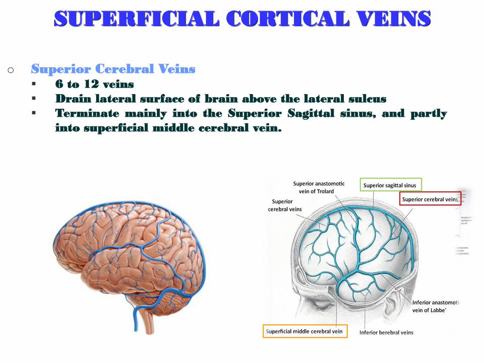
SUPERFICIAL CORTICAL VEINS
o Superior Cerebral Veins 6 to 12 veins
Drain lateral surface of brain above the lateral sulcus
Terminate mainly into the Superior Sagittal sinus, and partly
into superficial middle cerebral vein.
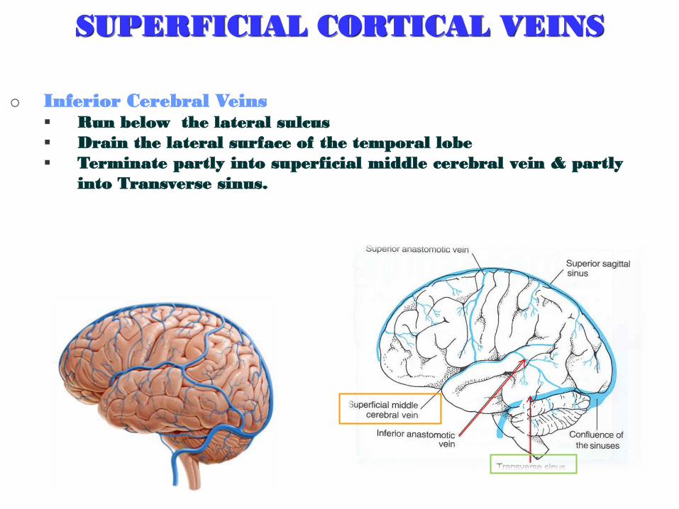
SUPERFICIAL CORTICAL VEINS
o Inferior Cerebral Veins Run below the lateral sulcus
Drain the lateral surface of the temporal lobe
Terminate partly into superficial middle cerebral vein & partly
into Transverse sinus.
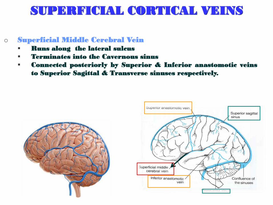
SUPERFICIAL CORTICAL VEINS
o Superficial Middle Cerebral Vein Runs along the lateral sulcus
Terminates into the Cavernous sinus
Connected posteriorly by Superior & Inferior anastomotic veins
to Superior Sagittal & Transverse sinuses respectively.
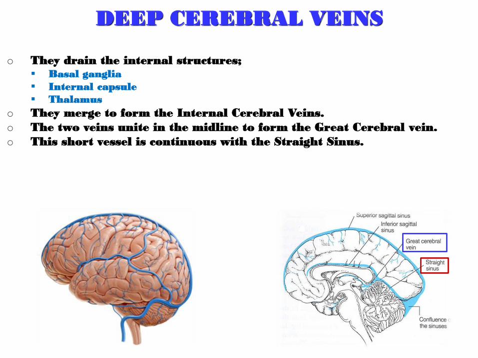
DEEP CEREBRAL VEINS
o They drain the internal structures; Basal ganglia
Internal capsule
Thalamus
o They merge to form the Internal Cerebral Veins.
o The two veins unite in the midline to form the Great Cerebral vein.
o This short vessel is continuous with the Straight Sinus.
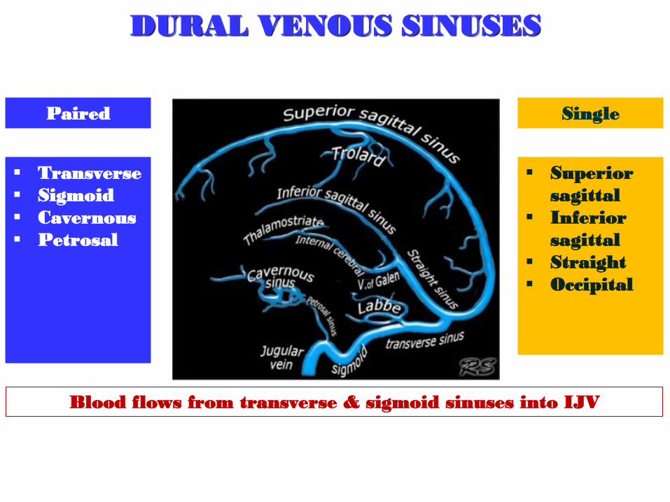
DURAL VENOUS SINUSES
Superior
sagittal
Inferior
sagittal
Straight
Occipital
Single
Transverse
Sigmoid
Cavernous
Petrosal
Paired
Blood flows from transverse & sigmoid sinuses into IJV
WATCH
VENOUS DISORDER
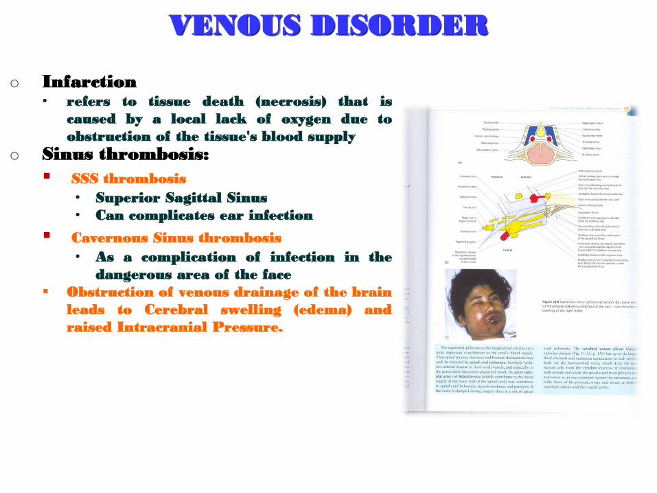
o Infarction • refers to tissue death (necrosis) that is
caused by a local lack of oxygen due to
obstruction of the tissue's blood supply
o Sinus thrombosis:
SSS thrombosis
• Superior Sagittal Sinus
• Can complicates ear infection
Cavernous Sinus thrombosis
• As a complication of infection in the
dangerous area of the face
Obstruction of venous drainage of the brain
leads to Cerebral swelling (edema) and
raised Intracranial Pressure.
ALSO, HOW WE ARE DOING ..?
o Which statement(s) of the following is Wrong?
1. Superior Cerebral Veins terminate mainly into the Superior Sagittal
sinus, and partly into superficial middle cerebral vein..!!
2. Infarction refers to tissue death (necrosis)..!!
3. Superior Cerebral Veins drain lateral surface of brain above the
lateral sulcus..!!
4. Inferior Cerebral Veins terminate partly into superficial middle
cerebral vein & partly into Transverse sinus..!!
5. Superficial Middle Cerebral Vein drains the lateral surface of the
temporal lobe..!!

QUESTION?


![Aktueller Stand der neurochirurgischen Therapie der ... · Circulus arteriosus Willisii -communicans anterior Aneurysma) [34] oder im Stromgebiet eines arterio-venösen Angioms (de](https://img.dokumen.tips/doc/110x75/5c9ebfb688c993502d8c10e4/aktueller-stand-der-neurochirurgischen-therapie-der-circulus-arteriosus.jpg)