Embed Size (px)
Citation preview

Int J Anat Res 2016, 4(2):2245-50. ISSN 2321-4287 2245
Original Research Article
DIAMETER OF ANTERIOR CEREBRAL ARTERY ON MRIANGIOGRAMSNavita Aggarwal *1, Molly M. Paul 2, Madhumita Mukherjee 3.
ABSTRACT
Address for Correspondence: Dr. Navita Aggarwal, Associate Professor, Department of Anatomy,Adesh Institute of Medical Sciences and Research, Bathinda, Punjab, India.Mobile no.: +919855424777 E-Mail: [email protected]
Background: Magnetic Resonance Imaging, by far, has been found to be the most sensitive and non-invasivemethod for detecting angiographic images on the circle of Willis. Arteries forming parts of circle of Willisfrequently vary in size. The haemodynamics of the circle of Willis is influenced by variations in the caliber of thesegments of the anterior and posterior cerebral arteries and their communicating arteries, thus affecting themajor role of the circulus arteriosus as an anastomotic channel.Materials and Methods: In the present study the diameter of A1 segment of anterior cerebral artery, forming theanterior part of the circle of Willis, is measured in the brains on MRI angiograms at two points ‘A’ and ‘B’respectively.Results: The left anterior cerebral artery at point ‘A’ has a larger diameter than the right, being 3.20mm and2.72mm respectively. In males and females both the left anterior cerebral artery at point ‘A’ has a larger diameterthan the right anterior cerebral artery, being 3.19mm and 2.86mm respectively in males and 3.21mm and 2.57mmrespectively in females. The diameter of anterior cerebral artery on MRI angiograms at point ‘B’ is also larger onleft side as compared to right side being 2.50mm and 2.31mm respectively. In males and females both it is largeron left side than on right side being 2.70mm and 2.42mm respectively in males and 2.30mm and 2.20mmrespectively in females.Discussion: The diameter was found to be higher on the left side than on right side and in males than in females.In view of this, the diameter which is presented here may provide reference values which will be specific to thethree dimensional time of flight MRI angiography.KEY WORDS: MRI angiograms, Diameter, Anterior cerebral artery.
INTRODUCTION
International Journal of Anatomy and Research,Int J Anat Res 2016, Vol 4(2):2245-50. ISSN 2321-4287
DOI: http://dx.doi.org/10.16965/ijar.2016.189
Access this Article online
Quick Response code Web site:
Received: 04 Apr 2016 Accepted: 18 Apr 2016Peer Review: 05 Apr 2016 Published (O): 30 Apr 2016Revised: None Published (P): 30 Apr 2016
International Journal of Anatomy and ResearchISSN 2321-4287
www.ijmhr.org/ijar.htm
DOI: 10.16965/ijar.2016.189
*1 Assosciate Professor, Department of Anatomy, Adesh Institute of Medical Sciences and Research,Bathinda, Punjab, India.2 Professor, Department of Anatomy, Christian Medical College and Hospital, Ludhiana, Punjab,India.3 Assistant Professor, Department of Anatomy, Christian Medical College and Hospital, Ludhiana,Punjab, India.
sides, complete the circle of Willis anteriorlythrough an anastomosis between them -theanterior communicating artery (A Com A.). The
The Anterior Cerebral Artery (ACA) , a branch ofthe internal carotid artery on both right and left
Int J Anat Res 2016, 4(2):2245-50. ISSN 2321-4287 2246
Navita Aggarwal, Molly M. Paul, Madhumita Mukherjee. DIAMETER OF ANTERIOR CEREBRAL ARTERY ON MRI ANGIOGRAMS.
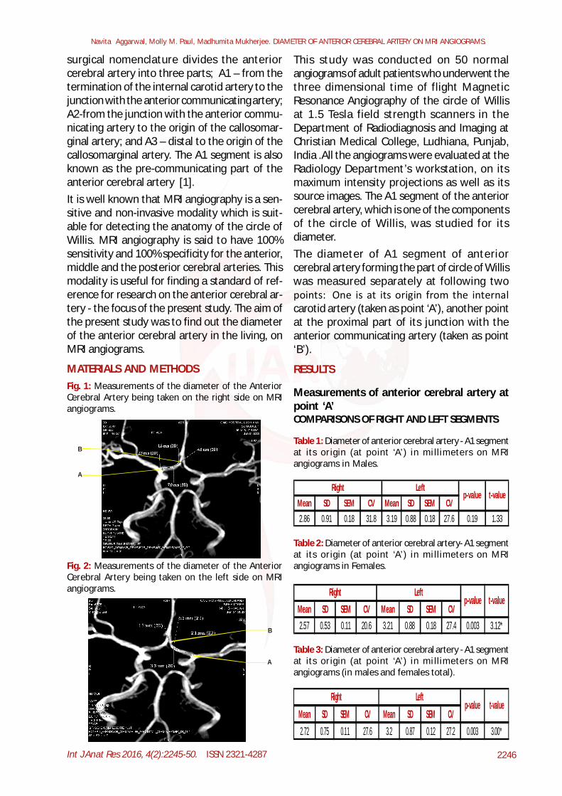
surgical nomenclature divides the anteriorcerebral artery into three parts; A1 – from thetermination of the internal carotid artery to thejunction with the anterior communicating artery;A2-from the junction with the anterior commu-nicating artery to the origin of the callosomar-ginal artery; and A3 – distal to the origin of thecallosomarginal artery. The A1 segment is alsoknown as the pre-communicating part of theanterior cerebral artery [1].It is well known that MRI angiography is a sen-sitive and non-invasive modality which is suit-able for detecting the anatomy of the circle ofWillis. MRI angiography is said to have 100%sensitivity and 100% specificity for the anterior,middle and the posterior cerebral arteries. Thismodality is useful for finding a standard of ref-erence for research on the anterior cerebral ar-tery - the focus of the present study. The aim ofthe present study was to find out the diameterof the anterior cerebral artery in the living, onMRI angiograms.
MATERIALS AND METHODS
This study was conducted on 50 normalangiograms of adult patients who underwent thethree dimensional time of flight MagneticResonance Angiography of the circle of Willisat 1.5 Tesla field strength scanners in theDepartment of Radiodiagnosis and Imaging atChristian Medical College, Ludhiana, Punjab,India .All the angiograms were evaluated at theRadiology Department’s workstation, on itsmaximum intensity projections as well as itssource images. The A1 segment of the anteriorcerebral artery, which is one of the componentsof the circle of Willis, was studied for itsdiameter.The diameter of A1 segment of anteriorcerebral artery forming the part of circle of Williswas measured separately at following twopoints: One is at its origin from the internalcarotid artery (taken as point ‘A’), another pointat the proximal part of its junction with theanterior communicating artery (taken as point‘B’).
Fig. 1: Measurements of the diameter of the AnteriorCerebral Artery being taken on the right side on MRIangiograms.
A
B
Fig. 2: Measurements of the diameter of the AnteriorCerebral Artery being taken on the left side on MRIangiograms.
B
A
RESULTS
Measurements of anterior cerebral artery atpoint ‘A’COMPARISONS OF RIGHT AND LEFT SEGMENTS
Table 1: Diameter of anterior cerebral artery - A1 segmentat its origin (at point ‘A’) in mi llimeters on MRIangiograms in Males.
Mean SD SEM CV Mean SD SEM CV2.86 0.91 0.18 31.8 3.19 0.88 0.18 27.6 0.19 1.33
Right Leftp-value t-value
Table 2: Diameter of anterior cerebral artery- A1 segmentat its origin (at point ‘A’) in mi llimeters on MRIangiograms in Females.
Mean SD SEM CV Mean SD SEM CV2.57 0.53 0.11 20.6 3.21 0.88 0.18 27.4 0.003 3.12*
LeftRightp-value t-value
Table 3: Diameter of anterior cerebral artery - A1 segmentat its origin (at point ‘A’) in mi llimeters on MRIangiograms (in males and females total).
Mean SD SEM CV Mean SD SEM CV2.72 0.75 0.11 27.6 3.2 0.87 0.12 27.2 0.003 3.00*
Right Leftp-value t-value

Int J Anat Res 2016, 4(2):2245-50. ISSN 2321-4287 2247
Navita Aggarwal, Molly M. Paul, Madhumita Mukherjee. DIAMETER OF ANTERIOR CEREBRAL ARTERY ON MRI ANGIOGRAMS.
COMPARISON WITH REGARDS TO SIDEGraph 1: Comparison of diameter of anterior cerebralartery – A1 segment at its orgin (point ‘A’).
0
1
2
3
4
Males MRI Females MRI Total MRI
in m
m
Right
Left
COMPARISON WITH REGARDS TO SEX
01234
Right Left
in m
m
Males
Females
Graph 2: Comparison of diameter of anterior cerebralartery – A1 segment at its origin (point ‘A’).
Measurements of anterior cerebral arteryat point ‘B’COMPARISON OF RIGHT AND LEFT SEGMENTS
Table 4: Diameter of anterior cerebral artery - A1 segmentat proximal part of its junction with anteriorcommunicating artery (at point ‘B’) in millimeters onMRI angiograms in Males.
Mean SD SEM CV Mean SD SEM CV2.42 0.85 0.17 35.1 2.7 0.95 0.19 35.2 0.28 1.08
Right Leftp-value t-value
Table 5: Diameter of anterior cerebral artery- A1 segmentat proximal part of its junction with anteriorcommunicating artery (at point ‘B’) in millimeters onMRI angiograms in Females.
Mean SD SEM CV Mean SD SEM CV2.2 0.55 0.11 25 2.3 0.82 0.16 35.7 0.62 0.49
LeftRightp-value t-value
Table 6: Diameter of anterior cerebral artery- A1 segmentat proximal part of its junction with anteriorcommunicating artery (at point ‘B’) in millimeters onMRI angiograms (in males and females total).
Mean SD SEM CV Mean SD SEM CV2.31 0.72 0.1 31.2 2.5 0.82 0.12 32.8 0.25 1.15
Right Leftp-value t-value
Comparison with regards to sideGraph 3: Comparison of diameter of anterior cerebralartery - A1 segment at proximal part of its junction withanterior communicating artery (point ‘B’).
0
1
2
3
4
Males MRI Females MRI Total MRI
in m
m Right Left
COMPARISON WITH REGARDS TO SEXGraph 4: Comparison of diameter of anterior cerebralartery-A1 segment at proximal part of its junction withanterior communicating artery (point ‘B’).
0
1
2
3
4
Right Left
in m
m
Males Females
DISCUSSION
On MRI angiograms the left anterior cerebralartery at point ‘A’ has a larger diameter than theright anterior cerebral artery at point ‘A’, 3.20mmand 2.72mm respectively, the difference being0.48mm. The left is significantly larger than theright and the difference has a highly significantp-value. In males the left anterior cerebralartery at point ‘A’ has a larger diameter than theright anterior cerebral artery at point ‘A’, 3.19mmand 2.86mm respectively, the difference being0.33mm, which is non-significant. In the femalesthe left anterior cerebral artery at point ‘A’ has

Int J Anat Res 2016, 4(2):2245-50. ISSN 2321-4287 2248
Navita Aggarwal, Molly M. Paul, Madhumita Mukherjee. DIAMETER OF ANTERIOR CEREBRAL ARTERY ON MRI ANGIOGRAMS.
a larger diameter than the right anteriorcerebral artery at point ‘A’, 3.21mm and 2.57mmrespectively, the difference being 0.64mm. Theleft being larger in diameter is a significantdifference from right with a significant p-value.The diameter of anterior cerebral artery on MRIangiograms at point ‘B’ (in all the cases takentogether) is larger on left side as compared toright side being 2.50mm and 2.31mm respec-tively. The difference of diameter on left andright side is 0.19mm, which is non-significant.The diameter of anterior cerebral artery at point‘B’ in the males is larger on left side than onright side. Diameter on left is 2.70mm whereasthat on right side it is 2.42mm. The diameter ofanterior cerebral artery at point ‘B’ in thefemale is larger on left side than on right side.Diameter on left is 2.30mm whereas that on rightside it is 2.20mm.According to Vohra et al (2006) [2] thediameter of anterior cerebral artery on MRIangiograms was found to range from 1.0 - 2.5mmand the mean diameter being 1.72 ± 0.45mm.The value is less as compared to that in presentstudy.Hartkamp et al (1998) [3] made followingobservations for the mean vessel diameter(in mm) according to age and sex:Table 7: Hartkamp et al (1998) [3] observations forthe mean vessel diameter (in mm) according to ageand sex:
the diameter on left side is larger than the rightside and that a statistically significant p-valueexists for the difference in diameter on the twosides.Pai et al (2005), Mandiola et al (2007), Kamath(1981) observed that diameters on left aregreater than right but they could not find asignificant difference between the two sides.Khade et al. (2008) [12] noted a statisticallysignificant difference of diameter of anteriorcerebral artery on right and left side in females.According to Kapoor et al (2008) [13] diameterand length of anterior cerebral artery do not showany differences on right and left side.According to Murray (1964) right side has largerdiameters than left side which is contrary to thefindings in the present study.It has been observed in the present study thatthe males have higher values for diameter ascompared to females in general, but thedifference is insignificant.This observation in present study is accordancewith observation by Orlandini et al (1985) andKapoor et al (2008) in cadaveric brains.Hartkamp et al (1998) who measured diameterof anterior cerebral artery on MRI angiogramsalso found that males have larger diameters thanfemales, but the difference is insignificant.
Male Female Male FemaleRight 2.3 2.3 1.9 1.8Left 2.2 2.2 1.8 1.7
Subjects aged 20-25 years
Subjects aged 60-85 years
The values observed by him are less on bothright and left sides as compared to those in thepresent study.The diameter of ACA has been measured bymany others but on cadaveric specimens of brainwith intact ACA.In the present study it has been observed thatthe diameters on left side are slightly larger bothat point ‘A’ and ‘B’ than that on the right side.The difference of diameter on right and left sidehas shown a significant p-value at point ‘A’. Thisfinding is in accordance with findings by:Orlandini et al (1985) [11] according to them
Table 8: Diameters of A1 segment given by variousauthors on cadaveric specimens of brain.
Author Year Diameter On Right Side Diameter On Left Side
Murray [4] 1964 1.475mm 1.425mm
Perlmutter and Rhoton[5] 1976 2.6mm 2.6mm
Kamath[6] 1981 2.2mm + 0.6mm 2.4mm + 0.05mm
Gomes et al[7] 1986 2.3mm + 1.0mm 2.5mm + 1.0mm
Stefani et al[8] 2000 2.61mm + 0.34mm 2.61mm + 0.34mm
Pai et al[9] 2005 2.8mm 2.9mm
Vohra et al[2] 2006 1.44 mm + 0.42 mm 1.44 mm + 0.42 mm
Mandiola et al[10] 2007 2.37mm + 0.68mm 2.42mm + 0.75mm
Among the studies reviewed for this presentwork, the diameter of anterior cerebralartery-A1 segment at proximal of its junctionwith anterior communicating artery that is atpoint B was not quoted by any of the authors. Ithas been measured in the present study becausethis site of anterior cerebral artery, that is the

Int J Anat Res 2016, 4(2):2245-50. ISSN 2321-4287 2249
anterior communicating artery-anterior cerebralartery complex, is the commonest site foroccurrence of aneurysms. Therefore it is a newaddition in the present work for studies onanterior cerebral artery.The range of diameter at point ‘B’ on right sideis 0.85mm – 4.52mm, the mean diameter being2.31mm ± 0.72mm whereas, on the left side therange is 1 mm - 5.13mm and the mean diameteris 2.50mm ± 0.82m.
CONCLUSION
From all the published work it has beenconcluded that the circle of Willis is essentialfor the maintenance of a stable and a constantblood flow to the brain. Any changes in itsmorphology may condition the appearance andthe severity of syndromes of vascularinsufficiency [14]. It must be emphasized that awider range of information on the size of theconsidered artery may be useful for a betterinterpretation of angiographic images and fordeeper understanding of cerebral pathology[11].Data obtained can provide precise microanat-omic information for surgical treatment ofaneurysm or vascular reconstructive proceduresin circle of Willis . Also the anatomic param-eters of anterior cerebral artery may be used toplan and design devices such as angiographicmicrocatheters and guides used in endovascularprocedures [8].Abnormal narrowing of vessels was a commonoccurrence on the right side than on the left inthis study. This may be related to the need for abetter blood supply to the left hemisphere. Thisis because of dominance of left cerebral hemi-sphere in most of the population [3] related tothe handedness of the person.The present study gives diameters of anteriorcerebral artery and their difference accordingto side and sex. In view of this, the morphologicvariation and diameters presented here mayprovide reference values specific to three di-mensional time of flight MRI angiography, aswell as be of value in studies in which three-dimensional time of flight MRI angiography isused to investigate other pathologic features ofthe circle of Wills.
ACKNOWLEDGEMENTSAcknowledge with deep gratitude the people inthe department of Radiology and in departmentof Anatomy, at CMC, Ludhian for theirsuggestions and support rendered to me incompletion of this work. I would like to thankthe MRI technicians for the long hours that theydevoted for helping me in the data collectionfor this study.
Conflicts of Interests: None
REFERENCES
[1]. Standring S, Ellis H, Healy JC, Johnson D, William A,Collins P, et al. Development of nervous system. In:.Gray’s Anatomy. 39th edn. Spain: Elsevier; 2005.241-274.
[2]. Vohra H, Singh P, Sood V. Study of circle ofWillis - anterior part. J Anat Soc India 2006;55:74.
[3]. Hartkamp K, Grond JV, Leeuw FE, Groot JC, Algra A,Hillen B, et al. Circle of Willis: Morphologicvariation on three-dimensional time-of-flight MRangiograms. Radiology 1998; 207:103-11.
[4]. Murray KD. Dimensions of the circle of Willis anddynamic studies using electrical analogy. JNeurosurg 1964; 21:26-34.
[5]. Perlmutter D, Rhoton AL. Microsurgical anatomy ofthe anterior cerebral–anterior communicatingrecurrent artery complex. J Neurosurg 1976;45:259-72.
[6]. Kamath S. Observations on the length and diameterof vessels forming the circle of Willis. J Anat 1981;133:419-23.
[7]. Gomes FB, Dujovny M, Umansky F, Berman SK, DiazFG, Ausman JI, et al. Microanatomy of the anteriorcerebral artery. Surg Neurol 1986;26:129-41.
[8]. Stefani MA, Schneider FL, Marrone ACH, Severino AG,Jackowski AP, Wallace MC. Anatomic variations ofanterior cerebral artery cortical branches. ClinicalAnatomy 2000;13:231-36.
[9]. Pai SB, Kulkarni RN, Varma RG. Microsurgicalanatomy of the anterior cerebral artery-anteriorcommunicating artery complex: An Indian study.Neurology Asia 2005;10:21-8.
[10]. Mandiola E, Alarcon E, Onate JC, Sanhueza P, Sol M,Olave E. Biometrical aspects of the anteriorcerebral artery in its proximal segment (A1) andinternal carotid artery. Int J Morphol 2007;25:915-18.
[11]. Orlandini GE, Ruggiero C, Orlandini SZ, Gulisano M.Blood vessel size of circulus arteriosus cerebri(circle of Willis): A statistical research on 100 hu-man subjects. Acta Anat 1985;123:72-6.
[12]. Khade AM, Sawant VG, Champaneri PC. Measure-ment of diameter of blood vessels forming circle ofWillis and their variations by magnetic resonanceimaging angiography. J Anat Soc India 2008; 57:53-89.
Navita Aggarwal, Molly M. Paul, Madhumita Mukherjee. DIAMETER OF ANTERIOR CEREBRAL ARTERY ON MRI ANGIOGRAMS.

Int J Anat Res 2016, 4(2):2245-50. ISSN 2321-4287 2250
[13]. Kapoor K, Singh B, Dewan IJ. Variations in the con-figuration of the circle of Willis. Anat Sci Int 2008;83: 96-106.
[14].Puchades-Orts A, Nombela-Gomez M, Ortuno-Pacheco G. Variation in form of circle of Willis :some anatomical and embryological consider-ations. Anat Rec 1976;185:119-123.
How to cite this article:Navita Aggarwal, Molly M. Paul, Madhumita Mukherjee.DIAMETER OF ANTERIOR CEREBRAL ARTERY ON MRIANGIOGRAMS. Int J Anat Res 2016;4(2):2245-2250. DOI:10.16965/ijar.2016.189
Navita Aggarwal, Molly M. Paul, Madhumita Mukherjee. DIAMETER OF ANTERIOR CEREBRAL ARTERY ON MRI ANGIOGRAMS.