Embed Size (px)
Citation preview
Use of Transcranial Doppler for Monitoring Hepatic EncephalopathyCacciatori A1,2*, Castelli J1,3 and Grecco G3
1Central Hospital of the Armed Forces, Uruguay2National Institute of Donations and Transplants, Uruguay3Bi-instutional unit of the Hospital de Clínicas and the Central Hospital of the Armed Forces, Uruguay*Corresponding author: Armando Cacciatori Castro, Medical Doctor in chief of the ICU, Central Hospital of the Armed Forces, Jaime Zudánez 2768, apto. 1001,Uruguay; Tel: 2710 02 9, PC: 1300; E-mail: [email protected]
Received date: February 23, 2015; Accepted date: March 30, 2015; Published date: April 06, 2015
Copyright: ©2015 Cacciatori A. This is an open-access article distributed under the terms of the Creative Commons Attribution License, which permits unrestricted use,distribution, and reproduction in any medium, provided the original author and source are credited.
Abstract
Fulminant hepatic failure (FHF) is an acute and severe disease that can lead to death by the development ofencephalopathy. Due to the severe alterations of coagulation that go along with this clinical picture, it may becontraindicated to place a sensor for monitoring intracranial pressure (ICP); it is for this reason that the option of thetranscranial doppler (TCD) arises as an auxiliary method for monitoring these patients. The aim of this work was topresent our first steps in the implementation of this technique in an intensive care unit (ICU) which is a referencecenter in liver transplant. Two clinical cases of young women, carriers of FHF with severe encephalopathy, wereanalyzed. The daily monitoring of these patients allowed us to identify changes in intracranial hemodynamics,expressed by “sistolized” patterns in the flow rates of cerebral arteries, coinciding with increases in the pulsatilityindex (PI) and the resistance index (RI). These findings suggest that an increased ICP coincides with thosepublished in other series analyzed. Monitoring of hepatic encephalopathy by TCD is useful, becoming a methodwhich is being implemented more and more every day in neurointensive treatment.
Keywords: Transcranial doppler; Liver failure; Liverencephalopathy; Liver transplant
IntroductionTranscranial doppler (TCD) is an auxiliary method which is useful
for measuring cerebral blood flow velocities, integratingneuromonitoring techniques in the intensive care unit (ICU).
It is a non-invasive, inexpensive, and simple to perform method,which does not require the patient to be transferred from the ICU toanother hospital sector and can be done at the foot of the bed.
The fulminant hepatic failure (FHF) is a condition characterized bya rapid deterioration of the liver function, which is accompanied by asevere impairment of the neurological status in previously healthypatients. These changes are caused by the necrosis of hepatocytes.Brain damage is caused by an increase in intracranial pressure (ICP)with consequent cerebral ischemia. This fact is very important becausebrain damage constitutes a criterion for deciding liver transplantation.
ObjectiveTo present the utility of DTC as a neuromonitoring technique to
monitor hepatic encephalopathy, knowing that crasis disorders that goalong with hepatic failure may be contraindicative of the ICP sensorplacement.
Materials and MethodsAnalysis of two patients that were admitted to the ICU of the
Central Hospital of the Armed Forces, because of a severe and acuteliver failure with encephalopathy. Both developed into a coma, with
reactivity in extensive-pronation, requiring artificial airway andmechanical respiratory assistance (MRA).
Tomographic images and TCD records will be discussed. The TCDequipment used for insonation of the cerebral vessels was a USSonaraTek 2007 model. The arteries of the anterior and posteriorsector of the Circle of Willis were insonated daily.
Medical Record 1I.L. Female. 22 years old. Admission date at the ICU: 7/03/2013.
Discharge date of the ICU: 15/03/2013. No highlighted PA. The weekprevious to admission she was feeling fatigue, weakness, malaise,nausea and was vomiting. In the evolution she develops jaundice andepisodes of mental confusion. She goes to a medical consultation in anInstitution of Mutual Health Care, and she enters a medical ward fortreatment and monitoring.
Of the complementary tests made, are highlighted: Total bilirubin:10.03 mg%, Direct Bilirubin: 8.08 mg%. GOT: 2611 Ul/ml, GTP: 2203Ul/ml. LDH: 1196 Ul/ml. Prothrombin time: 10%. INR: 8, 81.Ammoniemia: 429. Her family denied the intake of toxic substances.HBs Ag shows weak reactivity. HVC (-), HVA (-), HIV (-).
With a fulminant hepatic failure diagnosis, she was transferred tothe ICU of the Central Hospital of the Armed Forces. She is admittedin a coma; GSS: 4 (no eye opening to nociceptive stimulation,extensive-pronation of the members, no verbal response), keepingbrainstem reflexes (BS). Hypoglycemia was corrected withadministration of glucose solutions.
The computerized axial tomography (CAT) of the head showed adiffuse increase in the brain volume, with disappearance of the groovesof the convexity and compression of the lateral ventricles (Figure 1).
Neurology & NeurophysiologyCacciatori, et al., J Neurol Neurophysiol 2015, 6:2 DOI: 10.4172/2155-9562.1000283
Research Artilce Open Access
J Neurol NeurophysiolISSN:2155-9562 JNN, an open access journal
Volume 6 • Issue 2 • 1000283
Journal of Neurology & Neurophysiology
Jour
nal o
f Neu
rology & Neurophysiology
ISSN: 2155-9562

Figure 1: CAT scan of the skull where the increase in the volume ofthe brain is observed. I.L Female. 22 years old.
An endotracheal intubation (EI), a connection to MRA, femoralvenous access, and bladder catheter were performed. She evolves withpartial seizures. She develops respiratory sepsis by Staphylococcusaureus methicillin-sensitive (MSSA) with evolution to septic shock.
Due to the severe impairment of the blood crasis, the ICP invasivemonitoring could not be performed. TCD insonation was performed
daily.
Figure 2: CAT scan of the skull that shows a slight increase in thebrain volume. A.M. Female. 21 years old.
Medical record 2A.M. 21 years old. Female. From the department of Cerro Largo.
Admission date: 6/09/2013. Discharge date of the ICU: 10/09/2013.Shewas recently diagnosed with Wilson`s disease, she is sent toMontevideo for studying because of an acute and severe hepaticimpairment, she was admitted at the Medicine Room. The pre-transplant evaluation was initiated. She develops a fever andhyperleucocytis. Subsequentlty, her consciousness starts beingimpaired, expressed by somnolence, space-time disorientation,vomiting, abdominal pain and bloating. Diagnostic paracentesis wasperformed, and the study showed a polymorphonuclear count of theorder of 260, suggestive of spontaneous peritonitis.
She is admitted to the ICU, artificial airway was performed and aconnection to MRA. Neurological evaluation highlights reactivity in
extensive-pronation, without eye opening and without gesturalresponse. GSS: 4. CAT scan of the skull reveals a slight increase in thebrain volume and absence of intracranial bleeding (Figures 2 and 3).
Figure 3: CAT scan of the skull that shows a slight increase in thebrain volume. A.M. Female. 21 years old.
Due to the alteration of blood crasis with a Prothrombin time of theorder of 6%, it could not be indicated an ICP monitoring, therefore iswas requested to follow a TCD daily.
ResultsIn the first case, the first records showed at the anterior circle of
Wilis, middle cerebral artery (MCA) and anterior cerebral artery(ACA) bilaterally: a continuous flow, with a sharp drop in diastolicphase curve corresponding to a “sistolized” pattern with increasedpulsatility indices (PI) and resistance indices (RI), which suggest anincrease of brain resistances (Figure 4 and Table 1). This fact iscorrelated with an increase in the ICP. An aggressive treatment of ICHadministered boluses of hypertonic saline, optimizing sedation-analgesia was performed using muscle relaxants.
Figure 4: Insonation of left middle cerebral artery (MCA) andanterior cerebral artery (ACA). Bifurcation zone. “Sistolized”pattern. I.L- Female. 22 years old.
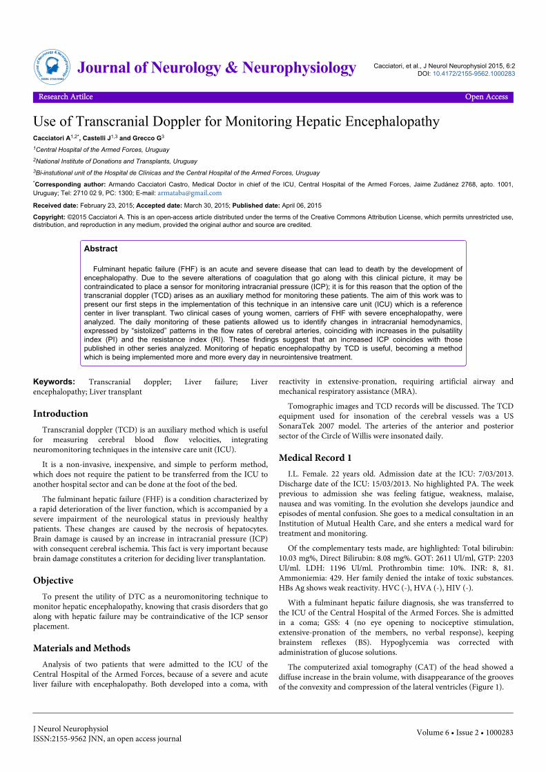
Despite the therapeutic measures, the patient developedunfavorably. On the last day of stay in the ICU, the study TCD showedat both the anterior and the posterior of the circle of Willis acirculatory pattern consistent with cerebral circulatory arrest (CCA)that accompanies brain death (BD) (Figures 5 and 6).
Citation: Cacciatori A, Castelli J and Grecco G (2015) Use of Transcranial Doppler for Monitoring Hepatic Encephalopathy. J NeurolNeurophysiol 6: 283. doi:10.4172/2155-9562.1000283
Page 2 of 6
J Neurol NeurophysiolISSN:2155-9562 JNN, an open access journal
Volume 6 • Issue 2 • 1000283
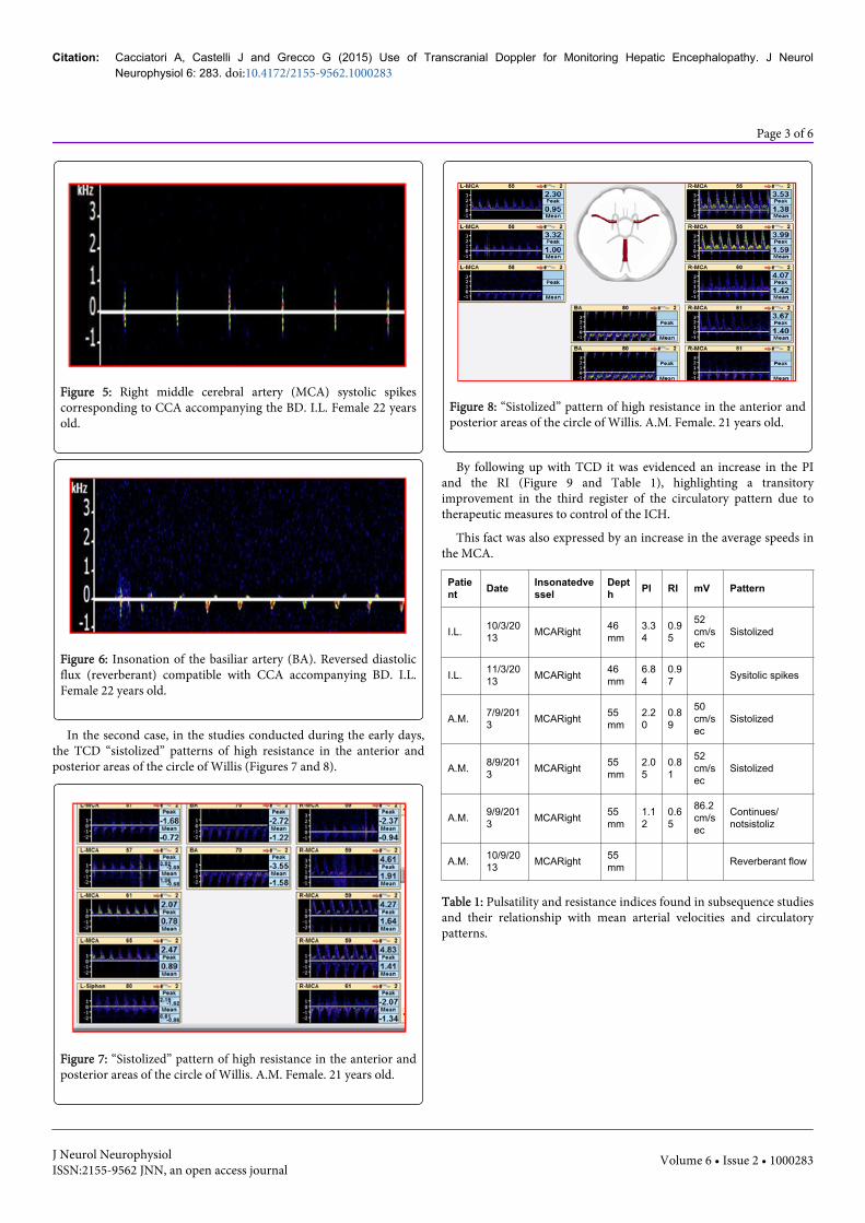
Figure 5: Right middle cerebral artery (MCA) systolic spikescorresponding to CCA accompanying the BD. I.L. Female 22 yearsold.
Figure 6: Insonation of the basiliar artery (BA). Reversed diastolicflux (reverberant) compatible with CCA accompanying BD. I.L.Female 22 years old.
In the second case, in the studies conducted during the early days,the TCD “sistolized” patterns of high resistance in the anterior andposterior areas of the circle of Willis (Figures 7 and 8).
Figure 7: “Sistolized” pattern of high resistance in the anterior andposterior areas of the circle of Willis. A.M. Female. 21 years old.
Figure 8: “Sistolized” pattern of high resistance in the anterior andposterior areas of the circle of Willis. A.M. Female. 21 years old.
By following up with TCD it was evidenced an increase in the PIand the RI (Figure 9 and Table 1), highlighting a transitoryimprovement in the third register of the circulatory pattern due totherapeutic measures to control of the ICH.
This fact was also expressed by an increase in the average speeds inthe MCA.
Patient Date Insonatedve
sselDepth PI RI mV Pattern
I.L. 10/3/2013 MCARight 46
mm3.34
0.95
52cm/sec
Sistolized
I.L. 11/3/2013 MCARight 46
mm6.84
0.97 Sysitolic spikes
A.M. 7/9/2013 MCARight 55
mm2.20
0.89
50cm/sec
Sistolized
A.M. 8/9/2013 MCARight 55
mm2.05
0.81
52cm/sec
Sistolized
A.M. 9/9/2013 MCARight 55
mm1.12
0.65
86.2cm/sec
Continues/notsistoliz
A.M. 10/9/2013 MCARight 55
mm Reverberant flow
Table 1: Pulsatility and resistance indices found in subsequence studiesand their relationship with mean arterial velocities and circulatorypatterns.
Citation: Cacciatori A, Castelli J and Grecco G (2015) Use of Transcranial Doppler for Monitoring Hepatic Encephalopathy. J NeurolNeurophysiol 6: 283. doi:10.4172/2155-9562.1000283
Page 3 of 6
J Neurol NeurophysiolISSN:2155-9562 JNN, an open access journal
Volume 6 • Issue 2 • 1000283

Figure 9: Graph that objectifies the variations in PI, RI and mV inMCA right, in subsequent days. 2nd case.
In the evolution it adds hemodynamic deterioration and distress,with clinical suspicion of multiple organ dysfunction (MOD), ofinfectious origin.
The study conducted on the last day of her stay in the ICU, showeda pattern consistent with CCA accompanying BD (Figure 10 and 11).
Figure 10: Systolic spikes in the territory of the right middlecerebral artery. A.M. Gender: Female. 21 years.
Figure 11: Reverse diastolic flow (reverberant) in the territory of thebasilar artery (BA). Pattern consistent with CCA. A.M. Gender:Female. 21 years.
DiscussionClinically, the hepatic encephalopathy is usually identified by the
impairment of consciousness and development of various motor andintellectual abnormalities, which can range from subtle cognitivedeficits to coma.
This neurological syndrome is accompanied by slow-pathelectroencephalogram (EEG) where triphasic waves can be recognized[1].
The most frequent cause of hepatic encephalopathy is thedecompensation of chronic liver diseases. However, the most seriousand rapid way of this encephalopathy is distinguished in the fulminantliver failure, either toxic, immune or viral in origin [1].
Acute liver failure, also known as fulminant hepatic failure (FHF),encompasses a spectrum of clinical entities characterized by: acuteliver injury, severe hepatic dysfunction and hepatic encephalopathy.Even though it is not very common, its prevalence is approximately2000 cases per year in the United States, with mortality ranging from50 to 90% [2].
The clinical evolution time has prognostic implication, due to itsrelation to the different etiologies and outcomes [3].
The loss of hepatocyte function triggers a systemic inflammatoryresponse (SIRS) with multiple organ dysfunction (MOD) that can leadto death. The orthotopic liver transplantation is the only definitivetreatment for those patients with FHF and for those whose livercannot recover its function spontaneously. The cerebral edema thatcauses the intracranial hypertension (ICH) complicates approximatelybetween 50 and 80% of patients with FHF (III or IV grade) andbecomes a death cause [4,5]. Cerebral edema and ICH do not occur inpatients with cirrhosis and chronic hepatic failure. Unfortunately,many patients die due to ICH and brain herniation. So, early diagnosisand aggressive therapy to control ICH prevent the evolution to exitus,waiting for an organ donor, or until spontaneous recover of hepaticfunction occurs [2].
Production brain edema and ICH mechanisms in the context ofFHF are multifactorial and so far, partially understood. They include:cytotoxicity as a result of osmotic effect caused by ammonia,glutamine, other amino acids and proinflammatory cytokines.Cerebral hyperemia and vasogenic edema can occur because of blood-brain barrier disruption with accumulation of low molecular weightsubstances. Dysfunction of the ATPase Na+-K+ bomb, withconsequent loss of autorregulation of cerebral blood flow (CBF), havebeen implicated as hyperemia causes [2] (Figure 12).
Cranial CT is indicated in those patients with acute liver failure,progressing to stages III/IV of encephalopathy, or experiencing acutechanges in consciousness, and before indicating ICP monitoring.
Although CT can often detect brain swelling in patients with acuteliver failure and with an advanced stage of encephalopathy, it does nothave the sensitivity to prove ICH; so its main value is to discardintracranial bleeding.
Many authors recommend ICP monitoring in patients withadvanced encephalopathy, believing that monitoring allows themanagement of cerebral edema, providing information aboutneurological recovery after orthotopic liver transplantation [6].Continuous measurements of ICP perioperatively in the managementof FHF, has been associated with a survival rate of 54%-74% in a seriesof six to 23 patients, wich is generally higher than with medical means,
Citation: Cacciatori A, Castelli J and Grecco G (2015) Use of Transcranial Doppler for Monitoring Hepatic Encephalopathy. J NeurolNeurophysiol 6: 283. doi:10.4172/2155-9562.1000283
Page 4 of 6
J Neurol NeurophysiolISSN:2155-9562 JNN, an open access journal
Volume 6 • Issue 2 • 1000283

and was a high as 92 % for the selected group who had undergone livertransplantation [7].
Figure 12: Pathophysiology of brain edema and intracranialhypertension in patients with fulminant hepatic failure.
The invasive monitoring, however, is specially risky in FHF patientswith coagulopathy, in whom the incidence of bleeding from ICPmonitoring ranges from 5%–22%, with a mortality rate of 60% [7].
Considering that crasis disorder constitute a severe impediment forplacing an ICP, is where the option of TCD arises in order to evaluatechanges in cerebral hemodynamic suggestive of ICH. The noninvasivetechnique provides adequate information when cerebral perfusion islow, comparable with the invasive technique, and allows the ICH isdiagnosed and treated effectively [7].
The TCD has been used extensively for monitoring of head injuryand cerebral circulatory arrest [8] and has been studied extensively inhead trauma patients [9].
Sidi and Mahla compared with simultaneous invasive monitoring ofICP sensor and TCD, in the pre, peri and postoperative orthotopicliver transplantation, in patients with fulminant hepatic failure. Theydemonstrated the usefulness of both to control ICP, and maintenancefor adequate cerebral perfusion pressure (CPP).
The extensive studies of TCD monitoring in head-trauma patientshave contributed some information about the relationship betweenICP and TCD. Diastolic flow velocity is influenced by cerebral vascularresistance, which is determined mainly by ICP and vessel diameter.TCD images show that diastolic flow velocity becomes zero when ICPequals diastolic blood pressure (10). This is a conclusive warning sign,at which time TCD images of the diastolic component should becompared with diastolic blood pressure (rather than systolic or meanpressure) [7].
PI, which also represents resistance to flow, can be correlated withICP or cerebral perfusion pressure in head-trauma patients.
CBF measurement with the TCD, present a good correlation withother direct measurements, as the xenon method. The attenuation ofthe diastolic phase of the curve constitutes a sign of ICH and theconsequent decrease in cerebral perfusion. Additionally, themorphology of the curve in the diastolic phase may indicate early or
late ICH signs with the consequent attenuation of the cerebral diastolicflow (2).
Aggarwal et al. published a study in the year 2008, in which, usingthe TCD, observed changes in the morphology of the velocity curve ofcerebral blood flow in patients with FHF, relating cerebral perfusionwith the different stages of this disease. In this study, changes wereobserved in the shape of the curve, as increased levels decreased ICPand CPP values, reaching the images of cerebral circulatory arrest :systolic spikes and retrograde flow during diastole.
The TCD secuence indicates, that TCD can provide informationabout the dynamic state of the intracranial circulation and perfusion.This study indicates that, other easily identifiable and calculablefeatures of TCD waveform that noticeably change as both ICP andCPP, change can be advantageously used to infer the state of cerebralperfusion with little addition of complexity [11].
The authors conclude that, the preliminary results on thecorrelation of TCD waveform features with the state of cerebralperfusion are promising.
Abdo et al. evaluated CBF by TCD in five patients with FHF andcompared with a control group that showed severe neurologicalalterations not associated with FHF. A pattern of cerebralhypoperfusion was found in 80% of the group with FHF, while in thecontrol group it corresponded to 40%.
Average values of the velocities and PI were 36.6 cm/sec and 2.4respectively in the FHF group, while in the control group they were47.8 cm/sec and 1.8 respectively. The authors conclude that patientswith FHF showed a predominant pattern of cerebral hypoperfusion(sistolized) with average speed values lower than normal and increasedPI [2].
Bindi et al., in a series of 5 patients with FHF, to whom TCD wasapplied in order to monitor, conclude that this technique allows theevaluation, non-invasive, repeatable and reliable of the changes inCBF, at the head of the patient, avoiding complications associated withan ICP placement. Likewise, it allows time to properly assess themoment for the hepatic transplant indication [12].
ConclusionFulminant hepatic failure (FHF) constitutes and acute and severe
nosological entity, which can lead to death, because of encephalopathydevelopment. Cerebral edema is responsible for generatingintracranial pressure (ICP), which can lead to brain herniation. FHFbrain commitment constitutes a criterion for deciding hepatictransplant.
Due to the fact that it is not always possible to place an ICP sensor,TCD arises as an auxiliary method for monitoring these patients. It hasthe advantage of being performed at the bedside of the patient; it isnoninvasive and can be repeated as many times as necessary. So far,there are few documented series with the use of this technique inmonitoring the hepatic encephalopathy.
Daily monitoring of the patients analyzed in this work, allowed thecare team to identify changes in intracranial hemodynamics, expressedby “sistolized” patterns in the velocity of cerebral arteries flow,coinciding with increases in pulsatility indices (PI) and resistanceindices (RI).
Citation: Cacciatori A, Castelli J and Grecco G (2015) Use of Transcranial Doppler for Monitoring Hepatic Encephalopathy. J NeurolNeurophysiol 6: 283. doi:10.4172/2155-9562.1000283
Page 5 of 6
J Neurol NeurophysiolISSN:2155-9562 JNN, an open access journal
Volume 6 • Issue 2 • 1000283

These findings, which suggest the increase in ICP, coincide withthose published in other analyzed series. The results of the studies,allowed conducting the therapeutic strategy. However, the evolution ofthe patients was not good at all, reaching the locking and cerebralcirculatory arrest (CCA), accompanying the BD, which is highlyexpressive of the extreme severity of this disease.
References1. Jiménez D, Cartier L (2012) Encefalopatía hepática fulminante
vinculadaa hiperintensidad de la corteza cerebral en la resonanciamagnética. Revista chilenade neuro-psiquiatría 50: 51-56.
2. Raghavan M, Marik PE (2006) Therapy of intracranial hypertension inpatients with fulminant hepatic failure. Neurocrit Care 4: 179-189.
3. Stravitz RT (2008) Critical management decisions in patients with acuteliver failure. Chest 134: 1092-1102.
4. O'Grady JG (1997) Paracetamol hepatotoxicity: how to prevent. J R SocMed 90: 368-370.
5. Butterworth RF (2003) Molecular neurobiology of acute liver failure.Semin Liver Dis 23: 251-258.
6. Todd Stravitz R, Kramer AH, Davern T, Shaikh O, Caldwell SH, et al.(2007). The Acute liver Failure Study Group. Intensive care of patients
with acute liver failure : Recommendations of the U.S. Acute LiverFailure Study Group. Critical Care Med 11: 2498-2508.
7. Sidi A, Mahla M (1995) Noninvasive Monitoring of Cerebral Perfusionby Transcranial Doppler During Fulminant Hepatic Failure and LiverTransplantation. Anesth Analg 80: 194- 200.
8. Newell DW, Seiler RW, Aaslid R (1992) Head injury and cerebralcirculatory arrest. In: Newell DW, Aaslid R (eds.) Transcranial Doppler.Raven Press, New York, pp. 109-121.
9. Homburg AM1, Jakobsen M, Enevoldsen E (1993) Transcranial Dopplerrecordings in raised intracranial pressure. Acta Neurol Scand 87:488-493.
10. Hassler W1, Steinmetz H, Gawlowski J (1988) Transcranial Dopplerultrasonography in raised intracranial pressure and in intracranialcirculatory arrest. J Neurosurg 68: 745-751.
11. Aggarwal S, Brooks DM, Kang Y, Linden PK, Patzer II JF (2008)Noninvasive Monitoring of Cerebral Perfusion Pressure in Patients withAcute Liver Failure Using Transcranial Doppler Ultrasonography. LiverTranspl 14: 1048-1057.
12. Bindi ML, Biancofiore G, Esposito M, Meacci l, Bisa M, et al. (2008)Transcraneal Doppler sonography is useful por the decision – making atthe ppoint of care in patients with acute hepatic failure : a single centre´sexperience. J Clin Monit Comput 22: 449-452.
Citation: Cacciatori A, Castelli J and Grecco G (2015) Use of Transcranial Doppler for Monitoring Hepatic Encephalopathy. J NeurolNeurophysiol 6: 283. doi:10.4172/2155-9562.1000283
Page 6 of 6
J Neurol NeurophysiolISSN:2155-9562 JNN, an open access journal
Volume 6 • Issue 2 • 1000283