Embed Size (px)
Citation preview

74 Short reportscases CNS involvement was diagnosed in theterminal stages on clinical evidence aloneheadaches, vomiting, and papilloedema in one case,blindness in another; in 2 further cases undiagnosedinvolvement of the CNS was found at necropsy(at weeks 38 and 74).Throughout this period the form of treatment was
varied, the commonest regimens being combina-tions such as those described by Gee, Yu, andClarkson (1969), Crowther, et al. (1970), theMedical Research Council (1974) and others. Asfar as can be determined no particular regimen wasassociated with a particularly high or low incidenceof CNS disease. However, among the 12 childrenwho received some sort of CNS prophylaxis therehave so far been no cases of meningeal leukaemia.Prophylaxis has included cranial (and spinal)irradiation and intrathecal methotrexate and/orcytosine arabinoside. Taking the period starting6 weeks after diagnosis, which was the usual pointat which CNS prophylaxis began, a comparisoncan be made of the relative incidence of CNSleukaemia in the time at risk between those who didand those who did not have CNS prophylaxis.Among 82 cases without prophylaxis, totalling2707 weeks at risk, there were 15 cases of CNSdisease, an incidence of 1 case per 180 weeks: thiscan be compared with 12 cases who received somesort of prophylactic treatment to the CNS, none ofwhom developed CNS leukaemia during a total of544 weeks at risk.
SummaryIn 130 cases of acute myeloid leukaemia in child-
ren below the age of 14 years in Great Britain, therewere 21 cases in which the central nervous systemwas involved. The incidence and timing is similarto that of acute lymphoblastic leukaemia; in a smallnumber of patients who received prophylactictreatinent, involvement of the central nervoussystem was prevented.
REFERENCESCrowther, D., Bateman, C. J. T., Vartan, C. P., Whitehouse,
J. M. A., Malpas, J. S., Fairley, G. H., and Scott, R. B. (1970).Combination chemotherapy using L-asparaginase, daunorubicinand cytosine arabinoside in adults with acute myelogenousleukaemia. British Medical Journal, 4, 513.
Gee, T. S., Yu, K-P., and Clarkson, B. D. (1969). Treatment ofadult acute leukaemia with arabinosylcytosine and thioguanineCancer, 23, 1019.
Report of the Medical Research Council's Working Party on Leukae-mia in Adults (1974). Treatment of acute myeloid leukaemiawith daunorubicin, cytosine arabinoside, mercaptopurine,L-asparaginase, prednisone and thioguanine: results oftreatmentwith five multiple-drug schedules. BritishJournal of Haemato-logy, 27, 373.
H. E. M. KAY* on behalf of the Medical ResearchCouncil's Working Partyt on Leukaemia in Child-hood
*Correspondence to H.E.M.K., Royal Marsden Hospital, FulhamRd., London SW3 6JJ.tDr. K. D. Bagshawe, Dr. J. M. Bridges, Professor Neville
Butler, Dr. J. M. Chessells, Dr. P. F. Deasy, Sir Richard Doll, Dr.P. Emerson, Dr. H. W. Everley Jones, Dr. D. I. K. Evans, Dr.D. M. T. Gairdner, Dr. D. A. G. Galton, Dr. R. J. Guyer, ProfessorR. M. Hardisty (Secretary), Dr. C. B. Howarth, Professor J. M.Hutchison (Chairman), Dr. E. M. Innes, Dr. P. Morris Jones,Dr. T. J. McElwain, Dr. I. C. M. MacLennan, Dr. J. Martin,Professor I. C. S. Nomand, Dr. P. G. Smith, Dr. J. Stuart, Dr. E. N.Thompson, Dr. M. L. N. Willoughby.
Mulkiple major cerebral arterythromboses with profoundthrombocytopenia in acute
leukaemiaCytotoxic, corticosteroid, and antibiotic therapy
in leukaemia are associated with an increase infungal infections (Bodey, 1966). Having to dealwith the clinical problems of thrombocytopeniaand bleeding in leukaemia is not uncommon. Inthis situation it is very unusual to find at necropsyextensive antemortem thrombosis in the cerebralarterial circulation.
Case reportA 5k-year-old girl was found to have acute lymphatic
leukaemia (ALL), with a haemoglobin of 7 4 g/dl, aplatelet count of 30 000/mm3, and leucocytes at 4900/mm3 with 2% neutrophils, 68% lymphocytes, and 30°%blast cells. Bone marrow biopsy showed that 96% ofthe nucleated cells were blasts.She was treated over a 9-week period, according to
the protocol oftheMRC UKALL III Ordinary Schedulewith intravenous vincristine and oral prednisone toinduce remission, followed by oral 6-mercaptopurine,intravenous asparaginase. intrathecal methotrexate, andcerebral irradiation. After 4 weeks her peripheral bloodand bone marrow had returned to normal During the10th week she had one dose of oral methotrexate afterwhich her leucocyte and platelet counts began to fallabruptly.
She was readmitted during the 12th week with fever,respiratory distress, widespread purpura, and rectalbleeding. She was anaemic (Hb 5 g/dl), leucopenic(total white blood count 2000/mm3) and thrombocyto-penic (platelets 20 000/mm3). A blood culture grewmeningococci. The bone marrow was hypoplastic.All cytotoxic therapy was stopped and she was givenpenicillin, sulphadimidine, and prednisone. While inthis thrombocytopenic state she fell out ofbed and deve-loped a large frontal haematoma, the blood later gravitat-ing down to surround both eyes. The skin overlying the
copyright. on N
ovember 26, 2021 by guest. P
rotected byhttp://adc.bm
j.com/
Arch D
is Child: first published as 10.1136/adc.51.1.74 on 1 January 1976. D
ownloaded from

Brain. The left frontal lobe was swollen and con-gested with a little yellowish exudate on the inferiorsurface. The intracranial portion of the left intemalcarotid, the left middle cerebral, both anterior cerebralarteries, and the first part of the right middle cerebralartery were totally occluded by thrombus. Thevertebral, basilar, and posterior cerebral arteries appear-ed normal.
After fixation in formol-saline multiple coronalsections showed extensive infarction of the cortex andadjacent white matter of the left inferior frontal lobe.In the parietal lobes there was extensive symmetricalhaemorrhagic necrosis of both intemal capsules, thecaudate and lentiform nuclei and the thalamus (Fig. 1).The necrotic areas broke away easily from the surround-ing cerebral tissue.There was partial collapse of the lower lobe of the
left lung and a white mucoid exudate in the trachea.The liver (830 g), spleen (50 g), and lymph nodes werenormal macroscopically. The upper gastrointestinaltract contained fresh blood with numerous petechialhaemorrhages in the gastric mucosa.
haematoma and at the inner canthus of each eye sloughed.leaving deep ulcers. Swabs from the sloughed areasgrew Esch. coli and Klebsiella aerogenes, but fungi werenot specifically sought.
For repeated rectal haemorrhages she was transfusedwith 16 units fresh blood and 40 units of platelets, with-out effect upon the thrombocytopenia or the bleeding.Pseudomonas aeruginosa was cultured from an infusionwound in her leg and she developed a few haemorrhagicvesicular skin lesions suggestive of pseudomonassepticaemia; she was treated with gentamicin andcarbenicillin. During the 18th week of her illness shedeteriorated rapidly, with fever, pain above the left eye,drowsiness deepening to coma, and signs of infection inthe left lung. At the time of death she was thought tohave had a large intracerebral haemorrhage.
Necropsy examination. Relevant findings in-cluded two areas of total skin loss without granulationtissue on either side of the nose, medial to each innercanthus, measuring 1-5 cm in diameter. Underlyingbone was visible. The skull and dura were normal.
FIG. 1.-Showing the extensive symmetrical haemorrhagic necrosis of both internal capsules and basal ganglia.
Short reports 75
copyright. on N
ovember 26, 2021 by guest. P
rotected byhttp://adc.bm
j.com/
Arch D
is Child: first published as 10.1136/adc.51.1.74 on 1 January 1976. D
ownloaded from
Short reports
$ ji~,Sb-+ o:;eStn *0 *Al aL
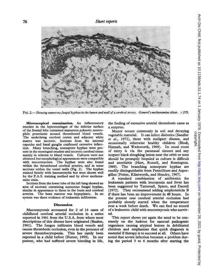
FIG. 2.-Showing numerousfungal hyphae in the lwnen andwal ofa cerebral artery. Gomori's methenamine silver. x 275.
Microscopical examination. An inflammatoryexudate in the leptomeninges of the inferior surfaceof the frontal lobe contained numerous pyknotic neutro-phils prominent around thrombosed blood vessels.The underlying cerebral cortex and adjacent whitematter was necrotic. Sections from the intemalcapsules and basal ganglia confirmed extensive infarc-tion. Many branching, nonseptate hyphae were pre-sent in the meningeal exudate and necrotic cerebral tissuemainly in relation to blood vessels. Cultures were notobtained but morphological appearances were compatiblewith mucormycosis. The hyphae were also foundwithin the thrombosed cerebral arteries, and in somesections within the vessel walls (Fig. 2). The hyphaestained faintly with haematoxylin but were shown wellby the P.A.S. staining method and by silver methena-mine stain.
Sections from the lower lobe of the left lung showed an
area of necrosis containing numerous fungal hyphae,similar in appearance to those in the brain and cerebralarteries. The bone marrow was hypoplastic. In nosystem was there evidence of leukaemic infiltration.
DiscussionMucormycosis accounted for 2 of 16 cases of
childhood cerebral arterial occlusion in a seriesreported in 1961 from the U.S.A. from where mostdescriptions of this disease have originated (Banker,1961). The fungus infiltrates blood vessels andcauses thrombotic occlusion, even in the presence ofsevere thrombocytopenia. This has rarely beenreported in a child before (Hutter, 1959). In ourpatient, who had suffered severe bleeding in life,
the finding of extensive arterial thrombosis came asa surprise.Mucor occurs commonly in soil and decaying
vegetable material. It can infect diabetics (Sandleret al., 1971), those with maligant disease, andoccasionally otherwise healthy children (Blodi,Hannah, and Wadsworth, 1969). Its usual routeof entry is via the paranasal sinuses and anysuspect black sloughing lesion near the orbit or noseshould be promptly biopsied as culture is difficultand unreliable (Hart, Russell, and Remington.1969). The branching nonseptate hyphae arereadily distinguishable from Penicillium and Asper-gillus (Fetter, Klintworth, and Hendry, 1967).A standard combination of antibiotics for
leukaemia patients with leucopenia and fever hasbeen suggested by Tattersall, Spiers, and Darrell(1972). They recommend adding amphotericin Bif there has been no improvement in 48 hours. Inthe present case cerebral arterial occlusion hadprobably already started when the temperaturerose a week before death. We can find no recordof a leukaemic child with mucormycosis who surviv-ed.
This report shows yet again the need to be con-stantly on the lookout for unusual pathogenicorganisms causing atypical lesions in debilitatedchildren and emphasizes that quick diagnosis isessential if therapy is to succeed at all. Others havenoted that severe infection becomes a problem dur-ing the period 3 to 6 months after starting the
76
copyright. on N
ovember 26, 2021 by guest. P
rotected byhttp://adc.bm
j.com/
Arch D
is Child: first published as 10.1136/adc.51.1.74 on 1 January 1976. D
ownloaded from
Short reports 77UKALL III schedule and special vigilance isneeded at that time.
SummaryA child with acute lymphoblastic leukaemia
complicated by prolonged gastrointestinal and skinhaemorrhages due to profound thrombocytopeniafinally died of thrombotic occlusions of majorcerebral arteries due to mucormycosis. Biopsy ofany suspect lesion is needed urgently before pro-longed therapy with amphotericin B is started.So far there have been no cures in childhood.
We are grateful to Miss Maureen McKeown for hersecretarial help.
REFERENCEsBanker, B. Q. (1961). Cerebral vascular disease in infancy and
childhood. Journal of Neuropathology and ExperimentalNeurology, 20, 127.
Blodi, F. C., Hannah, F. T., and Wadsworth, J. A. C. (1969).Lethal orbitocerebral phycomycosis in otherwise healthychildren. American Journal of Ophthalmology, 67, 698.
Bodey, G. P. (1966). Fungal infections complicating acute leuke-mia. J,ournal of Chronic Diseases, 19, 667.
Fetter, B. F., Klintworth, G. K., and Hendry, W. S. (1967).Mycoses of the Central Nervous System. Williams and Wilkins,Baltimore.
Hart, P. D., Russell, E., and Remington, J. S. (1969). The com-promised host and infection. II. Deep fungal infection.Journal of Infectious Disease, 120, 169.
Hutter, R. V. P. (1959). Phycomycetous infection (mucormycosis)in cancer patients:-a complication of therapy. Cancer, 12,330.
Sandler, R., Tallman, C. B., Keamy, D. G., and Irving, W. R. (1971).Successfully treated rhinocerebral phycomycoses in well con-trolled diabetes. New England Journal of Medicine, 285, 1180.
Tattersall, M. H., Spiers, A. S., and Darrell, J. H. (1972). Initialtherapy with combination of five antibiotics in febrile patientswith leukaemia and neutropoenia. Lancet, 1, 162.
D. G. SIMs,* D. J. SCOTT, and T. C. NOBLEDepartments of Child Health and Pathology, New-castle General Hospital, Newcastle upon TyneNE4 6BE.
*Correspondence to Dr. D. G. Sims.
Congenital rubella associatedwith hypsarrhythmia
Hypsarrhythmia, or infantile spasms, falls intotwo broad groups when considered on an aetiologicalbasis. In one-third of cases there is no knownpredisposing factor, while in the other two-thirds ahistory of cerebral insult is a possible causativefactor.Some factors implicated in causing hypsarrhyth-
mia have included cerebral birth injury, toxoplasmo-sis, postnatal head injury, meningitis, encephalitis,tuberous sclerosis, phenylketonuria, pyridoxine
deficiency (Millichap et al., 1962), and infectionwith cytomegalovirus (Stern, Latham, and Tizard1968). To our knowledge congenital rubellainfection has never been implicated as a contributingfactor.
Case reportIn the 16th week of gestation the mother was infected
with rubella. Diagnosis was confirmed by a rise inrubella antibody titre from 1:16 at the time of exposureto 1:2048 two weeks later. At 37 weeks' gestation a maleinfant was bom after a 5+-hour labour. No resuscita-tion was required and physical examination showed noabnormality. Birthweight was 3 - 35 kg.On the fifth day right-sided twitching, which later
became generalized, was noted but was easily controlledwith anticonvulsants. Examination of cerebrospinalfluid, blood culture, serum calcium, and blood glucose,was normal. Platelet count was not depressed andx-rays of the long bones were normal. Rubella viruswas not grown from urine and nasophargyneal secretions.Blood taken on the 6th day of life showed rubella HAItest titre of 1:2048 and a rubella specific IgM titre of1:40.The patient was hypotonic and lethargic for the next 3
days but by the 11th day of life physical examinationwas normal, and he was discharged with no medication.He was next seen at 6 weeks of age, was smiling andphysical examination was again normal. At 5 months hebegan to have episodes of quickly raising both arms andshuddering for 2 or 3 seconds. Infantile spasms weresuspected and confirmed by electroencephalogram(EEG), which showed the widespread slow activity withnumerous high voltage spike waves typical of hypsarrhy-thmia (Fig.). Physical examination, serum chemistry,and cerebrospinal fluid were normal.A course of ACTH (20 IU twice daily) was started 2
weeks after the spasms were first noticed by his mother.EEG 10 days after ACTH was started showed lowvoltage with irregular theta rhythm and some deltaactivity in all leads, no longer the pattern of hypsarrhy-thmia. After 6 weeks the ACTH dose was reduced insteps and was completely withdrawn 3 weeks later. At 9months of age his EEG was normal.Monthly assessments since birth showed that develop-
ment was normal up to the onset of hypsarrhythmia.Development then began to lag. By 10 months he wasfunctioning at a 5-month level using the Stycar tests(Sheridan, 1968). A review at 25 months showed nor-mal hearing and no evidence of cataracts or retinopathy.A Griffiths assessment (Griffiths, 1954) showed delay inall areas. Delays were most marked in languagedevelopment (9-month level) and hand-eye co-ordina-tion (12-month level).
DiscussionThere is no doubt that this child was exposed to
rubella at 16 weeks' gestation as shown by hismother's antibody response. The rubella specificIgM titre of 1:40 on the 6th day of life is not particu-
copyright. on N
ovember 26, 2021 by guest. P
rotected byhttp://adc.bm
j.com/
Arch D
is Child: first published as 10.1136/adc.51.1.74 on 1 January 1976. D
ownloaded from