Embed Size (px)
Citation preview

sigma.com
FOR LIFE SCIENCE RESEARCH
Cellular Mechanisms and Cancer
2007VOLUME 2NUMBER 4
INNOVATION @ WORK
ABC TRANSPORTERS AND CANCER DRUG RESISTANCE
NF-kB AND INFLAMMATION
DNA DAMAGE AND REPAIR NEW PRODUCTS
Cultured HeLa cells in metaphase stage
of mitosis.
sigma.com/yfgbiofiles
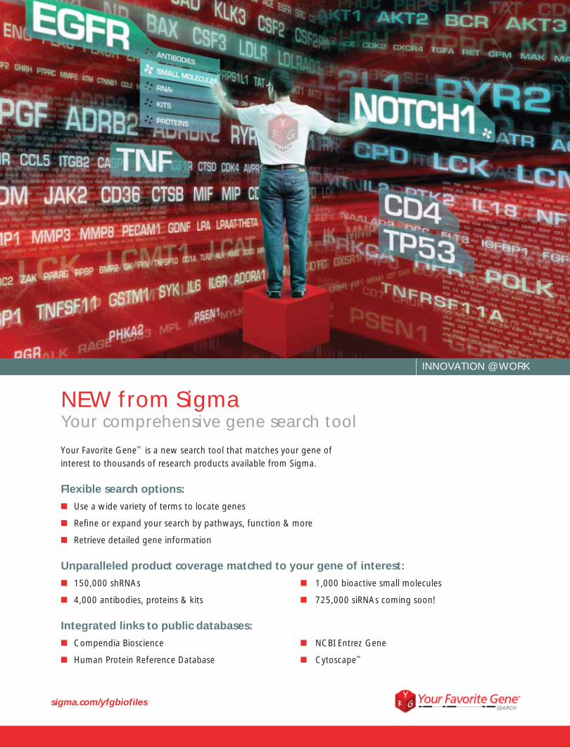
NEW from SigmaYour comprehensive gene search tool
Your Favorite Gene™ is a new search tool that matches your gene of interest to thousands of research products available from Sigma.
Flexible search options:■ Use a wide variety of terms to locate genes
■ Refi ne or expand your search by pathways, function & more
■ Retrieve detailed gene information
Unparalleled product coverage matched to your gene of interest:■ 150,000 shRNAs ■ 1,000 bioactive small molecules
■ 4,000 antibodies, proteins & kits ■ 725,000 siRNAs coming soon!
Integrated links to public databases:■ Compendia Bioscience ■ NCBI Entrez Gene
■ Human Protein Reference Database ■ Cytoscape™
INNOVATION @ WORK
Table of Contents
Cellular Mechanisms and Cancer
Innovation @ Work
MISSION® shRNA Human Tumor Suppressor Gene Panel ..................... 2
Panorama® Antibody Arrays ..................... 3
GenomePlex® Single Cell Whole Genome Amplification ........... 4
GenomePlex® Complete Whole Genome Amplification ........... 5
PhosphoProfile™ I Phosphopeptide Enrichment Kit .................................. 6
ABC Transporters and Cancer Drug Resistance
Role of ABC Transporters and Multi-Drug Resistance Reversal Using RNA Interference ...................................... 7 MDR-substrate Anticancer Agents ............. 9 ABC Transporter Membrane Proteins ...... 10 Antibodies to ABC Transporters ............... 11 Compounds for MDR Detection .............. 13
NF-kB and Inflammation
Inflammation and Cancer: The NF-kB Connection .............................14 Natural Product Inhibitors of NF-kB Activation ................................... 16 Antibodies and Kits ............................... 18
DNA Damage and Repair
DNA Damage and Repair Mechanisms....20 DNA Alkylating Agents ........................... 24 DNA Crosslinking Agents ........................ 26 DNA Synthesis Inhibitors ........................ 27 DNA Repair Enzymes and Antibodies ...... 28
New Products Antitumor Agents .................................. 30 Chemopreventative Agents ..................... 32 Enzymes ................................................. 32 Multi-Drug Resistance ............................ 32 Tumor Growth Regulation....................... 32
FOR LIFE SCIENCE RESEARCH
2007Volume 2Number 4
1
The medical definition of cancer appears simple and straightforward. According to the U.S. National Cancer Institute, cancer is “(a) term for diseases in which abnormal cells divide without control.”1 Behind this basic definition is a complex and unpredictable spectrum of over 100 types of cancer.
The human aspect of cancer cannot be completely separated from the scientific research. The World Health Organization recognizes cancer as a leading cause of death worldwide, and emphasizes prevention and early detection as crucial to reducing the global burden of the disease.2 The American Association for Cancer Research established its Scientist-Survivor program in 1999 to encourage communication between patients, patient advocates, and leading scientists in the field.3 Newspapers, magazines, and other media sources announce breakthrough discoveries to the public, increasing awareness that cancer is not an individual disease but a collection that has no single initiating event or defined evolution.
Cancer results from a cascade of abnormal cell reactions. When a cellular mechanism goes wrong, the resulting damage, if not repaired, may contribute to a cell’s evolu-tion into malignancy. Because cancers begin with a single cell, cancer investigators use genomics, proteomics, and signaling techniques to determine and evaluate cellular changes and contributing cause and effect. Discoveries such as the correlation between the human papilloma virus and cervical cancer encourage the scientific community to seek similar breakthroughs for other cancer types. The understanding of cellular muta-tions and signaling pathways involved in mutagenesis and abnormal cell function has been used to screen potential new drugs with more efficacy and/or less toxicity.
It’s impossible to comprehensively review current cancer research; the amount of informa-tion is enormous and the rate of discovery is increasing. In the preface to his book “The Biology of Cancer”, Robert Weinberg writes “…we are deluged with a vast amount of genetic, biochemical, and cell biological information about cancer development, far more almost than any human mind can assimilate and comprehend.”4 For this issue of BioFiles we have selected three aspects of cancer biology to review.
■ The exploitation of ABC transporter proteins by cancer cells to export chemotherapeutic drugs
■ The activation of NF−κB in response to inflammation and its role incancer progression
■ Genetic damage, mutagenesis, and cellular repair processes
Innovation @ Work - Innovative products from Sigma for genomics and proteomics stud-ies, including gene silencing, protein expression profiling, phosphopeptide enrichment, and whole genome amplification, are also highlighted.
References:1. World Health Organization, Cancer Fact Sheet No. 297, Feb. 2006, www.who.int/mediacentre/factsheets/fs297/en/index.html2. National Cancer Institute, U.S. National Institutes of Health, www.cancer.gov3. American Association for Cancer Research, www.aacr.org4. The Biology of Cancer, Robert A. Weinberg, Garland Science, Taylor & Francis Group, LLC, New York, NY (2007)
Cover image: Cultured HeLa cells labeled with anti-tubulin antibody and propidium iodide (to label the DNA) in metaphase stage of mitosis. Photographed by Dr. K. G. Murti, St. Jude Children’s Research Hospital, Memphis, TN.Technical content: Libby Yunger, Ph. D., Chloe McClanahan, B. Sc., Vicki Caligur, B. Sc.
For Safety Information please refer to the Sigma Biochemicals, Reagents and Kits for Life Science Research Catalog or Sigma.com
Introduction
sigma.com
INNOVATION @ WORK2
Silencing Tumor Suppressor Genes Using MISSION® TRC shRNA RNAi methodologies have been utilized in small- to large-scale screening projects, allowing researchers to perform gene-silencing experiments in timeframes and target cells not previously possible. While siRNA screens have become fairly com-mon, large-scale screening with shRNA is still evolving. Advantages of shRNA-based experiments include long-term knockdown and viral delivery to nontransfectable cell types.
The MISSION TRC shRNA Human Tumor Suppressor Gene Family Set is a panel of lentiviral-based shRNAs consisting of approximately 75 gene sets, each represented by 3–5 individual shRNA clones. The shRNA constructs included in the tumor sup-pressor set allow you to identify genes involved in cell survival by assisting in mak-ing a cell more sensitive, or more resistant, to an added chemotherapeutic agent through the knockdown of gene expression and resulting function.
Scientists at Sigma have utilized the MISSION TRC shRNA Human Tumor Suppressor Gene Family Set in order to address biologically relevant questions while simultane-ously developing screening strategies that can be used by researchers in the field. The tumor suppressor gene family set delivers a potential pharmacogenomics solu-tion, allowing you to profile the gene expression of a cancer cell line and compare it against the response of the cell line to chemotherapeutic treatment.1
In our study of paclitaxel resistance by the human lung cancer cell line, A549, various transcripts were down-regulated in the lung cancer cells using the tumor suppressor set. Several genes that are known to correlate with paclitaxel resistance, including p53 and VHL, were identified by the panel. In addition, some genes, when down-regulated by these shRNAs, led to increased sensitivity of the cells to paclitaxel treatment.
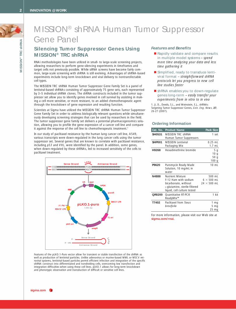
5' -
3' - UU
Sense Strand
Antisense Strand
Sense StrandCCGGNNNNNNNNNNNNNNNNNNNNNCTCGAGNNNNNNNNNNNNNNNNNNNNNT T T T TGGCCNNNNNNNNNNNNNNNNNNNNNGAGCTCNNNNNNNNNNNNNNNNNNNNNAAAAA
Antisense StrandLoop
Loo
p
cppt
pLKO.1-puro7,091 bp
hPGK
puroR
pUC ori
RRE
SIN/3’ LTR
RSV/5' LTR
U6
f1 ori
ampR
(Ψ) Psi
Features of the pLKO.1-Puro vector allow for transient or stable transfection of the shRNA as well as production of lentiviral particles. Unlike adenovirus or murine-based MML or MSCV ret-roviral systems, lentiviral-based particles permit efficient infection and integration of the specific shRNA construct into differentiated and nondividing cells, overcoming low transfection and integration difficulties when using these cell lines. pLKO.1 allows for long-term knockdown and phenotypic observation and transduction of difficult or sensitive cell lines.
Features and Benefi ts■ Rapidly validate and compare results
in multiple model systems – spend more time analyzing your data and less time gathering it
■ Simplified, ready to transduce lenti-viral format – straightforward shRNA protocols let you progress to new cell line studies faster
■ shRNA enables you to down-regulate genes long-term – easily transfer your experiments from in vitro to in vivo
1. Ji, D., Deeds, S.L., and Weinstein, E.J., shRNAs Targeting Tumor Suppressor Genes, Gen. Eng. News, 27, 26-27 (2007).
Ordering Information
Cat. No. Product Name Pack Size
SH0531 MISSION TRC shRNA Human Tumor Suppressors
1 set
SHP001 MISSION Lentiviral Packaging Mix
0.25 mL 1.7 mL
H9268 Hexadimethrine bromide 5 g 10 g 50 g
100 g
P9620 Puromycin Ready Made Solution, 10 mg/mL in water
10 mL
N4888 Nutrient Mixture F-12 Ham with sodium bicarbonate, without L-glutamine, sterile-filtered liquid, cell culture tested
500 mL6 3 500 mL
24 3 500 mL
QR0200 Quantitative RT-PCR ReadyMix™
1 kit
T7402 Paclitaxel from Taxus brevifolia
1 mg5 mg
25 mg
For more information, please visit our Web site at sigma.com/rnai.
MISSION® shRNA Human Tumor Suppressor Gene Panel
MIS
SIO
N® T
RC
sh
RN
A
INNOVATION @ WORK 3
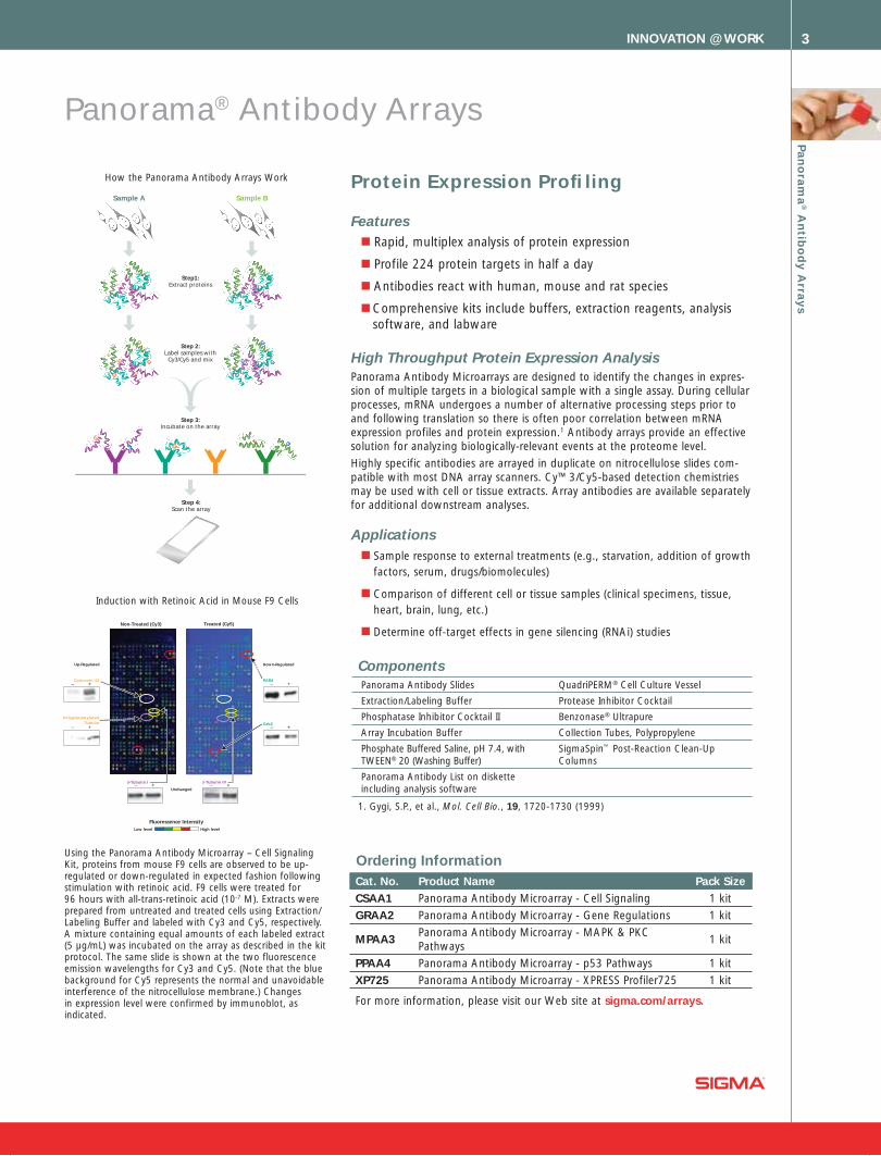
Step 2:Label samples with
Cy3/Cy5 and mix
Step 3:Incubate on the array
Step 4:Scan the array
Sample BSample A
Step1:Extract proteins
Panorama® Antibody Arrays
Protein Expression Profi ling
Features■ Rapid, multiplex analysis of protein expression
■ Profile 224 protein targets in half a day
■ Antibodies react with human, mouse and rat species
■ Comprehensive kits include buffers, extraction reagents, analysis software, and labware
High Throughput Protein Expression AnalysisPanorama Antibody Microarrays are designed to identify the changes in expres-sion of multiple targets in a biological sample with a single assay. During cellular processes, mRNA undergoes a number of alternative processing steps prior to and following translation so there is often poor correlation between mRNA expression profiles and protein expression.1 Antibody arrays provide an effective solution for analyzing biologically-relevant events at the proteome level.
Highly specific antibodies are arrayed in duplicate on nitrocellulose slides com-patible with most DNA array scanners. Cy™3/Cy5-based detection chemistries may be used with cell or tissue extracts. Array antibodies are available separately for additional downstream analyses.
Applications■ Sample response to external treatments (e.g., starvation, addition of growth
factors, serum, drugs/biomolecules)
■ Comparison of different cell or tissue samples (clinical specimens, tissue, heart, brain, lung, etc.)
■ Determine off-target effects in gene silencing (RNAi) studies
Connexin 43
Non-Treated (Cy3) Treated (Cy5)
Up-Regulated Down-Regulated
PolyglutamylatedTubulin
– +
– +Grb2
– +
PAR4– +
Unchanged
β-Tubulin I– + – +
β-Tubulin IV
High levelLow level
Fluorescence Intensity
Using the Panorama Antibody Microarray – Cell Signaling Kit, proteins from mouse F9 cells are observed to be up-regulated or down-regulated in expected fashion following stimulation with retinoic acid. F9 cells were treated for 96 hours with all-trans-retinoic acid (10-7 M). Extracts were prepared from untreated and treated cells using Extraction/Labeling Buffer and labeled with Cy3 and Cy5, respectively. A mixture containing equal amounts of each labeled extract (5 µg/mL) was incubated on the array as described in the kit protocol. The same slide is shown at the two fluorescence emission wavelengths for Cy3 and Cy5. (Note that the blue background for Cy5 represents the normal and unavoidable interference of the nitrocellulose membrane.) Changes in expression level were confirmed by immunoblot, as indicated.
Induction with Retinoic Acid in Mouse F9 Cells
Pan
ora
ma
® An
tibo
dy A
rrays
Ordering InformationCat. No. Product Name Pack SizeCSAA1 Panorama Antibody Microarray - Cell Signaling 1 kit
GRAA2 Panorama Antibody Microarray - Gene Regulations 1 kit
MPAA3Panorama Antibody Microarray - MAPK & PKC Pathways
1 kit
PPAA4 Panorama Antibody Microarray - p53 Pathways 1 kit
XP725 Panorama Antibody Microarray - XPRESS Profiler725 1 kit
For more information, please visit our Web site at sigma.com/arrays.
ComponentsPanorama Antibody Slides QuadriPERM® Cell Culture Vessel
Extraction/Labeling Buffer Protease Inhibitor Cocktail
Phosphatase Inhibitor Cocktail II Benzonase® Ultrapure
Array Incubation Buffer Collection Tubes, Polypropylene
Phosphate Buffered Saline, pH 7.4, with TWEEN® 20 (Washing Buffer)
SigmaSpin™ Post-Reaction Clean-Up Columns
Panorama Antibody List on diskette including analysis software
1. Gygi, S.P., et al., Mol. Cell Bio., 19, 1720-1730 (1999)
How the Panorama Antibody Arrays Work
sigma.com
INNOVATION @ WORK4
GenomePlex® Single Cell Whole Genome Amplifi cation KitWhole Genome Amplifi cation from a Single Cell
Ordering InformationCat. No. Product Name Pack Size
WGA2 GenomePlex Complete Whole Genome Amplification (WGA) Kit 10 reactions50 reactions
WGA4 GenomePlex Single Cell Whole Genome Amplification Kit 10 reactions50 reactions
WGA3 GenomePlex WGA Reamplification Kit 50 reactions
WGA5 GenomePlex Tissue Whole Genome Amplification Kit 10 reactions50 reactions
NA1020 GenElute PCR Clean-Up Kit 1 kit
G1N10 GenElute Mammalian Genomic DNA Miniprep Kit 1 kit
S4438 SYBR® Green JumpStart™ Taq ReadyMix™ 100 reactions500 reactions
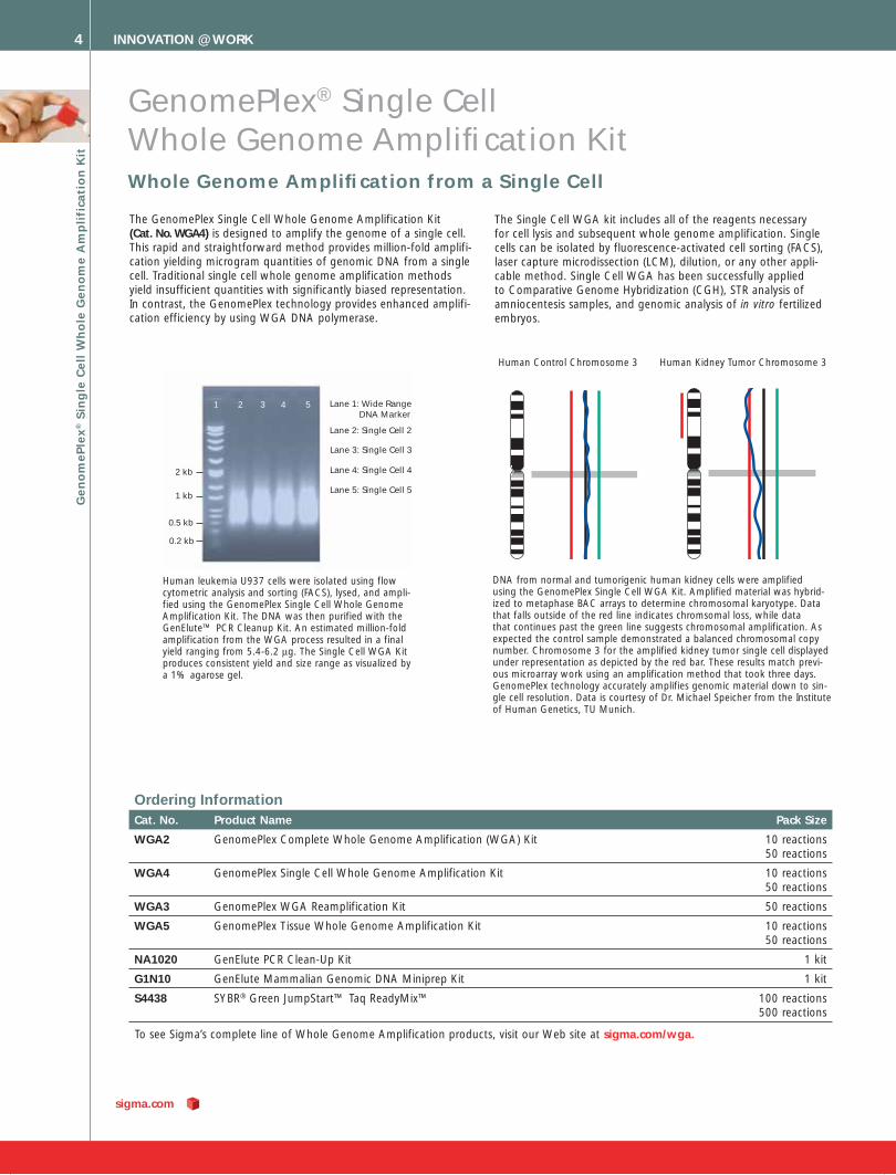
2 kb
1 kb
0.5 kb
0.2 kb
Lane 1: Wide Range DNA Marker
Lane 2: Single Cell 2
Lane 5: Single Cell 5
Lane 4: Single Cell 4
Lane 3: Single Cell 3
1 2 3 4 5
Human leukemia U937 cells were isolated using flow cytometric analysis and sorting (FACS), lysed, and ampli-fied using the GenomePlex Single Cell Whole Genome Amplification Kit. The DNA was then purified with the GenElute™ PCR Cleanup Kit. An estimated million-fold amplification from the WGA process resulted in a final yield ranging from 5.4-6.2 μg. The Single Cell WGA Kit produces consistent yield and size range as visualized by a 1% agarose gel.
To see Sigma’s complete line of Whole Genome Amplification products, visit our Web site at sigma.com/wga.
Gen
om
ePle
x® S
ing
le C
ell
Wh
ole
Gen
om
e A
mp
lifi
cati
on
Kit
The GenomePlex Single Cell Whole Genome Amplification Kit (Cat. No. WGA4) is designed to amplify the genome of a single cell. This rapid and straightforward method provides million-fold amplifi-cation yielding microgram quantities of genomic DNA from a single cell. Traditional single cell whole genome amplification methods yield insufficient quantities with significantly biased representation. In contrast, the GenomePlex technology provides enhanced amplifi-cation efficiency by using WGA DNA polymerase.
The Single Cell WGA kit includes all of the reagents necessary for cell lysis and subsequent whole genome amplification. Single cells can be isolated by fluorescence-activated cell sorting (FACS), laser capture microdissection (LCM), dilution, or any other appli-cable method. Single Cell WGA has been successfully applied to Comparative Genome Hybridization (CGH), STR analysis of amniocentesis samples, and genomic analysis of in vitro fertilized embryos.
Human Control Chromosome 3 Human Kidney Tumor Chromosome 3
DNA from normal and tumorigenic human kidney cells were amplified using the GenomePlex Single Cell WGA Kit. Amplified material was hybrid-ized to metaphase BAC arrays to determine chromosomal karyotype. Data that falls outside of the red line indicates chromsomal loss, while data that continues past the green line suggests chromosomal amplification. As expected the control sample demonstrated a balanced chromosomal copy number. Chromosome 3 for the amplified kidney tumor single cell displayed under representation as depicted by the red bar. These results match previ-ous microarray work using an amplification method that took three days. GenomePlex technology accurately amplifies genomic material down to sin-gle cell resolution. Data is courtesy of Dr. Michael Speicher from the Institute of Human Genetics, TU Munich.

INNOVATION @ WORK 5
Amplifi cation of Genome-Representative DNA from Limited Starting Material
Achieve Robust Amplifi cation Representative of the Original Input Genome
GenomePlex® Complete Whole Genome Amplifi cation Kit
M 1 2 3 4 5 6 7 8 9 10 11 12 13 14
GenomePlex WGA was performed on genomic DNA isolated from HT29 colon carcinoma cells and from a healthy human male. 2.5 µg of WGA product was labeled with Cy™3 or Cy5 dye using the Genomic DNA Labeling Kit PLUS (Agilent). The entire labeled sample was loaded onto an Agilent Human Genome CGH Microarray 105A. Specific activities were between 28 and 43 for all samples, and always within 50% of samples being compared. The dye swaps (A & B) demonstrate that there was no bias in the DNA labeling and the aberrations detected are consistent with the HT-29 karyotype.
WGA was performed on increasing concentrations of human genomic DNA. Amplification product can be detected on an agarose gel with as little as 10 pg of input DNA. Optimal performance is found with 1 to 10 ng of starting material. Increasing the amount of input DNA to 100 ng is not recommended.
Lane M: DNA MarkerLanes 1,2: no template LanesLanes 3,4: 1 pg DNA LaneLanes 5,6: 10 pg DNA Lanes
Lanes 7,8: 100 pg DNALanes 9,10: 1 ng DNALanes 11,12: 10 ng DNALanes 13,14: 100 ng DNA
Gen
om
ePlex® C
om
plete W
ho
le Gen
om
e Am
plifi catio
n K
it
The GenomePlex Complete Whole Genome Amplification Kit (Cat. No. WGA2) contains everything required for whole genome amplification including an optimized enzyme, WGA DNA Polymerase. The WGA DNA Polymerase provides increased accu-racy in amplification, as evidenced by producing no amplicon in the negative control reactions. WGA has been used in a variety of applications, and is suitable for use with purified genomic DNA from a variety of sources including blood cards, whole blood, buccal swabs, tissue, soil, plant, and serum. GenomePlex WGA uses nanogram quantities of starting genomic DNA, which after PCR yields on average 10 µg of amplified DNA. After purification, the WGA product can be analyzed in a manner similar to any genomic or chromosomal DNA sample. A number of downstream applications may be performed including TaqMan® assays, CGH analysis, SNP analysis, and sequencing.
NEW! Technical note on Agilent Array CGH with WGA, visit sigma.com/wgacgh.
Looking for a high-throughput system for the rapid and highly representative amplification of genomic DNA from trace amounts of starting material? Visit sigma.com/wgaautomation for auto-mated WGA protocols and methods.

sigma.com
INNOVATION @ WORK6
Kit
Recovery of phosphopeptide standards
Specificity*1 2 3 Total
Sigma 59% 52% 74% 59% 50%
Competitor A 6% 19% 11% 13% 28%
Competitor B 39% 56% 17% 42% 28%
Competitor C 37% 65% 37% 46% 25%
Performance summary of the IMAC technologies tested within this study.*Specificity was measured as a percentage of the total HPLC peak area corresponding to phosphorylated peptides that appeared in the elution fractions.
PhosphoProfi le™ Phosphopeptide Enrichment Kit
Phosphoproteomics — Phosphopeptide EnrichmentMatrix Assisted Laser Desorption/Ionization, Time of Flight (MALDI-TOF) or Electrospray Ionization (ESI) Mass Spectrometry of phosphopeptides from tryp-tic protein digests are powerful tools for characterization and identification of phosphorylation sites. A combination of low intrinsic abundance, inefficient ionization, and/or signal suppression of most phosphopeptides may limit or even prevent detection, unless the phosphopeptide content is significantly enriched prior to analysis. This kit conveniently includes all materials needed to enrich phosphopeptides from digested samples in a robust and unbiased manner (see binding comparison). The phosphopeptide capture matrix is a novel Ga+3 chelate silica based on a proprietary nitriloacetic acid (NTA) analog. The silica beads are approximately 20 microns in diameter with a pore size of 1,000 Angstroms. The matrix is packed into spin columns for easy, microscale affinity capture of phos-phopeptides.
Binding Comparison: Demonstrating enrichment of phosphopeptides for IMAC technologies. Standard phosphopeptides representing the three most common sites of phos-phorylation (phosphoserine, phosphothreonine, and phosphotyrosine) were used. The lyophilized solids were first dissolved in water, and an approximately equimolar mixture of the peptides was formulated. Each phosphopeptide was added to a BSA digest at a weight ratio of ~1.7% to produce a total phospho-peptide content of ~5% by weight. Quantitation results are given in the table below. Note that competitor A, B, and C technologies were biased in selecting Peptide 2, while Sigma’s technology bound and eluted the peptides in approxi-mately the same ratio as applied to the column.
The Complete Solution Kit 24 samples up to 25 nmoles phosphopeptide each.
Features and Benefi ts■ Mass spec compatible – save time,
reduce sample handling and potential loss
■ Proteomics Grade Trypsin – for clean and complete digests
■ Phos-SelectTM Gallium Spin Columns – high capacity, novel Ga+3 silica media for fast, unbiased capture and recovery
■ Controls – validate your process for confidence and reporting
■ Buffers – enzyme reaction, binding, washing and elution formulations optimized for robust performance
■ Consumable equipment included – matched equipment means no riskof sample loss, additional purchases,or waste
TrypticDigest
PhosphopeptideEnrichment
Flow throughfraction
Elutedfraction
Workflow highlighting the alternate use of trypsin spin columns (Cat. No. TT0010) for digestion fol-lowed by selective enrichment of phosphopeptides from sample.
Ph
osp
ho
Pro
file
™ P
ho
sph
op
ep
tid
e E
nri
chm
en
t K
it
Ordering InformationCat. No. Product Name Pack Size
PP0410 PhosphoProfi le I Phosphopeptide Enrichment Kit 1 kit
TT0010 Trypsin Spin Columns Proteomics Grade 10 each
For more information, please visit our Web site at sigma.com/pep.

7
si
gm
a-
al
dr
ic
h.
co
m/
bi
of
il
es
Role of ABC Transporters and Multi-Drug Resistance Reversal Using RNA Interference (RNAi)*
Chemotherapy is the treatment of choice against many types of cancer. However, over time chemotherapeutic drugs can become less effective due to the development of resistance that involves a group of membrane proteins. These multi-drug transporters expel cytotoxic molecules from the cell, thus keeping intracellular drug concentrations below the cell-killing threshold. These transporters belong to the superfamily of ATP Binding Cassette (ABC) proteins that are present in all organisms from bacteria to humans. The transport activity of ABC proteins has an important effect on the efficacy of pharmaceuticals by modulating the absorption, distri-bution, and excretion of these xenobiotics.
ABC transporter proteins are located in the plasma membrane of cells and in the membranes of cellular organelles where they mediate the transport of various substrate molecules. These sub-strates exhibit a wide variety of chemical structures. Most ABC proteins are active transporters, which utilize the energy generated by ATP hydrolysis; however, some ABC transporters form trans-membrane channels.
Cancer Multi-Drug Resistance - The PlayersNumerous clinical data revealed that the multi-drug resistance phenotype in tumors is associated with the overexpression of certain ABC transporters, termed multi-drug resistance (MDR) proteins. P-glycoprotein (P-gp, MDR1, ABCB1) was the first discov-ered ABC transporter1-3 and is likely to be responsible for the most widely observed mechanism in clinical multi-drug resistance.4-7 Soon after the cloning and characterization of MDR1, it became evident that other efflux pumps also play significant roles in trans-port-associated drug resistance. Two other ABC transporters have definitively demonstrated participation in the multi-drug resistance of tumors: the multi-drug resistance protein 1 (MRP1, ABCC1), and the mitoxantrone resistance protein (MXR/BCRP, ABCG2).7-11
Basic Mechanism of Cancer Multi-Drug Resistance and Substrate Specifi city of MDR-ABC TransportersThe generally accepted mechanism of multi-drug resistance is that the MDR proteins actively expel the cytotoxic drugs from cells, maintaining the drug concentration within the cells below the toxic level. The drug efflux mediated by these primary active transporters is driven by the energy of ATP hydrolysis. Tumors with MDR protein overexpression (e.g., hepatomas, lung or colon carcinomas) often show primary (or intrinsic) resistance to chemo-therapy treatment. In addition, chemotherapy itself may induce the overexpression of these proteins, resulting in the multi-drug resistant clones becoming less sensitive to treatment (secondary drug resistance).12-14
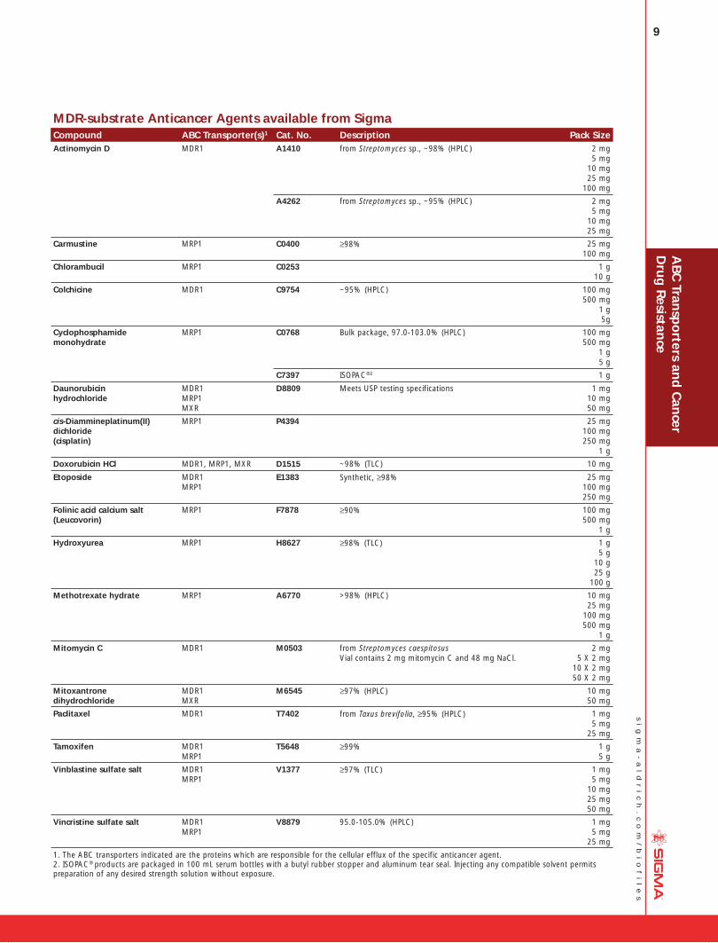
The most intriguing characteristic distinguishing the MDR proteins from other mammalian transporters is their broad substrate speci-ficity. Unlike other selective (classical) transport proteins, multi-drug transporters recognize and handle a large number of struc-turally diverse, mainly hydrophobic compounds, which explains cross-resistance to several chemically unrelated compounds, a characteristic feature of the multi-drug resistance phenotype.4-7 In addition to their overlapping substrate specificity, each transporter can handle unique compounds. The following table of MDR-sub-strate anticancer agents provides a selection of anticancer agents available from Sigma and identifies the key ABC transporter(s) responsible for each agent’s cellular efflux.
Circumvention of Cancer Multi-Drug ResistancePrevention of clinical multi-drug resistance should significantly improve therapeutic response in a large number of cancer patients. The initial search for pharmacological modulators of MDR transporters yielded two generations of compounds having poor clinical response profiles. Therefore, there has been a shift to structure-based drug design to synthesize modulator com-pounds characterized by a high affinity to MDR transporters.15,16 Additionally, research that utilizes siRNA and shRNA-mediated RNAi-based gene silencing methodology has recently delivered promising results.
MDR Modifying Agents MDR modifying agents, which competitively or non-competitively inhibit the MDR proteins, may increase the cytotoxic action of multi-drug resistant related drugs by preventing the active efflux of these drugs from the target cells. The co-application of an “MDR-modulating” compound in combination with chemotherapy would be expected to significantly improve the cancer cure rate. The first-generation modulators consisted of compounds that were already in clinical use. Calcium channel blockers, quinine derivatives, calmodulin inhibitors, and the immunosuppressive agent cyclosporin A, were all shown to interact with the MDR transporters in vitro and in vivo. These modulators were not spe-cifically developed for MDR protein inhibition, and their inherent low affinity for MDR transporters resulted in a high toxicity profile and were never shown to inhibit P-gp in patients.15-18
Most of the second-generation modulators were derivatives of the first-generation compounds that retained MDR modulatory effects, but with reduced activity toward other physiological targets. Prominent examples of this group are R-verapamil, biricodar (VX-710), and valspodar (PSC-833). These modulators were shown to inhibit P-gp in patients, but further study revealed significant phar-mokinetic interaction with several anticancer drugs, which delayed excretion of the anticancer agent, resulting in toxicity requiring reduction of anticancer drug doses.15-18
The third-generation MDR modulators are designed to interact with specific MDR transporters7, 11-13, 19, 20 with high affinity and with efficacy at nanomolar concentrations. Development of this class of MDR modulators employed combinatorial chemistry to produce potent and selective inhibitors. Examples are the small hydrophobic peptide derivatives named reversins, which were shown to have a strong inhibitory effect on P-gp/MDR1-mediated drug efflux without any toxic effect in the control cells.21 Ongoing clinical trials using third-generation MDR modulators for specific cancer types include: elacridar (GF120918), tariquidar (XR9576), zosuquidar (LY335979), laniquidar (R101933), and ONT-093. Still the shortcomings of earlier generation modulators continue to exist.17, 22 Other approaches to prevent the expression or function of multi-drug transporters are being considered, including the use of MDR protein targeted antibodies, the use of carriers that deliver these drugs selectively to tumor tissues, and the use of RNA inter-ference.
ABC Transporters and Cancer Drug Resistance A
BC
Transp
orters an
d C
ancer
Dru
g R
esistance
8
si
gm
a-
al
dr
ic
h.
co
m/
bi
of
il
es
Multi-Drug Resistance Reversal Using RNAiThe stable reversal of MDR protein-mediated drug efflux by RNAi technology has been demonstrated in vitro for MDR1, MXR, MRP2, and MDR3. One of the early multi-drug resistance stud-ies using RNAi technology reported a complete suppression of MDR1 expression on the mRNA and protein level in human gastric carcinoma cells.23 A subsequent study further demonstrated inhi-bition of both MDR1 and MDR3 expression in conjunction with the reversal of paclitaxel resistance in human ovarian cancer cells. Treatment of ovarian cancer cell lines with either chemically syn-thesized siRNAs or transfection with specific vectors that express targeted siRNAs resulted in decreased mRNA and protein levels. In this study, MTT assays of siRNA-treated cells demonstrated 7 to 12.4-fold reduction of paclitaxel resistance in the lines treated with the synthesized siRNA of MDR1 and 4.7 to 7.3-fold reduc-tion of paclitaxel resistance in the cell lines transfected with siRNA of MDR1 expressing vectors.24 A more recent study surprisingly showed that the MDR1 phenotype in human hepatoma cells was completely reversed by using two transfected clones.25 Aside from the more frequently studied MDR1 phenotype, reversal of the drug-resistant MXR and MRP2 phenotype using both siRNA and shRNA-mediated approaches was also demonstrated in human carcinoma cells.26, 27
In a pre-clinical study the ablation of MDR1 in cells stably trans-duced with shRNA was functionally confirmed by increased sensitivity of MDR1-transfected cells toward the cytotoxic drugs vincristine, paclitaxel, and doxorubicin as well as by transport of 99mTc-sestamibi. In the same study, shRNA-mediated down-regula-tion of MDR activity in tumor implants in living animals was fol-lowed by direct noninvasive bioluminescence imaging using the fluorophore coelenterazine, a known MDR1 transport substrate. Additionally, a MDR1-firefly luciferase (MDR1-FLuc) fusion con-struct was used to document the effect of shRNA delivered in vivo on MDR1-FLuc protein levels with D-luciferin bioluminescence imaging.28 A similar study validated selective MRP2 gene function inhibition: after the intravenous delivery of siRNA effectors into mice, researchers observed a significantly reduced calcein excretion rate and resultant siRNA accumulation in the kidney.29
RNAi is proving to be a powerful laboratory tool for better under-standing the multi-drug resistance genotype and phenotype. Its future therapeutic utility in suppressing gene expression in cancer patients will likely be dependent on the availability of effective RNAi delivery systems. Lessons can be learned from the history of gene therapy and antisense technologies. These technologies ultimately failed to produce successful clinical outcomes due to potentially harmful and inefficient delivery systems. The use of RNAi in complex genetic diseases, such as cancer, will not see a quick and straightforward transition from research to clinical suc-cess, but with time the promise of viable RNAi therapies may be realized.30 Additionally, innovative technologies combined with new directions in the study of ABC transporters will lead to an understanding of whether or not ABC transporters are important molecular targets for anticancer drug development.
*Technical content provided by: Balázs Sarkadi1, Gergely Szakács1,2 and András Váradi3
1. National Medical Center, Institute of Haematology and Immunology, Budapest, Hungary2. National Cancer Institute, NIH, Bethesda, MD, USA3. Institute of Enzymology, Hungarian Academy of Sciences, Budapest, Hungary
AB
C T
ran
spo
rter
s an
d C
ance
r D
rug
Res
ista
nce
ABC Transporters and Cancer Drug Resistance
References:
1. Juliano, R.L. and Ling, V., Biochim. Biophys. Acta, 455, 152-162 (1976).2. Chen, D., et al., Cell, 47, 381-389 (1986).3. Ueda, K., et al., Proc. Natl. Acad. Sci. USA, 84, 3004-3008 (1987).4. Endicott, J.A. and Ling, V., Annu. Rev. Biochem., 58, 137-171 (1989).5. Higgins, C.F., Ann. Rev. Cell Biol., 8, 67-113 (1992).6. Gottesman, M.M. and Pastan, I., Annu. Rev. Biochem., 62, 385-427 (1993).7. Gottesman, M.M., et al., Nat. Rev. Cancer, 2, 48-58 (2002).8. Cole, S.P.C., et al., Science, 258, 1650-1654 (1992).9. Borst, P., et al., J. Natl. Cancer Inst., 92, 1295-1302 (2000).10. Deeley, R.G. and Cole, S.P.C., Sem. Cancer Biol., 8, 193-204 (1997).11. Litman, T., et al., Cell. Mol. Life Sci., 58, 931-959 (2001).12. List, A.F., et al., Blood, 98, 3212-3220 (2001).13. Ambudkar, S.V., et al., Annu. Rev. Pharmacol. Toxicol., 39, 361-398 (1999).14. Sarkadi, B., et al., Semin. Cancer Biol., 8, 171-182 (1997).15. Perez-Tomas, R., Curr. Med. Chem., 13(16), 1859-76 (2006).16. McDevitt, C.A. and Callaghan, R., Pharmacol. Ther., 113(2), 429-441 (2007).17. B. A. Teicher (ed.), Humana Press, Totowa, NJ, Cancer Drug Resistance, (2006), 2.15.18. Leonard, G.D., et al., Curr. Opin. Investig. Drugs, 3, 1652-1659 (2002).19. Krishna, R., et al., Eur. J. Pharm. Sci., 11, 265-283 (2000).20. Leslie, E.M., et al., Toxicology, 5, 3-23 (2001).21. Sharom, F.J., et al., Biochem. Pharmacol., 58, 571-586 (1999).22. Nobili, S., et al., Curr. Drug Targets, 7(7), 861-879 (2006).23. Stege, A., et al., Cancer Gene Ther., 11(11), 699-706 (2004).24. Zhenfeng, D., et al., Mol. Cancer Ther., 3(7), 833-838 (2004).25. Chen, X.P., et al., World J. Gastroenterol., 12(21), 3332-3337 (2006).26. Priebsch, A., et al., Oligonucleotides, 16(3), 263-274 (2006).27. Materna, V., et al., Biochem. Biophys. Res. Commun., 348(1), 153-157 (2006).28. Pichler, A., et al., Clin. Cancer Res., 11(12), 4487-4494 (2005).29. van de Water, F.M., et al., Drug Metab. Dispos., 34(8), 1393-1397 (2006).30. Downward, J., BMJ, 328(7450), 1245-1248 (2004).
For more information on siRNA available through Sigma-Genosys and the comprehensive MISSION TRC shRNA library, targeting 15,000 human and 15,000 mouse genes, visit our Web site at sigma.com/rnai.
To obtain more information on specific ABC trans-porter shRNA clones, email [email protected].
9
si
gm
a-
al
dr
ic
h.
co
m/
bi
of
il
es
MDR-substrate Anticancer Agents available from SigmaCompound ABC Transporter(s)1 Cat. No. Description Pack SizeActinomycin D MDR1 A1410 from Streptomyces sp., ~98% (HPLC) 2 mg
5 mg10 mg25 mg
100 mg
A4262 from Streptomyces sp., ~95% (HPLC) 2 mg 5 mg10 mg
25 mg
Carmustine MRP1 C0400 ≥98% 25 mg 100 mg
Chlorambucil MRP1 C0253 1 g 10 g
Colchicine MDR1 C9754 ~95% (HPLC) 100 mg 500 mg
1 g 5g
Cyclophosphamide monohydrate
MRP1 C0768 Bulk package, 97.0-103.0% (HPLC) 100 mg 500 mg
1 g 5 g
C7397 ISOPAC®2 1 g
Daunorubicin hydrochloride
MDR1MRP1MXR
D8809 Meets USP testing specifications 1 mg 10 mg50 mg
cis-Diammine platinum(II) dichloride(cisplatin)
MRP1 P4394 25 mg 100 mg250 mg
1 g
Doxorubicin HCl MDR1, MRP1, MXR D1515 ~98% (TLC) 10 mg
Etoposide MDR1MRP1
E1383 Synthetic, ≥98% 25 mg 100 mg250 mg
Folinic acid calcium salt(Leucovorin)
MRP1 F7878 ≥90% 100 mg 500 mg
1 g
Hydroxyurea MRP1 H8627 ≥98% (TLC) 1 g 5 g
10 g25 g
100 g
Methotrexate hydrate MRP1 A6770 >98% (HPLC) 10 mg 25 mg
100 mg500 mg
1 g
Mitomycin C MDR1 M0503 from Streptomyces caespitosusVial contains 2 mg mitomycin C and 48 mg NaCl.
2 mg5 X 2 mg
10 X 2 mg50 X 2 mg
Mitoxantrone dihydrochloride
MDR1MXR
M6545 ≥97% (HPLC) 10 mg50 mg
Paclitaxel MDR1 T7402 from Taxus brevifolia, ≥95% (HPLC) 1 mg 5 mg25 mg
Tamoxifen MDR1MRP1
T5648 ≥99% 1 g 5 g
Vinblastine sulfate salt MDR1MRP1
V1377 ≥97% (TLC) 1 mg 5 mg
10 mg25 mg
50 mg
Vincristine sulfate salt MDR1MRP1
V8879 95.0-105.0% (HPLC) 1 mg 5 mg25 mg
1. The ABC transporters indicated are the proteins which are responsible for the cellular efflux of the specific anticancer agent.2. ISOPAC® products are packaged in 100 mL serum bottles with a butyl rubber stopper and aluminum tear seal. Injecting any compatible solvent permits preparation of any desired strength solution without exposure.
AB
C Tran
spo
rters and
Can
cer D
rug
Resistan
ce
10
si
gm
a-
al
dr
ic
h.
co
m/
bi
of
il
es
ABC Transporter Membrane Proteins
MDR1 humanABCB1; Pgp
The MDR1 protein is involved in cancer drug resistance and in the transport of hydrophobic drugs and xenobiotics in the bowel, kid-ney, liver, and the blood-brain barrier. Drugs interacting with this protein may be useful for the reversal of cancer drug resistance or increasing the absorption or brain entry of various pharmacologi-cal agents.
Membrane preparation, for ATPase, recombinant, expressed in Sf9 cellsSupplied as isolated Sf9 cell membranes containing human MDR1 (Pgp) suspended in TMEP solution.Distributed for SOLVO Biotechnology, Inc.
A DRY ICE
M9194 500 µL
Mdr1b from ratThe MDR1 protein is involved in cancer drug resistance and in the transport of hydrophobic drugs and xenobiotics in the bowel, kidney, liver, and the blood-brain barrier. In rodents, there are two MDR1 genes, mdr1a and mdr1b, while in human, there is a single MDR1 gene. Based on function and tissue distribution in rodents, the equivalent of the human MDR1 gene product (PgP) is the product of the rodent mdr1b gene. There have been no reported significant differences in function, substrate specificity, or substrate affinity between these two proteins.
Membrane preparation, for ATPase, recombinant, expressed in Sf9 cells. Supplied as isolated Sf9 cell membranes containing rat Mdr1b suspended in TMEP solution.Distributed for SOLVO Biotechnology, Inc.
A DRY ICE
M9319 500 µL
MRP2 humanMRP2 (ABCC2) is an organic anion transporter found in the liver, kidney, and gut epithelium apical membranes. The transport of glucuronate conjugates plays a role in the detoxification of endog-enous and xenobiotic substances, and may cause multidrug resis-tance (MDR) in tumor cells.
Membrane preparation, for Vesicular Transport, recombinant, expressed in Sf9 cellsSupplied as isolated Sf9 cell membranes containing human MRP2 suspended in TMEP solution. Distributed for SOLVO Biotechnology, Inc.
A DRY ICE
M9069 500 µL
ABC Transporters and Cancer Drug Resistance A
BC
Tra
nsp
ort
ers
and
Can
cer
Dru
g R
esis
tan
ce
Mrp2 from ratMRP2 (ABCC2) is an organic anion transporter found in liver, kidney, and gut epithelium apical membranes. The transport of glucuronate conjugates plays a role in the detoxification of endogenous and xenobiotic substances, and may cause multidrug resistance (MDR) in tumor cells. The rat Mrp2 transporter shows 72.3% sequence identity and 85.6% sequence similarity with human MRP2. Both transporters are expressed on the canalicular membrane of the liver and are known to be responsible for the transport of some organic molecules and their conjugates to the bile.
Membrane preparation, for ATPase, recombinant, expressed in Sf9 cellsSupplied as isolated Sf9 cell membranes containing rat Mpr2 suspended in TMEP solution. Distributed for SOLVO Biotechnology, Inc.
A DRY ICE
M9694 500 µL
MXR humanMXR membrane vesicles are purified from recombinant baculovirus transduced Sf9 cells or selected, MXR over-expressing mammalian cells.Distributed for SOLVO Biotechnology, Inc.
Membrane preparation, wild type variant, for Vesicular TransportSupplied as isolated mammalian cell membranes containing human MXR (wild type variant) suspended in TMEP solution.
The MXR transporter can be produced in sufficient quantity by selected, MXR over-expressing mammalian cell lines.
A DRY ICE
M9569 500 µL
Membrane preparation, wild type variant, for Vesicular Transport, recombinant, expressed in Sf9 cellsSupplied as isolated Sf9 cell membranes containing wild type human MXR suspended in TMEP solution.The MXR transporter can be expressed in Sf9 insect cells using the baculoviral expression system, yielding high protein levels (up to 5% of total membrane protein) in the cell membrane of infected cells.
A DRY ICE
M9444 500 µL
MXR ControlMembrane preparation, from Sf9 cellsControl for ATPase and vesicular transport assays.
Supplied as isolated Sf9 cell membranes containing a defective MXR gene suspended in TMEP solution.Distributed for SOLVO Biotechnology, Inc.
A DRY ICE
M9944 500 µL
MXR ControlMembrane preparation, mammalianControl for ATPase and vesicular transport assays.
Supplied as isolated mammalian cell membranes (not selected for transport expression) suspended in TMEP solution. Distributed for SOLVO Biotechnology, Inc.
A DRY ICE
C3992 500 µL
11
si
gm
a-
al
dr
ic
h.
co
m/
bi
of
il
es
Antibodies to ABC Transporters
Monoclonal Anti-Breast Cancer Resistance Protein antibody produced in mouseAnti-ABCG2; Anti-BCRP
250 µg/mL, clone BXP-21, tissue culture supernatantImmunogen: fusion protein containing human BCRP (amino acids 271-396) and maltose-binding protein. Reacts with an internal epitope of BCRP. Does not cross-react with the human MDR1, MRP1, MRP2 gene products.
Supplied in serum-free medium containing 0.7% bovine serum albumin and 0.1% sodium azide.
Species reactivity: human
Antigen mol wt ~70 kDa
Application(s)Immunocytochemistry ............................ 1:20-1:50 using acetone-fixed cytospin preparationsImmunohistochemistry (frozen sections) ......... 1:20 using acetone-fixed frozen sectionsImmunohistochemistry (formalin-fixed, paraffin-embedded sections).................................................suitable using pretreated human tissueImmunoblotting .......................................................................suitableIsotype ........................................................................................ IgG2aB DRY ICE
B7059 1 mL
Anti-Breast Cancer Resistance Protein antibody produced in rabbitAnti-BCRP
Affinity isolated antibodyImmunogen: synthetic peptide corresponding to amino acid resi-dues 150-167 of human breast cancer resistance protein with C-terminal added cysteine, conjugated to KLH.
Solution in 0.01 M phosphate buffered saline containing 15 mM sodium azide.
Species reactivity: human, mouse
Antigen mol wt ~70 kDa
Application(s)Immunoblotting .............................2.5-5 µg/mL using whole extract of
human term placenta or mouse kidney and a chemiluminescent detection reagent
Immunohistochemistry (formalin-fixed, paraffin-embedded sections) ............................... 20-40 µg/mL using heat-retrieved tissue
sections from human term placenta by indirect immunoperoxidase staining of syncytiotrophoblasts.
B DRY ICE
B7185 200 µL
AB
C Tran
spo
rters and
Can
cer D
rug
Resistan
ce
Monoclonal Anti-P-Glycoprotein (MDR) antibody produced in mouseClone F4, ascites fluidImmunogen: mixture of human and hamster drug-resistant whole cells and crude plasma membranes.
The antibody recognizes an epitope located in the amino terminal half of P-glycoprotein (Pgp), at the third extra cellular loop of the molecule. The epitope is resistant to formalin fixation and peri-odate oxidation.1 The antibody detects specifically human MDR1 P-glycoprotein, but does not appear to recognize the human MDR3 product1, nor the mouse mdr1a, mdr1b or the mdr3 P-gly-coprotein.2
Contains 15 mM sodium azide
Species reactivity: human, hamster
Antigen mol wt 170-180 kDaApplication(s)Immunoblotting ........................................................................suitableRadioimmunoassay ............................... suitable using cell-surface RIAImmunoprecipitation ............................................................... suitableImmunohistochemistry (frozen sections) ...................................suitableImmunocytochemistry ............................................................. suitableFlow cytometry ........................................................................suitableIndirect ELISA ...........................................................................suitableImmunohistochemistry (formalin-fixed, paraffin-embedded sections)......................................................1:500 using human kidney sectionsIsotype ........................................................................................ IgG1
Lit. cited: 1. Chu, T.M., et al., Hybridoma 12, 417 (1993)2. Chu, T.M., et al., Biochem. Biophys. Res. Commun. 203, 506 (1994)B DRY ICE
P7965 0.2 mL
Monoclonal Anti-MDR3 P-Glycoprotein antibody produced in mouse250 µg/mL, clone P3II-26, tissue culture supernatantImmunogen: MDR3 P-gp (amino acids 629-692) GST fusion pro-tein.
Reacts with an internal epitope of MDR3. Does not cross-react with human MDR1 P-gp.
Supplied in serum-free medium containing 0.7% bovine serum albumin and 0.1% sodium azide.
Species reactivity: human
Application(s)Immunocytochemistry ............................ 1:20-1:50 using acetone-fixed cytospin preparationsImmunohistochemistry (frozen sections) ............................... 1:20 using acetone-fixed sectionsImmunohistochemistry (formalin-fixed, paraffin-embedded sections)..........................................................................................Not suitableImmunoblotting ........................................................................suitableIsotype ........................................................................................ IgG2bB DRY ICE
M7317 1 mL
12
si
gm
a-
al
dr
ic
h.
co
m/
bi
of
il
es
Monoclonal Anti-MRP1 antibody produced in mouseAnti-Multidrug Resistance Associated Protein 1
~2 mg/mL, clone QCRL-1, purified immunoglobulinImmunogen: non-denatured membrane preparations of H69AR human small cell lung cancer cell line, which highly expresses MRP1. The epitope resides within amino acids 918-924 of human MRP1. Does not cross-react with the human MDR1 and MDR3, mouse MRP1, and human MRP2, MRP3, MRP4, MRP5, and MRP6 gene products. The antibody may cross-react with canine MRP1.
Solution in 0.01 M phosphate buffered saline, pH 7.4, containing 1% BSA and 15 mM sodium azide.
Species reactivity: human
Antigen mol wt ~190 kDa
Application(s)Flow cytometry ........................ 0.5-2 µg/mL using human H69AR cellsImmunoblotting ........................................................................suitableImmunoprecipitation .................................................................suitableImmunohistochemistry ..............................................................suitableImmunocytochemistry ...............................................................suitableIsotype .......................................................................................... IgG1B DRY ICE
M9067 200 µL
Anti-MRP2 antibody produced in rabbitAnti-ABCC2; Anti-cMOAT; Anti-cMRP; Anti-Multidrug Resistance Associated Protein 2
Affinity isolated antibodyImmunogen: synthetic C-terminal peptide corresponding to amino acids 1528-1545 of human MRP2 conjugated to KLH.
Additional lower bands including an approx. 175 kDa band rep-resenting an immature unglycosylated form may be detected in various extract preparations.
Solution in 0.01 M phosphate buffered saline, pH 7.4, containing 1% bovine serum albumin and 15 mM sodium azide.
Species reactivity: human, rat
Antigen mol wt ~190 kDa
Application(s)Immunoblotting ....................................1:1,000 using whole extract of
HepG2 human hepatoblastoma cellsImmunoblotting .............................1:1,000 using 293T cells expressing
recombinant human MRP2Indirect immunofluorescence ............................................. 1:100 using
paraformaldehyde-fixed HepG2 cellsIndirect immunofluorescence ........ 1:100 using rat liver frozen sectionsB DRY ICE
M8316 200 µL
AB
C T
ran
spo
rter
s an
d C
ance
r D
rug
Res
ista
nce
ABC Transporters and Cancer Drug Resistance
Monoclonal Anti-MRP2 antibody produced in mouseAnti-ABCC2; Anti-cMOAT; Anti-cMRP; Anti-Multidrug Resistance Associated Protein 2
~1.5 mg/mL, clone CPR96, purified immunoglobulinImmunogen: synthetic C-terminal peptide corresponding to amino acid residues 1528-1545 of human MRP2 with N-terminal added cysteine conjugated to KLH.
Solution in 0.01 M phosphate buffered saline, pH 7.4, containing 15 mM sodium azide.
Species reactivity: human
Antigen mol wt ~180 kDa
Application(s)Immunoblotting ...................................1-2 µg/mL using cell extracts of
293T cells transfected with human MRP2Indirect ELISA ............................................................................suitableArray ........................................................................................suitableIsotype .......................................................................................... IgG1B DRY ICE
M3692 200 µL
Anti-MRP3 antibody produced in rabbitAnti-Multidrug Resistance Associated Protein 3
Affinity isolated antibodyImmunogen: synthetic C-terminal peptide of human MRP3 (amino acids 1507-1527) with N-terminal cysteine conjugated to KLH. The sequence in mouse and rat differs by three residues.
Solution in 0.01 M phosphate buffered saline, pH 7.4, containing 15 mM sodium azide.
Species reactivity: human, rat
Antigen mol wt ~180 kDa
Application(s)Immunoblotting .................................................................2.5-5 µg/mL
using whole extract of cultured human colon carcinoma HCT-116 cells
Indirect immunofluorescence .......... 15-30 µg/mL using frozen sections of rat skin (outer root sheaths
of hair follicles)B DRY ICE
M0318 200 µL
Monoclonal Anti-MRP5 antibody produced in ratAnti-Multidrug Resistance Associated Protein 5
250 µg/mL, clone M5I-1, tissue culture supernatantImmunogen: human MRP5 (amino acids 82-168) bacterial fusion protein. Reacts with an internal epitope of MRP5. Does not cross-react with the human MDR1, MRP1, MRP2, MRP3 gene products.
Supplied in serum-free culture medium containing 0.7% bovine serum albumin and 0.1% sodium azide.
Species reactivity: human
Antigen mol wt 190-200 kDa
Application(s)Immunocytochemistry .............. 1:20-1:50 using acetone-fixed cytospin
preparationsImmunohistochemistry (frozen sections) ............................... 1:20 using
acetone-fixed frozen sectionsImmunoblotting .................................................................... 1:20-1:50Isotype ........................................................................................ IgG2aB DRY ICE
M6067 1 mL
13
si
gm
a-
al
dr
ic
h.
co
m/
bi
of
il
es
Compounds for MDR Detection
bisBenzimide H 33342 trihydrochloridebisBenzimide; 2’-(4-Ethoxyphenyl)-5-(4-methyl-1-piperazinyl)-2,5’-bi-1H-benzimidazole trihydrochloride; HOE 33342; Hoechst 33342C27H28N6O · 3HCl · xH2O FW 561.93 (Anh) [23491-52-3]
Membrane-permeable, fluorescent DNA stain with low cytotoxicity that intercalates in A-T regions of DNA.
Useful for staining DNA, chromosomes and nuclei. May be used for fluorescence microscopy or flow cytometry.
Excitation max. = 346 nm
Emission max. = 460 nm
≥95% (TLC)
B EC No. 2456911 BRN 1234011
B2261 25 mg, 100 mg, 500 mg, 1 g
CalceinBis[N,N−bis(carboxymethyl)aminomethyl]fluorescein; Fluorescein-bis(methyliminodiacetic acid); Fluorexon
C30H26N2O13 FW 622.53 [1461-15-0]
Used for the fluorometric determination of calcium and EDTA titration of calcium in the presence of magnesium.
B EC No. 2159571 BRN 8181068
C0875 5 g, 10 g, 25 g
Calcein AM solutionC46H46N2O23 FW 994.86 [148504-34-1]
;4 mM in DMSO, ;90% (TLC)Fluorescent cell permeable derivative of calcein.
B
C1359 100 µL
Coelenterazine, nativeCLZN; 3,2-Dihydro-2-(p-hydroxybenzyl)-6-(p-hydroxy phenyl)-8-benzylimidazo-lo[1,2-a]pyrazin -3-oneC26H21N3O3 FW 423.46 [55779-48-1]
Luminophore of the aequorin complex which is oxidized by oxy-gen to illuminate at 465 nm when Ca2+ binds to the complex; used to measure Ca2+ concentration in cells with high sensitivity and large dynamic range.
B
C2230 50 µg
AB
C Tran
spo
rters and
Can
cer D
rug
Resistan
ce
Luciferase from Photinus pyralis (firefly)[61970-00-1] E.C. 1.13.12.7
Lyophilized powder, 15-30 × 106 light units/mg proteinArsenate free.
Lyophilized powder approximately 20% protein; balance is primar-ily NaCl, HEPES buffer salts, and carbohydrate.
Mol wt 120 kDa (two non-identical subunits, each containing four free thiol groups, one of which is necessary for activity)
Composition Protein ~20% 1%280E(
(
Chromatographically prepared and crystallized.
Sold on the basis of protein content
One light unit produces a biometer peak height equivalent to 0.02 µCi of 14C in PPO/POPOP cocktail. Light units measured in 50 µl assay mixture containing 5 pmol ATP and 7.5 nmol luciferin in Tris-glycine buffer, pH 7.6, at 25 °C.Sensitivity ................................................................:1 femtomole ATP
(using 0.2 µg of luciferase and suitably sensitive liquid scintillation counters or luminometers)
ATPase ................................................<5 nanomolar units/mg proteinNucleoside diphosphokinase .............. <20 nanomolar units/mg proteinB EC No. EINECS
L9506 1 mg, 2 mg, 10 mg
D-Luciferin4,5-Dihydro-2-(6-hydroxy-2-benzothiazolyl)-4-thi azole carboxylic acid; Firefly luciferin; (S)-2-(6-Hydroxy-2-benzo thiazolyl)-2-thiazoline-4-carboxylic acidC11H8N2O3S2 FW 280.32 [2591-17-5] EC No. 2199813 BRN 30484
Synthetic, SigmaUltra, ≥99% (HPLC)Highly purified grade with reduced levels of inhibitor relative to L9504
L6152 1 mg, 10 mg
SyntheticL9504 1 mg, 5 mg, 10 mg, 50 mg, 100 mg
Rhodamine 1232-(6-Amino-3-imino-3H-xanthen-9-yl)benzoic acid methyl ester; Rh 123C21H17ClN2O3 FW 380.82 [62669-70-9]
Fluorescent dye most commonly used in flow cytometry as func-tional reporter for P-glycoprotein (P-gp). Functional assays for MDR proteins are better prognostic indicators in cancer therapy than levels of MDR protein expression. Rh 123 can be used in multipa-rameter analyses without fluorescence interference in combination with common protein labeling dyes such as PE-Cy™5 and AMCA (7-amino-4-methyl coumarin-3-acetic acid). Recent reports indicate Rh 123 may also be a substrate of MRP1. Used as a laser dye and for selective cell growth effects.
EC No. 2636878 BRN 6030951
R8004 5 mg, 25 mg, 100 mg

14
si
gm
a-
al
dr
ic
h.
co
m/
bi
of
il
es
Infl ammation and Cancer: the NF-κB Connection
Chronic inflammation is an underlying factor in the development and progression of many of the chronic diseases of aging, such as arthritis, atherosclerosis, diabetes, and cancer. Oxidative cel-lular stress induced by environmental factors, such as cigarette smoke, UV or ionizing radiation, bacterial or viral infection, or any number of oxidizing xenobiotic compounds, triggers a wide range of cellular responses, some of which are proinflammatory and proapoptotic, while others protect the cell against apoptosis and enhance cellular adhesion, cell proliferation, and angiogenesis. The inappropriate induction or constitutive activation of these protec-tive responses in mutated or damaged cells appears to be a major factor in the transformation and proliferation of cancer cells.1–5
NF-κB and Infl ammation N
F-κB
an
d In
flam
mat
ion
Two nuclear transcription factors that are involved in mediating the cellular responses to oxidative cell stress and proinflamma-tory stimuli are activator protein-1 (AP-1) and nuclear factor-κB (NF-κB). The role of these transcription factors on cancer ini-tiation and progression has been studied in cell culture and in vivo models.5,6 The activation of two AP-1 components, c-Jun and c-Fos, by JNK and by ERK1/2 or p38 MAPK, respectively, is involved in the malignant transformation of cells stimulated by the tumor promotor phorbol 12-myristate 13-acetate (PMA). The proinflammatory and antiapoptotic response to tumor promotion is primarily mediated through activation of NF-κB by the IKK fam-ily of serine/threonine kinases. The following discussion will focus on the NF-κB pathway as a target for cancer chemotherapy and chemoprevention.
Figure 1. Many diverse stimuli utilize intracellular signaling pathways to activate NF-κB, a nuclear transcription factor that regulates proinflammatory and cell survival pathways.

15
si
gm
a-
al
dr
ic
h.
co
m/
bi
of
il
es
NF-κB Transcription Factors NF-κB refers to a family of transcription factors that has been highly conserved through evolution and is present in the cyto-plasm of all cells. NF-κB has been called a “stress sensor” because its activity is induced by a wide variety of stimuli,7 including tumor necrosis factor (TNF-α), PMA and other tumor promoters, ciga-rette smoke extract (CSE), lipopolysaccharide (LPS), oxidants, and pathogenic bacteria. The NF-κB family comprises five members: p50 (NF-κB1), p52 (NF-κB2), RelA (p65), RelB, and c-Rel. p50 and p52 are cleaved from inactive precursor proteins, p105 and p100, respectively, prior to translocation to the nucleus. NF-κB family members are characterized by having:
• a Rel homology domain that binds to DNA
• a dimerization domain
• the ability to bind to the intracellular inhibitor complex, IκB
The most widely studied NF-κB heterodimers are p50/p65 and p50/c-Rel (both associated with the classical or canonical pathway) and p52/RelB (alternative pathway). The classical pathway is acti-vated by inflammatory cytokines, bacterial and viral infections, and oxidative stimuli and induces gene expression responsible for the antiapoptotic actions of NF-κB. The alternative pathway is primar-ily involved in B cell survival.2,7–9 The classical pathway is illustrated in Figure 1.
Cytoplasmic NF-κB is sequestered as an inactive complex with its regulatory subunit, IκB. The most abundant member of the IκB family of proteins is IκBα. Phosphorylation of two conserved ser-ine residues in the N-terminal domain of the NF-κB/IκB complex induces the rapid dissociation and polyubiquitination of IκB fol-lowed by its degradation by the 26S proteasome. Activated NF-κB translocates to the nucleus where specific subunit lysines are acet-ylated by SRC-1 and p300 histone acetyltransferases. Acetylation promotes DNA binding and NF-κB-induced gene transcription.10 A list of more than 200 proteins that are regulated by NF-κB is given by Ahn and Aggarwal.7 Many of the genes regulated by NF-κB code for inflammatory cytokines and proteins that mediate cell survival, cellular adhesion, cell cycle activation, cell prolifera-tion, angiogenesis, and oncogenesis. However, not all the actions of NF-κB promote cell survival. Activation of NF-κB also appears to be essential for p53-induced apoptosis in response to oxidative stress or to the anticancer agents, doxorubicin and etoposide.2,11
Activation of NF-κB via PhosphorylationPhosphorylation of the NF-κB/IκB complex is catalyzed by IKK, a protein complex that contains two homologous kinase subunits (IKKα and IKKβ) and a regulatory subunit IKKγ/NEMO.17 Activation of the IKK complex can be initiated by any one of several intracell-ular phosphorylation pathways, including NF-κB-inducing kinase (TRAF/NIK),8,12,13 MEK1,11,14 ERK5,15 and PI3K/Akt.16 T cell, B cell and lysophosphatidic acid receptors activate a kinase cascade that results in the activation of the IKK complex by Bcl10, Malt1 and CARMA-1.9,17 Acetylation of serines and threonines in the activa-tion loop of the IKKα and IKKβ subunits can prevent phosphoryla-tion and activation of the IKK complex.18 In addition, regulation of the NF-κB/IκB complex can also occur independently of IKK activation or inhibition. Both the PI3K/Akt and JAK/STAT/Pim kinase pathways activate NF-κB by phosphorylating Cot, a serine-threonine kinase that can induce the proteasomal degradation of IκB.19 Upregulation or over-expression of mitogen activated pro-tein kinase phosphatase-5 (MKP5) can decrease cytokine-induced phosphorylation of NF-κB/IκB and of p38 MAP kinase.20
NF-κB
and
Inflam
matio
n
There are two mechanisms by which the NF-κB-induced gene transcription is terminated. Genes coding for IκB complex proteins are upregulated by NF-κB. Newly formed IκBα subunits can enter the nucleus where they bind to and inactivate NF-κB, and the NF-κB/IκBα complex is exported back to the cellular cytoplasmic compartment.9 Alternatively, the translational action of DNA-bound NF-κB can be terminated by deacetylation of the p65 subunit at Lys310 by the histone deacetylases SIRT1 or HDAC1.10,21 Inhibition of p300 histone acetyltransferase or the overexpression or activation of SIRT1 has been shown to inhibit NF-κB-mediated gene expression.10,22 Conversely, decreased histone deacetylase level or activity has been shown to increase the expression of inflammatory cytokines, presumably through enhancement of NF-κB-mediated gene transcription.23
NF-κB and its Relationship to DiseaseAltered regulation of NF-κB activity is observed in many geneti-cally-linked diseases and chronic diseases of aging, including cancer.7,9 NF-κB activation has been linked to inflammation-driven tumor promotion and progression. In addition, many solid and hematopoietic cancers express constitutively active NF-κB that contributes to the pathogenesis of the disease by inducing factors that promote proliferation, invasiveness, angiogenesis, and resis-tance to chemotherapeutic agents and radiation. Researchers have hypothesized that inhibition of NF-κB activation or transcriptional activity may delay cancer onset or may be used as an adjunct to more traditional chemotherapeutics. Many compounds, including many phytochemicals and micronutrients having putative chemo-preventive properties, inhibit NF-κB activation or constitutive NF-κB activity in cellular or in vivo models of cancer.2,5,7,24 The anticancer activities of some of these phytochemicals are summarized in the table on page 16.
Many of the inducers of NF-κB also regulate other intracellular pathways that mediate cell cycle arrest or apoptosis. One hypoth-esis holds that inhibition of prosurvival pathways mediated via NF-κB allows expression of proapoptotic mechanisms that may also be mediated by the various cell signaling pathways. For example, TNF-α is an inflammatory cytokine that mediates a broad spectrum of biological actions via activation of the TNFR1 recep-tor and, depending on the cellular environment, can promote either cell survival or programmed cell death. On the one hand, TNF-α/TNFR1-induced activation of TRAF2 initiates kinase cascades that lead to phosphorylation and activation of AP-1 and NF-κB, thereby promoting gene expression, cell survival and proliferation. Conversely, through its interactions with TRADD or FADD, TNF-α/TNFR1 initiates signaling pathways that activate caspase 8 and the proteolytic cascade that ends in apoptotic cell death. Furthermore, some of the genes induced by NF-κB activation, such as Gadd45β and XIAP, are inhibitors of prolonged JNK activation. JNK activa-tion facilitates mitogen-induced and oxidative stress-induced apop-tosis. Thus, in the presence of NF-κB inhibitors, TNFR1 activation would favor the proapoptotic, anticancer actions of TNF-α.2,13,25
It should be noted that many phosphorylation pathways, e.g., PI3K-Akt, JNK, and ERK, are involved in both prosurvival and pro-apoptotic cellular processes. Therefore, the physiological response to procarcinogenic and anticarcinogenic xenobiotics represents the sum of all the specific intracellular signaling pathways that are up-regulated and down-regulated in response to these stimuli. While chemopreventive natural products may tip the balance toward cell death in some damaged or transformed cells, their actions may promote carcinogenesis or drug resistance in other cells or organisms.
16
si
gm
a-
al
dr
ic
h.
co
m/
bi
of
il
es
NF-
κB a
nd
Infl
amm
atio
n
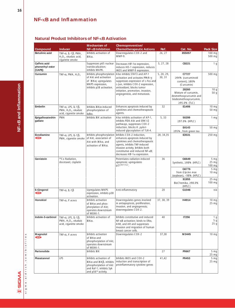
Natural Product Inhibitors of NF-κB Activation
Compound InducerMechanism of NF-κB Inhibition
Chemopreventive/Chemotherapeutic Actions Ref. Cat. No. Pack Size
Betulinic acid TNF-α, IL-1β, PMA, H2O2, okadaic acid, cigarette smoke
Inhibits activation of IKKα.
Downregulates COX-2 and MMP-9.
26, 27 855057 100 mg500 mg
Caffeic acid phenethyl ester (CAPE)
Suppresses p65 nuclear translocalization; inhibits MAPK.
Decreases HIF-1α expression, induces HO-1 expression, reduces iNOS and MMP-9 expression.
5, 27, 28 C8221 1 g
Curcumin TNF-α, PMA, H2O2 Inhibits phosphorylation of Akt and activation of IKKα; upregulates MKP5 expression, inhibits p38 activation.
Also inhibits STAT3 and AP-1 activation and activates PPAR-γ; suppresses expression of c-Fos and c-Jun, inhibits COX-2 expression, antioxidant; blocks tumor initiation, promotion, invasion, angiogenesis, and metastasis.
5, 20, 29, 30, 31
C7727≥94% (curcuminoid
content), ≥80% (Curcumin)
500 mg
28260Mixture of curcumin,
desmethoxycurcumin andbisdesmethoxycurcumin,
≥95.0% (TLC)
10 g50 g
Embelin TNF-α, LPS, IL-1β, PMA, H2O2, okadaic acid, cigarette smoke
Inhibits IKKα-induced phosphorylation of IκBα.
Enhances apoptosis induced by cytokines and chemotherapeutic agents.
32 E1406 10 mg50 mg
Epigallocatechin gallate
PMA Inhibits IKK activation Also inhibits activation of AP-1, inhibits PI3K-Akt and ERK1/2 pathways, suppresses COX-2 induction, blocks H. pylori-induced glycosylation of TLR-4.
5, 33 50299≥97.0% (HPLC)
1 mg
E4143≥95%, from green tea
50 mg
Evodiamine8
TNF-α, LPS, IL-1β, PMA, cigarette smoke
Inhibits phosphorylation of Akt, association of Akt with IKKα, and activation of IKKα.
Inhibits COX-2 induction, enhances apoptosis induced by cytokines and chemotherapeutic agents, inhibits TNF-induced invasive activity. Inhibits both constitutive and induced NF-κB; decreases HIF-1α expression.
28, 34,35 E3531 250 mg
Genistein 60Co Radiation, docetaxel, cisplatin
Potentiates radiation-induced apoptosis; upregulates p21WAF1/Cip1.
36 G6649Synthetic, ≥98% (HPLC)
5 mg 25 mg
100 mgG6776
from Glycine max (soybean), ~98% (HPLC)
5 mg 10 mg
91955BioChemika, ≥98.0%
(HPLC)
25 mg100 mg
6-Gingerol8
TNF-α, IL-1β Upregulates MKP5 expression, inhibits p38 activation.
Anti-infl ammatory 20 G1046 n/a
Honokiol TNF-α, P. acnes Inhibits activation of IKKα and phos-phorylation of Akt; operates downstream of MEKK-1.
Downregulates genes involved in antiapoptosis, proliferation, invasion, and angiogenesis; downregulates COX-2.
37, 38, 39 H4914 10 mg25 mg
Indole-3-carbinol TNF-α, LPS, IL-1β, PMA, H2O2, okadaic acid, cigarette smoke
Inhibits activation of IKKα.
Inhibits constitutive and induced NF-κB activation; binds to ERα, AhR, and AR and suppresses invasion and migration of human breast cancer cells.
40 I7256 1 g 5 g
25 g
Magnolol8
TNF-α, P. acnes Inhibits activation of IKKα and phosphorylation of Akt, operates downstream of MEKK-1.
Downregulates COX-2 37,38 M3445 10 mg
Partenolide Inhibits IKK 27 P0667 5 mg 25 mg
Piceatannol LPS Inhibits activation of IKKα and IKKβ, inhibits phosphorylation of Akt and Raf-1; inhibits Syk and p56lck activity.
Inhibits iNOS and COX-2 induction and transcription of proinfl ammatory cytokine genes
41,42 P0453 5 mg25 mg
NF-κB and Infl ammation
17
si
gm
a-
al
dr
ic
h.
co
m/
bi
of
il
es
NF-κB
and
Inflam
matio
n
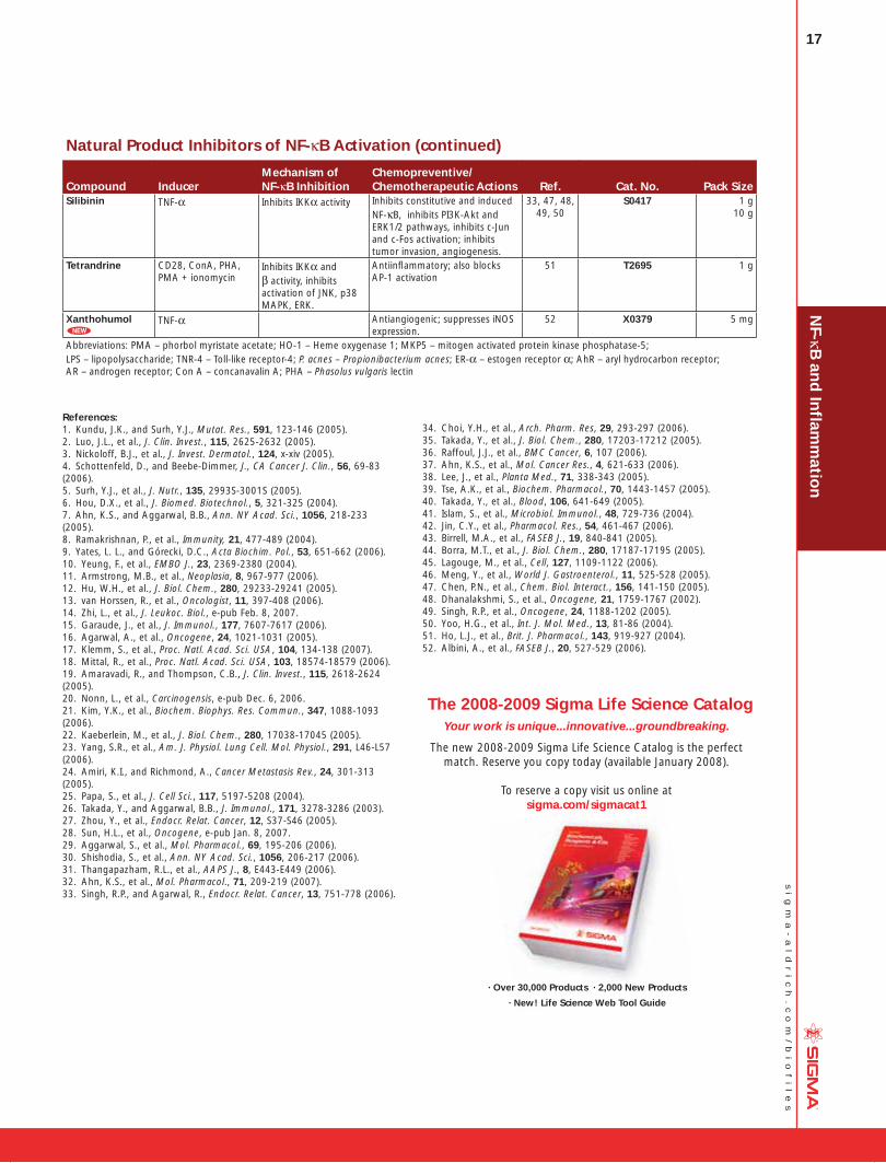
Natural Product Inhibitors of NF-κB Activation (continued)
Compound InducerMechanism of NF-κB Inhibition
Chemopreventive/Chemotherapeutic Actions Ref. Cat. No. Pack Size
Silibinin TNF-α Inhibits IKKα activity Inhibits constitutive and induced NF-κB, inhibits PI3K-Akt and ERK1/2 pathways, inhibits c-Jun and c-Fos activation; inhibits tumor invasion, angiogenesis.
33, 47, 48, 49, 50
S0417 1 g 10 g
Tetrandrine CD28, ConA, PHA, PMA + ionomycin
Inhibits IKKα and β activity, inhibits activation of JNK, p38 MAPK, ERK.
Antiinfl ammatory; also blocks AP-1 activation
51 T2695 1 g
Xanthohumol8
TNF-α Antiangiogenic; suppresses iNOS expression.
52 X0379 5 mg
Abbreviations: PMA – phorbol myristate acetate; HO-1 – Heme oxygenase 1; MKP5 – mitogen activated protein kinase phosphatase-5; LPS – lipopolysaccharide; TNR-4 – Toll-like receptor-4; P. acnes – Propionibacterium acnes; ER-α – estogen receptor α; AhR – aryl hydrocarbon receptor; AR – androgen receptor; Con A – concanavalin A; PHA – Phasolus vulgaris lectin
References:1. Kundu, J.K., and Surh, Y.J., Mutat. Res., 591, 123-146 (2005).2. Luo, J.L., et al., J. Clin. Invest., 115, 2625-2632 (2005).3. Nickoloff, B.J., et al., J. Invest. Dermatol., 124, x-xiv (2005).4. Schottenfeld, D., and Beebe-Dimmer, J., CA Cancer J. Clin., 56, 69-83 (2006).5. Surh, Y.J., et al., J. Nutr., 135, 2993S-3001S (2005).6. Hou, D.X., et al., J. Biomed. Biotechnol., 5, 321-325 (2004).7. Ahn, K.S., and Aggarwal, B.B., Ann. NY Acad. Sci., 1056, 218-233 (2005).8. Ramakrishnan, P., et al., Immunity, 21, 477-489 (2004).9. Yates, L. L., and Górecki, D.C., Acta Biochim. Pol., 53, 651-662 (2006).10. Yeung, F., et al., EMBO J., 23, 2369-2380 (2004). 11. Armstrong, M.B., et al., Neoplasia, 8, 967-977 (2006).12. Hu, W.H., et al., J. Biol. Chem., 280, 29233-29241 (2005).13. van Horssen, R., et al., Oncologist, 11, 397-408 (2006).14. Zhi, L., et al., J. Leukoc. Biol., e-pub Feb. 8, 2007.15. Garaude, J., et al., J. Immunol., 177, 7607-7617 (2006).16. Agarwal, A., et al., Oncogene, 24, 1021-1031 (2005).17. Klemm, S., et al., Proc. Natl. Acad. Sci. USA, 104, 134-138 (2007).18. Mittal, R., et al., Proc. Natl. Acad. Sci. USA, 103, 18574-18579 (2006).19. Amaravadi, R., and Thompson, C.B., J. Clin. Invest., 115, 2618-2624 (2005).20. Nonn, L., et al., Carcinogensis, e-pub Dec. 6, 2006.21. Kim, Y.K., et al., Biochem. Biophys. Res. Commun., 347, 1088-1093 (2006).22. Kaeberlein, M., et al., J. Biol. Chem., 280, 17038-17045 (2005).23. Yang, S.R., et al., Am. J. Physiol. Lung Cell. Mol. Physiol., 291, L46-L57 (2006).24. Amiri, K.I., and Richmond, A., Cancer Metastasis Rev., 24, 301-313 (2005).25. Papa, S., et al., J. Cell Sci., 117, 5197-5208 (2004).26. Takada, Y., and Aggarwal, B.B., J. Immunol., 171, 3278-3286 (2003).27. Zhou, Y., et al., Endocr. Relat. Cancer, 12, S37-S46 (2005).28. Sun, H.L., et al., Oncogene, e-pub Jan. 8, 2007.29. Aggarwal, S., et al., Mol. Pharmacol., 69, 195-206 (2006).30. Shishodia, S., et al., Ann. NY Acad. Sci., 1056, 206-217 (2006).31. Thangapazham, R.L., et al., AAPS J., 8, E443-E449 (2006).32. Ahn, K.S., et al., Mol. Pharmacol., 71, 209-219 (2007). 33. Singh, R.P., and Agarwal, R., Endocr. Relat. Cancer, 13, 751-778 (2006).
34. Choi, Y.H., et al., Arch. Pharm. Res, 29, 293-297 (2006).35. Takada, Y., et al., J. Biol. Chem., 280, 17203-17212 (2005).36. Raffoul, J.J., et al., BMC Cancer, 6, 107 (2006).37. Ahn, K.S., et al., Mol. Cancer Res., 4, 621-633 (2006).38. Lee, J., et al., Planta Med., 71, 338-343 (2005).39. Tse, A.K., et al., Biochem. Pharmacol., 70, 1443-1457 (2005).40. Takada, Y., et al., Blood, 106, 641-649 (2005).41. Islam, S., et al., Microbiol. Immunol., 48, 729-736 (2004).42. Jin, C.Y., et al., Pharmacol. Res., 54, 461-467 (2006).43. Birrell, M.A., et al., FASEB J., 19, 840-841 (2005).44. Borra, M.T., et al., J. Biol. Chem., 280, 17187-17195 (2005). 45. Lagouge, M., et al., Cell, 127, 1109-1122 (2006).46. Meng, Y., et al., World J. Gastroenterol., 11, 525-528 (2005).47. Chen, P.N., et al., Chem. Biol. Interact., 156, 141-150 (2005).48. Dhanalakshmi, S., et al., Oncogene, 21, 1759-1767 (2002).49. Singh, R.P., et al., Oncogene, 24, 1188-1202 (2005).50. Yoo, H.G., et al., Int. J. Mol. Med., 13, 81-86 (2004).51. Ho, L.J., et al., Brit. J. Pharmacol., 143, 919-927 (2004).52. Albini, A., et al., FASEB J., 20, 527-529 (2006).
The 2008-2009 Sigma Life Science CatalogYour work is unique...innovative...groundbreaking.
The new 2008-2009 Sigma Life Science Catalog is the perfect match. Reserve you copy today (available January 2008).
To reserve a copy visit us online at sigma.com/sigmacat1
· Over 30,000 Products · 2,000 New Products
· New! Life Science Web Tool Guide

18
si
gm
a-
al
dr
ic
h.
co
m/
bi
of
il
es
NF-
κB a
nd
Infl
amm
atio
nNF-κB and Infl ammation
Antibodies and Kits
Anti-IKKα (699-715) antibody produced in rabbitAnti-IκB Kinase αAffinity isolated antibodyImmunogen: synthetic peptide corresponding to amino acids 699-715 of human IκB Kinase α (IKK α) (C2)1,2. This sequence contains one amino acid substitution with the mouse sequence.
Solution in phosphate buffered saline containing 0.02% sodium azide.
Species reactivity: human
Antigen mol wt 85 kDa
Application(s)Immunoblotting ....................... 1 µg/mL using human HeLa cell extractLit Cited: 1. DiDonato, J.A., et al., Nature, 388, 548-554, (1997).2. Regnier, C.H., et al., Cell, 90, 373-383 (1997)B DRY ICE
I7778 0.1 mg
Anti-IKKα (716-734) antibody produced in rabbitAnti-IκB Kinase αAffinity isolated antibodyImmunogen: synthetic peptide corresponding to amino acids 716-734 of human IκB Kinase α (IKKα) (C1).
Solution in phosphate buffered saline containing 0.02% sodium azide.
Species reactivity: human
Antigen mol wt 85 kDa
Application(s)Immunoblotting .................... 0.5 µg/mL using human HeLa cell extractB DRY ICE
I7903 0.1 mg
Anti-IKKβ antibody produced in rabbitAnti-IKK2; Anti-IκB Kinase βAffinity isolated antibodyImmunogen: synthetic peptide located near the C-terminal region of human IKKβ (amino acids 720-735).
Solution in 0.01 M phosphate buffered saline, pH 7.4, containing 1% bovine serum albumin and 15 mM sodium azide.
Antigen mol wt 85 kDa
Application(s)Immunoblotting ........................................ 1:500 using a whole extract of human epitheloid carcinoma HeLa cells
B DRY ICE
I9767 0.2 mL
Anti-IKKγ/NEMO (N-Terminal) antibody produced in rabbitAnti-IKKAP1
~0.5 mg/mL, affinity isolated antibodyImmunogen: synthetic peptide corresponding to amino acids 400-416 of human IKKγ.
Solution in phosphate buffered saline containing 0.02% sodium azide.
Species reactivity: human, mouse, rat
Antigen mol wt ~52 kDa
Application(s)Immunoblotting .................................................................0.5-1 µg/mLB DRY ICE
I5032 100 µg
Monoclonal Anti-NF-κB antibody produced in mouseclone NF-12, ascites fluidImmunogen: recombinant mouse NF-κB p65 fragment (C-terminal, 151 amino acids).
Contains 15 mM sodium azide
Species reactivity: mouse, human
Antigen mol wt 65 kDa
Application(s)Immunocytochemistry .............................................................. suitableImmunoblotting ............................... 1:1,000 using a whole cell extract from a mouse fibroblast cell lineArray ........................................................................................suitableIsotype .................................................................................. IgG1
B DRY ICE
N8523 0.2 mL
NF-κB (p50) humanFull length, 453 amino acid protein from spliced cDNA containing the p105 precursor polypeptide. For use in gel shift and footprint-ing assays, as well as in vitro transcriptional activation.
recombinant, expressed in Escherichia coli, buffered aque-ous glycerol solutionSolution in 50 mM NaCl, 5 mM DTT, 0.5 mM PMSF, 20 mM HEPES, 10 µM zinc acetate, 0.1% NP-40, and 10% glycerol.
Unit Definition : One gel shift unit is defined as the amount of p50 required to shift 0.38 pmol of NF-κB oligonucleotide.
A DRY ICE
N9909 50 un
8 Histone Deacetylase Assay Kit, Fluorometric Histone deacetylases (HDACs) are enzymes that remove acetyl groups from histone proteins. HDACs are important regulators of gene expression and genome function. They are implicated in a number of human disease states, namely several cancers, neu-rological disorders, and aging. Therefore, HDAC are relevant key targets for therapeutic intervention. Moreover, histone deacetylase inhibitors have been shown to serve as antitumor agents.
The Histone Deacetylase Assay Kit provides a simple method for the detection of HDAC activity based on a two-step enzymatic reaction. The substrate for this enzymatic assay is a substituted peptide with an acetylated lysine residue and a bound fluorescent group. The first step of the reaction is deacetylation of the acety-lated lysine side chain by the HDAC containing sample (HeLa cell extract, purified enzyme, etc.). The second step is the cleavage of the deacetylated substrate by the developer solution and the release of the free highly fluorescent group. The measured fluo-rescence is directly proportional to the deacetylation activity of the sample.
The Histone Deacetylase Assay Kit has been tested on HeLa, HEK 293T, NIH 3T3, and U 973 cell extracts. The kit includes all the reagents required for the fast and easy measurement of HDAC activity in cell or nuclear extracts, or with purified enzyme prepara-tions. In addition, the kit provides HeLa cell lysate as a source of HDAC activity for inhibitor screening or as a positive control, a HDAC inhibitor (Trichostatin A), and a standard to enable activity quantitation.
Sufficient for 100 assays (96 well plates)
B DRY ICE
CS1010 1 kit
19
si
gm
a-
al
dr
ic
h.
co
m/
bi
of
il
es
8 Sirt1 human
SIR2α; SIR2L1; Sirtuin1
Sirtuins are a family of NAD+ dependent deacetylases that remove an acetyl group from the e-amino group of lysine residues. The proteins within this family are named after the first protein dis-covered, from yeast, called Sir2 (Silent Information Regulator 2). The proteins are conserved from bacteria to higher eukaryotes. In humans, there are seven Sir2 family members (SIRT1 to SIRT7). SIRT1 plays a pivotal role in the regulation of cellular differentia-tion, metabolism, cell cycle, apoptosis and regulation of p53. Several targets for SIRT1 were identified, among them Lys382 of p53.1 Using RNA interference, additional targets were identified. It was demonstrated that reduced levels of human SIRT1 led to increased acetylation of Histone H4-Lys16, H4-Lys20, and Histone H3-Lys9 as well as histone H1-Lys26.2
Recombinant, expressed in Escherichia coli, N-terminal histidine tagged, >90% (SDS-PAGE)Solution containing 50 mM Tris, pH 7.4, 100 mM NaCl, 1 mM DTT, protease inhibitors (Cat. No. P8340) 1:200, and 10% glycerol (w/v).Lit cited: 1. Vaziri, H., et al., Cell 107, 149-159 (2001) 2. Vaquero, A., et al., Mol. Cell. 16, 93-105 (2004) B DRY ICE
S8446 150 µg
Monoclonal Anti-Sirt1 antibody produced in mouseAnti-mouse homologue of yeast Sir2; Anti-Sir2α; Anti-Sirtuin1
~2 mg/mL, clone SIR11, purified immunoglobulinImmunogen: synthetic peptide corresponding to amino acids 722-737 at the C-terminus of mouse Sir2 with C-terminal added lysine.
Solution in 0.01 M phosphate buffered saline, pH 7.4, containing 15 mM sodium azide.
Species reactivity: mouse
Antigen mol wt ~105 kDa
Application(s)Immunoblotting .............. 1-2 µg/mL using total cell extract of C-2 cellsIndirect ELISA ............................................................................suitableArray ........................................................................................suitableIsotype .......................................................................................... IgG1B DRY ICE
S5196 200 µL
NF-κB
and
Inflam
matio
n
8 SIRT1 Assay KitThe assay procedure is based on a two-step enzymatic reaction. The first step is deacetylation by SIRT1 of a substrate that contains an acetylated lysine side chain. The second step is the cleavage of the deacetylated substrate by the Developing Solution and the release of a highly fluorescent group. The measured fluorescence is directly proportional to the deacetylation activity of the enzyme in the sample. The kit offers all the reagents required for the fast and easy measurement of purified SIRT1 activity and for screen-ing of inhibitors/activators. Moreover, the kit contains an inhibitor (nicotinamide) and an activator (resveratrol) as negative and posi-tive controls, respectively.Sufficient for 100 assaysComponents Assay Buffer ..........................................................................20 mL SIRT1 Substrate (Fluorometric) ..............................................100 µL Standard (non-acetylated) 20 mM ........................................100 µL Developer Solution ...............................................................1.5 mL Nicotinamide Solution (inhibitor) ..........................................100 µL Sirt1 human ........................................................................ 150 µg NAD+ Solution .........................................................................1 mL Resveratrol Solution (activator) .............................................100 µLB DRY ICE
CS1040 1 kit
Anti-Sir2 (AS-16) antibody produced in rabbit Affinity isolated antibodyImmunogen: synthetic peptide corresponding to mouse Sir2 sequence (amino acids 722-737) with N-terminal cysteine added conjugated to KLH. This sequence is 62% homologous to the cor-responding human sequence.
Solution in 0.01 M phosphate buffered saline, pH 7.4 containing 1% bovine serum albumin and 15 mM sodium azide
Antigen mol wt ~110 kDa
Species reactivity: mouse
Application(s)Array ........................................................................................suitableImmunoblotting .............................................................. suitable using mouse NIH 3T3 cellsImmunoprecipitation .................................................................suitableIndirect immunofluorescence .......................................... suitable using mouse NIH 3T3 cellsB DRY ICE
S5313 0.2 mL
Splitomicin ≥98% (HPLC)Sir2p (silent information regulator) and HDAC inhibitor.
E
S4068 5 mg

20
si
gm
a-
al
dr
ic
h.
co
m/
bi
of
il
es
DN
A D
amag
e an
d R
epai
rDNA Damage and Repair
DNA Damage and Repair Mechanisms
Damage to cellular DNA is involved in mutagenesis and the devel-opment of cancer. The DNA in a human cell undergoes several thousand to a million damaging events per day, generated by both external (exogenous) and internal metabolic (endogenous) processes. Changes to the cellular genome can generate errors in the transcription of DNA and ensuing translation into proteins necessary for signaling and cellular function. Genomic mutations can also be carried over into daughter generations of cells if the mutation is not repaired prior to mitosis.
Once cells lose their ability to effectively repair damaged DNA, there are three possible responses (see Figure 1).
1. The cell may become senescent, i.e., irreversibly dormant. In 2005, multiple laboratories reported that senescence could occur in cancer cells in vivo as well as in vitro, stopping mitosis and preventing the cell from evolving further.1-4
2. The cell may become apoptotic. Sufficient DNA damage may trigger an apoptotic signaling cascade, forcing the cell into pro-grammed cell death.
3. The cell may become malignant, i.e., develop immortal charac-teristics and begin uncontrolled division.
DNA DamageExogenous/Endogenous
DNA DamageExogenous/Endogenous
DNA Repair
Healthy Cell
Cell with Damaged DNA
Cell with AccumulatedDNA Damage
Senescent Cell
Apoptotic Cell
Cancerous Cell
UnrepairedDNA damage
Figure 1. The pathway of cellular DNA damage and repair that leads to senescence, apoptosis, or cancer.
To compensate for the degree and types of DNA damage that occur, cells have developed multiple repair processes including mis-match, base excision, and nucleotide excision repair mechanisms, with little process redundancy. Cells may have evolved to proceed into apoptosis or senescence if overwhelming damage occurs rather than expend energy to effectively repair the damage. The rate at which a cell is able to make repairs is contingent on factors including cell type and cell age.
Sources of DNA DamageFor many years, exogenous sources of damage have been thought to be the primary cause of DNA mutations leading to cancer. However, Jackson and Loeb proposed that endogenous sources of DNA damage also contribute significantly to mutations that lead to malignancy.5 Both environmental and cellular sources can result in similar types of DNA damage.
DNA can be attacked by physical and chemical mutagens. Physical mutagens are primarily radiation sources, including UV (200-300 nm wavelength) radiation from the sun. UV radiation produces covalent bonds that crosslink adjacent pyrimidine (cytosine and thymine) bases in the DNA strand. Ionizing radiation (X-rays) initi-ates DNA mutations by generating free radicals within the cell that create reactive oxygen species (ROS) and result in single-strand and double-strand breaks in the double helix. Chemical muta-gens can attach alkyl groups covalently to DNA bases; nitrogen mustard compounds that can methylate or ethylate the DNA base are examples of DNA alkylating agents. Procarcinogens are chemi-cally inert precursors that are metabolically converted into highly reactive carcinogens. These carcinogens can react with DNA by forming DNA adducts, i.e., chemical entities attached to DNA. Benzo[a]pyrene, a polyaromatic heterocycle, is not itself carcino-genic. It undergoes two sequential oxidation reactions mediated by cytochrome P450 enzymes, which results in benzo[a]pyrenediolepoxide (BPDE), the carcinogenic metabolite that is able to form a covalent DNA adduct (see Figure 2).
OHO
OH
O
HO
OHBenzo[a]pyrene(procarcinogen)
+ H2O
CYP
+ NADPH + O2
Benzo[a]pyrenediolepoxide (carcinogen)
CYP
+ NADPH + O2
Figure 2. Benzo[a]pyrene is oxidized by P450 enzymes to create the highly carcinogenic benzo[a]pyrenediolepoxide.

21
si
gm
a-
al
dr
ic
h.
co
m/
bi
of
il
es
DN
A D
amag
e and
Rep
air
DNA damage can also result from endogenous metabolic and biochemical reactions, some of which are not well understood.6 Hydrolysis reactions can partially or completely cleave the nucleo-tide base from the DNA strand. The chemical bond connecting a purine base (adenine or guanine) to the deoxyribosyl phosphate chain can spontaneously break in the process known as depurina-tion. An estimated 10,000 depurination events occur per day in a mammalian cell.7 Depyrimidination (loss of pyrimidine base from thymine or cytosine) also occurs, but at a rate 20 to 100-fold lower than depurination.
Deamination occurs within the cell with the loss of amine groups from adenine, guanine, and cytosine rings, resulting in hypoxan-thine, xanthine, and uracil, respectively. DNA repair enzymes are able to recognize and correct these unnatural bases. However, an
uncorrected uracil base may be misread as a thymine during sub-sequent DNA replication and generate a C→T point mutation.
DNA methylation, a specific form of alkylation, occurs within the cell due to a reaction with S-adenosyl methionine (SAM). SAM is an intracellular metabolic intermediate that contains a highly reac-tive methyl group. In mammalian cells, methylation occurs at the 5-position of the cytosine ring of a cytidine base (C) that is 5’ to a guanosine base (G), i.e., sequence CpG. A significant source of mutation error is the spontaneous deamination of the 5-methyl-cytosine product of methyl-ation. Loss of the amine group results in a thymine base, which is not detected by DNA repair enzymes as an unnatural base. The resulting substitution is retained in DNA replication, creating a C→T point mutation (see Figure 3).
NH
N
O
NH2
NH
N
O
NH2
H3C
NH
NH
O
OH3CMethylation Deamination
Cytosine 5-Methylcytosine Thymine
Figure 3. The 2-phase mutation of cytosine results in thymine, creating a C→T point mutation.
Normal metabolic processes generate reactive oxygen species (ROS), which modify bases by oxidation. Both purine and pyrimi-dine bases are subject to oxidation. The most common mutation is guanine oxidized to 8-oxo-7,8-dihydroguanine, resulting in the nucleotide 8-oxo-deoxy guano sine (8-oxo-dG). The 8-oxo-dG is capable of base pairing with deoxyadenosine, instead of pairing with deoxycytotidine as expected. If this error is not detected and corrected by mismatch repair enzymes, the DNA subsequently replicated will contain a C→A point mutation. ROS may also cause depurination, depyrimidination, and single-strand or double strand breaks in the DNA.
Other genomic mutations may be introduced during DNA replica-tion in the S phase of the cell cycle. Polymerases that duplicate template DNA have a small but significant error rate, and may incorporate an incorrect nucleotide based on Watson-Crick pairing versus the template DNA. Chemically altered nucleotide precursors may be incorporated into the generated DNA by the polymerase, instead of normal bases. In addition, polymerases are prone to “stuttering” when copying sections of DNA that contain a large number of repeating nucleotides or repeating sequences (micro-satellite regions). This enzymatic “stuttering” is due to a strand slippage, when the template and replicated strands of DNA slip out of proper alignment. As a result, the polymerase fails to insert the correct number of nucleotides indicated by the template DNA, resulting in too few or too many nucleotides in the daughter strand.
Single strand and double strand cleavage of the DNA may occur. Single strand breaks may result from damage to the deoxyribose moiety of the DNA deoxyribosylphosphate chain. Breaks also result as an intermediate step of the base excision repair pathway after the removal of deoxyribose phosphate by AP-endonuclease 1.8 When a single strand break occurs, both the nucleotide base and the deoxyribose backbone are lost from the DNA structure. Double strand cleavage most often occurs when the cell is passing through S-phase, as the DNA may be more susceptible to break-age while it is unraveling for use as a template for replication.
Mechanisms of DNA RepairWhile the cell is able to evolve into either an apoptotic or senes-cent state, these actions are performed as a last resort. For each type of DNA damage, the cell has evolved a specific method of repairing the damage or eliminating the damaging compound.
O6-Methylguanine DNA methyltransferase (MGMT; DNA alkyltrans-ferase) cleaves both methyl and ethyl adducts from guanine bases on the DNA structure. The reaction is not a catalytic (enzymatic) reaction but is stoichiometric (chemical), consuming one molecule of MGMT for each adduct removed. Cells that have been engi-neered to overexpress MGMT are more resistant to cancer, likely because they are able to negate a larger amount of alkylating damage. A recent study by Niture, et al., reports an increase in MGMT expression by use of cysteine/glutathione enhancing drugs and natural antioxidants.9
DNA polymerases such as polymerase-δ contain proofreading activities and are primarily involved in replication error repair. When an error is detected, these polymerases halt the process of DNA replication, work backward to remove nucleotides from the daughter DNA chain until it is apparent that the improper nucleo-tide is gone, and then reinitiate the forward replication process. Mice with a point mutation in both copies of the Pold1 gene dem-onstrated a loss of proofreading activity by DNA polymerase-δ and developed epithelial cancers at a significantly higher rate than did mice with wild-type genes or with a single copy mutation.10
A group of proteins known as mismatch excision repair (MMR) enzymes is capable of correcting errors of replication not detected by the proofreading activities of DNA polymerase. MMR enzymes excise an incorrect nucleotide from the daughter DNA and repair the strand using W-C pairing and the parent DNA strand as the correct template.11 This is especially crucial for errors generated during the replication of microsatellite regions, as the proofread-ing activity of DNA polymerase does not detect these errors. To a lesser degree, MMR enzymes also correct a variety of base pair anomalies resulting from DNA oxidation or alkylation. These muta-tions include modified base pairs containing O6-methylguanine and 8-oxoguanine, and carcinogen and cisplatin adducts.12,13 Mutations in the human mismatch excision repair genes MSH2 and MLH1 are associated with hereditary non-polyposis colorectal cancer (HNPCC) syndrome.14
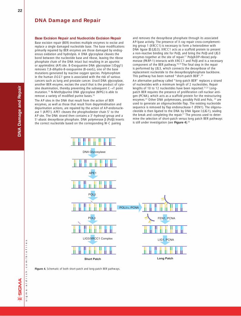
22
si
gm
a-
al
dr
ic
h.
co
m/
bi
of
il
es
DN
A D
amag
e an
d R
epai
rDNA Damage and Repair
Base Excision Repair and Nucleotide Excision RepairBase excision repair (BER) involves multiple enzymes to excise and replace a single damaged nucleotide base. The base modifications primarily repaired by BER enzymes are those damaged by endog-enous oxidation and hydrolysis. A DNA glycosylase cleaves the bond between the nucleotide base and ribose, leaving the ribose phosphate chain of the DNA intact but resulting in an apurinic or apyrimidinic (AP) site. 8-Oxoguanine DNA glycosylase I (Ogg1) removes 7,8-dihydro-8-oxoguanine (8-oxoG), one of the base mutations generated by reactive oxygen species. Polymorphism in the human OGG1 gene is associated with the risk of various cancers such as lung and prostate cancer. Uracil DNA glycosylase, another BER enzyme, excises the uracil that is the product of cyto-sine deamination, thereby preventing the subsequent C→T point mutation.15 N-Methylpurine DNA glycosylase (MPG) is able to remove a variety of modified purine bases.16
The AP sites in the DNA that result from the action of BER enzymes, as well as those that result from depyrimidination and depurin ation actions, are repaired by the action of AP-endonucle-ase 1 (APE1). APE1 cleaves the phosphodiester chain 5’ to the AP site. The DNA strand then contains a 3’-hydroxyl group and a 5’-abasic deoxyribose phosphate. DNA polymerase β (Polβ) inserts the correct nucleotide based on the corresponding W-C pairing
and removes the deoxyribose phosphate through its associated AP-lyase activity. The presence of X-ray repair cross-complement-ing group 1 (XRCC1) is necessary to form a heterodimer with DNA ligase III (LIG3). XRCC1 acts as a scaffold protein to present a non-reactive binding site for Polβ, and bring the Polβ and LIG3 enzymes together at the site of repair.17 Poly(ADP-ribose) poly-merase (PARP-1) interacts with XRCC1 and Polβ and is a necessary component of the BER pathway.18,19 The final step in the repair is performed by LIG3, which connects the deoxyribose of the replacement nucleotide to the deoxyribosylphosphate backbone. This pathway has been named “short-patch BER”.20
An alternative pathway called “long-patch BER” replaces a strand of nucleotides with a minimum length of 2 nucleotides. Repair lengths of 10 to 12 nucleotides have been reported.21,22 Long-patch BER requires the presence of proliferation cell nuclear anti-gen (PCNA), which acts as a scaffold protein for the restructuring enzymes.23 Other DNA polymerases, possibly Polδ and Polε, 24 are used to generate an oligonucleotide flap. The existing nucleotide sequence is removed by flap endonuclease-1 (FEN1). The oligonu-cleotide is then ligated to the DNA by DNA ligase I (LIG1), sealing the break and completing the repair.17 The process used to deter-mine the selection of short-patch versus long patch BER pathways is still under investigation (see Figure 4).25
DNA Glycosylase
APE1
POLβ
POLδ/ε, PCNA
FEN1, PCNAPOLβ
Short Patch Long Patch
LIG3/XRCC1 Complex LIG1, PCNA
Figure 4. Schematic of both short-patch and long-patch BER pathways.

23
si
gm
a-
al
dr
ic
h.
co
m/
bi
of
il
es
DN
A D
amag
e and
Rep
air
While BER may replace multiple nucleotides via the long-patch pathway, the initiating event for both short-patch and long-patch BER is damage to a single nucleotide, resulting in minimal impact on the structure of the DNA double helix. Nucleotide excision repair (NER) repairs damage to a nucleotide strand containing at least 2 bases and creating a structural distortion of the DNA. NER acts to repair single strand breaks in addition to serial damage from exogenous sources such as bulky DNA adducts and UV radia-tion.26 The same pathway may be used to repair damage from oxidative stress.27 Over 20 proteins are involved in the NER path-way in mammalian cells, including XPA, XPC-hHR23B, replication protein A (RPA), transcription factor TFIIH, XPB and XPD DNA heli-cases, ERCC1-XPF and XPG, Polδ, Polε, PCNA, and replication fac-tor C.28 Overexpression of the excision repair cross-complementing (ERCC1) gene has been associated with cisplatin resistance by non-small-cell lung cancer cells29 and corresponds to enhanced DNA repair capacity.30 Global genomic NER (GGR) repairs dam-age throughout the genome, while a specific NER pathway called Transcription Coupled Repair (TCR) repairs genes during active RNA polymerase transcription.31
Repair of Double-Strand BreaksDouble-strand breaks in DNA can result in loss and rearrangement of genomic sequences. These breaks are repaired by either non-homologous end-joining (NHEJ) or by homologous recombination (HR), also called recombinational repair or template–assisted repair.
The HR pathway is activated when the cell is in late S/G2 phase and the template has recently been duplicated. This mechanism requires the presence of an identical or nearly identical sequence linked to the damaged DNA region via the centromere for use as a repair template. Double-stranded breaks repaired by this mecha-nism are usually caused by the replication machinery attempting to synthesize across a single-strand break or unrepaired lesion, resulting in the collapse of the replication fork.
Non-homologous end-joining (NHEJ) is used at other points of the cell cycle when sister chromatids are not available for use as HR templates. When these breaks occur, the cell has not yet rep-licated the region of DNA that contains the break, so unlike the HR pathway, there is no corresponding template strand available. In NHEJ, the Ku heterodimeric protein positions the two ends of the broken DNA strands for repair without an available template, losing sequence information in the process. Multiple enzymes are involved in the rejoining process, including DNA ligase IV, XRCC4, and DNA-dependent protein kinase (DNA-PK).32,33 NHEJ is inher-ently mutagenic as it relies on chance pairings, called microhomol-ogies, between the single-stranded tails of the two DNA frag-ments to be joined (see Figure 5). In higher eukaryotes, DNA-PK is required for NHEJ repair, both via the primary mechanism and via an alternative back-up mechanism (D-NHEJ).34
Future ApplicationsWhile DNA damage is a key factor in the development and evolu-tion of cancer cells, continued damage is used as part of clinical treatments for cancer, forcing malignant cells into apoptosis or senescence. Many chemotheraputic drugs such as bleomycin, mitomycin, and cisplatin, are effective because they cause further DNA damage in cancer cells that replicate at a faster rate than surrounding tissue. Cellular DNA repair mechanisms are a double-edged sword; by reducing mutations that may lead to cancer, these processes strive for genomic integrity, but the same mecha-nisms in malignant cells allow those cells to survive additional DNA damage and continue uncontrolled growth. In order to block this survival mechanism within cancer cells, clinical trials are now being performed using inhibitors to specific DNA repair enzymes, includ-ing MGMT, PARP, and DNA-PK.35-38
Double-strand break
End binding by KuEnd alignment
Trimming of 5' and 3'overhangs
Ligation by XRCC4 andDNA Ligase IV
Template-dependentfilling-in
Loss of base pairs present in original wild-type sequence
Figure 5. General mechanism of NHEJ repair of double-strand breaks in DNA.
References:1. Collado, M., et al., Nature, 436, 642 (2005).2. Chen, Z., et al., Nature, 436, 725 (2005).3. Michaloglou, C., et al., Nature, 436, 720 (2005).4. Braig, M., et al., Nature, 436, 660 (2005).5. Jackson, A.L., and Loeb, L.A., Mutat. Res., 477, 7-21, (2001).6. De Bont, R., and van Larebeke, N., Mutagenesis, 19, 169-85 (2004).7. Lindahl, T. and Nyberg, B., Biochemistry, 11, 3610-3618 (1972).8. Brem., R. and Hall, J., Nucleic Acids Res., 33, 2512-2520 (2005).9. Niture S.K., et al, Carcinogenesis, Epub ahead of print (Aug. 31, 2006)10. Goldsby, et al., Proc. Natl. Acad. Sci. USA, 99, 15560 (2002).11. Yang, W., Mutat. Res., 460, 245-56 (2000).12. Iyer, R.R., et al., (Review), Chem. Rev., 106, 302-23 (2006).13. Modrich, P., J. Biol. Chem., 281, 30305-30309 (2006). 14. Muller, A. and Fishel, R., Cancer Invest., 20, 102-9 (2002).15. Lindahl, T., Proc. Natl. Acad. Sci. USA, 71, 3649-3653 (1974).16. Singer, B., and Hang, B., Chem. Res. Toxicol., 10, 713-32 (1997).17. Lindahl, T. and Wood, R.D., Science, 286, 1897-1905 (1999).18. Caldecott, K.W., et al., Nucleic Acids Res., 24, 4387-4394 (1996).19. Dantzer, F., et al., Biochemistry, 39, 7559-7569 (2000).20. Srivastava, D.K., et al., J. Biol. Chem., 273, 21203-9 (1998). 21. Ranalli, T.A., et al., J. Biol. Chem., 277, 41714-41724 (2002). 22. Sattler, U., et al., EMBO Reports, 4, 363-367 (2003). 23. Fortini, P., et al., Biochemistry, 37, 3575-3580 (1998). 24. Klungland, A. and Lindahl, T., EMBO Journal, 16, 3341-8 (1997)25. Sung, J.S. and Demple, B., (Review), FEBS Journal, 273, 1620-1629 (2006).26. Balajee, A.S. and Bohr, V.A., Gene, 250, 15-30 (2000).27. Gros, L., et al., Oncogene, 21, 8905-8925 (2002).28. You, J.S., J. Biol. Chem., 278, 7476-7485 (2003).29. Rosell, R., et al., Cancer Control, 10, 297-305 (2003).30. Vogel, U., et al., Mutat. Res., 461, 197-210 (2000).31. Hanawalt, P.C., Oncogene, 21, 8949-8956 (2002).32. Critchlow, S.E. and Jackson, S.P., Trends Biochem. Sci., 23, 394-398 (1998). 33. Wang, H., et al., Nucleic Acids Res., 31, 5377-88 (2003). 34. Perrault, R., et al., J. Cell. Biochem., 92, 781-794 (2004). 35. Sanchez-Perez, I., Clin. Transl. Oncol., 8, 642-6 (2006). 36. Madhusudan, S. and Hickson, I.D., Trends Mol. Med., 11, 503-11 (2005). 37. Plummer, E.R., Curr. Opin. Pharmacol., 6, 364-8 (2006).38. Sabharwal, A. and Middleton, M.R., Curr. Opin. Pharmacol., 6, 355-63 (2006).
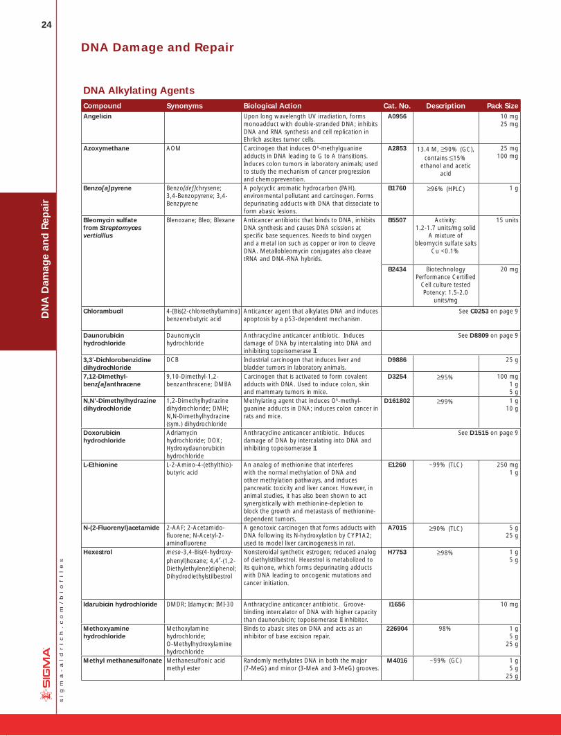
24
si
gm
a-
al
dr
ic
h.
co
m/
bi
of
il
es
DN
A D
amag
e an
d R
epai
r
DNA Alkylating AgentsCompound Synonyms Biological Action Cat. No. Description Pack SizeAngelicin Upon long wavelength UV irradiation, forms
monoadduct with double-stranded DNA; inhibits DNA and RNA synthesis and cell replication in Ehrlich ascites tumor cells.
A0956 10 mg25 mg
Azoxymethane AOM Carcinogen that induces O6-methylguanine adducts in DNA leading to G to A transitions. Induces colon tumors in laboratory animals; used to study the mechanism of cancer progression and chemoprevention.
A2853 13.4 M, ≥90% (GC), contains ≤15%
ethanol and acetic acid
25 mg100 mg
Benzo[a]pyrene Benzo[def]chrysene; 3,4-Benzopyrene; 3,4-Benzpyrene
A polycyclic aromatic hydrocarbon (PAH), environmental pollutant and carcinogen. Forms depurinating adducts with DNA that dissociate to form abasic lesions.
B1760 ≥96% (HPLC) 1 g
Bleomycin sulfate from Streptomyces verticillus
Blenoxane; Bleo; Blexane Anticancer antibiotic that binds to DNA, inhibits DNA synthesis and causes DNA scissions at specifi c base sequences. Needs to bind oxygen and a metal ion such as copper or iron to cleave DNA. Metallobleomycin conjugates also cleave tRNA and DNA-RNA hybrids.
B5507 Activity: 1.2-1.7 units/mg solid
A mixture of bleomycin sulfate salts
Cu <0.1%
15 units
B2434 Biotechnology Performance Certifi ed
Cell culture testedPotency: 1.5-2.0
units/mg
20 mg
Chlorambucil 4-[Bis(2-chloroethyl)amino] benzenebutyric acid
Anticancer agent that alkylates DNA and induces apoptosis by a p53-dependent mechanism.
See C0253 on page 9
Daunorubicin hydrochloride
Daunomycin hydrochloride
Anthracycline anticancer antibiotic. Induces damage of DNA by intercalating into DNA and inhibiting topoisomerase II.
See D8809 on page 9
3,3′-Dichlorobenzidine dihydrochloride
DCB Industrial carcinogen that induces liver and bladder tumors in laboratory animals.
D9886 25 g
7,12-Dimethyl-benz[a]anthracene
9,10-Dimethyl-1,2-benzanthracene; DMBA
Carcinogen that is activated to form covalent adducts with DNA. Used to induce colon, skin and mammary tumors in mice.
D3254 ≥95% 100 mg1 g 5 g
N,N’-Dimethylhydrazine dihydrochloride
1,2-Dimethylhydrazine dihydrochloride; DMH; N,N-Dimethylhydrazine (sym.) dihydrochloride
Methylating agent that induces O6-methyl-guanine adducts in DNA; induces colon cancer in rats and mice.
D161802 ≥99% 1 g 10 g
Doxorubicin hydrochloride
Adriamycin hydrochloride; DOX; Hydroxydaunorubicin hydrochloride
Anthracycline anticancer antibiotic. Induces damage of DNA by intercalating into DNA and inhibiting topoisomerase II.
See D1515 on page 9
L-Ethionine L-2-Amino-4-(ethylthio)-butyric acid
An analog of methionine that interferes with the normal methylation of DNA and other methylation pathways, and induces pancreatic toxicity and liver cancer. However, in animal studies, it has also been shown to act synergistically with methionine-depletion to block the growth and metastasis of methionine-dependent tumors.
E1260 ~99% (TLC) 250 mg1 g
N-(2-Fluorenyl)acetamide 2-AAF; 2-Acetamido-fl uorene; N-Acetyl-2-aminofl uorene
A genotoxic carcinogen that forms adducts with DNA following its N-hydroxylation by CYP1A2; used to model liver carcinogenesis in rat.
A7015 ≥90% (TLC) 5 g25 g
Hexestrol meso-3,4-Bis(4-hydroxy-phenyl)hexane; 4,4′-(1,2-Diethylethylene)diphenol; Dihydrodiethylstilbestrol
Nonsteroidal synthetic estrogen; reduced analog of diethylstilbestrol. Hexestrol is metabolized to its quinone, which forms depurinating adducts with DNA leading to oncogenic mutations and cancer initiation.
H7753 ≥98% 1 g5 g
Idarubicin hydrochloride DMDR; Idamycin; IMI-30 Anthracycline anticancer antibiotic. Groove-binding intercalator of DNA with higher capacity than daunorubicin; topoisomerase II inhibitor.
I1656 10 mg
Methoxyamine hydrochloride
Methoxylamine hydrochloride; O-Methylhydroxylamine hydrochloride
Binds to abasic sites on DNA and acts as an inhibitor of base excision repair.
226904 98% 1 g 5 g
25 g
Methyl methanesulfonate Methanesulfonic acid methyl ester
Randomly methylates DNA in both the major (7-MeG) and minor (3-MeA and 3-MeG) grooves.
M4016 ~99% (GC) 1 g 5 g
25 g
DNA Damage and Repair
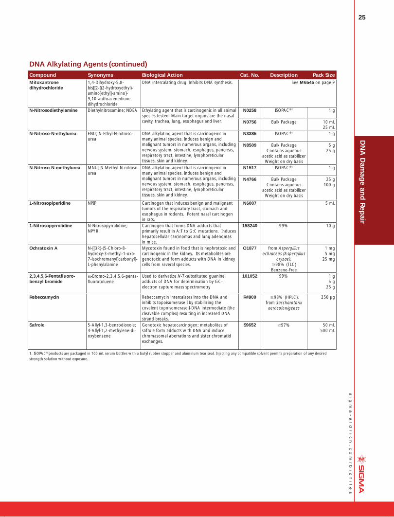
25
si
gm
a-
al
dr
ic
h.
co
m/
bi
of
il
es
DN
A D
amag
e and
Rep
air
DNA Alkylating Agents (continued)Compound Synonyms Biological Action Cat. No. Description Pack SizeMitoxantrone dihydrochloride
1,4-Dihydroxy-5,8-bis[[2-[(2-hydroxyethyl)-amino]ethyl]-amino]-9,10-anthracenedione dihydrochloride
DNA intercalating drug. Inhibits DNA synthesis. See M6545 on page 9
N-Nitrosodiethylamine Diethylnitrosamine; NDEA Ethylating agent that is carcinogenic in all animal species tested. Main target organs are the nasal cavity, trachea, lung, esophagus and liver.
N0258 ISOPAC®1 1 g
N0756 Bulk Package 10 mL25 mL
N-Nitroso-N-ethylurea ENU; N-Ethyl-N-nitroso-urea
DNA alkylating agent that is carcinogenic in many animal species. Induces benign and malignant tumors in numerous organs, including nervous system, stomach, esophagus, pancreas, respiratory tract, intestine, lymphoreticular tissues, skin and kidney.
N3385 ISOPAC®1 1 g
N8509 Bulk PackageContains aqueous
acetic acid as stabilizerWeight on dry basis
5 g 25 g
N-Nitroso-N-methylurea MNU; N-Methyl-N-nitroso-urea
DNA alkylating agent that is carcinogenic in many animal species. Induces benign and malignant tumors in numerous organs, including nervous system, stomach, esophagus, pancreas, respiratory tract, intestine, lymphoreticular tissues, skin and kidney.
N1517 ISOPAC®1 1 g
N4766 Bulk PackageContains aqueous
acetic acid as stabilizerWeight on dry basis
25 g 100 g
1-Nitrosopiperidine NPIP Carcinogen that induces benign and malignant tumors of the respiratory tract, stomach and esophagus in rodents. Potent nasal carcinogen in rats.
N6007 5 mL
1-Nitrosopyrrolidine N-Nitrosopyrrolidine; NPYR
Carcinogen that forms DNA adducts that primarily result in A:T to G:C mutations. Induces hepatocellular carcinomas and lung adenomas in mice.
158240 99% 10 g
Ochratoxin A N-[(3R)-(5-Chloro-8-hydroxy-3-methyl-1-oxo-7-isochromanyl)carbonyl]-L-phenylalanine
Mycotoxin found in food that is nephrotoxic and carcinogenic in the kidney. Its metabolites are genotoxic and form adducts with DNA in kidney cells from several species.
O1877 from Aspergillus ochraceus (Aspergillus
oryzae), ;98% (TLC) Benzene-Free
1 mg 5 mg
25 mg
2,3,4,5,6-Pentafl uoro-benzyl bromide
a-Bromo-2,3,4,5,6-penta-fl uorotoluene
Used to derivatize N-7-substituted guanine adducts of DNA for determination by GC-electron capture mass spectrometry
101052 99% 1 g 5 g
25 g
Rebeccamycin Rebeccamycin intercalates into the DNA and inhibits topoisomerase I by stabilizing the covalent topoisomerase I-DNA intermediate (the cleavable complex) resulting in increased DNA strand breaks.
R4900 ;98% (HPLC),from Saccharothrix aerocolonigenes
250 µg
Safrole 5-Allyl-1,3-benzodioxole; 4-Allyl-1,2-methylene-di-oxybenzene
Genotoxic hepatocarcinogen; metabolites of safrole form adducts with DNA and induce chromasomal aberrations and sister chromatid exchanges.
S9652 ;97% 50 mL 500 mL
1. ISOPAC® products are packaged in 100 mL serum bottles with a butyl rubber stopper and aluminum tear seal. Injecting any compatible solvent permits preparation of any desired strength solution without exposure.

26
si
gm
a-
al
dr
ic
h.
co
m/
bi
of
il
es
DN
A D
amag
e an
d R
epai
rDNA Damage and Repair
DNA Crosslinking AgentsCompound Synonyms DNA Crosslinking Action Cat. No. Description Pack Size
4′-Aminomethyltrioxsalen hydrochloride
4′-Aminomethyl-4,5′,8-trimethylpsoralen hydrochloride
Used to inactivate DNA and RNA viruses, including HIV-1, by nucleic acid crosslinking followed by UV irradiation.
A4330 ≥98% 5 mg
Carboplatin cis-Diammine(1,1-cyclo-butanedicarboxylato) platinum
A platinum-based antineoplastic drug that forms intrastrand crosslinks with neighboring guanine residues in DNA. Tumors acquire resistance to these drugs through the loss of DNA-mismatch repair (MMR) activity.
C2538 ≥98% (HPLC) 100 mg250 mg
Carmustine BCNU; 1,3-Bis(2-chloro-ethyl)-1-nitrosourea
DNA alkylating agent causing DNA interstrand crosslinks.
See C0400 on page 9
CB 1954 5-(1-Aziridinyl)-2,4-di-nitrobenzamide
Anticancer prodrug that is activated by nitroreductase to form a potent DNA crosslinking agent.
C2235 50 mg
Cyclophosphamide Cytoxan Cytotoxic nitrogen mustard derivative used in cancer chemotherapy. Cyclophosphamide crosslinks DNA, causes strand breakage, and induces mutations.
See C0768, C7397 on page 9
cis-Diammineplatinum(II) dichloride
Cisplatin Potent platinum-based antineoplastic agent. Forms cytotoxic adducts with the DNA dinucleotide d(pGpG), inducing intrastrand crosslinks.
See P4394 on page 9
Melphalan 4-[Bis(2-chloro-ethyl)amino]-L-phenyl-alanine; L-PAM; L-Phenyl-alanine mustard
Antineoplastic agent that forms DNA intrastrand crosslinks by bifunctional alkylation of 5’-GGC sequences.
M2011 ≥95% (TLC) 100 mg250 mg
1 g
Mitomycin C MMC Following enzymatic reduction, activated mitomycin C forms covalent adducts and crosslinks with CpG sequences in DNA.
See M0503 on page 9
Nimustine hydrochloride N’-[(4-Amino-2-methyl-5-pyrimidinyl)methyl]-N-(2-chloroethyl)-N-nitrosourea hydrochloride; ACNU
Antineoplastic compound that is a bifunctional DNA alkylating agent.
N8659 1 g
Oxaliplatin [SP-4-2-(1R-trans)]-(1,2-Cyclohexanediamine-N,N’)[ethanedioata(2–)-O,O’] platinum
Platinum-based antitumor agent that forms interstrand crosslinks with neighboring guanine residues in DNA.
O9512 5 mg
Psoralen 7H-Furo[3,2-g]benzopyran-7-one; Furo[3,2-g]coumarin
When activated by UV light, psoralen induces interstrand crosslinks in DNA. Photochemical reagent for the investigation of nucleic acid structure and function.
P8399 ≥99% 10 mg 25 mg
100 mg
Trioxsalen TMP; 4,5’,8-Trimethyl-psoralen; Trisoralen
Photochemical crosslinker of DNA has been used as a probe for nucleic acid structure and function.
T6137 ≥98% (HPLC) 100 mg500 mg
1 g
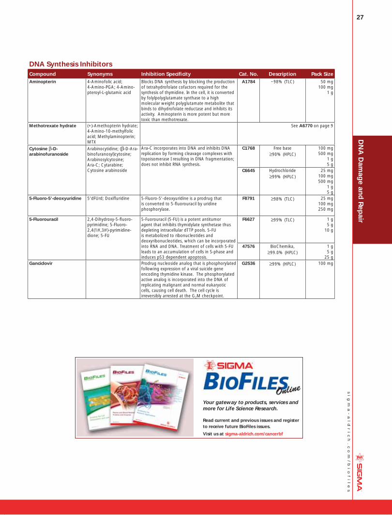
27
si
gm
a-
al
dr
ic
h.
co
m/
bi
of
il
es
DN
A D
amag
e and
Rep
air
DNA Synthesis InhibitorsCompound Synonyms Inhibition Specifi city Cat. No. Description Pack SizeAminopterin 4-Aminofolic acid;
4-Amino-PGA; 4-Amino-pteroyl-L-glutamic acid
Blocks DNA synthesis by blocking the production of tetrahydrofolate cofactors required for the synthesis of thymidine. In the cell, it is converted by folylpolyglutamate synthase to a high molecular weight polyglutamate metabolite that binds to dihydrofolate reductase and inhibits its activity. Aminopterin is more potent but more toxic than methotrexate.
A1784 ~98% (TLC) 50 mg 100 mg
1 g
Methotrexate hydrate (+)-Amethopterin hydrate; 4-Amino-10-methylfolic acid; Methylaminopterin; MTX
See A6770 on page 9
Cytosine β-D-arabinofuranoside
Arabinocytidine; (β-D-Ara-binofuranosyl)cytosine; Arabinosylcytosine; Ara-C; Cytarabine; Cytosine arabinoside
Ara-C incorporates into DNA and inhibits DNA replication by forming cleavage complexes with topoisomerase I resulting in DNA fragmentation; does not inhibit RNA synthesis.
C1768 Free base≥90% (HPLC)
100 mg 500 mg
1 g 5 g
C6645 Hydrochloride≥99% (HPLC)
25 mg 100 mg500 mg
1 g5 g
5-Fluoro-5’-deoxyuridine 5’dFUrd; Doxifl uridine 5-Fluoro-5’-deoxyuridine is a prodrug that is converted to 5-fl uorouracil by uridine phosphorylase.
F8791 ≥98% (TLC) 25 mg100 mg250 mg
5-Fluorouracil 2,4-Dihydroxy-5-fl uoro-pyrimidine; 5-Fluoro-2,4(1H,3H)-pyrimidine-dione; 5-FU
5-Fuorouracil (5-FU) is a potent antitumor agent that inhibits thymidylate synthetase thus depleting intracellular dTTP pools. 5-FU is metabolized to ribonucleotides and deoxyribonucleotides, which can be incorporated into RNA and DNA. Treatment of cells with 5-FU leads to an accumulation of cells in S-phase and induces p53 dependent apoptosis.
F6627 ≥99% (TLC) 1 g 5 g
10 g
47576 BioChemika, ≥99.0% (HPLC)
1 g 5 g
25 gGanciclovir Prodrug nucleoside analog that is phosphorylated
following expression of a viral suicide gene encoding thymidine kinase. The phosphorylated active analog is incorporated into the DNA of replicating malignant and normal eukaryotic cells, causing cell death. The cell cycle is irreversibly arrested at the G2M checkpoint.
G2536 ≥99% (HPLC) 100 mg
Your gateway to products, services and more for Life Science Research.
Read current and previous issues and register to receive future BioFiles issues. Visit us at sigma-aldrich.com/cancerbf
28
si
gm
a-
al
dr
ic
h.
co
m/
bi
of
il
es
DN
A D
amag
e an
d R
epai
r
DNA Repair Enzymes and Antibodies
Monoclonal Anti-AP Endonuclease antibody produced in mouseAnti-APE1; Anti-APEN; Anti-APE/Ref1; Anti-APEX1; Anti-APEX nuclease; Anti-AP lysase; Anti-Apurinic/Apyrimidinic Endonuclease; Anti-HAP1
~2 mg/mL, clone APEREF, purified immunoglobulinImmunogen: recombinant human AP endonuclease.
Solution in 0.01 M phosphate buffered saline, pH 7.4, containing 15 mM sodium azide.
Species reactivity: human, dog, rat, mouse
Antigen mol wt ~37 kDa
Application(s):Immunoblotting ........... 0.5-1 µg/mL using total cell extract of Raji cellsIndirect ELISA ............................................................................suitableImmunocytochemistry ...............................................................suitableArray ........................................................................................suitableIsotype .......................................................................................... IgG1B DRY ICE
A2105 200 µL
Monoclonal Anti-DNA Ligase I antibody produced in mouse~1 mg/mL, clone 1A9, purified immunoglobulinImmunogen: bovine DNA ligase I.
Solution in phosphate buffered saline, pH 7.4, containing 0.08% sodium azide.
Species reactivity: human, bovine
Antigen mol wt ~125 kDa
Application(s):Immunoblotting ............................... 1-5 µg/mL using HeLa or DiFi cellsDirect ELISA ..............................................................................suitableIsotype .......................................................................................... IgG1B WET ICE
D7690 100 µg
Monoclonal Anti-Excision Repair Cross Comple-menting Protein-1 antibody produced in mouse Anti-ERCC-1
~1 mg/mL, clone 8F1, purified immunoglobulinImmunogen: recombinant full length human ERCC1 protein.
Solution in phosphate buffered saline pH 7.4, containing 0.08% sodium azide.
Species reactivity: human, rat
Antigen mol wt 33-36 kDa
Application(s):Immunoblotting ........................... 1-5 mg/mL using A431 or HeLa cellsImmunoprecipitation ............................. 2 µg using 1 mg protein lysateImmunohistochemistry ................2-4 µg/mL using human tonsil tissuesIsotype .......................................................................................... IgG1B WET ICE
E8903 100 µg
Methylpurine DNA Glycosylase humanAAG; MPG
MPG is a base excision repair (BER) protein that removes the mutated N-methylpurine nucleotide from alkylated DNA, creating an apurinic/apyrimidinic site. MPG recognizes and excises 3-meth-yladenine, 7-methylguanine, 3-methylguanine, N-6-ethanolad-enine, and hypoxanthine.
≥80% (SDS-PAGE), recombinant, expressed in Escherichia coli (as a MBP fusion protein), activity: ≥5000 units/mg proteinSolution in 50% glycerol (w/v) containing 20 mM Tris-HCl, pH 7.4, 1 mM DTT, 1 mM EDTA, 250 mM NaCl, 0.25% CHAPS, 1% prote-ase inhibitor cocktail (v/v) (Cat. No. P8340).
Apparent mol wt ~70 kDa
Unit definition: One unit will cleave 50% of 0.5 pmole of double stranded DNA oligomer substrate containing a hypoxanthine site lesion in 10 min. at 37 °C.
A DRY ICE
M7940 25 µg
8 Monoclonal Anti-Methylpurine DNA N-Glycosylase antibody produced in mouseAnti-AAG; Anti-3-Alkyladenine DNA glycosylase; Anti-Alkylpurine DNA N-glycosylase; Anti-APNG; Anti-Methyladenine DNA Glycosylase; Anti-MPG
~2 mg/mL, clone MPG80, purified immunoglobulinImmunogen: recombinant human MPG protein.
Solution in 0.01 M phosphate buffered saline, pH 7.4, containing 15 mM sodium azide.
Species reactivity: human
Antigen mol wt ~32 kDa
Application(s):Immunoblotting ..............................0.5-1 µg/mL using total cell extract of 293T cells expressing MPGIndirect ELISA ........................................................................... suitableImmunocytochemistry ...............................................................suitableIsotype .......................................................................................... IgG1B DRY ICE
M6195 25 µl, 200 µl
Monoclonal Anti-Mismatch Repair Protein 2 antibody produced in mouseAnti-MSH2
~1 mg/mL, clone 2MSH01, tissue culture supernatantImmunogen: recombinant human mismatch repair protein 2 (MSH2).
Contains 15 mM sodium azide.
Species reactivity: human
Antigen mol wt 102 kDa
Application(s):Immunohistochemistry (formalin-fixed, paraffin-embedded sections) ....................................................................................... 1:25Isotype .......................................................................................... IgG1B WET ICE
M6315 500 µL
DNA Damage and Repair
29
si
gm
a-
al
dr
ic
h.
co
m/
bi
of
il
es
Ogg1 Assay KitThe assay is based on Ogg1 glycosylase activity that recognizes and removes the mutated base (8-oxo-G). The substrate is a 23 oligonucleotide containing 8-oxo-dG at its 11th base, labeled with 32P at its 5’ end, and annealed to its complementary strand (con-taining dC at the opposite base position to the 8-oxo-dG). Upon cleavage of the substrate by the Ogg1 enzyme, the oligonucle-otide strands are run on a denaturing gel and a 10 base fragment (labeled cleavage product) is revealed in addition to the original 23 base oligonucleotide band. The detection is performed by autora-diography.
The Ogg1 Assay Kit:
• Provides a tool for measurement of Ogg1 enzyme activity
• Contains control Ogg1 enzyme enabling screening of Ogg1 modulators
• Can be used with cell lysates and purified enzyme preparation
Sufficient for 180 assays Components Ogg1 Substrate 8-oxo-G strand .............................................. 1 vial Ogg1 Substrate complementary strand ................................... 1 vial Reaction Buffer ............................................................ 10 X 1.5 mL Stop Solution ...........................................................................1mL Ogg1 (control enzyme) .......................................................... 10 µg Putrescine ................................................................................1 mL SigmaSpin™ Post-Reaction Purification Columns .................. 2 each
CS0710 1 kit
Ogg1 from mouse8-Oxoguanine-DNA glycosylase
≥90% (SDS-PAGE), recombinant, expressed in Escherichia coli, activity: >20,000 units/mg protein
Solution in 50% (w/v) glycerol containing 20 mM Tris, pH 7.5, 200 mM sodium chloride, 1 mM EDTA, and 1 mM DTT.
Apparent mol wt 38.8 kDa (345 amino acid protein)
Unit definition: One unit is the amount of protein that cleaves 50% of 0.5 pmol double-stranded, 8-oxoguanine-mutated DNA oligomer substrate in 10 min at 37 °C.
B WET ICE
O2135 10 µg
Uracil DNA Glycosylase from Escherichia coliDNA Uracil Glycosylase; UDG; Uracil N-glycosylase[59088-21-0]
UDG catalyzes the removal of uracil residues from both single- and double-stranded DNA, but not RNA. This reaction leaves the DNA sugar-phosphodiester backbone intact. The resulting DNA is not suitable for use as a hybridization target or as a template for DNA polymerases.
Solution in 30 mM Tris-HCl, pH 7.5, 1 mM DTT, 0.05% (w/v) Tween® 20, 1 mM EDTA, 150 mM NaCl, 50% (v/v) glycerol.
Unit definition: One unit catalyzes the release of 1 nmol of free uracil from 3H-poly(dU) in 1 hr at 37°C.Concentration .........................................................................1 unit/µLPurchase of this product conveys the licensed right under U.S. Patent No. 5,035,996 and foreign equivalents to use this product only in internal research conducted by the purchaser. No rights under the aforementioned patents are conveyed which permit the resale, transfer, or use for purposes other than research. Rights under the aforementioned patents, for purposes other than inter-nal research, may be obtained by contacting Life Technologies, Inc.
B DRY ICE
U1257 100 units
DN
A D
amag
e and
Rep
air
Sigma-Aldrich cytochemistry assays are used in thediagnosis of acute myeloid leukemia and in the stainingof inflammatory tissue exudates for characterizing neu-trophils, lymphocytes and macrophages. The prepackagedkits are a cost effective way to bring testing in-house withno capital equipment requirements.
Classic methods include:• Naphthol AS-D Chloroacetate Esterase• Tartrate Resistant Acid Phosphatase• a-Naphthyl Acetate Esterase• Myeloperoxidase• Nitroblue Tetrazolium
• Periodic Acid Schiff• Alkaline Phosphatase• a-Naphthyl Butyrate Esterase
From traditional hematoxylin and eosin stains to specialstain kits designed to visualize specific tissue features,Sigma-Aldrich offers a comprehensive solution of qualityhistology reagents you need and the support you demand.
Please visit sigma-aldrich.com/handh for all your histology needs.
CytochemistryKits
De l i ve r ing S c i ence Through Bus ine s s Under s tand ing
30
si
gm
a-
al
dr
ic
h.
co
m/
bi
of
il
es
Sigma is committed to continually adding new products for use in cancer research and cell biology, as well as new technology plat-forms for cellular studies. We welcome your recommendations for new products. Please tell us your suggestions by visiting Sigma.com/product_suggestions.
Antitumor Agents
CephalomannineC45H53NO14 FW 831.90 [71610-00-9]
Antitumor; antiproliferative. Promotes the formation of highly sta-ble microtubules that resist depolymerization but does not induce cytokine secretion.
≥97% (HPLC)
C4991 1 mg, 5 mg
Chrysomycin AC28H28O9 FW 508.52 [82196-88-1]
Antibiotic from Streptomyces sp. Inhibits the catalytic activity of human topoisomerase II. Exhibits antitumor activity against human cell lines K562, HT29, MCF7, PC6, and MKN28.
≥98% (HPLC)
E
C6616 100 µg
Chrysomycin BC27H28O9 FW 496.51 [83852-56-6]
Antibiotic from Streptomyces sp. Inhibits the catalytic activity of human topoisomerase II. Exhibits antitumor activity against human cell lines K562, HT29, MCF7, PC6, and MKN28.
≥98% (HPLC)
E
C6491 100 µg
CIL-1021-[4-(Furo[2,3-b]quinolin-4-ylamino)phenyl]ethanoneC19H14N2O2 FW 302.33 [479077-76-4]
Tubulin polymerization inhibitor: apoptosis inducer.
US Patent No. 6,750,223. Sold for non-human research purposes under a non-exclusive license agreement with Kaohsiung Medical University.
≥95% (HPLC)
B WET ICE
C5492 10 mg
17-Dimethylaminoethylamino-17-demethoxy-geldanamycin17-DMAGC32H48N4O8 FW 616.75
17-DMAG is a more potent water soluble analog of geldanamy-cin. 17-DMAG has shown more antitumor activity than 17-AAG. Inhibits cancer growth and promotes apoptosis in multiple cell lines.
B
D5193 1 mg
FinasterideN-tert-Butyl-3-oxo-4-aza-5α-androst-1-en-17β-carboxamide; N-(2-Methyl-2-propyl)-3-oxo-4-aza-5α-androst-1-ene-17β-carboxamide; MK-906 C23H36N2O2 FW 372.54 [98319-26-7]
Selective 5α-reductase inhibitor; antiandrogen.
≥97% (HPLC)
B
F1293 100 mg
ICRF-193meso-4,4’-(3,2-butanediyl)-bis(2,6-piperazinedione)C12H18N4O4 FW 282.30 [21416-68-2]
ICRF-193 induces a G2 checkpoint that is associated with an ATR dependent inhibition of polo-like kinase 1 (Plk1) activity and a decrease in cyclin B1 phosphorylation. Induces apoptosis in several cell lines including K562 and Molt-4 cells. ICRF-193 is a topoi-somerase II inhibitor that targets topoisomerase II-β to a greater extent than it targets topoisomerase II-α and does not cause DNA damage.
≥95%
B
I4659 1 mg, 5 mg
Kazusamycin AC33H48O7 FW 556.73 [92090-94-3]
Kazusamycin A, an unsaturated branched chain fatty acid with a terminal lactone ring, is a hydroxy analog of Leptomycin B. It has significant in vitro cytotoxic activity against various human and mouse tumor lines en compassing a wide range of tissue types. Kazusamycin A exhibits in vivo antitumor activity against experi-mental murine tumors. It inhibits nuclear export and Rev transloca-tion, a regulatory gene product in the HIV genome, at nanomolar concentrations.
from Streptomyces sp., ≥95% (HPLC) 70% methanol solution.
B DRY ICE
K1764 1 µg
LinomideLS-2616; N-Phenylmethyl-1,2-dihydro-4-hydroxyl-1-methyl-2-oxoquinoline-3-carboxamide; Quinoline-3-carboxamideC18H16N2O3 FW 308.33 [84088-42-6]
Immunomodulator; antiangiogenic. Upregulates IL-10 and decreases CXC chemokine production. Upregulates mRNA of IL-23, IL-12p35, and IFNγ. Also, antagonizes hepatic effects of TNFα by blocking recruitment of inflammatory cell infiltration.
≥98% (HPLC)
E
L1793 10 mg
Myoseverin B2,6-Bis(4-methoxybenzylamino)-9-cyclohexylpurineC27H32N6O2 FW 472.58 [361431-27-8]
Tubulin polymerization inhibitor.
≥97% (HPLC)
B DRY ICE
M3316 1 mg, 5 mg
New
Pro
du
cts
New Products

31
si
gm
a-
al
dr
ic
h.
co
m/
bi
of
il
es
Neocarzinostatin from Streptomyces carzinostaticus Holoneocarzinostatin; NCS; NSC-69856; Zinostatin [9014-02-2]
Protein-small molecule complex composed of an enediyne chro-mo-phore tightly bound to a 113-amino acid single chain protein. Chemotherapeutic agent which inhibits DNA synthesis, possesses antitumor activity, and induces apoptosis.
Supplied as 100 µg (~9 nmoles) in a solution containing 20 mM MES buffer, pH 5.5.
≥90% (SDS-PAGE), ~0.5 mg/mL
E WET ICE
N9162 100 µg
Nutlin-3(±)-4-[4,5-Bis(4-chlorophenyl)-2-(2-isopropoxy-4-methoxyphenyl)-4,5-dihydroimidazole-1-carbonyl]-piperazin-2-oneC30H30Cl2N4O4 FW 581.49 [548472-68-0]
Mdm2 (mouse double minute 2) antagonist, p53 pathway activa-tor, apoptosis inducer.
Sold under license from Hoffman-La Roche, Inc. US patent 6,734,302
≥98% (HPLC)
B WET ICE
N6287 1 mg, 5 mg
p53 humanp53 gene is highly conserved and expressed in normal tissues. It is the most commonly mutated gene in human cancer and more than 500 gene mutations have been described in various types of malignancies, hematologic as well as solid tumors. Intact p53 function is essential for the maintenance of the non-tumorogenic phenotype of cells. Thus, p53 plays a vital role in suppressing the development of cancer. Useful for the study of postranslational modification of p53, gel-shift assays, or protein-protein interaction studies.
Supplied at a concentration of 0.2 mg/ml in PBS, pH 7.0.
recombinant, expressed in Escherichia coli>90% (SDS-PAGE)
B WET ICE
P6249 50 µg
>95% (SDS-PAGE)
B WET ICE
P6374 20 µg
PTACHCpd 51; S-[6-(4-Phenyl-2-thiazolylcarbamoyl)hexyl] thioisobutyrate C20H26N2O2S2 FW 390.56 [848354-66-5]
HDAC inhibitor; more potent than the majority of HDAC inhibitors except for SAHA.
≥98% (HPLC)
E
P5874 2 mg
RG108N-Phthalyl-1-tryptophanC19H14N2O4 FW 334.33 [48208-26-0]
DNA methyltransferase (DMNT) inhibitor. Reactivates tumor sup-pressor gene expression (p16, SFRP1, secreted frizzled related protein-1, and TIMP-3) in tumor cells by DNA demethylation. Inhibits human tumor cell line (HCT116, NALM-6) proliferation and increases doubling time in culture.
≥98% (HPLC)
B
R8279 10 mg
SKI II4-[[4-(4-Chlorophenyl)-2-thiazolyl]amino]phenolC15H11ClN2OS FW 302.78 [312636-16-1]
Sphingosine kinase (SK) plays a pivotal role in regulating tumor growth and SK can act as an oncogene. Expression of SK RNA is significantly elevated in a variety of solid tumors, compared with normal tissue from the same patient. A number of novel inhibitors of human SK were identified, and several representative com-pounds were characterized in detail. These compounds demon-strated activity at sub- to micromolar concentrations, making them more potent than any other reported SK inhibitor, and were selec-tive toward SK compared with a panel of human lipid and protein kinases. Kinetic studies revealed that the compounds were not competitive inhibitors of the ATP-binding site of SK. SKI-II inhibitor is orally bioavailable, detected in the blood for at least 8 h, and showed a significant inhibition of tumor growth in mice with IC50 = 0.5 µM; SKI II does not act at ATP-binding site. Displays no inhibition of ERK2, PI 3-kinase, or PKCa at concentrations up to 60 µM. SKI II induces apoptosis and inhibits proliferation in sev-eral other tumor cell lines in vitro (IC50 = 0.9-4.6 µM).
≥98% (HPLC)
S5696 10 mg, 50 mg
SulochrinC17H16O7 FW 332.30 [519-57-3]
Antibiotic from Aspergillus and Penicillium sp. Fungal metabolite; VEGF inhibitor and antiangiogenic that inhibits the VEGF-induced tube formation of human umbilical vein endothelial cells.
≥98% (HPLC)
E
S4570 1 mg
New
Prod
ucts

32
si
gm
a-
al
dr
ic
h.
co
m/
bi
of
il
es
Chemopreventitive Agents
2-Amino-N-quinolin-8-yl-benzenesulfonamideQBSC15H13N3O2S FW 299.35 [16082-64-7]
Inhibitor of cell cycle at G2 phase; apoptosis inducer.
≥98% (HPLC)
A3105 5 mg
BorrelidinBorrelidine; 2-(7-Cyano-8,16-dihydroxy-9,11,13,15-tetra methyl-18-oxooxacyclooctadeca-4,6-dien-2-yl)-cyclo pentanecarboxylic acid; Cyclopentanecarboxylic acid; NSC 216128; TreponemycinC28H43NO6 FW 489.64 [7184-60-3]
Potent angiogenesis inhibitor that induces apoptosis of the capil-lary tube-forming cells. Also, displays antimalarial activity against drug-resistant Plasmodia. Antimicrobial and selective threonyl t-RNA synthetase inhibitor.
from Streptomyces parvulus, ≥98% (HPLC)B WET ICE
B3061 1 mg
CarnosolC20H26O4 FW 330.42 [5957-80-2]
A phenolic diterpene with antioxidant and anticarcinogenic activi-ties.
from Rosemarinus officinalis L.B DRY ICE
C9617 5 mg
Etidronate disodiumDihydrogen (1-hydroxyethylidene)bisphosphonate disodiumC2H6Na2O7P2 FW 249.99 [7414-83-7]
Bisphosphonate antiresorptive agent. Less potent inhibitor of farnesyl diphosphate synthase (IC50 = 80 µM) as compared to the nitrogen containing bisphosphonates
E WET ICE
P5248 10 mg, 100 mg
IfosfamideN,3-Bis(2-chloroethyl)tetrahydro-2H-1,3,2-oxaza phos phorin-2-amine-2-oxide; Ifex C7H15Cl2N2O2P FW 261.09 [3778-73-2]
Ifosfamide is a nitrogen mustard compound that is a structural isomer of cyclophosphamide. Ifosfamide is a prodrug that must be transformed by cytochrome P450 to the biologically active compo-nent. It is used as an antineoplastic agent in cancer chemotherapy, but ifosfamide is more likely to cause renal toxicity than cyclophos-phamide.
≥98%
E WET ICE
I4909 1 g, 5 g
Lomustine1-(2-Chloroethyl)-3-cyclohexyl-1-nitrosoureaC9H16ClN3O2 FW 233.70 [13010-47-4]
Antineoplastic agent with cellular DNA effects. Lomustine induc-es p53 expression in A2870 cells.
≥98%B
L5918 100 mg
Enzymes
Breast Tumor Kinase Active humanBrk; Protein Tyrosine Kinase 6; PTK6
recombinant, expressed in Escherichia coli, ≥85% (SDS-PAGE)Breast Tumor Kinase (Brk, PTK6) is a member of the non-recep-tor tyrosine kinases (PTKs). Brk is expressed in breast tumors. Brk promotes cell motility and invasion and functions as a mediator of EGF-induced migration and invasion.
Protein apparent mol wt ~80 kDa
A DRY ICE
B9810 5 µg
Multi-Drug Resistance
Fumitremorgin CFTC C22H25N3O3 FW 379.45 [118974-02-0]
from Neosartorya fischeri, >98% (HPLC and TLC)Fumitremorgin C (FTC) is a fungal toxin of the diketopiperazines family of compounds. In mammalian cells, FTC is tremorgenic and causes cell cycle arrest. Fumitremorgin C has been shown to reverse resistance to doxorubicin, mitoxantrane, and topotecan in non-Pgp (P-glycoprotein), non-MRP (multidrug resistance protein) multidrug-resistance (MDR) cells. This reversal of resistance is asso-ciated with an increase in drug accumulation. Fumitremorgin C is a specific, selective, and potent inhibitor at micromolar concen-trations of the breast cancer resistant protein (BCRP/ABCG2), an ABC transporter associated with chemotherapy resistance. FTC, in combination with mitoxantrone, can be used for the detection of ABCG2 functional activity in several cell lines.
E WET ICE
F9054 250 µg
Tumor Growth Regulation
(E/Z)-4-HTA monohydrate(E,Z)-2-[4-[1-(p-Hydroxyphenyl)-2-phenyl]-1-butenyl] phenoxyacetic acid; 4-Hydroxytamoxifen acid C24H22O4 · H2O FW 392.44
Non-steroidal estrogen.
≥90% (HPLC)
E
H5039 2 mg, 10 mg
PD 173074N-[2-[4-(Diethylamino)butyl]amino-6- (3,5-di methoxy-phenyl)pyrido[2,3-d]pyrimidin-7-yl]-N’-(1,1-di methyl ethyl)ureaC28H41N7O3 FW 523.67 [219580-11-7]
Fibroblast growth factor receptor 3 (FGFR3) inhibitor: IC50 = 5 nM in inhibition of FGFR3 autophosphorylation; arrested the G0/G1 phase of the cell cycle of FGFR3-expressing cells. 100-fold more selective for FGFR3 than for VEGF receptors, IGF-1 receptors, and MAPKs.
≥98% (HPLC)
E
P2499 5 mg, 25 mg
Reveromycin AC36H52O11 FW 660.79 [134615-37-5]
Polyketide antibiotic from Streptomyces sp. Epidermal growth fac-tor (EGF) inhibitor; apoptosis inducer; G1 phase cell cycle inhibitor having antiproliferative behavior against human cell lines KB and K562 as well as antifungal activity.
≥98% (HPLC)
B WET ICE
R0654 100 µg
New
Pro
du
cts
New Products

PathFinder
INNOVATION @ WORK
Discover Your Path to Innovation
Accelerating Customers' success through Leadership in Life Science, High Technology and ServiceS I G M A - A L D R I C H C O R P O R AT I O N • B O X 1 4 5 0 8 • S T. L O U I S • M I S S O U R I 6 3 1 7 8 • U S A
sigma.com/pathfinder
On your path to innovation, Sigma is with you every step of the way. PathFinder is an online collection of interactive, interconnected maps showing biological signaling and metabolic pathways. For you to explore the relationships between different pathway elements, individual components are linked with related high-quality products.
You know your destination. PathFinder will get you there.
With Sigma’s broad range of products, you will discover that we offer everything from small molecules to antibodies and enzymes. You will also be able to use PathFinder to locate qPCR components and siRNAs for gene knockdown. A valuable resource, PathFinder provides fast and accurate information on numer-ous levels, all in one place – and all linked to the important products that are key to the success of your research. In addition to products and services, you’ll have immediate access to these helpful tools:
Learn how PathFinder can help you discover your path to innovation by visiting us at:
• Specific workflow analysis• In-depth technical information
• Detailed product descriptions • Relevant technical articles
ArgentinaSIGMA-ALDRICH DE ARGENTINA S.A.Free Tel: 0810 888 7446Tel: (+54) 11 4556 1472Fax: (+54) 11 4552 1698
AustraliaSIGMA-ALDRICH PTY LTD.Free Tel: 1800 800 097 Free Fax: 1800 800 096Tel: (+61) 2 9841 0555Fax: (+61) 2 9841 0500
AustriaSIGMA-ALDRICH HANDELS GmbHTel: (+43) 1 605 81 10Fax: (+43) 1 605 81 20
BelgiumSIGMA-ALDRICH NV/SA.Free Tel: 0800 14747Free Fax: 0800 14745Tel: (+32) 3 899 13 01Fax: (+32) 3 899 13 11
BrazilSIGMA-ALDRICH BRASIL LTDA.Free Tel: 0800 701 7425Tel: (+55) 11 3732 3100Fax: (+55) 11 5522 9895
CanadaSIGMA-ALDRICH CANADA LTD.Free Tel: 1800 565 1400Free Fax: 1800 265 3858Tel: (+1) 905 829 9500Fax: (+1) 905 829 9292
ChinaSIGMA-ALDRICH (SHANGHAI) TRADING CO. LTD.Free Tel: 800 819 3336Tel: (+86) 21 6141 5566Fax: (+86) 21 6141 5567
Czech RepublicSIGMA-ALDRICH S.R.O.Tel: (+420) 246 003 200Fax: (+420) 246 003 291
DenmarkSIGMA-ALDRICH DENMARK A/STel: (+45) 43 56 59 10Fax: (+45) 43 56 59 05
FinlandSIGMA-ALDRICH FINLAND OYTel: (+358) 9 350 9250Fax: (+358) 9 350 92555
FranceSIGMA-ALDRICH CHIMIE S.à.r.l.Free Tel: 0800 211 408Free Fax: 0800 031 052Tel: (+33) 474 82 28 00Fax: (+33) 474 95 68 08
GermanySIGMA-ALDRICH CHEMIE GmbHFree Tel: 0800 51 55 000Free Fax: 0800 64 90 000Tel: (+49) 89 6513 0Fax: (+49) 89 6513 1160
GreeceSIGMA-ALDRICH (O.M.) LTD.Tel: (+30) 210 994 8010Fax: (+30) 210 994 3831
HungarySIGMA-ALDRICH KftIngyenes zöld telefon: 06 80 355 355Ingyenes zöld fax: 06 80 344 344Tel: (+36) 1 235 9055Fax: (+36) 1 235 9050
IndiaSIGMA-ALDRICH CHEMICALS PRIVATE LIMITEDTelephoneBangalore: (+91) 80 6621 9600New Delhi: (+91) 11 4165 4255Mumbai: (+91) 22 2570 2364Hyderabad: (+91) 40 6684 5488FaxBangalore: (+91) 80 6621 9650New Delhi: (+91) 11 4165 4266Mumbai: (+91) 22 2579 7589Hyderabad: (+91) 40 6684 5466
IrelandSIGMA-ALDRICH IRELAND LTD.Free Tel: 1800 200 888Free Fax: 1800 600 222Tel: (+353) 1 404 1900Fax: (+353) 1 404 1910
IsraelSIGMA-ALDRICH ISRAEL LTD.Free Tel: 1 800 70 2222Tel: (+972) 8 948 4100Fax: (+972) 8 948 4200
ItalySIGMA-ALDRICH S.r.l.Numero Verde: 800 827018Tel: (+39) 02 3341 7310Fax: (+39) 02 3801 0737
JapanSIGMA-ALDRICH JAPAN K.K.Tokyo Tel: (+81) 3 5796 7300Tokyo Fax: (+81) 3 5796 7315
KoreaSIGMA-ALDRICH KOREAFree Tel: (+82) 80 023 7111Free Fax: (+82) 80 023 8111Tel: (+82) 31 329 9000Fax: (+82) 31 329 9090
MalaysiaSIGMA-ALDRICH (M) SDN. BHDTel: (+60) 3 5635 3321Fax: (+60) 3 5635 4116
MexicoSIGMA-ALDRICH QUÍMICA, S.A. de C.V.Free Tel: 01 800 007 5300Free Fax: 01 800 712 9920Tel: 52 722 276 1600Fax: 52 722 276 1601
The NetherlandsSIGMA-ALDRICH CHEMIE BVFree Tel: 0800 022 9088Free Fax: 0800 022 9089Tel: (+31) 78 620 5411Fax: (+31) 78 620 5421
New Zealand
SIGMA-ALDRICH NEW ZEALAND LTD.
Free Tel: 0800 936 666
Free Fax: 0800 937 777
Tel: (+61) 2 9841 0555
Fax: (+61) 2 9841 0500
Norway
SIGMA-ALDRICH NORWAY AS
Tel: (+47) 23 17 60 60
Fax: (+47) 23 17 60 50
Poland
SIGMA-ALDRICH Sp. z o.o.
Tel: (+48) 61 829 01 00
Fax: (+48) 61 829 01 20
Portugal
SIGMA-ALDRICH QUÍMICA, S.A.
Free Tel: 800 202 180
Free Fax: 800 202 178
Tel: (+351) 21 924 2555
Fax: (+351) 21 924 2610
Russia
SIGMA-ALDRICH RUS, LLC
Tel: +7 (495) 621 6037
Fax: +7 (495) 621 5923
Singapore
SIGMA-ALDRICH PTE. LTD.
Tel: (+65) 6779 1200
Fax: (+65) 6779 1822
South Africa
SIGMA-ALDRICH
SOUTH AFRICA (PTY) LTD.
Free Tel: 0800 1100 75
Free Fax: 0800 1100 79
Tel: (+27) 11 979 1188
Fax: (+27) 11 979 1119
Spain
SIGMA-ALDRICH QUÍMICA, S.A.
Free Tel: 900 101 376
Free Fax: 900 102 028
Tel: (+34) 91 661 99 77
Fax: (+34) 91 661 96 42
Sweden
SIGMA-ALDRICH SWEDEN AB
Tel: (+46) 8 742 4200
Fax: (+46) 8 742 4243
Switzerland
SIGMA-ALDRICH CHEMIE GmbH
Free Tel: 0800 80 00 80
Free Fax: 0800 80 00 81
Tel: (+41) 81 755 2828
Fax: (+41) 81 755 2815
United Kingdom
SIGMA-ALDRICH COMPANY LTD.
Free Tel: 0800 717 181
Free Fax: 0800 378 785
Tel: (+44) 1747 833 000
Fax: (+44) 1747 833 313
SAFC (UK) Free Tel: 0800 71 71 17
United States
SIGMA-ALDRICH
P.O. Box 14508
St. Louis, Missouri 63178
Toll-Free: 800 325 3010
Toll-Free Fax: 800 325 5052
Call Collect: (+1) 314 771 5750
Tel: (+1) 314 771 5765
Fax: (+1) 314 771 5757
Internet
sigma-aldrich.com
JGY02047-504243
0057
Accelerating Customers’ Success
through Leadership in Life Science,
High Technology and Service
The SIGMA-ALDRICH Group
World Headquarters3050 Spruce St., St. Louis, MO 63103(314) 771-5765sigma-aldrich.com
Order/Customer Service (800) 325-3010 • Fax (800) 325-5052
Technical Service (800) 325-5832 • sigma-aldrich.com/techservice
Development/Bulk Manufacturing Inquiries (800) 244-1173
©2007 Sigma-Aldrich Co. All rights reserved.SIGMA, -, SAFC, , SIGMA-ALDRICH, ), ISOTEC, ALDRICH, ^, FLUKA, T, and SUPELCO are trademarks belonging to Sigma-Aldrich Co. and its affiliate Sigma-Aldrich Biotechnology, L.P. Riedel-de Haën® trademark under license from Riedel-de Haën GmbH. MISSION, Panorama, Isopac are registered trademarks, Your Favorite Gene, PhosphoProfile, SigmaSpin, ReadyMix, JumpStart, GenElute, Phos-Select, are trademarks of Sigma-Aldrich Biotechnology LP. GenomePlex is a registered trademark of Rubicon Genomics. QuadriPERM is a registered trademark of Heraeus Instruments GmbH. Benzonase is a registered trademark of Merck KGaA, Darmstadt, Germany. TWEEN is a registered trademark of Uniqema. SYBR is a registered trademark of Molecular Probes, Inc. TaqMan is a registered trademark of Roche Molecular Systems, Inc. Cy is distributed under license from Amersham Biosciences Limited. Cytoscape is a trademark of Cytoscape Consortium Corporation. Cy ™ is a registered trademark of Amersham Biosciences. Sigma brand products are sold through Sigma-Aldrich, Inc. Sigma-Aldrich, Inc. warrants that its products conform to the information contained in this and other Sigma-Aldrich publications. Purchaser must determine the suitability of the product(s) for their particular use. Additional terms and conditions may apply. Please see reverse side of the invoice or packing slip.
3050 Spruce Street • St. Louis, MO 63103 USA