Embed Size (px)
Citation preview

MOLECULAR AND CELLULAR MECHANISMSOF BRAIN REPAIRIII Neurobiology Conference
This Conference has been co-fundedby the European Commission, within the
V Framework Programme for Research and Development
October 11-14, 2000Villa Gualino, Turin, Italy

BRAIN REPAIR INTERNATIONAL CONFERENCETECHNICAL ANNEX
Lesion of central nervoussystem creates permanentdisabilities and therefore representsa high priority research area for itsimpact in the quality of life and alsounder the socio-economical point ofview.
The aim of BRAIN conferenceis to bring together differentexpertises and to discuss the recentadvances obtained at basic level inorder to plan future researchstrategies for the development ofeffective treatment.
Six sessions are offered, which will face up to the following topics: i) growth-associated genes; ii) neuronal response to injury; iii) environmental factors promotingor inhibiting regeneration; iv) axon-glia interactions; v) neuroprotection; vi)transplantation. Four speakers will intervene in each session, for a total of 24 leadingexperts from all over Europe.
The format of the meeting will allow young scientists and students to directly interactand discuss with leading experts. Comparison and interaction between differentexpertises and approaches may hopefully produce new ideas and collaborative effortsleading to substantial advances in the comprehension of the basic mechanisms ofbrain repair and in the creation of new effective treatments.
BRAIN REPAIR will take place from 11 to 14 October 2000, for a total of 3 days.
The location is Villa Gualino, in Turin, Italy, which offers both congress and hotelfacilities.
Prof. Piergiorgio Strata, member of Biotechnolgy Foundation Scientific Committee,will be the scientific co-ordinator of the project.
Biotechnology Foundation is in charge with all organisational aspects, of EU andprivate funding administration and of the production of content and economic reports.
BRAIN REPAIR official language is English. Simultaneous translation will not beprovided
Attendance is limited to 100 participants.
Registration for researchers and students aged 35 years or less (before 31 December2000) is free.
For the other participants registration is 100 Euro or Lit. 200.000 and includes:
registration fees
attendance certificate
coffee breaks
3 luncheons
proceedings
BRAIN REPAIR INTERNATIONAL CONFERENCETECHNICAL ANNEX
All participants are requested to fill registration form in all parts and to send it to theOrganising Secretariat.
Deadline for all applications (registration, posters and short communications, travelgrants request) is September 10th, 2000.
Registered participants may present either a poster or a short communication, to bediscussed during dedicated sessions.
Poster size: max 90 cm x 120 cm (w/h).
Short communications: max 1 page (Times New Roman, 12, justified, line 1, margin 2cm.) reporting speakers details, 3 keywords and 15 lines abstract.
20 travel grants will be available for young researchers or students aged less than 35years old interested in attending the meeting. The grants will cover travel fares bytrain, for distance up to 500 km, or by air (weekend fare), over 500 km. Applicantsmust send a curriculum vitae with a publication list and a photocopy of an identity cardto the secretary of the course.
The social dinner will be offered to all speakers.
Proceedings on paper and CD-ROM will be produced and distributed to all speakersand participants. Their addresses, short and long abstracts of interventions and relativekey-words, pictures and slides, short communications and posters will be available inhtml format.
Updated program and useful links are already available on Fobiotech WEB page(www.fobiotech.org/programma2000.html#brain_repair) and on a dedicated WEB page,www.brainrepair.org.
CONTENTS
Short communications
Poster presentation
GROWTH-ASSOCIATED PROTEINS
NEURONAL RESPONSE TO INJURY
MODIFYING THE AXONAL MICROENVIRONMENT TO
PROMOTE REGENERATION
AXON-GLIA INTERACTIONS
NEUROPROTECTION
TRANSPLANTATION
1st session
2nd session
3rd session
4th session
5th session
6th session

CONTENTS
GROWTH-ASSOCIATED PROTEINS1st session
Signalling pathways that controlanatomical plasticity in the adultsPico Caroni (Switzerland)
Axon regeneration in the CNS of fishand frogsClaudia Stuermer (Germany)
Repair in the olivo-cerebellar systemPiergiorgio Strata (Italy)
Growth-associated genes required forthe initiation of regenerative axongrowthJ. Pate Skene (USA)

The dynamic properties of the cell cortex and its actin cytoskeleton determine importantaspects of cell behavior and are a major target of cell regulation. GAP43, MARCKS, andCAP23 (GMC) are functionally related, locally abundant, plasmalemma associated PKCsubstrates that affect theactin cytoskeleton. GMC proteins are widely and abundantly expressed during development,maintained in selected brain structures and neurons in the adult, and reinduced during nerveregeneration. Loss and gain of function experiments in cultured cells and mice establishedcritical roles for these proteins in mediating growth cone steering and target innervation, andin promoting anatomical plasticity in the adult. GMC proteins accumulate at plasmalemmal lipid microdomains (rafts), where theycodistribute with the lipid second messenger PI(4,5)P2, and promote its retention and clustering.Binding and modulation of PI(4,5)P2 depends on the basic effector domain (ED) of theseproteins, and constructs lacking the ED function as dominant inhibitors of plasmalemmalPI(4,5)P2 modulation. In the neuron-like cell line PC12, stimulus-induced actin recruitmentand neurite outgrowth were greatly augmented by any of the three proteins, and suppressed by•ED mutants. Agents that globally mask PI(4,5)P2 mimicked the effects of GMC on peripheralactin recruitment and cell spreading, but interfered with polarization and process formation.PI(4,5)P2 modulation by GMC proteins specifically affected actin cytoskeleton regulation,but not PI-3-kinase nor PKC activation. The results suggest that GMC proteins aremechanistically related PI(4,5)P2 modulating pipmodulins, upstream of actin and cell cortexdynamics regulation.
Pico Caroni
Pico CaroniFriedrich Miescher InstituteMaulbeerstrasse 66CH-4058 Basel, Switzerland
Short abstract:
Full abstract:
Title:SIGNALLING PATHWAYS THAT CONTROL ANATOMICALPLASTICITY IN THE ADULT
key words: actin cytoskeleton, nerve sprouting, synapse, spines, intrinsic
GAP43, MARCKS, and CAP23 (GMC) are functionally related growth-associated PKCsubstrates that promote anatomical plasticity. GMC accumulate at subplasmalemmal rafts,where they modulate PI(4,5)P2, to promote and regulate actin dynamics in response to calciumand PKC signals. Expression, targeting and regulation of GMC proteins play important rolesin growth cone guidance, stimulus-induced nerve sprouting, synaptic growth, and dendriticspine maturation.

GMC protein expression and targeting are highly regulated, and GMC-ED domain binding toPI(4,5)P2 is negatively regulated by calcium/calmodulin and PKC-mediated phosphorylation.In this manner, GMC expression and targeting provides an intrinsic mechanism to predeterminethe extent of anatomical plasticity in axons, dendrites and synapses. In addition, through theirparticular ED domain regulation properties, GMC proteins couple local calcium and PKC-mediated signaling to actin cytoskeleton regulation. These regulatory mechanisms playimportant roles in growth coneguidance, stimulus-induced nerve sprouting, synaptic growth, and dendritic spine maturation.
Overexpression of dominant-negative GAP43(ED) in mouse neurons postnatally interferedwith peripheral nerve regeneration and stimulus-induced nerve sprouting. In addition,GAP43(ED) caused a dramatic disruption in the control of anatomical plasticity at theneuromuscular junction. Thus, in wild type mice, a defined subtype of synapses exhibitsrobust paralysis-induced ultraterminal sprouting, whereas a different subtype of synapsesdoes not sprout. Overexpression of GMC proteins in motoneurons affected the extent, butnot the specificity of these sprouting patterns. In contrast, in GAP43(ED) overexpressingmice all synapses sprouted to a comparable extent. Thus, like in growth cone steering,mechanisms that control local actin recruitment may play a central role in regulating thespecificity of anatomical plasticity in the
Pico Caroni

There is a striking difference in the ability of neurons to regenerate their axons in the CNS ofwarmblooded and coldblooded vertebrates. To elucidate the cellular and molecular mechanismsthat account for this difference we analysed: A) the properties of CNS myelin andoligodendrocytes in fish and frogs (Xenopus); and B) the neuron-intrinsic properties of retinalganglion cells (RGCs) in fish, in comparison to rat RGCs following optic nerve lesion andgraft-assisted regeneration. A brief survey is given below.Properties of oligodendrocytes in fish. As was demonstrated earlier, oligodendrocytes andCNS myelin in fish posses axon growth permissive properties, and no signs for the presenceof inhibitors were observed [1,2]. Moreover, we demonstrated that oligodendrocytes in fishundergo dramatic changes in morphology and protein expression following optic nerve lesion[3]. Within the first week after lesion, oligodendrocytes lose their myelinating processes (Fig.1), cease expressing myelin proteins such as the teleost-specific 36K myelin protein, GAL-Cand MBP, and turn into elongate cells which express the L1-related and axon growth promotingcell surface protein E587 antigen. These de-differentiating fish oligodendrocytes extendprocesses in the direction of the regenerating axons. The regenerating RGC axons also expressthe E587 antigen and in vitro experiments have shown that this L1-like protein contributes tothe growth of RGC axons along the oligodendrocyte surface [4]. This as well as other surfacecomponents on fish oligodendrocytes also promote regrowth of RGC axon from the adult ratretina indicating they are growth supportive across species boundaries [4]. Furthermore, fisholigodendrocytes secrete the Axogenesis factors AF-1 and AF-3 [5]. These factors when addedto single RGCs in culture, induce axon outgrowth and elongation and stimulate gene re-
Claudia A. O. Stuermer
Claudia A.O. Stuermer,Dept. of Biology, University of Konstanz,78 457 Konstanz,Germany
Short abstract:
Full abstract:
Title:Axon regeneration in the CNS of fish and frogs
Key words: oligodendrocytes; non-mammalian vertebrates; adaptive plasticity; growth-related genes; re-expression
This contribution briefly surveys correlates of axonal regeneration in the CNS of fish: i.e., thegrowth-supportive properties of oligodendrocytes and the ability of the axotomized neuronsto re-express growth-related proteins. Moreover, differences in the properties between forebrainand hindbrain oligodendrocytes in frogs are demonstrated in conjunction with success andfailure of axon regeneration in the amphibian visual system versus spinal cord.

expression in RGCs. Thus, goldfish oligodendrocytes actively support axon regeneration.When regenerating RGC axons arrive in the optic tectum and reform synaptic connections,fish oligodendrocytes begin to re-express myelin molecules [3]. Single cell injections revealedthat oligodendrocytes reform myelinating processes around the regenerating axons andgradually regain their normal morphology typical of myelinating oligodendrocytes (Fig.1).Thus, due to their unique adaptive plasticity, goldfish oligodendrocytes contribute to the successof axonal regeneration and to the repair of CNS fiber tracts to an extent not seen in mammals.Properties of oligodendrocytes in frogs. Morphological changes and transient downregulationof myelin proteins was also observed in oligodendrocytes in the visual pathway of Xenopuswhere axons regenerate successfully [6]. In the Xenopus spinal cord, however, axons fail toregenerate. Here, oligodendrocytes do not undergo the changes seen in case of their forebraincounterparts. Moreover, Xenopus oligodendrocytes and CNS myelin of the hindbrain/spinalcord posses nonpermissive substrate properties which can be partially neutralised by IN-1 [6],the antibody against mammalian neurite growth inhibitors, now known as Nogo-A [7]. Bycontrast, oligodendrocytes and CNS myelin of the frog optic nerve/forebrain are growthpermissive [6]. This raises the question if there is a differential expression of Nogo-A in theXenopus CNS. Quite recently, we have identified a Nogo-A homolog from Xenopus[unpublished]. This may allow us to determine if there is a difference in the expression ofNogo-A between the optic nerve/forebrain and hindbrain/spinal cord in Xenopus.The RGC‘s response to injury in fish and rats. In fish, optic nerve lesion induces upregulationof growth-related proteins in all RGCs which correlates with axon regeneration [8]. Amongthe many proteins being re-expressed by RGCs are Gap-43, the L1-like E587 antigen, NCAM,neurolin, reggie-1 and -2, and as was found more recently TAG-1, F3 and netrin receptors [9].All of these appear to subserve specific functions for axon growth and guidance, and areexpressed in quite specific patterns in the normal fish visual pathway. Based on the hypothesisthat proteins that are re-expressed by axon-regenerating RGCs in fish may likewise be neededfor axon regrowth in mammals we have analyzed if rat RGCs are capable of reexpressing thehomologous mRNAs and proteins, respectively, particularly after optic nerve lesion and duringaxon regeneration in the presence of a sciatic nerve graft. We found that only some of themRNAs and proteins under consideration are re-expressed by axon-regenerating rat RGCs(i.e., Gap-43, L1, reggie-1 and -2, F3), whereas TAG-1 [10] and the netrin receptors DCC,Unc5H1 and H2 [9] were downregulated directly after optic nerve lesion and were not re-expressed in grafted rats.This shows that major differences exists in the expressional control of growth-related genes inrats and fish.
1. Bastmeyer, Beckmann, Schwab, Stuermer (1991) J.Neurosci.11, 626-640
2. Wanner, Lang, Bandtlow, Schwab, Stuermer (1995) J. Neurosci. 15, 7500-7508
3. Ankerhold, Stuermer (1999) J. Neurobiol. 41, 572-584
4. Ankerhold, Leppert, Bastmeyer, Stuermer (1998) Glia 23, 257-270
5. Schwalb, Gu, Stuermer, Bastmeyer, Hu, Baulis, Irvine, Benowitz (1996) Neurosci.72, 901-911
6. Lang, Rubin, Schwab, Stuermer (1995) J. Neurosci. 15, 99-109
Claudia A. O. Stuermer
10 selectedreferences

Figure Legend
Adult sensory axons regenerate within degenerating adult spinal cord white matter.A confocal scan at a distance of 4mm from an 8 day survival microtransplant showing EGFP labeled adult sensoryaxons (green channel) that have grown within acutely degenerating white matter distal to a large lesion of the adultrat dorsal columns. Reactive adult astrocytes (GFAP: red channel) within the degenerating white matter mayprovide trophic and substrate support for the rapidly growing axons.
Claudia A. O. Stuermer
7. Chen, Huber, van der Haar, Frank, Schnell, Spillmann, Christ, Schwab (2000) Nature 403, 434-439
8. Stuermer, Leppert (2000) In: Nerve Regeneration, Marcel Dekker Inc. in press
9. Petrausch, Jung, Leppert, Stuermer (2000) Mol. Cell. Neurosci. in press
10. Jung, Petrausch, Stuermer (1997) Mol. Cell. Neurosci. 9, 116-131
This work was supported by the BMBF, the DFG, ISRT, IRP and Stiftung Querschnittlähmung

GAP-43, like many others growth-associated proteins, is considered important for axonal
plasticity both under the form of collateral sprouting and for long-distance axonal elongation1.
In the somatic motoneurons, that are a paradigm of peripheral nervous system, GAP-43 is
down-regulated at the end of development, but it is overexpressed following axotomy,
independent from the distance from the cell body. In addition, a GAP-43 overexpression is
observed when there is a remodeling of their terminal arbor at muscular level, even in the
absence of injury10. In many neurons of the central nervous system, this protein is absent in
the mature brain, but overexpressed only when the axotomy is made close to cell body, but
not when the lesion is more distally located. However, there are neurons where the protein is
constitutively present. It is assumed that this protein is important for the terminal arbor
remodeling associated with learning and memory1.
One structure where GAP-43, together with other growth-associated proteins, is highly
expressed in the mature state is the olivocerebellar system whose neurons originate in the
inferior olivary nucleus and project mainly to the proximal dendritic arbor of the Purkinje
cells through the climbing fibers. They represent a neuronal population with a high degree of
plastic capabilities7.
Following a subtotal lesion of the inferior olive in adult rats they present vigorous collateral
Piergiorgio Strata
Piergiorgio StrataRita Levi Montalcini Center for Brain Repair,Department of NeuroscienceC.so Raffaello 30,10125 Torino,Italy
Short abstract:
Full abstract:
Title:A HOMEOSTATIC ROLE FOR THE GROWTH-ASSOCIATED PROTEINS OF THEOLIVOCEREBELLAR NEURONS
Key words: Growth-associated proteins, GAP-43, cerebellum, climbing fibers, heterologouscompetition
The constitutive presence of GAP-43 and other growth associated proteins of the olivocerebellar
axons in the mature cerebellum, are suggested to be important not, or not only, for synaptic
remodeling, but for a homeostatic control of the two excitatory afferents to the Purkinje cells
in order for each of them to maintain the proper postsynaptic territory.
sprouting that reinnervate the climbing fiber deprived Purkinje cells6. A similar growth of
axon collaterals occurs also when an embryonic cerebellum is grafted onto the surface of the
cerebellar cortex5. CF collaterals elongate towards the cerebellar surface, they perforate the
pia, undergo a profuse branching to innervate several Purkinje cells in the graft. In a similar
way, they innervate those embryonic Purkinje cells that migrate inside the host cerebellum.
By electrophysiological recording it has been shown that the new synapses are fully functional
and go through the different stages of maturation already described during development9.
Following axotomy at the level of the inferior cerebellar peduncle the severed axons are able
to regenerate into a graft of Schwann cells taken from neonatal sciatic nerves3. When the new
fibers reach the cerebellar white matter, no sign of regeneration is observed. However, when
in contact with the cortical gray matter, they undergo a rich branching pattern that prevent the
fibers to further grow towards the Purkinje cells, though a number of Purkinje cells are
reinnervated.
All these examples are related to postlesional plasticity and they do not account for a possible
physiological plasticity. In fact, all current hypotheses of cerebellar learning refer to plasticity
of the parallel fiber to Purkinje cell synapses under a form of LTD, while the climbing fiber
synapse is considered static with no remodeling properties4 despite the high expression of
GAP-43.
To find out whether the latter synapses are modulated by activity, we have administered
tetrodotoxin (TTX) to the cerebellar parenchyma through a minipump for seven days. We
found that in the proximal dendritic compartment of the Purkinje cells, the target area of the
climbing fibers, spine density increased by 35 times, while spine density in the branchlets, the
target region of the parallel fibers, was unchanged. Surprisingly, despite the large change in
spine density in the proximal dendritic compartment, the climbing fiber terminal arbor became
highly atrophic with a large reduction in the total fiber length, number of branching and
varicosities2,8. The new spines were heavily innervated by parallel fibers and some by inhibitory
neurons (Morando, Cesa, Rasetti, Harvey, Strata, unpublished).
Glutamate δ-2 receptors are normally found only on spines innervated by parallel fibers and
not by climbing fibers. Following TTX administration, δ-2 receptors were present in the entire
complement of new spines innervated by parallel fibers and inhibitory neurons and in the
climbing fibers synapses still innervated by the climbing fibers before their withdrawal.
Climbing fiber activity exerts a powerful repressing action on δ-2 receptors and on spine
formation (Morando, Cesa, Rasetti, Harvey, Strata, unpublished). Thus, CFs by their activity
repress the cues of the competitor afferents and by this mechanism they acquire and maintain
the proper target territory. We propose that the constitutive presence of GAP-43 and/or other
growth associated proteins that are present in both these types of afferents, has the role to
control an ongoing competition and therefore these proteins have a homeostatic function in
order for each input to maintain the proper territory.
Piergiorgio Strata

1. Benowitz LI, Routtenberg A (1997) GAP-43: an intrinsic determinant of neuronal
development and plasticity. Trends Neurosci 20: 84-91
2. Bravin M, Morando L, Vercelli A, Rossi F, Strata P (1999) Control of spine formation by
electrical activity in the adult cerebellum. Proc Natl Acad Sci 96: 1704-1709
3. Bravin M, Savio T, Strata P, Rossi F (1997) Olivocerebellar axon regeneration and target
reinnervation following dissociated Schwann cell grafts in surgically injured cerebella of
adult rats. Eur J Neurosci 9: 2634-2649
4. Ito M (1984) The cerebellum and neural control. Raven Press, New York
5. Rossi F, Borsello T, Strata P (1994) Embryonic Purkinje cells grafted on the surface of the
adult uninjured rat cerebellum migrate in the host parenchyma and induce sprouting of
intact climbing fibres. Eur J Neurosci 6: 121-136
6. Rossi F, Van der Want JJL, Wiklund L, Strata P (1991) Reinnervation of cerebellar Purkinje
cells by climbing fibres surviving a subtotal lesion of the inferior olive in the adult rat. II.
Synaptic organization on reinnervated Purkinje cells. J Comp Neurol 308: 536-554
7. Strata P, Rossi F (1998) Plasticity in the olivocerebellar pathway.
Trends Neurosci 21:407-413
8. Strata P, Morando L, Bravin M, Rossi F (2000) Dendritic spine density in Purkinje cells.
Trends Neurosci 23, 198
9. Tempia F, Bravin M, Strata P (1996) Postsynaptic currents and short-term synaptic plasticity
in Purkinje cells grafted onto an uninjured adult cerebellar cortex.
Eur J Neurosci 8: 2690-2701
10. Verzè L, Buffo A, Rossi F, Oestreicher AB, Gispen WH, Strata P (1996) Increase in GAP-
43 immunoreactivity in uninjured muscle nerves of the mdx mice.
Neuroscience 70: 807-815
10 selectedreferences
Piergiorgio Strata

Regeneration of axons in the brain or spinal cord requires adult neurons to carry out a type ofaxon growth that is more common in the developing nervous system. Upon completion ofdevelopmental axon outgrowth, however, most neurons dramatically reduce their expressionof many growth-related genes, including those coding for major protein components of axonalgrowth cones (Skene, 1989; Caroni, 1997). In adults, peripheral nerve damage re-activatesexpression of these growth-associated genes, and the injured axons regenerate vigorously.Brain or spinal cord injuries, on the other hand, often fail to activate similar gene expression,leaving the injured neurons deficient in multiple growth cone components and other growth-related genes. We have been exploring the extent to which the continued suppression ofgrowth-associated genes limits that ability of adult neurons to support axon regeneration, andthe minimal set of growth-associated genes that must be restored to permit effective regenerationof CNS axons.The importance of differential gene responses to peripheral and CNS injury has beendemonstrated most elegantly in the case of dorsal root ganglion (DRG) neurons. These cellsare unique in having an axon that bifurcates, extending one branch into a peripheral nerve andthe other centrally into the spinal cord. Some of these centrally projecting axons ascend thefull length of the spinal cord in the dorsal columns, forming one of the principal somatosensorypathways to the brain. Previous work from this laboratory and others has shown that peripheralnerve injury evokes robust re-expression of many developmentally regulated genes in DRGcell bodies, including the genes for a variety of cytoskeletal components and growth coneproteins. In contrast, spinal cord lesions fail to activate these growth-associated genes in
J. H. Pate Skene
J. H. Pate Skene, Ph.D.Associate ProfessorDepartment of NeurobiologyDuke UniversityDurham, North Carolina 27710USA
Short abstract:
Full abstract:
Title:Growth-associated genes required for initiation of regenerative axon growth
Keywords: growth-associated proteins, gene induction, elongation, regeneration, dorsal rootganglion
To identify specific genes required for regeneration of CNS axons, we have used transgenicmice that maintain expression of specific growth-associated genes in adult neurons. In thesemice, expression of two major growth cone components, GAP-43 and CAP-23, is sufficientto trigger an elongating mode of axon extension in vitro and a 50-fold increase in the ability ofDRG neurons to support regeneration of their spinal axons in vivo.

DRG cell bodies (Schreyer and Skene, 1993; Chong, et al., 1994). Richardson and Issa (1984)first showed that these cell body responses are critical in determining whether DRG neuronscan support effective axon regeneration. They grafted segments of a peripheral nerve into alesion site in the spinal cord, providing DRG axons in the dorsal columns with a favorableenvironment for elongation. Neurons subjected to the spinal lesion alone were unable toextend axons, even into the favorable environment of the peripheral nerve graft. When thespinal lesion was combined with a peripheral nerve injury, however, DRG neurons were ableto regenerate not only their peripheral axons but also their axons in the spinal cord (Richardsonand Issa, 1984). More recently, Woolf and colleagues showed that peripheral nerve injuryallows DRG neurons to regrow their dorsal column axons for a substantial distance beyond aspinal cord lesion, even in the absence of a peripheral nerve graft (Neumann and Woolf,1999). These observations suggest that genes induced by peripheral, but not spinal, axondamage are both necessary and sufficient to permit adult DRG neurons to support effectiveregeneration of CNS axons. Of the many genes induced by peripheral nerve injury, however,which are needed to trigger regeneration?
We previously showed that an acute culture system of adult DRG neurons can be used tomonitor the roles of axotomy-induced genes in regenerative axon grow (Smith and Skene,1997). When DRG neurons are removed from adult rats or mice with no prior injury, thenaïve adult cells are initially able to carry out a limited type of axon growth, even in thecontinuous presence of transcription inhibitors to prevent new gene induction. This showsthat genes activated by peripheral nerve injury are not required for all types of axon extension.Axons extended under these conditions, however, are relatively short and highly branched.This arborizing outgrowth resembles the local sprouting of axons within a target region duringthe remodeling of synaptic connections. In contrast, neurons that have responded to peripheralnerve injury in vivo support a dramatically different form of axon extension when assayed inour short-term culture system. They extend very long, sparsely branched axons, resemblingthe outgrowth of axons in a regenerating nerve in vivo (Smith and Skene, 1997). This suggeststhat peripheral nerve injury triggers the onset of an elongating mode of growth adequate tosupport regeneration of long fiber tracts. When naïve neurons are maintained in culture morethan 24 hours after removal from the animal, they initiate a similar extension of long, unbranchedaxons. This transition in vitro, from arborizing to elongating growth, can be delayed byapplication of a reversible inhibitor of transcription. Together, these studies indicated that theonset of effective axon elongation following nerve injury requires the expression of criticalgenes induced in response to peripheral nerve injury. But which of the many genes inducedby peripheral axotomy are responsible for activating this regenerative growth?
To identify specific genes involved in the onset of regeneration, we used our acute cultureassays to monitor the effects of individual genes on axon outgrowth by adult DRG neurons.Of the many genes induced by peripheral nerve injury, one of the most intensively studied isthe gene for GAP-43, a major component of axonal growth cones (Skene, 1989). Severalgroups have reported, however, that overexpression of GAP-43 in adult neurons of transgenicanimals can enhance local axon sprouting (Aigner, et al., 1995; Buffo, et al., 1997), but is notsufficient to trigger regeneration of long axons in vivo (Buffo, et al., 1997; Neumann andWoolf, 1999). We used our short-term in vitro assay to monitor outgrowth by DRG neuronsisolated from transgenic mice in which expression of GAP-43 is maintained into adult life(Aigner, et al., 1995). Consistent with previous observations, we found that GAP-43 increasesthe propensity of adult sensory neurons for axon extension, and reduces branching by individualaxons. However, expression of GAP-43 alone failed to trigger the dramatic increase in axon
J. H. Pate Skene

length characteristic of pre-axotomized neurons. This indicates that other axotomy-inducedgenes are required to trigger the elongating mode of growth required for effective axonregeneration.
Caroni and colleagues had shown that another major growth cone protein, CAP-23, can actsynergistically with GAP-43 in promoting the local sprouting at axon terminals (Aigner, et al.,1995; Caroni, et al., 1997). Like GAP-43, CAP-23 is widely expressed during developmentand then suppressed in the majority of adult neurons. Also like GAP-43, CAP-23 is among thegrowth-associated proteins induced in response to peripheral nerve injury (Caroni, 1997; Caroni,et al., 1997). We therefore tested whether CAP-23 contributes to the onset of regenerativegrowth by adult neurons after injury. As we had found for GAP-43, we found that persistentexpression of CAP-23 in transgenic mice increases the propensity of adult DRG neurons foraxon arborization in our in vitro assay, but fails to elicit the extension of long axons. However,combined expression of GAP-43 and CAP-23 triggered the emergence of very long, sparselybranched axons, closely resembling those extended in response to peripheral nerve injury.
These results suggest that expression of GAP-43 and CAP-23 can, to a large degree, mimic theeffects of the full complement of axotomy-induced genes in stimulating an elongating mode ofaxon growth. To test the extent to which these genes can enhance axon regeneration in vivo,we introduced peripheral nerve grafts into the spinal cords of wild-type mice or transgenicanimals that maintain expression of both GAP-43 and CAP-23 in adult DRG neurons. We thenused retrograde tracing to identify DRG neurons that were able to regenerate their spinal axonsat least 5 mm through the graft. In wild-type animals, the spinal axons were able to regenerateinto the nerve graft only when neurons were also subjected to a peripheral nerve injury at thesame time as the spinal injury. This confirms previous evidence that genes induced by peripheralaxon damage are required for effective regeneration of spinal axons. In the transgenic animalsexpressing GAP-43 and CAP-23, however, DRG neurons were able to regenerate their spinalaxons into the peripheral nerve grafts in the absence of peripheral axon damage. The numberof neurons able to support regeneration of their spinal axons was 50 times higher in the transgenicanimals compared to wild-type controls.
Our findings argue that genes induced in response to peripheral nerve injury are critical indetermining the success or failure of axon regeneration by adult neurons. Continued suppressionof these genes following many types of brain or spinal cord lesions precludes the effectiveregeneration of long axons, even when axons have access to a favorable environment forextension. On the other hand, our results indicate that replacement of a limited number ofcritical growth cone proteins can dramatically enhance the ability of adult neurons to supportregeneration of damaged brain or spinal cord axons.
1. Aigner, L., S. Arber, J. P. Kapfhammer, T. Laux, C. Schneider, F. Botteri, H.-R. Brennerand P. Caroni. 1995. Overexpression of the neural growth-associated protein GAP-43induces nerve sprouting in the adult nervous system of transgenic mice.
Cell. 83: 269-278. 2. Buffo, A., A. J. Holtmaat, T. Savio, J. S. Verbeek, J. Oberdick, A. B. Oestreicher, W. H.
Gispen, J. Verhaagen, F. Rossi and P. Strata. 1997. Targeted overexpression of theneurite growth-associated protein B-50/GAP-43 in cerebellar Purkinje cells inducessprouting after axotomy but not axon regeneration into growth-permissive transplants.
J. Neurosci. 17: 8778-91.
10 selectedreferences
J. H. Pate Skene

3. Caroni, P. 1997. Intrinsic neuronal determinants that promote axonal sprouting andelongation. Bioessays. 19: 767-75.
4. Caroni, P., L. Aigner and C. Schneider. 1997. Intrinsic neuronal determinants locallyregulate extrasynaptic and synaptic growth at the adult neuromuscular junction.
J. Cell Biol. 136: 679-692. 5. Chong, M. S., M. L. Reynolds, N. Irwin, R. E. Coggeshall, P. C. Emson, L. I. Benowitz and
C. J. Woolf. 1994. GAP-43 expression in primary sensory neurons following centralaxotomy. J. Neurosci. 14: 4375-84.
6. Neumann, S. and C. J. Woolf. 1999. Regeneration of dorsal column fibers into and beyondthe lesion site following adult spinal cord injury. Neuron. 23: 83-91.
7. Richardson, P. M. and V. M. K. Issa. 1984. Peripheral injury enhances central regenerationof primary sensory neurons. Nature. 309: 791-793.
8. Schreyer, D. J. and J. H. P. Skene. 1993. Injury-associated induction of GAP-43 expressiondisplays axon branch specificity in rat dorsal root ganglion neurons.
J. Neurobiol. 24: 959-970. 9. Skene, J. H. P. 1989. Axonal growth-associated proteins. Annual Review of Neuroscience. 12: 127-156.10. Smith, D. S. and J. H. P. Skene. 1997. A transcription-dependent switch controls competence of adult neurons for distinct modes of axon growth. J. Neurosci. 17: 646-658.

CONTENTS
2st session
Stimulating the cell body response ofCNS neurons after acute and chronicspinal cord injury to promote sproutingand regenerationWolfram Tetzlaff (Canada)
Axotomy and the signal-transcriptioncouplingThomas Herdegen (Germany)
Regeneration of Purkinje cellsConstantino Sotelo (France)
Extrinsic regulation of intrinsic growthpotential in cerebellar neuronsFerdinando Rossi (Italy)
NEURONAL RESPONSE TO INJURY

Our work supports the concept that the neuronal cell body response to axotomy plays a crucialrole in the CNS regeneration failure (Tetzlaff et al., 1994; Fernandes and Tetzlaff, 2000). Wehave previously shown that rubrospinal neurons (RSN) express regeneration associated genes,e.g. GAP-43, after cervical but not after thoracic axotomy (Fernandes et al.; 1999). Thiscorrelated with their ability to regenerate into cervical but not thoracic peripheral nervetransplants. Infusion of BDNF into the vicinity of the axotomized rubrospinal neuronsovercomes that failure, i.e. promotes regeneration into cervical (Kobayashi et al., 1997) aswell as thoracic transplants (Kobayashi et al., in prep.). This treatment of the neuronal cellbody stimulates regeneration associated gene expression (e.g. GAP-43) and that the cell bodyresponse is a crucial determinant of the neuronal propensity to regenerate. Here we show thattreatment of the neuronal cell body is still effective one year after cervical axotomy and “bringsback” the highly atrophic rubrospinal neurons which have hitherto reported to be dead. Themassive atrophy of an axotomized rubrospinal neuron imposes great difficulties to discriminateit from a glial cell in cresyl violet sections. We have therefore used the neuron specific antibody,NeuN, and counted rubrospinal neurons one year after axotomy in the cervical spinal cordusing the disector method. This counting method requires a series of sections and a cell scoresif it no longer appears in the adjacent section. We established that counting every 4th section(i.e. 3-4; 7-8; 11-12; 15-16; 19-20; 23-24) yielded cell counts ranging plus/minus 20% aroundthe true value and a mean error of less than 5%. We found that the neuronal antigen NeuN wasstill detectable in highly atrophic rubrospinal neurons as late as one year after spinal cordinjury. Many NeuN stained cell profiles were not detectable in cresyl violet staining at this
Wolfram Tetzlaff
Wolfram Tetzlaff, J. Liu, B. Kwon, N.R. Kobayashi,C. Messerer and G.W. HiebertCollaboration on Repair Discoveries (CORD),University of British Columbia,Vancouver, BC,Canada
Short abstract:
Full abstract:
Title:Stimulating the Cell Body Response of CNS neurons after Acute and ChronicSpinal Cord Injury to Promote Sprouting and Regeneration
Keywords: Rubrospinal Neurons, Brain Derived Neurotrophic Factor, GAP-43, Rat,
Treatment of rubrospinal neurons at the level of the cell body (but not the spinal cord) withBrain Derived Neurotrophic Factor (BDNF) reverses their atrophy, even if this is initiated oneyear after spinal cord injury (cervical) in adult rats. Most rubrospinal neurons survive thisinjury in a highly atrophic state – contrary to previous reports in the literature – and some canbe stimulated to regenerate into peripheral nerve transplants even one year after spinal cordinjury.

time point. Using the disector method we found comparable numbers of NeuN positiverubrospinal neurons on the 1-year-axotomized and non-axotomized side of the red nucleus.Infusion of BDNF for 7 days (12 mg/day) at the end of the 10 months period (almost completely)reversed the atrophy of the chronically axotomized RSN. Again the number of NeuN stainedneurons on the axotomized & BDNF treated side was not reduced. Treatment with BDNF alsostimulated the expression of GAP-43 and T-alpha-1-tubulin mRNA in the chronically injuredRSN. Application of BDNF to the spinal cord injury side has no effect on the chronicallyinjured RSN.We tested further whether chronically injured RSN can be enticed to regenerate by this cellbody treatment with BDNF. Reports from the literature indicate that the ablitity of RSN toregenerate into peripheral nerve transplants (PN) is greatly diminished when the PNtransplantation is delayed and no regeneration has been observed beyond 8 weeks after injury.In our own experiments, in order to label those RSN projecting into the low cervical spinalcord and below, Fast Blue (100nl) was injected into the rubrospinal tract at the low cervicalspinal cord (C 6-7). 7 days later the left lateral funiculus was transected at C 3-4 and the ratsallowed to survive for 1 year. After this year, the right sciatic nerve was cut and ligated. 10days later a 35 mm long segment of the distal pre-degenerated nerve was excised and insertedinto the refreshed (C 3) cervical spinal cord injury site. A small crystal of DiI was depositedinto the free end of the PN transplant and an osmotic minipump was installed infusing 12 mgBDNF/day (for 2 weeks) into the vicinity of the red nucleus. The rats were allowed to survivefor 2 more months. We found between 20 and 40 RSN were double labeled with FB and DiI,indicating that chronically injured RSN can be enticed to regenerate by cell body treatmenteven one year after spinal cord injury.
In conclusion, these studies indicate that cell death after chronic injury and perhaps in chronicneurodegenerative disorders has been overestimated and that effective treatments for chronicallyinjured patients may become possible.Supported by the Spinal Cord Research Foundation of the Paralyzed Veterans of America, theRicK Hansen Institute and the MRC of Canada. BDNF was kindly supplied by RegeneronPharmaceuticals & AMGEN.
1. Fernandes KJ, Kobayashi NR, Jasmin BJ, and W Tetzlaff (1999)Influence of the axotomy to cell body distance in rat rubrospinal and spinal motoneurons:differential regulation of GAP-43, tubulins, and neurofilament-M.J Comp Neurol. 414:495-510.
2. Fernandes KJL and W Tetzlaff (2000)Gene expression in axotomized neurons: Identifying the intrinsic determinants of axon growthReview; In: Regeneration in the Central Nervous System, Ed. N.A. Ingoglia and M. Murray,Marcel Dekker
3. Kobayashi NR, Fan DP, Giehl KM, Bedard AM, Wiegand SJ and W Tetzlaff (1997)BDNF and NT-4/5 prevent atrophy of rat rubrospinal neurons after cervical axotomy, stimulateGAP-43 and Talpha1-tubulin mRNA expression, and promote axonal regeneration.J Neurosci.17:9583-95.
4. Tetzlaff W, Kobayashi NR, Giehl KM, Tsui BJ, Cassar SL, Bedard AM (1994)Response of rubrospinal and corticospinal neurons to injury and neurotrophins.Prog Brain Res.103:271-86. Review.
4 selectedreferences
Wolfram Tetzlaff

Axotomy provokes a typical cellular response of the damaged neuron, the so calledcell body response (CBR). The intensity of this reaction is positively linked with the shortnessof the remaining axonal stump ranging from cell death and regenerative efforts followingaxotomy close to the perikaryon to complete absence of visible reactions following (far) distalinjuries. Interest deserves the pivotal reaction of the CBR following proximal axotomy whichlinks the regenerative capacity with the risk of neuronal cell death. The nature of themasterswitch, however, which realizes the decision for death or regeneration, remains to beelucidated. The expression and activation of the c-Jun transcription factor protein and theupstream JNK stresskinase which are closely linked with the intensity of the CBR, appear tobe candidate modulators for the dichotomous CBR. The parallel axotomy of (a) mamillaryneurons (MM) which survive axotomy, and (b) neurons of substantia nigra compacta (SNC)which rapidly degenerate (but display a capacity for fast regeneration), present a model toanalyse the different responses. The CBR program in MM and SNC comprises rather congruentincrease in c-Jun and JNK activation and JAB1 (Jun activation domain binding protein 1)expression, as well as downregulation of ATF-2 and CREB. However, MKP-1 (MAP kinasephosphatase 1), a counterplayer of JNK, and CREM (CRE response element modulator), aputative transcriptional effector for neurotrophins, are expressed in surviving MM but notdegenerating SNC neurons. Knockout of JNK1 isoform increases the survival of SNC neurons,and these findings attribute to JNK a major role in axotomy induced cell death.
In PC-12 cells, both, NGF-induced differential sprouting and NGF-withdrawal inducedcell death is companied by increase in JNK activity but with different kinetics whereas p38
Thomas Herdegen
Thomas Herdegen,Institute of Pharmacology,University of Kiel,Hospitalstrasse 4,24105 Kiel, Germany.
Short abstract:
Full abstract:
Title:The signal-transcription coupling following nerve fiber transection
Keywords: axotomy, cell body response, JNK, Jun
Neurons differentially react to axotomy ranging from inactivity, regeneration to cell death.The initial stereotypic cell body response CBR includes as central signal-transcription cascadethe activation of the transcription factor c-Jun and its upstream kinase JNK. The moleculesmight be seen as stand-by modules which help the effectors to switch between death andregeneration.

activity remains unchanged.Taken together, our findings indicate that c-Jun and JNK are stand-by modules which
do not decide on the outcome of the CBR but which assist the program of death, survival orregeneration (conditio sine qua non).
1. Herdegen T, Skene P, Bähr M (1997).The c-Jun protein - transcriptional mediator of neuronal survival, regeneration and death.Trends in Neuroscience 20: 227-231.
2. Herdegen T, Leah J (1998).Inducible and constitutive transcription factors in the mammalian nervous system: control ofgene expression by Jun, Fos, Krox and CREB/ATF proteins.Brain Research Reviews 28: 370-490.
3. Herdegen T, Claret FX, Kallunki T, Martin A, Winter C, Hunter T, Karin M (1998).Lasting N-terminal phosphorylation of c-Jun and activation of JNK/SAPK kinases followingneuronal injury. Journal of Neuroscience 18: 5124-5135.
4. Kaplan DR, Miller FD (2000)Neurotrophin signal transduction in the nervous system.Current Opinion in Neurobiology 10: 381-391.
5. Mielke K, Herdegen T (2000).JNK and p38 stress kinases – degenerative effectors of signal transduction cascades in thenervous system.Progess in Neurobiology, 61: 45-60.
5 selectedreferences
Thomas Herdegen

Costantino Sotelo
Constantino Sotelo and Isabelle Dusart,INSERM U-106, Hôpital de la Salpétrière,75651 Paris Cedex 13, France.
Full abstract:
Title:ARE AXONAL REGENERATION AND NEURONAL DEGENERATIONLINKED PROCESSES ? EVIDENCE FROM CEREBELLAR PURKINJECELLS
It is generally accepted that successful regeneration is the result of an interplay between intrinsicand environmental factors. The first requirement for neurons to regenerate is to surviveaxotomy ; the second is to initiate a genetic cascade allowing them to elongate their severedaxons. It has been known for years that intense perikaryal reaction prepares injured neuronsfor regeneration, although it often provokes a retrograde cell death (Cragg, 1970). Thus, it hasbeen proposed that the genetic pathways leading to regeneration and neuronal cell death couldbe interconnected (Herdegen et al., 1997). We have addressed this issue using cerebellarPurkinje cells.In the adult rat cerebellum, most if not all Purkinje cells survive a long or short axotomy,without apparent somatic changes (no retrograde degeneration) but with important axonicalterations. With the exceptions of a delayed sprouting reaction, axonal regeneration is neverobserved, even when the axons were confronted to permissive environments (Dusart and Sotelo,1994 ; Rossi et al., 1995).The precise geometric arrangement of cerebellar elements makes it possible to use organotypiccultures of cerebellar slices, containing Purkinje cells and their postsynaptic neurons. This,offers an optimal in vitro system to study Purkinje cell regeneration. Cerebellar slices takenfrom 10 day-old mice (P10) and kept in culture for 7 to 14 days, resemble mature in vivocerebellum. Most Purkinje cells survive, and the glial maturation (astrogliosis, myelinatedaxons and oligodendrocytes, and reactive microglia) mimics that encountered in adult lesionedcerebellum. Moreover, despite the absence of a lesion cavity, these mature-looking and highlyresistant Purkinje cells are unable to regenerate, corroborating their in vivo great resistance toaxotomy and lack of spontaneous regeneration. On the contrary, when the cultured explantsare taken from perinatal animals (E17-P1), immature Purkinje cells regenerate, even whentheir lesioned axons are confronted with mature cerebellar explants (Dusart et al., 1997). Ofinterest for our topic, most Purkinje cells degenerate in the cultures when taken from P2 to P5animals. We have found that Purkinje cells in these explants die by apoptosis, and thatoverexpression of bcl-2 and/or inhibition of Caspase-3 activity can either rescue or delay thePurkinje cell death in these young cultures (Ghoumari et al., 2000), but without increasingtheir regenerative potential.GAP-43 is the protein that has been the most often associated with axonal growth duringdevelopment and axonal regeneration in the adult (Skene, 1989). It is well established thatmature Purkinje cells do not constitutively express GAP-43. However, 18 months after axotomythese neurons display a protracted and mild GAP-43 expression ; this expression is correlated

with an increasing terminal sprouting of their lesioned axons (Wehrlé et al., 2000). Thus, GAP-43 expression in adult Purkinje cells seems to be associated with some remote process of axonoutgrowth. Nevertheless, GAP-43 alone does not suffice to promote Purkinje cell regeneration,as demonstrated in a transgenic mouse line (L7 promoter and GAP-43 transgene), in whichalthough Purkinje cells express high levels of the transgene, they remain unable to regenerate.(Buffo et al., 1997). We used, for our culture experiments, a different transgenic mouse line, inwhich chick GAP-43 is expressed constitutively and selectively in neurons by the use of amouse Thy1.2 expression casette. In P10 tansgenic cerebellar explants, Purkinje cells do notsurvive to the in vitro conditions and massively die. Thus, the expression of GAP-43 after theperiod of axonal elongation is associated with a state of vulnerability of the Purkinje cells(Wehrlé et al., 2000).The conflicting results obtained with two transgenic mouse lines overexpressing respectivelybcl-2 or GAP-43 are now under examination, using new in vitro experiments aimed at unravelingthe presumptive interrelations between the two cell decisions : regeneration and cell death.
1. BUFFO A, HOLMAAT AJ, SAVIO T, VERBEEK JS, OBERDICK J, OESTREICHERAB, GISPEN WH, VERHAAGEN J, , ROSSI F , STRATA P (1997) Targeted overexpressionof the neurite growth-associated protein B-50/GAP-43 in cerebellar Purkinje cells inducessprouting after axotomy but not axon regeneration into growth-permissive transplants. J Neurosci17 : 8778-8791.
2. CRAGG BG (1970) What is the signal for chromatolysis ? Brain Res. 23 :1-21.
3. DUSART I, SOTELO C (1994) Lack of Purkinje cell loss in adult rat cerebellum followingprotracted axotomy : degenerative changes and regenerative attempts of the severed axons. JComp Neurol 347 : 211-232.
4. DUSART I, AIRAKSINEN MS, SOTELO C (1997) Purkinje cell survival and axonalregeneration are age dependent : an in vitro study. J Neurosci 17 : 3710-3726.
5. GHOUMARI AM, WEHRLE R, BERNARD O, SOTELO C, DUSART I (2000) Implicationsof Bcl-2 and Caspase-3 in the age-related Purkinje cell death in murine organotypic culture : anin vitro model to study apoptosis. Eur J Neurosci (in press).
6. HERDEGEN T, SKENE P, BAEHR M (1997) The c-Jun transcription factor : bipotentialmediator of neuronal death, survival and regeneration. Trends Neurosci 20 : 227-231.
7. ROSSI F, JANKOVSKI, A, SOTELO C (1995) Differential regenerative response of Purkinjecell and inferior olivary axons confronted with embryonic grafts : environmental cues versusintrinsic neuronal determinants. J Comp Neurol 359 : 663-677.
8. SKENE PJH (1989) Axonal growth-associated proteins. Ann Rev Neurosci 12 127-156.
9. WEHRLE R, CARONI P, SOTELO C, DUSART I (2000) Role of GAP-43 in mediatingthe responsiveness of cerebellar neurons to axotomy (submitted for publication)
9 selectedreferences
Costantino Sotelo

Although axon regeneration in the mammalian CNS is primarily hampered by adverseenvironmental conditions (Schwab et al., 1993), it is also dependent on the ability of theinjured neurons to express a specific set of genes required to sustain neurite growth andnavigation (Skene, 1989; Fawcett, 1991). Most of such genes are developmentally regulatedand their expression following injury in the adult brain results from a complex interplay betweenintrinsic neuron properties and the balance between positive and negative environmental signals.
To investigate the mechanisms that control the intrinsic growth properties of adult CNSneurons we have examined and compared the response to injury and the regenerative potentialof different neuron populations of the rodent cerebellum. Following a surgical transection ofthe cerebellar white matter, severed mossy fibres or olivocerebellar axons vigorously regenerateinto growth-permissive transplants, whereas Purkinje neurites show extremely poor growthcapabilities (Rossi et al., 1995; Bravin et al., 1997). The different regenerative behaviour ofthese neurons is paralleled by the features of their response to injury (Buffo et al., 1998;Zagrebelsky et al., 1998): inferior olive nerve cells as well as mossy fibre neurons (e.g. neuronsin the lateral reticular nucleus or in the deep cerebellar nuclei) upregulate several injury/growth-associated molecules, including c-Jun, JunD, nNOS and GAP-43. In addition, if axonregeneration is prevented they undergo atrophy and slow degeneration. In contrast, mostaxotomised Purkinje cells fail to upregulate growth-associated genes (Zagrebelsky et al., 1998),but they are strongly resistant to injury (Dusart and Sotelo, 1994). To increase the intrinsicgrowth potential of Purkinje cells, we produced transgenic mice in which GAP-43 wasoverexpressed under the control of the Purkinje cell-specific L7 promoter. These transgenic
Ferdinando Rossi
Short abstract:
Full abstract:
Title:Extrinsic regulation of intrinsic growth potential of cerebellar axons
Keywords: axon regeneration, Purkinje cell, growth-associated genes, Nogo-A, myelin,sprouting
The response to injury and the regenerative properties of different cerebellar axon populationshave been examined to investigate the role played by environmental factors in the control ofthe intrinsic growth properties of CNS neurons. The results of recent experiments will bediscussed, which indicate that myelin-associated molecules, among which Nogo-A,constitutively inhibit the cell body response to axotomy and the regenerative potential ofadult Purkinje cells.
Department of Neuroscience,Rita Levi Montalcini Center for Brain Repair,University of Turin,Corso Raffaello 30,I-10125 Turin,Italy.

Purkinje cells show enhanced regenerative capabilities, but they degenerate after axotomy,indicating that the strength of the cell body response influences both the regenerative potentialand the survival probability of the injured neuron (Buffo et al., 1997).
The weak cell body response shown by axotomised Purkinje cells may be due to anintrinsic inability to express growth-associated genes or to the activity of regulatoryenvironmental cues. Colchicine application to the intact cerebellum induces the upregulationof several injury/growth-associated markers in Purkinje cells, suggesting that the expressionof these molecules is constitutively regulated by retrogradely transported inhibitory signals.Such signals may be issued either by target neurons (e.g. deep nuclear neurons or other Purkinjecells) or by non-neuronal elements along the neurite. The Purkinje axon and its recurrentcollateral branches in the cerebellar cortex are covered by a thick myelin sheath, suggestingthat myelin-associated molecules might be responsible to regulate Purkinje cell growthpotential. Indeed, a single injection of neutralising antibodies (IN-1, 472) against the myelin-associated protein Nogo-A induces a strong upregulation of growth-associated markers inboth intact and axotomised Purkinje cells (Zagrebelsky et al., 1998). The cell body modificationsare accompanied by a profuse sprouting of Purkinje axons in the granular layer (Buffo et al.,2000). All these changes are transitory and they are completely reversed within a few weeksafter antibody application. These results indicate that environmental factors, among which aremyelin associated molecules such as Nogo-A, constitutively inhibit the intrinsic growthpotential of Purkinje cells to restrict axon plasticity within defined regions of the cerebellarcortical layers.
Developing Purkinje cells lose the ability to regenerate their axon during early postnatallife (Dusart et al., 1997), suggesting that regulatory environmental cues become active duringthis period. Hence, we examined the response to injury, survival and regenerative capabilitiesof Purkinje cells after axotomy made at different ages between postnatal day 3 and adulthood(Gianola et al., 1999). Purkinje cells injured during the first postnatal week show a strongupregulation of c-Jun and a moderate expression of GAP-43. These injured neurons massivelydegenerate within a few days after axotomy, but they are able to regenerate their axon into thelesioned cerebellum or into growth-permissive transplants. The reaction to injury becomesweaker during the second postnatal week when the affected neurons gradually become resistantand their regenerative potential declines. Thus, the peculiar features of the adult Purkinje cellresponse to injury and regenerative behaviour progressively develop during the second postnatalweek. The evolution of these phenomena parallels the time course of myelin formation and ofthe development of the intracortical Purkinje axon plexus. These observations suggest intrinsicchanges occurring in the maturing Purkinje cell together with the progressive appearance ofregulatory cues in their microenvironment progressively restrict the plastic capability of Purkinjeaxons. These regulatory mechanisms, which are likely required to stabilise the matureconnectivity and prevent unwanted growth, eventually lead to inhibit the cell body responseto injury and, hence, determine the strong survival and poor regenerative properties of adultPurkinje cells.
1. Bravin M, Savio T, Strata P, Rossi F (1997) Olivocerebellar axon regeneration and targetreinnervation following dissociated Schwann cell grafts in surgically injured cerebella of adultrats. Eur J Neurosci 9:2634-2649.
2. Buffo A, Fronte M, Oestreicher AB, Rossi F (1998) Degenerative phenomena and reactivemodifications of the adult rat inferior olivary neurons following axotomy and disconnectionfrom their targets. Neuroscience 85: 587-604.
12 selectedreferences
Ferdinando Rossi

3. Buffo A, Holtmaat AJDG, Savio T, Verbeek S, Oberdick J, Oestreicher AB, Gispen WH,Verhaagen J, Rossi F, Strata P (1997) Targeted overexpression of the neurite growth-associatedprotein B-50/GAP-43 in cerebellar Purkinje cells induces sprouting in response to axotomy,but does not allow axon regeneration into growth permissive transplants.J Neurosci 17:8778-8791.
4. Buffo A, Zagrebelsky M, Huber A, Skerra A, Schwab ME, Strata P, Rossi F (2000) Applicationof neutralising antibodies against NI-35/250 myelin-associated neurite growth inhibitory proteinsto the adult rat cerebellum induces sprouting of uninjured Purkinje cell axons.J Neurosci 20: 2275-2286.
5. Dusart I, Sotelo C (1994) Lack of Purkinje cell loss in adult rat cerebellum following protractedaxotomy: degenerative changes and reactive attempts of the severed axons.J Comp Neurol 347:211-232.
6. Dusart I, Airaksinen MS, Sotelo C (1997) Purkinje cell survival and axonal regeneration areage dependent: an in vitro study. J Neurosci 17:3710-3726.
7. Fawcett JW (1992) Intrinsic neuronal determinants of regeneration.Trends Neurosci 15:5-8.
8. Gianola S, Felice B, Pollo A, Rossi F (1999) Reaction to injruy and regenerative potential ofdeveloping Purkinje cells in the rat cerebellum. Soc Neurosci Abstr 25, 603.1
9. Rossi F, Jankovski A, Sotelo C (1995a) Differential regenerative response of Purkinje celland inferior olivary axons confronted with embryonic cerebellar grafts: environmental cuesversus intrinsic neuronal determinants. J Comp Neurol 359:663-677.
10. Schwab ME, Kapfhammer JP, Bandtlow CE (1993) Inhibitors of neurite growth. Annu RevNeurosci 16:565-595.
11. Skene JHP (1989) Axonal growth-associated proteins. Annu Rev Neurosci 12:127-156.
12. Zagrebelsky M, Buffo A, Skerra A, Schwab M, Strata P, Rossi F (1998) Retrograde regulationof growth-associated gene expression in adult rat Purkinje cells by myelin-associated neuritegrowth inhibitory proteins. J Neurosci 18:7912-7929.
Ferdinando Rossi

CONTENTS
MODIFYING THE AXONAL MICROENVIRONMENT TO
PROMOTE REGENERATION
3rd session
Axonal reinnervation of the pretectumin adul ratsManuel Vidal-Sanz (Spain)
Neural reconnection following repairof pathways by transplantation of glialcellsGeoffrey Raisman (Great Britain)
Role of basal membrane asimpediment of axon regeneration inthe CNSHans Werner Müller (Germany)
Functional and histological repair ofinjured spinal cords by olfactoryensheating glial transplantsAlmudena Ramon Cueto (Spain)

Adult injured rat retinal ganglion cell axons can regenerate for long distances along a surrogatepathway towards the superior colliculus (Vidal-Sanz et al., 1987), their main target territoriesin the brain, where they form well differentiated synaptic connections that persist for longperiods of time (Vidal-Sanz et al., 1991; Carter et al., 1994). Moreover, these connections arefunctionally active and may rely information onto postsinaptic target neurons (Keirstead etal., 1989; Sauvé et al., 1995) that mediate simple visual behaviours (Thanos et al., 1997) suchas light-induced EEG desynchronization (Sasaki et al., 1996) and pupillary light reflex (Thanos,1992).To further investigate axonal regeneration in the mammalian adult central nervous system wehave investigated the patterns of axonal regeneration, specificity of innervation and terminalarborization of injured RGC axons in the brainstem. In addition, the functional reinnervationof the olivary pretectal nucleus, which subserves the pupillary light reflex was analised withpupillometry techniques.In adult rats, the left optic nerve was intraorbitally transected close to its origin, and anautologous segment of peripheral nerve (PN) graft was used to bridge the retina with differentregions of the ipisilateral brainstem, including the lateral aspect of the superior colliculus.Following different survival intervals (4-13 months), the fate of regenerated RGC axons wereinvestigated in coronal sections stained to identify the cholera toxin B subunit, which hadbeen previously injected in the PN-grafted eye to label orthogradely the regenerated retinofugalprojections. Approximately two-thirds of the animals showed regenerated RGC axons; thesewere forming a neuroma-like formation at the end of the PN graft, or were seen extending forshort distances into the adjacent neuropil. In the remaining one-third of the experiments,RGC axons extended into the ipsilateral brainstem for distances of up to six milimeters. Withinthe pretectum axons tended to innervate preferentially the olivary pretectal nucleus (OPN) aswell as the nucleus of the optic tract, where they formed two types of terminal arbors. Withinthe superior colliculus, axons extended laterally and formed a different type of terminalarborization within the visual layers. These studies suggest that regenerating RGC axons canreinnervate selectively denervated retino-recipient nuclei where they form typical terminalarborisations that persist for long periods of time (Avilés-Trigueros et al., 2000).To further characterise the potential of regenerated RGC axons to re-establish functionalconnections we analysed the pupillary light response in an additional group of animals with
Manuel Vidal - Sanz
Manuel Vidal-SanzProfessor of Experimental OphthalmologyDepartamento de OftalmologíaFacultad de MedicinaUniversidad de MurciaE-30.100 Espinardo, MurciaSpain
Full abstract:
Title:AXONAL REINNERVATION OF THE PRETECTUM IN ADULTRATS
Keywords: Peripheral nerve; axonal regeneration, retina, pupillary light reflex; functionalrecovery.

peripheral nerve grafts linking the eye with the ipsilateral olivary pretectal nuclei. Followingdifferent survival intervals (1-15 months), functional reinnervation was assessed quantitativelywith established pupillometry techniques. Pupillary light reflex was observed in over one-halve of the animals and in the best cases, the response obtained was comparable to thatrecorded from control intact animals. The amplitude of the response tended to diminish withrepeated stimulus and also appeared to deteriorate with age. Nevertheless pupillary lightreflex was observed in 3 out of 6 animals analysed 15 month after PN-grafting (Whiteley etal., 1998). Taken together these results indicate that a similar to normal pupillary light reflexfunction may be restored under these experimental conditions.
1. Carter D, Bray GM, Aguayo AJ (1994) Long-term growth and remodeling of regeneratedretino-collicular connections in adult hamsters. J Neurosci 14:590-598.
2. Keirstead SA, Rasminsky M, Fukuda Y, Carter DA, Aguayo AJ, Vidal-Sanz M (1989)Electrophysiologic responses in hamster superior colliculus evoked by regenerating retinalaxons. Science 246:255-7.
3. Sasaki H, Coffey P, Villegas-Pérez MP, Vidal-Sanz M, Young M, Lund RD, Fukuda Y.(1996) Light induced EEG desynchronization and behavioral arousal in rats with restoredretinocollicular projection by peripheral nerve graft. Neurosci Lett 218:45-48.
4. Sauvé Y, Sawai H, Rasminsky M (1995) Functional synaptic connections made byregenerated retinal ganglion cell axons in the superior colliculus of adult hamsters.J Neurosci 15: 665-675.
5. Thanos S (1992) Adult retinofugal axons regenerating through peripheral nerve grafts canrestore the light-induced pupilloconstriction reflex. Eur J Neurosci 4:691-699.
6. Thanos S, Naskar R, Heiduschka P (1997) Regenerating ganglion cell axons in the adultrat establish retinofugal topography and restore visual function. Exp Brain Res 114:483-491.
7. Vidal-Sanz M, Bray GM, Villegas-Pérez MP, Thanos S, Aguayo AJ (1987) Axonalregeneration and synapse formation in the superior colliculus by retinal ganglion cells in theadult rat. J Neurosci 9:2894-2909.
8. Vidal-Sanz M, Bray GM, Aguayo AJ (1991) Regenerated synapses persist in the superiorcolliculus after the regrowth of retinal ganglion cell axons. J Neurocytol 20:940-952.
9. Whiteley SJO, Avilés-Trigueros M, Sauvé Y, Vidal-Sanz M, Lund RD (1998) Extent andduration of recovered pupillary light reflex following retinal ganglion cell axon regenerationthrough peripheral nerve grafts directed to the pretectum in adult rats.Exp Neurol 154:560-572.
9 selectedreferences
Manuel Vidal - Sanz

The concept of the glial pathway for neural regeneration will be discussed.Lesions made in the corticospinal tract at the level of the upper cervical segments in the adultrat destroyed all central nervous tissue elements (axons, astrocytes, oligodendrocytes, microglia,and microvessels) in a highly circumscribed area. Immediately after lesioning, a suspensionof cultured adult olfactory ensheathing cells was transplanted into the lesion site. Within thefirst week after transplantation, the cut corticospinal axons (identified by anterograde transportof biotin dextran) extended caudally along the axis of the corticospinal tract as single, fine,minimally branched sprouts which ended in a simple tip. By 3 weeks, the regenerating axons,ensheathed by P0 positive peripheral myelin had accumulated into parallel bundles, whichnow extended across the full length of the lesioned area and re-entered the caudal part of thehost corticospinal tract. The point of re-entry of the axons into the central nervous territory ofthe caudal host corticospinal tract was marked by the resumption of oligodendrocyticmyelination. Thus the effect of the transplant was to form a patch of peripheral type tissueacross which the cut central axons regenerated and then continued to grow along their originalcentral pathway.
Dr. Geoffrey Raisman
Y. Li, P.M. Field and G. RaismanDivision of NeurobiologyThe Norman and Sadie Lee Research CentreNational Institute for Medical ResearchThe Ridgeway, Mill HillLondon NW7 1AA, UK
Short abstract:
Full abstract:
Title:Repair of Corticospinal Axons by Transplantation of Olfactory EnsheathingCells
Keywords: Repair CNS spinal cord transplantation olfactory ensheathing cells
Transplantation of adult olfactory ensheathing cells into a complete unilateral lesion of theupper cervical corticospinal tract in the adult rat leads to complete regrowth of cut axonsacross the lesion and restoration of a directed forepaw reaching task.

Hans W. Müller, Susanne Hermanns, Friederike Lausberg and Christine C. StichelMolecular Neurobiology Laboratory, Department of Neurology, University of Düsseldorf,
D-40225 Düsseldorf, Germany
Traumatic injury to the adult mammalian central nervous system (CNS) initiates a cascade ofhistopathological reactions leading to the formation of a lesion scar that, besides activation ofglial cells, includes profound remodelling of the extracellular matrix. Scarring is consideredas one of the major extrinsic constraints to effective axonal regeneration in the CNS (Fawcettand Asher, 1999). The histology of the lesion scar is dominated by the activation of astrocytesand the deposition of a basement membrane (BM) (Reier and Houle, 1988). The BM representsan extracellular matrix constituent composed of collagen type IV networks arranged in parallellayers forming a scaffold to which laminin as well as numerous other molecules (e.g.glycoproteins and proteoglycans) are attached. In the present investigation we determinedwhether specific pharmacological or immunochemical modulation of BM biosynthesis ordeposition would provide a means to diminish the axon growth inhibitory activity of thelesion scar and promote axon regeneration.In our brain lesion model the postcommissural fornix of adult rat was transected by stereotacticmicrosurgery using a Scouten-wire knife. The axons of the postcommissural fornix originatein the subiculum and project into the mammillary body in the hypothalamus. Within 5 daysthe cut axons of the proximal stump were retracted from the lesion zone for approximately500mm. Subsequently a significant proportion of the fibers spontaneously sprouted again but
Prof. Hans W. Müller, PhD
Prof. Hans W. Müller, PhDMolecular Neurobiology LaboratoryDepartment of NeurologyUniversity of DüsseldorfMoorenstr. 5D-40225 DüsseldorfGermany
Short abstract:
Full abstract:
Title: ROLE OF BASEMENT MEMBRANE AS IMPEDIMENT OF AXONREGENERATION IN THE CNS
Key words: Basement membrane, collagen, lesion scar, prolyl 4-hydroxylase, regeneration
Scarring after traumatic injury is a major impediment of axon regeneration in the CNS. Thecollagenous basement membrane of the extracellular matrix has recently been identified asan axonal growth barrier. We have developed pharmacological and immunochemicaltreatment strategies to successfully inhibit both the lesion-induced biosynthesis of collagen(IV) and the deposition of basement membrane following brain and spinal cord lesions.

stopped growing when they reached the lesion area at approx. 9-14 days postinjury. CollagenIV and laminin immunopositive sheets of BM developed within 1 week after lesion and werealigned perpendicular to the course of the tract in spatio-temporal synchronism with the abruptgrowth arrest of sprouting axons (Stichel et al., 1995; Stichel et al., 1999).In an effort to modulate postlesion BM deposition we locally injected either polyclonalantibodies directed to collagen IV or the iron chelator 2,2´-bipyridine (BPY) into the transectionsite immediately after the lesion. BPY is a competitive inhibitor of prolyl 4-hydroxylase, akey enzyme in collagen biosynthesis, and has been shown to prevent collagen triple helixformation (Kivirikko et al., 1989), which results in a feedback inhibition of procollagensynthesis and enhanced procollagen degradation. Animals receiving a single injection (1.6µl)of anti-collagen IV antibodies (50-100µg/ml) or BPY (1.6-16nmoles) showed a massive andspecific inhibition of BM formation at the lesion site (Stichel et al., 1999). Interestingly,preformed vascular BM remained intact. The inhibition of lesion-induced BM was transientand formation of the latter structure was delayed for at least 2 weeks. Apparently, this delay inBM deposition was sufficient to allow a large proportion of the injured axons to elongateacross the lesion zone. Anterograde tracing provided evidence that the regenerating fornixaxons follow their original pathway into the appropriate target, the mammillary body (MB),where synaptic contacts are developed (Stichel et al., 1999).Our results indicate that lesioned-induced BM is an impediment for axon regeneration in theCNS and prevention or significant reduction of BM deposition promotes axon elongationacross the lesion.The molecular nature of the BM-associated axon growth inhibitory activity or activities,however, still remains to be identified. Thus far, the local application of (a) glycosaminoglycandegrading enzymes such as chondroitinase ABC or heparitinase and (b) antibodies directed toNG2, an abundant proteoglycan in the lesion scar that is expressed by oligodendrocyte precursorcells, failed to promote axon regeneration in the postcommissural fornix.To examine whether the collagen/BM reducing approach to improve axon regeneration couldbe transfered to other CNS injury models, we recently have chosen a traumatic spinal cordlesion paradigm in adult rat comprising dorsal corticospinal tract (CST) and bilateral dorsalcolumn transections that were performed with a Scouten wire knife at the T8 level. Due to theclose vicinity of the lesion zone to meningeal fibroblasts, a cell type that secretes enormousamounts of collagen/ECM, BM deposition was much more extensive in the spinal cord thanin the fornix lesion (approx. 2500µm vs 50-200µm in rostro-caudal extension, respectively).Neither immediate injections nor continuous application of BPY using osmotic minipumps orBPY-soaked gel foam resulted in a detectable reduction of BM formation in the lesionedspinal cord in a total of 60 treated animals. However, only a combination of several anti-scarring approaches was successful to markedly reduce the lesion-induced BM in spinal cord(Fig. 1). The improved anti-scarring protocol includes (i) multiple immediate post-lesioninjections of 2,2´-bipyridine-5,5´-dicarboxylic acid (BPY-DCA), a much more potent inhibitorof prolyl-4-hydroxylase, (ii) continuous local release of BPY-DCA encapsulated in an ethylenevinyle acetate copolymer (ELVAX) and (iii) selective inhibition of fibroblast ECM/collagenproduction and proliferation by 8-Br-cAMP. Elevation of intracellular cAMP levels blocksthe TGFb regulatory element of the CTGF(Connective Tissue Growth Factor)-promoter infibroblasts thus inhibiting CTGF-mediated cell proliferation and ECM production (Duncan etal., 1999). Our recent findings clearly demonstrate that significant reduction of the extensivelesion-induced BM following spinal cord trauma can be achieved. However, it is important tonote that by no means a single injection of BPY, that is sufficient to suppress BM formation inpostcommissural fornix lesion, will reduce the enormous amounts of BM deposited in thelesion scar of a spinal cord injury. Furthermore, appropriate tissue processing (perfusion with
Prof. Hans W. Müller, PhD

4% paraformaldehyde followed by paraffin-embedding) is critical for preservation of BMproteins, since, e.g., in paraformaldehyde-fixed cryostat sections (see Weidner et al., 1999)collagen IV immunoreactivity remains undetectable in the lesion zone of injured rat spinalcord.
A B
F ig . 1 : C o llag e n IV e xp re ss io n a fte r trau m atic sp in a l c o rd les io n in ra t (A con tro l, B re a te d by m u ltip le in je c tio n so f 3 0 m M B P Y -D C A , so lid 8 -B r-cA M P , an d an E lv ax c o po ly m er co n ta in ing 9 0 m M B P Y -D C A ). S ca le b a rs 1 0 0
m .
1. Duncan, M.R., Frazier, K.S., Abramson, S., Williams, S., Klapper, H., Huang, X. andGrotendorst, G.R. (1999) Connective tissue growth factor mediates transforming growthfactor beta-induced collagen synthesis: down-regulation by cAMP.FASEB J. 13, 1774-1786.
2. Fawcett, J.W. and Asher, R.A. (1999) The glial scar and central nervous system repair.Brain Res. Bull. 49, 377-391.
3. Kivirikko, K.I., Myllylä, R. and Pihlajaniemi, T. (1989) Protein hydroxylation: prolyl4-hydroxylase, an enzyme with four cosubstrates and a multifunctional subunit.FASEB J. 3, 1609-1617.
4. Reier, P.J. and Houle, J.D. (1988) The glial scar: its bearing on axonal elongation andtransplantation approaches to CNS repair: In: Waxman, S.G. (ed.) Functional recovery inNeurological Disease. Raven Press, NY, pp. 87-138.
7 selectedreferences
Prof. Hans W. Müller, PhD

5. Stichel, C.C., Wunderlich, G., Schwab, M. and Müller, H.W. (1995) Clearance of myelinconstituents and axonal sprouting in the transected postcommissural fornix of the adult rat.Eur. J. Neurosci. 7, 401-411.
6. Stichel. C.C., Hermanns, S., Luhmann, H.J., Lausberg, F., Niermann, H., D´Urso, D.,Servos, G., Hartwig, H.-G. and Müller, H.W. (1999) Inhibition of collagen IV depositionpromotes regeneration on injured CNS axons. Eur. J. Neurosci. 11, 632-646.
7. Weidner, N., Grill, R.J. and Tuzynski, M.H. (1999) Elimination of basement lamina andthe collagen „scar“ after spinal cord injury fails to augment corticospinal tract regeneration.Exp. Neurol. 160, 40-50.
Prof. Hans W. Müller, PhD

In the central nervous system (CNS) of adult mammals neurons are unable to regenerate theiraxons upon a damage. The molecules released and expressed by reactive astrocytes andmicroglia at the injury site, and by oligodendroglial cells are responsible for this inhibition.This absence of “self-repair” in the CNS leads to a permanent and irreversible loss of thefunction carried by damaged neurons, and in most cases after time, to degeneration and deathof these neurons. After a damage to the spinal cord (traumatic, isquemic, compressive, etc.),there is an interruption of the ascending and descending fibers that communicate the cordwith supraspinal structures. This causes a loss of motor and somatosensory function belowthe injury site, whose severity depends on the extension of the damaged area and the numberof fibers affected. Several scientists have focused their efforts on finding a strategy to repairspinal cord injuries in experimental animals, in an attempt to find a treatment to this prevalentand devastating affliction in humans. Most of the experimental repair strategies have tried tocircumvent and block the hostile CNS environment, and to provide injured spinal cord axonswith the appropriate conditions for their regeneration (reviewed in 1, 2).
This absence of axonal regeneration does not occur in the mature olfactory bulb. Thisis a CNS structure where olfactory axons can elongate and make synaptic contact withappropriate target neurons, in the olfactory glomeruli, throughout life (3). Olfactory ensheathingglia (OEG) seem responsible for the axonal growth-promoting properties of the olfactorybulb to axonal growth. They enfold olfactory axons preventing their exposure to repulsivemolecules, and produce a variety of molecules known to be involved in neuronal survival andaxonal elongation (4, and reviewed in 5, 6). We have transplanted pure OEG in regions of the
Almudena Ramon-Cueto
Almudena Ramon-Cueto, M.D., Ph.D.Director of Neural Regeneration Group,Institute of Biomedicine, Spanish Councilfor Scientific Research,Jaime Roig 11,46010 Valencia, Spain
Short abstract:
Full abstract:
Title:
REPAIR OF INJURED SPINAL CORDS BY OLFACTORY ENSHEATHING GLIA
TRANSPLANTS
Keywords: Spinal cord injury, axonal regeneration, paraplegia, transplantation, glia
We have transplanted pure olfactory ensheathing glia (OEG) into the completely transected
spinal cords of nine adult rats. From three to seven months post surgery, all OEG-transplanted
paraplegic rats recovered voluntary movement, light touch and proprioception in their
hindlimbs. Moreover, injured corticospinal, raphespinal, and serotonergic fibers regenerated
for long distances within caudal cord stumps. Therefore, OEG transplants could open new
avenues in the search for a repair strategy to treat spinal cord lesions in humans.

mature CNS where axonal regeneration does not spontaneously occur. In a first pioneer studypublished in 1994, we observed that OEG transplants were able to promote the regenerationof sectioned dorsal root axons, into adult rat spinal cords (7). More recently we have usedOEG transplants to promote the repair of completely sectioned adult rat spinal cords (8, 9). Ina first study (8), OEG enhanced the regenerative ability of Schwann cell-filled guidancechannels, and promoted long-distance axonal regeneration of injured serotonergic andpropriospinal fibers into caudal (1.5 cm) and rostral (2.5 cm) spinal cord stumps, respectively.In our last study, OEG were transplanted into completely transected spinal cords (9). Twentyone paraplegic rats were used in this study. Immediately after lesioning, nine of the paraplegicrats received stereotaxic injections of pure OEG into four sites of the midline of both cordstumps, at 1 mm from the transection site. The remaining twelve rats received only culturemedium in the same spinal cord sites, and were considered the injured non-transplanted group.From three to seven months post surgery the improvement of locomotion was evaluated in allrats using a new climbing test, and the recovery of sensoriomotor responses using conventionaltests. During this period, all OEG transplanted rats presented voluntary movements of theirhindlimbs, body weight support and their hindlimbs responded to light skin contact andproprioceptive stimuli. The spinal cords of these animals presented a repair of the lesionedregion that was visible macroscopically. Moreover, corticospinal, raphaespinal (serotonergic),and coeruleospinal (noradrenergic) fibers were able to elongate for long distances distally (upto 3 cm). By comparison, none of the twelve injured non-transplanted rats presented anyfunctional outcome or histological repair after the same post surgery period. In all theseexperimental paradigms (7, 8, 9), OEG migrated from the injection sites, intermingled withreactive glia, and were found in the same locations as regenerating axons.
In conclusion, OEG transplantation has led to a dramatic functional improvement andhistological repair after complete spinal cord transection, and provides a useful repair strategyin adult mammals with spinal cord traumatic injuries. Therefore, our results with these cellscould lead to new therapies for the treatment of spinal cord lesions in humans.
1. L. Olson, Nat. Med., 3: 1329-1335 (1997)
2. B. S. Bregman, Curr. Op. Neurobiol., 8: 800-807 (1998)
3. P. Graziadei and G. Monti Graziadei, J. Neurocytol., 9: 145-162 (1980)
4. J. R. Doucette, Glia, 3: 433-449 (1990)
5. A. Ramón-Cueto and F. Valverde, Glia, 14: 163-173 (1995)
6. A. Ramón-Cueto and A. Avila, Brain Res. Bull., 46: 175-187 (1998)
7. A.Ramón-Cueto and M.Nieto-Sampedro, Exp. Neurol., 127: 232-244 (1994)
8. A. Ramón-Cueto et al., J. Neurosci., 18: 3803-3815 (1998
9. A. Ramón-Cueto et al., Neuron, 25: 425-435 (2000)
9 selectedreferences
Almudena Ramon-Cueto

Figure legend
One OEG-transplanted rat performing the climbing test, three months after surgery. This rat is climbingonto a vertical grid, voluntarily moving the hindlimbs, properly placing the paws on the rungs, and suporting itsbody weight.
Almudena Ramon-Cueto
CONTENTS
AXON-GLIA INTERACTIONS4th session
The glial scar, chondroitinsulphateproteoglycans and axon regenerationJames Fawcett (Great Britain)
Glial extracellular matrix in axon growth andguidanceAndreas Faissner (France)
Regeneration and plastic growth of axons ininjured central nervous systemMartin Schwab (Switzerland)
Beyond the glial scar: Adult axon growth inthe adult central nervous systemStephen Davies (USA)
Regeneration of axons in the CNS is prevented by the poor regenerative response of damagedaxons, and by inhibitory factors in the environment of the damaged CNS. These inhibitoryfactors can be divided into inhibitory molecules that surround the damaged axon, and thosethat form discrete barriers to growth. This talk focusses on the inhibitory molecules foundthroughout the extracellular matrix of the damaged CNS, which are mostly chondroitin sulphateproteoglycans. Also discussed will be in vitro models two types of barrier to regeneration, atboundaries between Schwann cells and astrocytes, and between astrocytes and meningealcells.
Wherever the CNS is damaged a reactive gliotic process is started, known as glial scarring.Since axons never normally regenerate after CNS injury, by definition axon regenerationmust fail in the environment of the glial scar. The failure of regeneration is partly due to thepoor regenerative response of the axon, but also due to inhibitory molecules found in the glialscar.
In order to find the inhibitory molecules responsible for the inhibition of regeneration in thescar, we first set up a tissue culture model of scar tissue. The mature glial scar is made uplargely of astrocytes, yet astrocytes in monolayer culture are permissive to the regeneration ofmany types of axon. However, astrocytes in a glial scar are never found as a monolayer, butinstead as a dense three-dimensional tissue. We therefore created three-dimensional astrocytecultures, and found that these cultures were extremely inhibitory to axon regeneration.
The next step was to find which astrocyte-related molecules were responsible for this inhibition.Work with astrocytes is complicated by the large variety of astrocyte types found in the CNS,and by their responsiveness to a large number of stimuli. We therefore created a number ofclonal astrocyte cell lines, and selected a range of cells which were permissive or inhibitory toaxon regeneration. Various cell surface molecules were assayed in these cells, but the onlysecure correlation we found was that inhibitory cells produced inhibitory extracellular matrix,and permissive cells produced permissive matrix. Since both inhibitory and permissive matricescontained large amounts of laminin, fibronectin and other molecules that promote axon growth,it was clear that the inhibitory cells must produce some inhibitory molecules. These moleculeswere also secreted in soluble form, since the conditioned medium made by inhibitory cellswas also inhibitory. This inhibitory activity was heat stable, able to block the growth-promotingproperties of laminin, sensitive to digestion by chondroitinase and co-purified with the
James W. Fawcett
James W. Fawcett
Dept. Physiology and Centre for Brain Repair,Cambridge University.
Title:THE GLIAL ENVIRONMENT AND CNS REGENERATION
Full abstract:

chondroitin sulphate proteoglycan fraction of the conditioned medium. We therefore concludedthat the inhibitory activity was a chondroitin sulphate proteoglycan (CSPG). Experiments inwhich CSPG synthesis was interfered with using chlorate or b-D-xylosides applied to theinhibitory cell line Neu7 showed that the cells became more permissive to axon growth afterthese treatments.
In order to identify the inhibitory CSPGs produced in the injured CNS we took two paths. Thefirst was to find the differences between inhibitory and permissive astrocyte cell lines. Thesecond was to observe the production of inhibitory CSPGs in the injured CNS.
The cell line A7 is permissive to axon growth, and Neu7 is more inhibitory. Various CSPGswere investigated in these cells, but the main difference was in the production of NG2. ThisCSPG was present on the cell surface, and also shed into the medium. Moreover, it wasproduced both with and without chondroitin sulphate chains. Only Neu7 produced the CSPGform of NG2, and only fractions containing this form were inhibitory to axon growth. Moreovera function blocking antibody to NG2 made Neu7 cells more permissive to axon growth, andalso allowed axons to cross from permissive A7 cells to inhibitory Neu7 cells. We thereforeconcluded that the main inhibitory molecule produced by this cell line was NG2. NG2 ishighly expressed in the damaged CNS, mostly on the surface of oligodendrocyte precursorcells, which are recruited in large numbers to CNS injuries, and which upregulate productionof NG2.
Apart from NG2 various other CSPGs are upregulated in the damaged CNS. We have examinedtwo CSPGs of the aggrecan family, namely neurocan and versican, using cortical stab injuriesas the model. Neurocan is greatly upregulated around cortical injuries. In the normal brainmost of the versican is in the form of the two processed forms, created by proteolytic cleavageof the molecule. In the damaged CNS much of the upregulation is of the intact form, whichhas not been cleaved. In culture neurocan is produced both by astrocytes, and by cells of theoligodendrocyte lineage, and its production is upregulated by various cytokines, particularlyTGFb and TGFa. Neurocan is inhibitory to the growth of cerebellar axons, as seen in a stripeassay on an L1 surface. Also upregulated is versican, although not over such a large distanceas neurocan. Versican exists in three splice variants, of which the main form in the normal andinjured CNS is V2. In culture versican V2 is not made by astrocytes, but only by cells of theoligodendrocyte lineage. Bipolar oligodendrocyte precursors do not produce versican, butcells at the late A2B5-expressing multipolar oligodendroblast stage do produce it, as dopre-oligodendrocytes and mature oligodendrocytes. The only other glial type that producesversican is the meningeal cell, but these cells produce the V0 and V1 form of the molecule.
In a CNS glial scar we conclude, therefore, that the main inhibitory molecules are thoseassociated with myelin, particularly Nogo and MAG, together with the inhibitory CSPGsNG2, versican and neurocan, and also probably brevican. Neurocan and brevican can be madeby astrocytes, but the versican and NG2 are probably produced only by the oligodendrocyteprecursors which are recruited and activated in all CNS injuries, and which also produceneurocan.
If many of the inhibitory molecules are CSPGs, and many of these are produced byoligodendrocyte precursors, then various possibilities for modifying the injured CNS so as topromote regeneration become apparent. The first possibility is that it may be possible to degradethe inhibitory molecules. Many of the CSPGs rely for their inhibitory properties on the highlycharged chondroitin sulphate chains that they carry. These can be removed by treatment withchondroitinase. We therefore made lesions of the nigrostriatal tract, and injected chondroitinasedaily to the region of the lesion. This treatment allowed considerably more axons to regenerate
James W. Fawcett

to the striatum, although most of these axons subsequently retracted. A second possibility is tokill oligodendrocyte precursors. We do not at present have a specific method of doing this, sowe treated animals with injuries to the nigrostriatal tract with cytosine arabinoside, to kill alldividing cells in the region of the injury. This prevented recruitment of oligodendrocyteprecursors and also most microglia. In these animals there was again a substantial short termregenerative response, with axons reaching the striatal target, but again most of the axonssubsequently retracted.
In addition to the general surround inhibition that affects axon growth in the injured CNS, thereare places where axons meet barriers that they are unable to cross. Two important examples ofthis are Schwann cell-astrocyte boundaries, and astrocyte-meningeal cell boundaries.
Schwann cell-astrocyte boundaries are found in two situations that affect CNS repair. Animportant experimental model for working out mechanisms of CNS regeneration has been thedorsal root entry zone. When the dorsal root is crushed axons can grow through the Schwanncell environment of the root, but stop growing where they contact the CNS environment at thedorsal root entry zone (DREZ). At the DREZ axons growing on Schwann cells suddenlyencounter astrocytes, and fail to make the transition from growing on one cell type to growingon another. A similar situation occurs where a Schwann cell containing graft is inserted into alesion in the CNS. Axons will regenerate into the graft, but seldom exit the graft back intoastrocyte-containing CNS tissue. Schwann cell-astrocyte boundaries can be modelled in tissueculture, because these two cell types are unwilling to mix together. Thus when both cell typesare cultured in the same dish, they separate out into patches. Axons grown on these mixedcultures show very distinctive behaviour. Both cell types are permissive to axon growth asmonolayers, but when axons that are growing on astrocytes encounter Schwann cells theyinvariably cross onto them. However, when axons growing on Schwann cells encounter aboundary with astrocytes almost all of them either stop growing, or turn so as to remain on theSchwann cells.
Astrocyte-meningeal cell boundaries are found in most types of CNS injury, because meningealcells rapidly migrate into lesions to line them, and recreate the glia limitans. Thus any axonattempting to grow straight through an injury would have to pass from astrocytes to meningealcells, then back to astrocytes. Meningeal cells and astrocytes separate out into individual areasin culture, thereby creating boundaries. When axons approach these boundaries only around20% will cross from astrocytes to meningeal cells, whereas almost all will cross from meningealcells to astrocytes. In addition, at the boundary between the cell types there is a region whichappears to be particularly attractive to axons.
For both types of boundary, there are two potential ways in which axons might be persuaded tocross. The first way is to analyse the molecular differences between the surfaces of the differentcell types, identify the changes in molecular terrain that causes axons to avoid crossing theboundary, then modify the cell surfaces so as to eliminate these differences. The second potentialtechnique is to modify the signalling pathways within the growth cone so that the change inenvironment does not affect behaviour.
The cell surface changes between Schwann cell and astrocyte are relatively few. Astrocytesproduce the inhibitory CSPG neurocan, but this is not retained at the cell surface, and the sameis probably true of brevican, so there may not be great changes in CSPG at the interface. Manyof the cell surface adhesion molecules are shared, particularly N-cadherin and NCAM. However,L1 is present on Schwann cells but is not present on astrocytes. Treatments that modify CSPGbehaviour were not effective in getting axons to cross from Schwann cells to astrocytes. Howeveradding L1 to the medium in the form of L1-Fc at least doubled the proportion of axons that
James W. Fawcett

could cross to astrocytes. This L1-Fc binds to Schwann cells but not astrocytes, so we suspectthat it is acting as a blocker of Schwann cell L1, making the Schwann cell L1 invisible to axonsand thereby removing the L1 gradient across the boundary.
Meningeal cells and astrocytes have many cell surface differences. The main adhesion moleculedifference is that astrocytes have N-cadherin on their surface, but meningeal cells do not.Meningeal cells have two inhibitory CSPGs attached to their cell surface, NG2 and versican.Disturbing CSPG production with chlorate or degrading CSPGs with chondroitinase did notaffect boundary crossing. Blocking N-cadherin with blocking peptides also did not affectboundary behaviour. In addition, at the interface between the cell types there is a concentrationof matrix molecules, including laminin and fibronectin. However, blocking beta1 integrins, didnot permit axons to leave the boundary regions and cross onto meningeal cells. Since we havebeen unsuccessful in promoting boundary crossing by modifying the cell surface, we concludethat we are yet to identify the crucial molecules.
Growth cone guidance is affected by factors which alter levels of cyclic nucleotides. We havetherefore experimented with forskolin, which increased synthesis of cAMP by activation ofadenyl cyclase, and IBMX which is a phosphodiesterase inhibitor and therefore also increasescAMP levels. IBMX alone is able to treble the number of axons that cross from astrocytes tomeningeal cells. Forskolin alone has a smaller effect, but this effect is enhanced by NT3, whichmay also affect levels of cyclic nucleotides. The combination of NT3 and forskolin also allowsaxons to grow from Schwann cells onto astrocytes.
In conclusion, we believe that by careful analysis of the molecular terrain of the damaged CNS,it is possible to design treatments that will permit an increase in regeneration of cut axons. Inaddition, modification of the response of the axonal growth cone to its environment is also apromising method for allowing axons to grow through an inhibitory environment.
1. Asher RA, Fidler PS, Morgenstern DA, Adcock KH, Oohira A, Rogers JH, Fawcett JW(2000) Neurocan is upregulated in injured brain and in cytokine-treated astrocytes.J.Neurosci. 20:2427-2438.
2. Fawcett JW, Asher RA (1999) The glial scar and CNS repair. Brain Res.Bull. 49:377-391.
3. Fidler PS, Schuette K, Asher RA, Dobbertin A, Thornton SR, Calle-Patino Y, Muir E,Levine JM, Geller HM, Rogers JH, Faissner A, Fawcett JW (1999) Comparing astocyticcells lines that are inhibitory or permissive for axon growth: the major axon -inhibitoryproteoglycan is NG2. J.Neurosci. 19:8778-8788.
3 selectedreferences
James W. Fawcett

It has become increasingly clear that glycoproteins and proteoglycans of the extracellularmatrix (ECM) are involved in mediating astroglial functions during neurohistogenesis andregeneration [1]. Tenascin-C glycoproteins (TN-C) have gained increasing attention becausethey are transiently expressed by astrocytes during CNS development and delineate functionalprocessing units in some areas, e.g. the somatosensory barrel field and the basal ganglia. Thegeneration of libraries of monoclonal antibodies (mAbs) and bacterially expressed recombinantTN-C domains and their investigation using in vitro bioassays served to uncover at least fourfunctions of TN-C, namely neuron-binding, the control of neuron migration and neuriteoutgrowth and the repulsion of neurons, their processes and growth cones [2]. Two regionswith neurite outgrowth promoting properties for E18 hippocampal neurons could bedistinguished, the fibronectin type III (FNIII) repeats TNfnBD and TNfn6 which flank thedistal splice site of the glycoprotein. In contrast, repulsive properties could be attributed toTNfnA1,2,4 and TNegf. These localizations suggest functional significance of alternativesplicing of FNIII domains [2]. Therefore, the extent of TN-C diversity was examined on themRNA level. Current results suggest that a large variety of isoforms is expressed in neuraltissues. Thus, up to 6 additional FNIII-cassettes are inserted between the 5th and 6th domainby alternative splicing, at least 28 distinct isoforms are generated by combinatorial variationof alternatively spliced domains, and the prevalence of some of the variants seems to bedevelopmentally regulated. This opens the perspective that TN-C glycoproteins might conferlocal specificities to neural microenvironments. Another aspect concerns the functionalconsequences of structural combinatorial variation. In this regard, the applicability of optical
Andreas Faissner
Andreas Faissner,Centre de Neurochimie du CNRS,Laboratoire de Neurobiologie du Développement etde la Régénération (LNDR, UPR 1352),5, rue Blaise Pascal,F 67084 Strasbourg Cedex,France.
Short abstract:
Full abstract:
Title:GLIAL-DERIVED EXTRACELLULAR MATRIX IN THE CONTROL OF AXON GROWTH AND
GUIDANCE
Key words: Tenascin-C glycoproteins, DSD-1-proteoglycan/phosphacan, astroglial scar,fibronectin type III domain, chondroitin sulfate.
Recent years have shown that glial-derived extracellular matrix (ECM) constituents are involvedin histogenetic events of the developing nervous system such as cell migration and axonoutgrowth and guidance, and contribute to the astroglial-derived scar, a serious obstacle toregeneration. Studies on the tenascin-C glycoproteins and the proteoglycan DSD-1-PG/phosphacan illustrate versatile functional properties and structural variability of neural ECM.
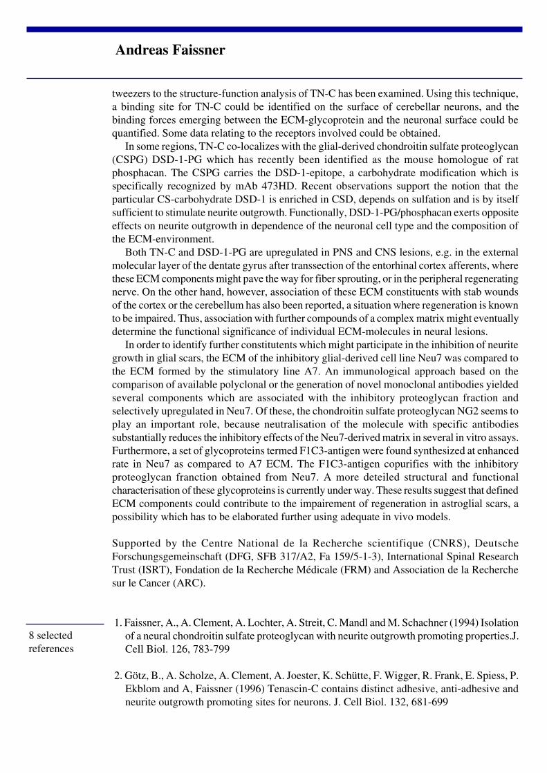
tweezers to the structure-function analysis of TN-C has been examined. Using this technique,a binding site for TN-C could be identified on the surface of cerebellar neurons, and thebinding forces emerging between the ECM-glycoprotein and the neuronal surface could bequantified. Some data relating to the receptors involved could be obtained.
In some regions, TN-C co-localizes with the glial-derived chondroitin sulfate proteoglycan(CSPG) DSD-1-PG which has recently been identified as the mouse homologue of ratphosphacan. The CSPG carries the DSD-1-epitope, a carbohydrate modification which isspecifically recognized by mAb 473HD. Recent observations support the notion that theparticular CS-carbohydrate DSD-1 is enriched in CSD, depends on sulfation and is by itselfsufficient to stimulate neurite outgrowth. Functionally, DSD-1-PG/phosphacan exerts oppositeeffects on neurite outgrowth in dependence of the neuronal cell type and the composition ofthe ECM-environment.
Both TN-C and DSD-1-PG are upregulated in PNS and CNS lesions, e.g. in the externalmolecular layer of the dentate gyrus after transsection of the entorhinal cortex afferents, wherethese ECM components might pave the way for fiber sprouting, or in the peripheral regeneratingnerve. On the other hand, however, association of these ECM constituents with stab woundsof the cortex or the cerebellum has also been reported, a situation where regeneration is knownto be impaired. Thus, association with further compounds of a complex matrix might eventuallydetermine the functional significance of individual ECM-molecules in neural lesions.
In order to identify further constitutents which might participate in the inhibition of neuritegrowth in glial scars, the ECM of the inhibitory glial-derived cell line Neu7 was compared tothe ECM formed by the stimulatory line A7. An immunological approach based on thecomparison of available polyclonal or the generation of novel monoclonal antibodies yieldedseveral components which are associated with the inhibitory proteoglycan fraction andselectively upregulated in Neu7. Of these, the chondroitin sulfate proteoglycan NG2 seems toplay an important role, because neutralisation of the molecule with specific antibodiessubstantially reduces the inhibitory effects of the Neu7-derived matrix in several in vitro assays.Furthermore, a set of glycoproteins termed F1C3-antigen were found synthesized at enhancedrate in Neu7 as compared to A7 ECM. The F1C3-antigen copurifies with the inhibitoryproteoglycan franction obtained from Neu7. A more deteiled structural and functionalcharacterisation of these glycoproteins is currently under way. These results suggest that definedECM components could contribute to the impairement of regeneration in astroglial scars, apossibility which has to be elaborated further using adequate in vivo models.
Supported by the Centre National de la Recherche scientifique (CNRS), DeutscheForschungsgemeinschaft (DFG, SFB 317/A2, Fa 159/5-1-3), International Spinal ResearchTrust (ISRT), Fondation de la Recherche Médicale (FRM) and Association de la Recherchesur le Cancer (ARC).
1. Faissner, A., A. Clement, A. Lochter, A. Streit, C. Mandl and M. Schachner (1994) Isolationof a neural chondroitin sulfate proteoglycan with neurite outgrowth promoting properties.J.Cell Biol. 126, 783-799
2. Götz, B., A. Scholze, A. Clement, A. Joester, K. Schütte, F. Wigger, R. Frank, E. Spiess, P.Ekblom and A, Faissner (1996) Tenascin-C contains distinct adhesive, anti-adhesive andneurite outgrowth promoting sites for neurons. J. Cell Biol. 132, 681-699
8 selectedreferences
Andreas Faissner

3. Rauch, U., A. Clement, C. Retzler, L. Fröhlich, R. Fässler, W. Göhring and A. Faissner(1997) Mapping of a defined neurocan binding site to distinct domains of tenascin-C.
J. Biol. Chem. 272, 26905-26912
4. Clement, A.M., S. Nadanaka, K. Masayama, C. Mandl, K. Sugahara and A. Faissner (1998)The DSD-1 carbohydrate epitope depends on sulfation, correlates with chondroitin sulfateD motifs, and is sufficient to promote neurite outgrowth. J. Biol. Chem. 273, 28444-28453
5. Garwood, J., O. Schnädelbach, A. Clement, K. Schütte, A. Bach and A. Faissner (1999)DSD-1-Proteoglycan is the mouse homologue of phosphacan and displays opposing effectson neurite outgrowth dependent on neuronal lineage. J. Neurosci. 19, 3888-3899
6. Joester, A. and A. Faissner (1999) Evidence for combinatorial variability of tenascin-Cisoforms and developmental regulation in the mouse central nervous system.
J. Biol. Chem. 24, 17144-17151
7. Kiernan, B.W., J. Ferguson, E.E. Frost, E.M. Torres, S.B. Dunnett, Y. Saga, S. Aizawa, R.Kaur, R.J.M. Franklin, A. Faissner, and C. ffrench-Constant (1999) Myelination andbehaviour of tenascin-C null transgenic mice. European J. Neurosci. 11, 3082-3092
8. Fidler, P.S., K. Schuette, R.A. Asher, A. Dobbertin, S.R. Thornton, Y. Calle-Patino, E.Muir, J.M. Levine, H.M. Geller, J.H. Rogers, A. Faissner and J.W. Fawcett (1999) Comparingastrocytic cell lines that are inhibitory or permissive for axon growth: the major axon-inhibitory proteoglycan is NG2.
J. Neuroscience 19, 8778-8788
Andreas Faissner

During postnatal development the capacity of the CNS to initiate repair processes upon lesionsdecreases dramatically. The developmental decrease in the levels of GAP-43 in specific regionsof the CNS is correlated in time and degree to myelin formation. In several types of cell andtissue culture experiments CNS myelin has been shown to be an inhibitory substrate for neuritegrowth and adhesion and to induce rapid and long lasting growth cone collapse. Severalmolecular constituents including MAG, proteoglycans, tenascin-R and the high molecularweight membrane protein NI-250/Nogo-A/IN-1 antigen have been shown to exert neuritegrowth inhibitory effects. In vivo, inhibition of myelin formation, removal of myelin, antibodiesagainst CNS myelin or a monocloncal antibody against NI-250/Nogo-A (mAB IN-1) havebeen shown to induce regeneration of lesioned adult CNS fiber tracts. Nogo-A is a noveloligodendrocyte membrane protein with very potent inhibitory effects which are neutralizedby mAB IN-1 or several new anti-Nogo-A antibodies. In vivo, these antibodies upregulateexpression of growth-associated genes in the adult CNS (see abstract Rossi et al.). They enhancelong distance regeneration of lesioned corticospinal axons. mAB IN-1 also induces sproutingof corticofugal and rubrospinal fibers in brainstem and spinal cord following CST lesions.These antibody-treated rats show a high degree of functional recovery for locomotion andfine motor control of the hand (food pellet reaching). These results suggest that neurite growthinhibitory proteins, in particular Nogo-A, are crucially involved in down-regulating plasticityand repair during maturation of the mammalian CNS.
Martin E. Schwab
Schwab, Martin E.,Dept. Neuromorphology,Brain Research Institute,University of Zurich and Swiss Federal Institute ofTechnology Zurich, Winterthurerstr. 190,8057 Zurich,Switzerland
Short abstract:
Full abstract:
Title:REGENERATION AND PLASTIC GROWTH OF AXONS IN INJUREDCENTRAL NERVOUS SYSTEM
Keywords: regeneration, plasticity, repair, inhibitors of neurite growth, Nogo-A
Components in CNS myelin efficiently inhibit neurite growth in vitro and in vivo. Theoligodendrocyte protein Nogo-A is a potent collapse-inducing and growth inhibitorycomponent. Its neutralization by antibodies induces regeneration and large scale plasticchanges in the lesioned spinal cord and brain which are accompanied by a high degree offunctional recovery in adult rats.

1. Schnell, L., Schneider, R., Kolbeck, R., Barde, Y.-A. and Schwab, M.E.: Neurotrophin-3enhances sprouting of corticospinal tract during development and after adult spinal cord lesion.Nature 367: 170-173, 1994.
2. Thallmair, M., Metz, G.A.S., Z’Graggen, W.J., Raineteau, O., Kartje,G.L. and Schwab,M.E.: Neurite growth inhibitors restrict plasticity and functional recovery followingcorticospinal tract lesions. Nature Neurosci. 1: 124-131, 1998.
3. Chen, M.S., Huber, A.B., van der Haar, M.E., Frank, M., Schnell, L., Spillmann, A.A.,Christ, F. and Schwab, M.E.: Nogo-A is a myelin-associated neurite outgrowth inhibitor andan antigen for monoclonal antibody IN-1. Nature 403: 434-439, 2000.
4. Buffo, A., Zagrebelsky, M., Huber, A.B., Skerra, A., Schwab, M.E., Strata, P. and Rossi,F.: Application of neutralizing antibodies against NI-35/250 myelin-associated neurite growthinhibitory proteins to the adult rat cerebellum induces sprouting of uninjured Purkinje cellaxons. J. Neurosci. 20: 2275-2286, 2000.
4 selectedreferences
Martin E. Schwab

It has become widely accepted that in addition to inhibitory influences presented byglial scarring and cavitation at a site of injury, the environment of the adult mammalian CNSbeyond the glial scar is generally non-permissive for the long distance growth of axons. Anumber of theories suggest that as part of normal development, the brain and spinal cordeither lose axon growth promoting trophic and substrate molecules (Ramon y Cajal, 1928;Kawaja and Gage, 1991) or acquire molecules that are potently inhibitory to regeneration(McKerracher et al., 1994; Chen et al., 2000), or a combination of both (Schnell et al., 1994).My research interests for the past few years have therefore focused on designing in vivoexperiments that allow a clearer distinction of the relative contributions of the glial scar, theadult environment and intrinsic neuronal limitations to the ultimate failure of axon regenerationin the adult mammalian CNS after injury.
MicrotransplantationThe microtransplantation technique, originally developed by Dr. John Seeley at Mill
Hill (Emmett et al., 1989) has proven to be a powerful in vivo research tool in that it allowsthe ability to efficiently assess the degree of axon growth support provided by normal ordegenerating adult CNS white and grey matter for known numbers of neurons of differentages and types, all in the absence of glial scarring. White matter distal to a large lesionundergoes many cellular and molecular changes from acute to chronic stages after trauma,including axon degeneration, myelin degradation, glial cell death, reactive glial responses andglial precursor invasion as well as a cascade of inflammatory cell and cytokine responses
Stephen Davies
Stephen Davies Ph.D.Assistant Professor,Department of Neurosurgery,Baylor College of Medicine,Scurlock Tower, Suite 944,6560 Fannin Street,Houston, Texas, USA 77030
Short abstract:
Full abstract:
Title:Regeneration of adult axons in the intact and degenerating adult mammalian
central nervous system.
Keywords: Regeneration, Scar, Astrocyte, Proteoglycan, Growth cone
A lack of growth supportive molecules, myelin associated inhibitors, intrinsic neuronallimitations, and the glial scar have all been proposed as major factors preventing axonregeneration in the adult mammalian CNS. In order to test the ability of the intact anddegenerating adult CNS environment to support axon growth in the absence of glial scarring,a minimally traumatic microtransplantation technique was used to inject adult neurons directlyinto white matter pathways of the brain and spinal cord.

whose effects on the ability of the damaged CNS to support axon growth are at present poorlyunderstood. The minimally traumatic microtransplantation technique prevents scar formationaround the site of implantation and therefore allows newly formed axons of transplanted neuronsdirect access to the environment of the adult CNS without first having to grow through thepotentially inhibitory glial scar. The use of intrinsic markers of transplanted neurons notexpressed by surrounding host tissues, such as M2 (mouse specific antigen), calcitonin generelated peptide and most recently, enhanced green fluorescent protein (EGFP transgenic mousedonor), unequivocally labels regenerating axons and thus avoids the complication of axonsparing commonly associated with partial lesion experimental paradigms.
The permissive nature of the adult CNS environment Using this technique it has been possible to demonstrate that not only is minimally
disturbed adult CNS white matter of the fimbria and corpus callosum permissive for the rapidand long distance directed growth of axons from microtransplanted embryonic CNS (Davieset al., 1993, 1994) and adult sensory neurons (Davies et al., 1997), but that acute and evenchronically (2 weeks to 3 months) degenerating adult spinal cord white matter, distal to alarge lesion of the dorsal columns, is also similarly supportive of the long distance (10 mm in10 days) regeneration of significant numbers (~900) of adult EGFP labeled sensory axons(Davies et al., 1999). The close association of the regenerating axons with host white matterastrocytic processes and their contact with myelin observed with confocal microscopy andimmuno-EM (Davies et al., 1997,1999), strongly suggests that the adult astrocyte, away froma site of injury, plays an important role in supporting axon growth and overcoming the effectsof the myelin associated inhibitors. Adult astrocytes are known to express growth-supportivemolecules such as laminin and neurotrophins in the injured adult CNS that might be involvedin tipping the balance in favour of axon growth. Interestingly, unlike the unbranched adultsensory axons that regenerated from intra-callosal grafts (Davies et al., 1997), the EGFP labeleddonor axons within white matter of the adult rat spinal cord dorsal columns extended numerouscollaterals directed towards the adjacent dorsal horn grey matter. This demonstrates that adultsensory axons are capable of responding to correctly matched terminal field guidance cuesthat are still present within the adult spinal cord even when undergoing wallerian degeneration.
Experiments currently underway will determine if degenerating spinal cord white matterand grey matter distal to the scar retain their axon growth permissive nature at time pointsbeyond 3 months when challenged with freshly dissociated microtransplanted adult donorneurons. This approach permits the effects of wallerian degeneration on the capacity of theCNS environment to support axon growth to be distinguished from changes in the ability ofaxotomized neurons to mount a regenerative response from acute to chronic time points afterinjury.
Inhibition of regeneration at the glial scarImportantly, in a second series of experiments (Davies et al., 1999) in which substantial
lesions of the dorsal columns were moved closer to the adult EGFP labeled DRGmicrotransplants, donor axons rapidly regenerated 5 mm through acutely and chronically (3month) degenerating white matter only to abruptly stop upon entering the vicinity of the glialscar, a distance effectively half that normally covered from the site of implantation in bothunlesioned and lesioned white matter. The change from rapidly elongating, streamlined growthcones to dystrophic endings closely resembling those classically described by Ramon y Cajal,strongly correlated with the arrival of axons at the highest concentrations of scar associatedinhibitory molecules such as the chondroitin sulfate proteoglycans (reviewed in Hoke and
Stephen Davies

Silver, 1996). Physical constraints and disorganization of the adult CNS cytoarchitecture mayhave also contributed to regeneration failure within these large lesions. However, a similarinhibition of axon regeneration upon contacting high levels of CSPGs even in the presence ofnormally aligned astrocytic processes, was also observed at the boundaries of a small numberof failed adult DRG microtransplants (Davies et al., 1997), and in the cingulate microlesionmodel of adult CNS regeneration failure (Davies et al., 1996, & unpublished data).
Scar associated inhibitors of axon growthIt seems likely that in both the latter minimally traumatic studies, a critical threshold of
damage to the adult CNS had been exceeded, triggering the upregulation and deposition ofinhibitory ECM molecules such as the CSPGs (e.g. neurocan, phosphacan, brevican, aggrecanand NG2) as well as other scar associated inhibitors such as Tenascins-C & R, and SemaphorinIII at the site of injury. One group of candidate triggers of scar formation are the transforminggrowth factor-beta pro-inflammatory molecules which have been shown to induce inhibitorymatrix production in cultured astrocytes (Smith and Hale, 1997). However, at present, thesignaling cascades that promote the deposition of inhibitory matrix at a site of injury within theCNS have not been fully established. Ongoing and future studies within my laboratory andthose of other researchers will determine which molecules inhibitory to axon growth and celltypes expressing them are present within stab and contusion lesions of the adult brain andspinal cord at acute to chronic time points after injury.
ConclusionsTaken together, observations from the adult to adult microtransplantation experiments
constitute compelling evidence that the glial scar and hence those inhibitory factors such asCSPGs found within its boundaries, constitute the major environmental impediment toregeneration in the adult CNS, certainly at acute to 3 month chronic stages after injury. Anunderstanding of the biology of scar formation and the molecular and cellular mechanismsintrinsic to the adult CNS beyond the glial scar that can promote and guide adult axons to theircorrect targets is vitally important if we are to design efficient regeneration strategies for thefuture.
1. Chen, MS, Huber, AB, van der Haar, ME, Frank, M, Schnell, L, Spillmann, AA, Christ, F,Schwab, ME (2000) Nature 403:434-9.
2. Davies, SJA, Field, PM & Raisman, G (1993) Eur J Neurosci 5:95-106.
3. Davies, SJA, Field, PM & Raisman, G (1994) J Neurosci 14:1596-1612.
4. Davies, SJA, Field PM & Raisman, G (1996) Exp Neurol 142:203-216.
5. Davies, SJA, Fitch MT, Memberg SP, Hall AK, Raisman, G & Silver, J (1997) Nature 390:680-683.
6. Davies, SJA., Gaucher DR., Doller, C & Silver, J (1999) J Neurosci 19(14):5810-5822.
7. Emmett, CJ, Jaques-Berg, W & Seeley, PJ (1989) Neuroscience 38: 213-222.
8. Hoke, A & Silver, J (1996) Prog Br Res 108:149-163.
9. Kawaja, MD, & Gage, FH (1991) Neuron 7:1019-1030.
10. McKerracher, L, David, S, Jackson, D L, Kottis, V, Dunn, R & Braun PE (1994) Neuron 13: 805-811.
13 selectedreferences
Stephen Davies

11. Ramon y Cajal, S (1928) Degeneration and regeneration of the nervous system (Oxford University Press, London).
12. Schnell, L, Schneider, R, Kolbeck, R, Barde, YA, & Schwab, ME (1994) Nature 367:170-173.
13. Smith GM, Hale JH (1997) J Neurosci 24:9624B9633.
Figure Legend
Adult sensory axons regenerate within degenerating adult spinal cord white matter.A confocal scan at a distance of 4mm from an 8 day survival microtransplant showing EGFP labeled adultsensory axons (green channel) that have grown within acutely degenerating white matter distal to a large lesionof the adult rat dorsal columns. Reactive adult astrocytes (GFAP: red channel) within the degenerating whitematter may provide trophic and substrate support for the rapidly growing axons.
Stephen Davies

CONTENTS
NEUROPROTECTION5th session
Molecular mechanisms of lesion-induced celldeath in the adults CNSMathias Bähr (Germany)
Bcl2 gene therapy and BDNF rescuegeniculate neurons following cortical lesionsLamberto Maffei (Italy)
Inhibitory of apoptosis proteins (IAP) in braindevelopment and injuryDan Lindholm (Sweden)
Neurosteroid and hormone metabolism in thenervous system: Regulation by neurones androle during regenerationMichael Schumacher (France)

GENE EXPRESSION PATTERN IN DE- AND REGENERATING CNS NEURONES
A hallmark of axonal injury is secondary neuronal death, which may occur withinhours (ischemia), days (trauma) or years (demyelination and neurodegeneration) from onsetof the disease. Transection or crush-axotomy of long projection tracts like the optic nerveinduces retrograde death of approximately 80% of the affected neurones, the retinal ganglioncells (RGCs) within two weeks after lesion. This secondary cell loss is mainly apoptotic andinvolves specific changes in gene expression pattern of transcription factors (e.g. c-jun orATF-2), pro- and anti-apoptotic genes (e.g. bcl-2 or bax) and growth-associated genes (likeGAP-43). Thus, long term survival and initiation of regeneration programmes of projectionneurones such as RGCs critically depends on inhibition of apoptotic cell death which is triggeredby axonal lesions and initiation of developmentally regulated growth programmes, which canusually not be activated in adult terminally differentiated CNS neurones.
We have used a variety of techniques to interfere with the cell death cascades thatfollow lesions of the optic nerve in adult rats. Temporary inhibition of neuronal apoptosis canbe afforded by pharmacological administration of trophic factors or by gene therapy approachesusing adenovirus vectors that can deliver neurotrophic factors directly into neurons or intosurrounding glial cells. Application of inhibitors of pro-apoptotic proteins like NAIP, XIAP,CrmA or p35 or antisense-oligonucleotides that counteract pro-apoptotic proteins like baxcan also be used to rescue RGCs from secondary death. Other strategies to prevent secondarycell loss aim at caspase inhibition, or reduction of neuronal damage caused by free radicals,
Prof. Dr. Mathias Bähr
Prof. Dr. Mathias BährNeurologische UniversitätsklinikHoppe-Seyler-Str.3, 72076 Tübingen
Full abstract:
Long abstract:
Title:Molecular mechanisms of lesion induced cell death in the adult CNS
Keywords: Neuronal cell death – apoptosis – neuroprotection – retinal ganglion cells – genetherapy
The mechanisms of secondary neuronal cell death were examined in a highly standardisedmodel system of apoptosis in vivo. To that end, retinal ganglion cells of adult rats wereaxotomized by cutting the optic nerve, which leads to changes in the expression of transcriptionfactors, apoptosis-regulating genes, caspase activation and apoptotic cell death. Based onthese findings, neuro-protective strategies were designed which support neuronal survivalafter axonal lesions and allow regenerative axon growth.

including inhibition of PARP.Recently, we have studied the intersections between neuronal survival and cell death
programmes in more detail. Trophic factors like IGF-I and BDNF activate protein kinase B(PKB) via a phosphatidyl-inositol-3’-kinase (PI-3-K) – dependent mechanism and therebysupport neuronal survival by direct suppression of caspase-3 activation. BDNF also acts viathe mitogen-activated protein kinases (MAPKs) ERK1 and ERK2. Thus, we assume that BDNFdoes not depend on a single signal transduction pathway in exerting its survival promotingaction on lesioned CNS neurons.
In summary, important molecular mechanisms of the cell death cascade that followsaxonal lesions could be determined in a highly standardised in vivo model of neuronal apoptosis.The development of new neuroprotective therapy strategies including gene therapy approachesmay serve as a basis for effective brain repair in the future.
1. N.Klöcker, A. Cellerino and M. Bähr (1998) Free radical scavenging and inhibition ofnitric oxide synthase potentiates the neurotrophic effects of BDNF on axotomized retinalganglion cells in vivo. J. Neurosci.18(3): 1038-1046
2. S.Isenmann, N. Klöcker, C.Gravel and M. Bähr (1998) Protection of axotomized retinalganglion cells by adenovirally delivered BDNF in vivo. Eur. J. Neurosci.10: 2751-2756
3. P.Kermer, N.Klöcker, M. Labes and M. Bähr (1998) Inhibiton of CPP-32-like proteasesrescues axotomized retinal ganglion cells from secondary death in vivo.: J.Neurosci.18(12):4656-4662.
4. Isenmann, S. Engel, E. Jost, E. Hoffmann, F. Gillardon and M. Bähr (1999) Bax antisenseoligonucleotides suppress bax expression in axotomized rat retinal ganglion cells in vivo.Cell Death & Differentiation 6 : 673-682.
5. Klöcker, P. Kermer, M. Gleichmann, M. Weller and M. Bähr (1999) Both the neuronaland inducible isoform contribute to upregulation of retinal NOS activity by BDNF. J.Neurosci.19:8517-8527.
6. S.Kügler, N. Klöcker, P. Kermer, S. Isenmann and M. Bähr (1999) Transduction ofaxotomized retinal ganglion cells by adenoviral vector administration at the optic nervestump: an in vivo model system for the inhibition of neuronal apoptotic cell death. GeneTher.6: 1759-1767
7. P. Kermer, N. Klöcker, M. Labes, S. Thomsen, A. Srinivasan, K. Tomaselli and M. Bähr(1999) Activation of caspase-3 in axotomized rat retinal ganglion cells in vivo: FEBSletters 453: 361-364
8. P.Kermer, N.Klöcker, M. Labes and M. Bähr (2000) Insulin-like growth factor-I protectsaxotomized rat retinal ganglion cells from secondary death via PI3K-dependent Akt-phosphorylation and inhibition of caspase-3 in vivo. J. Neurosci.20: 722-728.
9. J.Weise, S.Isenmann, N.Klöcker, S.Kügler, S.Hirsch, C.Gravel and M.Bähr (2000)Adenovirus-mediated expression of CNTF rescues axotomized rat retinal ganglion cellsbut does not support axonal regeneration in vivo. Neurobiol Dis. 7: 212-223.
10. S.Kügler, G.Straten, F.Kreppel, S.Isenmann and M.Bähr (2000) The X-linked inhibitorof apoptosis (XIAP) prevents cell death in axotomized CNS neurons in vivo. Cell Death& Differentation: accepted
10 selectedreferences
Prof. Dr. Mathias Bähr

The figure shows putative signal transduction pathways and intersections of apoptotic and neuroprotectivepathways. Cell death triggers lead to Cytochrome c release from mitochondria and caspase 9 activation via theapoptosome (Cytochrome c, Apaf-1 and procaspase-9). Active caspase 9 can activate caspase 3 which is able tocleave important cellular proteins like PARP finally resulting in apoptotic death of the cell.Stimulation of growth factor receptors like trks by neurotrophic factors leads to activation of the PI-3 kinase –Akt/PKB pathway which can suppress activation of caspase-3. Alternative or additional neuroprotective pathwaysinclude the ras-pathway and leading to enhanced transcription of survival-promoting genes. Intracellularly, anti-apoptotic members of the bcl-2 family are able to suppress the cell death cascades.
Prof. Dr. Mathias Bähr
Cortical traumatic lesions and cortical ischemia are known to produce retrogradeneuronal death in thalamic areas rojecting to the damaged cortex. This secondarythalamic damage has been described in both animal models and man (Tamura et al.,1991; de Bilbao et al., 2000) and is known n to produce well-characterized syndromesassociated with severe behavioural impairments (Bogousslavsky et al., 1988). Forexample, studies in patients with lesions of the cortex have shown that the post-traumatic decline of cognitive performance correlates with atrophy and reductions ofblood flow values in the thalamus and subcortical white matter (Tamura et al., 1991;Terayama et al., 1991). These data indicate that the development of strategies forlimiting thalamic death after a cortical injury is fundamental to achieve functionalrestoration in clinical settings.As a model system we have chosen the degeneration of the lateral geniculate bodyafter visual cortex ablation in the rat. A rapid and massive loss of geniculate neuronstakes place in both newborn and adult animals following the lesion (Cunningham etal., 1979; Al-Abdulla et al., 1998). Recent experiments have provided ultrastructuraland biochemical evidence that this death occurs by apoptosis, suggesting potentialtargets for therapeutic intervention (Al-Abdulla et al., 1998; de Bilbao et al., 2000).We have developed two novel anti-apoptotic strategies that may be suitable for clinicalapplications:
1) Trophic factor supply to the thalamus by means of intraocular injections. We havefound that one neurotrophin, brain-derived neurotrophic factor (BDNF), is taken upby retinal ganglion cells after intraocular injection and transported anterogradely alongthe optic nerve. The factor is then released to post-synaptic neurons in the lateralgeniculate nucleus. We have performed intraocular injections of BDNF in rats withvisual cortex ablation and we have found that this treatment decreases by 50% thenumber of degenerating neurons in the geniculate. The rescue effect is dependenton axonal transport (since it is completely blocked by intraocular administration ofcolchicine) but not on BDNF signalling within the retina (as shown by blocking theBDNF transduction cascade at the retinal level).These data demonstrate that the BDNF delivery into the eye represents an effectivestrategy for promoting neuronal survival after lesions of the geniculo-striate pathway.
Full abstract:
Title:Bcl2 gene terapy and BDNF rescue geniculate neurons following corticallesions.
Lamberto Maffei
Prof of Neurobiology at the Scuola Normale,director of the institute of Neurophysiology of CNR

The clinical application of neurotrophins has been limited by the fact that these factorsdo not cross the blood-brain barrier following systemic administration (Lindsay et al.,1994). Obviously this is a serious snag for their use in humans. Our experimentsdemonstrate the feasibility of neurotrophin delivery to the visual thalamus (and possiblyother retino-recipient districts) via a minimally invasive eye approach.
2) Over-expression of anti-apoptotic genes (bcl-2) in thalamic neurons via adeno-associated virus (AAV) vectors.It is known that Bcl-2 overexpression represents a very effective strategy for protectingcentral neurons from trauma-induced cell death (Bonfanti et al., 1996; Porciatti et al.,1996), and AAV vectors are particularly attractive for gene therapy applications in thecentral nervous system due to the high efficiency of neuronal transduction and thelong-term stability of gene expression (Hermens and Veerhagen, 1999). We haveconstructed recombinant AAV vectors harboring a human bcl-2 gene under the controlof a CMV promoter. The vectors have been stereotaxically injected into thegeniculate of the adult rat. Three weeks after inoculation, clusters of cells stronglyimmunopositive for the human Bcl-2 protein can be found within the injected geniculate(Fig. 1A). All the transduced cells display a neuronal morphology (Fig. 1B) and indeed75% of the Bcl-2-positive units are geniculo-cortical neurons (Fig. 1C). No expressioncan be detected in glial cells (Fig. 1D). Using stereological techniques we havecalculated that, on average, 10-15% of the geniculate neurons are transfected by theBcl-2 vector. These neurons survive visual cortex ablation and indeed theirnumber is not altered in lesioned rats (Fig. 1E). Injection of a control AAV vectorencoding green fluorescent protein (GFP) results in a similar efficiency of transductionbut has no effect on cell survival after lesion. This demonstrates that AAV-mediatedBcl-2 overexpression represents a very effective tool for preventing geniculate deathafter lesion.
REFERENCES1. Tamura et al. (1991) Thalamic atrophy following cerebral infarction in the territoryof the middle cerebral artery. Stroke 22: 615-618.
2. de Bilbao et al. (2000) Cell death is prevented in thalamic fields but not in injuredneocortical areas after permanent focal ischemia in mice overexpressing the anti-apoptotic protein Bcl-2. Eur J Neurosci 12: 921-934.
3. Bogousslavsky J, Regli F, Uske A (1988) Thalamic infarcts: clinical syndromes,etiology, and prognosis. Neurology 38: 837-848.
4. Terayama et al. (1991) Role of thalamus and white matter in cognitive outcomeafter head injury. J Cereb Blood Flow Metab 11: 852-860.
5. Cunningham TJ, Huddelston C, Murray M (1979) Modification of neuron numbersin the visual system of the rat. J Comp Neurol 184: 423-434.
6. Al-Abdulla NA, Portera-Caillau A, Martin LJ (1998) Neuroscience 86: 191-209.
7. Lindsay RM, Wiegand SJ, Altar CA, DiStefano P (1994) Neurotrophic factors:
8 selectedreferences
Lamberto Maffei

from molecule to man. Trends Neurosci 17: 182-189.
8. Bonfanti L, Strettoi E, Chierzi S, Cenni MC, Liu H-H, Martinou J-C, Maffei L, Rabacchi SA (1996)Protection of retinal ganglion cells from natural and axotomy-induced cell death in neonatal transgenicmice overexpressing bcl-2. J Neurosci 16: 4186-4194.
9. Porciatti V, Pizzorusso T, Cenni MC, Maffei L (1996) The visual response of retinal ganglion cells isnot altered by optic nerve transection in transgenic mice overexpressing Bcl-2. Proc Natkl Acad SciUSA 93: 14955-14959.
10. Hermens W, Veerhagen J (1998) Viral vectors, tools for gene transfer in the nervous system. Prog Neurobiol55: 399-432.
FIGURE LEGEND
(A,B) Low- and high-power photomicrograph of geniculate neurons labelled with an anti-human Bcl-2 antibody, three weeks after vector inoculation. (C) Geniculate neurons double-labelled with anti-human Bcl-2 (red) and Fluorogold (green) retrogradely transported fromthe visual cortex. (D) Astrocytes (labelled with GFAP, green) are not stained by anti-humanBcl-2 (red). (E) The stereological analysis shows that 15% of the geniculate neurons aretransduced by the Bcl-2 vector. All the transduced cells survive in animals with visual cortexlesion.
Lamberto Maffei

Lindholm D.
Lindholm D.*, Korhonen L.*, Mercer E.A.*, Belluardo N.#, Besselmann M** , Oláh L. **,Föcking M. **, Hossmann K.-A. **, Trapp, T. **
* Department of Neuroscience, Neurobiology, Uppsala University, BMC, Box 587, S-75123 Uppsala, Sweden# Department of Human Physiology, Faculty of Medicine, University of Palermo, I-94125 Palermo, Italy**/ Max-Planck-Institute for Neurological Research, Department of ExperimentalNeurology, Gleueler Str. 50, 50931 Cologne, Germany
Title:INHIBITORY OF APOPTOSIS PROTEIN (IAP) IN BRAINDEVELOPMENT AND INJURY
Key words: Nerve cell death, kainic acid, ischemia, XIAP, NAIP
IAP is a family of proteins which counteract cell death and suppress apoptosis in different celltypes. However, little is known about the presence and function of these proteins in the nervoussystem. We have studied the expression of X chromosome-linked inhibitor of apoptosis protein(XIAP) which inhibits caspases, in developing and adult rat brain using a specific antibody,XIAP is widely expressed in adult rat brain, among others within hippocampus, cerebellumand cereberal cortex. XIAP immunoreactivity co-localised with neuronal marker NeuN butalso with glial fibrillary acidic protein showing that both cell types can express XIAP. Kainicacid, which induces delayed cell death of specific neurons, first increased the number of XIAPpositive cells in hippocampus, but at later time XIAP was absent from cells undergoing celldeath. Inhibitors studies showed that XIAP is degraded in a caspase-3 dependent manner inneurons which contributes to nerve cell death in vulnerable regions of the hippocampus.Similarly, following occlusion of the middle cerebral artery XIAP immunoreactivity firstincreased and then decreased in the core of the infarction. However, outside the core, whereneurons will survive the ischemic insult, the XIAP upregulation was long lasting. These resultsindicate that endogenous XIAP has a protective function in brain and that the relative levelsmay determine whether a neuron will die or not following different insults.In contrast to XIAP, the mechanism by which Neuronal Apoptosis Inhibitory Protein (NAIP),deleted in many patients suffering from Spinal Muscular Atrophy, inhibit neuronal death isunknown. We observed that NAIP, through its third BIR 3 domain, binds the neuron-restrictedcalcium binding protein, hippocalcin, in an interaction promoted by calcium. Survival datausing motorneuron cell lines showed a synergistic effect between NAIP and hippocalcin infacilitating neuronal survival against calcium-induced death stimuli. Analysis of caspase-3activity in these cells showed that NAIP can protect against calcium-induced nerve cell deathin both caspase-3 activated and non-activated pathways. These results show that XIAP andNAIP can both inhibit neuronal death after various insults but that the mechanisms used andthe protected neuronal populations are partly different.
Full abstract:

Studies of steroid hormone actions on the nervous system have mainly focused on the controlof reproduction. However, the emerging picture is that steroid hormones are not only sexhormones and that their functions go far beyond reproduction. In fact, steroids regulate manyaspects of neuronal and glial functioning and may be considered as therapeutic agents toenhance reparative responses in the nervous system.Steroids acting on neurons and glial cells are produced by the steroidogenic endocrine glands,the gonads and adrenal glands, and they reach the brain and peripheral nerves via the bloodstream. In addition, some steroids can be formed locally in the brain and in peripheral nerves,either by the metabolism of circulating hormones or by the de novo synthesis from cholesterol.The latter have been named “neurosteroids” (Baulieu et al., 1999).The rodent sciatic nerve is an excellent model to study the synthesis, mechanisms of actionand trophic effects of steroids because of its simple structure, plasticity and regenerativecapacity. Thus, in response to local freezing, axons degenerate quickly in the region of thenerve distal to the lesion site. However, Schwann cells survive, proliferate and produce growthfactors and hormones. They then surround the regenerating axons with new myelin sheaths.This system has allowed us to show that the 3β-hydroxysteroid dehydrogenase (3β-HSD),enzyme which converts pregnenolone to progesterone, is present and functional in peripheralnerves and that its expression depends on the presence of intact axons.In the uninjured nerve, high levels of progesterone were measured, transcripts of the 3β-HSDare expressed and homogenates prepared from rat sciatic nerves actively convert[3H]pregnenolone to [3H]progesterone (Km = 1 µM, Vmax = 32 pmol/mg protein/min). After
Michael Schumacher
Michael Schumacher1, Rachida Guennoun1, HéctorCoirini2, Yasmina Benmessahel1, FlorenciaLabombarda2, Françoise Robert1, Alejandro DeNicola2, Etienne-Emile Baulieu1.(1) INSERM U488, 80 rue du Général Leclerc, 94276Bicêtre, France, (2) Instituto de Biologia y MedicinaExperimental, Buenos Aires, Argentina.
Short abstract:
Full abstract:
Title:Neurosteroid and hormone metabolism in the nervous system :regulation by neurons and role during regeneration.
Keywords : Neurosteroids, progesterone, myelin, Schwann cells, spinal cord.
Progesterone is synthesized by Schwann in response to an axonal signal. In the peripheralnervous system, progesterone plays an important role in myelination. As Schwann also expressintracellular progesterone receptors, the steroid may regulate myelination by autocrine actions.Indeed, progesterone was shown to activate the expression of Schwann cell genes involved inmyelin formation. Progesterone receptors are also present in the spinal cord, where progesteroneregulates expression of GFAP and of the NADPH-diaphorase.

cryolesion, 3β-HSD mRNA could no longer be detected by RT-PCR, recovering only as thenerves regenerate and as axon-Schwann cell relationships are re-established. If regenerationdoes not occur, as after nerve transection, 3β-HSD transcripts are not re-expressed. By usinga co-culture system in which Schwann cells and sensory neurons were separated by amicroporous membrane, allowing the diffusion of large molecules but no direct contact betweenthe two cell types, it was shown that 3β-HSD expression and activity in Schwann cells isdependent on a diffusible neuronal factor (Robert et al., 2000).The induction of 3β-HSD mRNA in the regenerating rat sciatic nerve coincided with theinduction of transcripts coding for the myelin protein zero (P0) and for the peripheral myelinprotein-22 (PMP-22), thus suggesting a role for locally synthesized progesterone in myelinformation. This was demonstrated in the regenerating male mouse sciatic nerve (in collaborationwith H. Koenig Univ. Bordeaux). Blocking either the local synthesis (trilostane) or the receptor-mediated action of progesterone (RU486) after cryolesion impaired the formation of newmyelin sheaths (Koenig et al., 1995).Schwann cells not only have the capacity to synthesize progesterone from pregnenolone, theyalso express an intracellular receptor for the steroid. In contrast to the progesterone receptorpresent in reproductive tissues, the receptor present in the sciatic nerve is not inducible byestrogens (Jung-Testas et al., 1996). Progesterone may thus be part of an autocrine signallingloop involved in the regulation of myelination. Indeed, in transient transfection experiments,it was shown that progesterone increases promoter activity of the genes coding for P0 andPMP-22 (Désarnaud et al., 1998). It is unlikely that the genes coding for P0 and PMP-22 aredirect targets for progesterone, because no classical hormone response elements could beidentified in their promoter regions. Instead, progesterone could activate genes involved inearlier steps of myelin formation. Our attention has focused on the transcription factor Krox-20 (Egr-2), which is a key component of the transduction cascade linking axonal signalling tomyelination. When added to the culture medium of the mouse Schwann cell line MSC80,progesterone very rapidly and transiently induced Krox-20 mRNA expression measured byRT-PCR. The effect of progesterone was hormone-specific, could be mimicked by the selectiveprogesterone receptor agonist ORG 2058 and blocked by the progesterone receptor antagonistRU486 (figure). The induction of both Krox-20 mRNA and protein could also be demonstrated
Michael Schumacher

Whether progesterone is a neurosteroid in the peripheral nervous system, which means a steroid synthesized denovo from cholesterol, remains to be determined. We have so far not been successful in demonstrating thepresence and activity in Schwann cells of the cytochrome P450scc, enzyme characteristic of all steroidogeniccells which converts cholesterol to pregnenolone. However, levels of pregnenolone are elevated in the rodentsciatic nerve and they remain unchanged after removal of the steroidogenic endocrine glands by combined castrationand adrenalectomy, thus suggesting a local synthesis of the steroid. In addition, proteins which play an obligatoryrole in cholesterol transport from intracellular stores to the inner mitochondrial membrane, where the cytochromeP450scc is located, are expressed and regulated in Schwann cells. These proteins include the peripheralbenzodiazepine receptor (Lacor et al., 1999) and the steroidogenic regulatory protein StAR.Progesterone also exerts major influences on the central nervous system. Progesterone receptors have recentlybeen identified by immunocytochemistry in the rat spinal cord. By using an antibody which recognizes the B-isoform, it was shown that ventral horn motoneurons, glial cells in grey and white matter and ependymal cellscontain progesterone receptors. As for the sciatic nerve, spinal cord progesterone receptors are not inducible byestrogen (Labombarda et al., 2000a). Progesterone was shown to regulate the expression of two proteins whichare affected by injury in the rat spinal cord : glial fibrillary acidic protein (GFAP) and NADPH-diaphorase, anenzyme with nitric oxide synthase activity (Labombarda et al., 2000b).
Figure legend
Effects of progesterone receptor agonist and anatgonist on Krox-20 mRNA expression in the MSC80 Schwanncell line. Top : a representative RT-PCR analysis; bottom : quantitative data after normalization with 18S obtainedfrom cultures treated with 10-6 M of the synthetic progestin Organon 2058 (ORG) , 10-7 M of progesterone(PROG) and/or with 10-5 M of the progesterone receptor antagonist RU486 (RU) (means of triplicates ± SD).
Michael Schumacher

1. Labombarda F, Guennoun R, Gonzalez S, Roig P, Lima A, Schumacher M, De Nicola AF(2000) Immunocytochemical evidence for a progesterone receptor in neurons and glial cells ofthe rat spinal cord. Neurosci.Lett. 288:29-32.
2. Labombarda F, Gonzalez S, Roig P, Lima A, Guennoun R, Schumacher M, De Nicola AF(2000) Progesterone-induced changes of NADPH-diaphorase and glial fibrillary acidic proteinin astrocytes from normal and injured rat spinal cord. J.Neurochem., in press.
3. Robert F, Guennoun R, Désarnaud F, Do Thi AN, Benmessahel Y, Baulieu EE, SchumacherM (2000) Synthesis of progesterone in Schwann cells: regulation by neurons, submitted.
4. Baulieu EE, Robel P, Schumacher M (1999) Neurosteroids. A new regulatory function inthe nervous system. Totowa, New Jersey: Humana Press.
5. Lacor P, Gandolfo P, Tonon MC, Brault E, Dalibert I, Schumacher M, Benavides J, FerzazB (1999) Regulation of the expression of peripheral benzodiazepine receptors and theirendogenous ligands during rat sciatic nerve degeneration and regeneration: a role for PBR inneurosteroidogenesis. Brain Res. 815:70-80.
6. Désarnaud F, Do T, Brown AM, Lemke G, Suter U, Baulieu EE, Schumacher M (1998)Progesterone stimulates the activity of the promoters of peripheral myelin protein-22 and proteinzero genes in Schwann cells. J Neurochem 71:1765-1768.
7. Jung-Testas I, Schumacher M, Robel P, Baulieu EE (1996) Demonstration of progesteronereceptors in rat Schwann cells. J Steroid Biochem Mol Biol 58:77-82.
8. Koenig H, Schumacher M, Ferzaz B, Do Thi AN, Ressouches A, Guennoun R, Jung-TestasI, Robel P, Akwa Y, Baulieu EE (1995) Progesterone synthesis and myelin formation by Schwanncells. Science 268:1500-1503.
8 selectedreferences
Michael Schumacher

CONTENTS
TRANSPLANTATION6th session
Cell therapies for neurodegenerativedisorders: neuronal replacement andreconstruction of brain circuitryAnders Björklund (Sweden
Development of neural and stem-cells intransplantation to the adult mammalian CNSOle Isacson (U.S.A.)
Functional repair of the neostriatumStephen Dunnett (Great Britain)
Rescue and reconstruction of degeneratedretinae with cell transplantsRay Lund (Great Britain)

Neuronal replacement in Parkinson´s disease.Neural grafting in Parkinson´s disease (PD) is based on the idea that dopamine supplied
from cells implanted into the striatum can substitute for the lost nigrostriatal neurons. In rodentand primate models of PD, embryonic dopamine neurons grafted to the denervated striatumcan establish a functional innervation and restore dopaminergic neurotransmission in the areasurrounding the transplant. However, to survive transplantation, dopamine neurons must beobtained at an early stage of development, and they must be placed ectopically (in the striatumrather than in the substantia nigra) to connect up with the dopamine-receptor-bearing targetcells and exert their functional effects.
Clinical trials have shown that mesencephalic dopamine neurons obtained from humanembryo cadavers can survive and function in the brains of patients with PD. PET scans haveshown significant increases in [18 F]fluorodopa uptake in the area around the graft that hasbeen maintained for at least six years in several patients. Long-lasting (up to over 10 years)symptomatic improvement with 30-50% reduction in the UPDRS motor scores has beenreported in a majority of the grafted patients, and in the most successful cases it has beenpossible to withdraw L-DOPA treatment. Two cases that have come to autopsy have showngood survival of grafted dopamine neurons and extensive axonal outgrowth into the graftedputamen. Immunological rejection of the human-to-human allografts has not been reported inany PD patient, even several years after withdrawal of immunosuppressive treatment. However,abundant macrophages and T and B lymphocytes were observed in otherwise healthy-lookingand functional transplants in the two autopsy cases, 12 months after withdrawal of cyclosporin
Anders Björklund
Wallenberg Neuroscience Center,Department of Physiological Sciences,Lund University,Sölvegatan 17, S-223 62 Lund, Sweden
Short abstract:
Full abstract:
Title:Use of neural progenitors and stem cells for dopamine neuron replacementin Parkinson´s disease
Key words: Transplantation, Parkinson´s disease, Dopamine, Stem cells, Functional recovery
Cell replacement has emerged as a promising approach for repair and functional recovery inthe damaged adult central nervous system. The lecture will summarize the progress made inthe use of fetal mesencephalic dopanine neuroblasts in Parkinson´s disease and discuss thepossibilites to use immature neural progenitors and stem cells for this purpose.

immunosuppression (18 months after transplantation), indicating the potential for a host-derivedimmune response in intra-cerebral allografts when immunosuppression is removed.
The demonstration that embryonic dopamine neurons can survive and function in thehuman brain represents a first important step towards a cell replacement therapy in PD. Currentresearch is aimed at improving the survival and growth of transplanted dopamine neurons,and finding alternative sources of cells for grafting. The main limitations of current cell-transplant procedures are the ethical, practical and safety issues associated with tissue derivedfrom aborted human fetuses, and the large amounts of embryonic mesencephalic tissue thatare needed to obtain therapeutic effects in patients, which severely restricts the possibility ofapplying this procedure outside highly specialized centres.
In current grafting protocols, no more than 5–20% of the expected numbers of grafteddopamine neurons survive. Consequently, tissue from at least 3–4 embryos, yielding about100,000 surviving dopamine neurons, needs to be implanted on each side of the patient’sbrain to induce significant therapeutic improvement. Addition of free-radical scavengers,caspase inhibitors or neurotrophic factors to the fetal cell preparation may increase dopamineneuron survival 2–3 fold. Moreover, in the rat PD model, administration in vivo of neurotrophicfactors (including GDNF, BDNF and bFGF) to the transplants during the first weeks afterimplantation enhances both survival and growth of intrastriatal dopamine neuron transplants.
Application of these principles to clinical protocols may reduce the need for multipledonor embryos and increase the functional efficacy of the grafted cells. The ethical issuessurrounding the use of human embryonic cells for grafting remains a matter of concern. Cellsfrom other species may offer a solution. Cells transplanted between species survive well inthe brain, which is partly protected from the body’s immune system, provided the recipient istreated with immunosuppressive drugs. Indeed, clinical trials using porcine cells are alreadyunder-way in both PD and Huntington’s disease patients. However, the use of xenotransplantsin humans remains controversial, not least because effective and acceptable techniques forlong-term immunosuppression across the species barrier are not yet fully established. Moreover,there remain concerns about a theoretical risk of cross-species transfer of infectious agents, inparticular animal retroviruses.
Expansion of neuronal progenitors.Until now, transplantation of dopamine neurons has focused primarily on differentiatedneuroblasts and young postmitotic neurons, at the stage of neuronal development which hasbeen found empirically to be optimal for survival, growth and establishment of functionalconnectivity of the explanted cells. However, precursor cells taken at earlier stages ofdevelopment, when they are in an active proliferative phase, might prove more effective.Thus, if it were possible to expand precursor cells in vitro and control their terminaldifferentiation into mature dopamine neurons, then large numbers of cells could be expandedand made available for transplantation as required. This would have the additional advantagethat such cells can be standardized, screened and manipulated (for example, by cell sorting orgene transduction) in ways that could never be possible with the limited quantities of freshtissue that are available. It is now possible to expand the precursors of mesencephalic dopamineneurons in vitro by stimulation with high concentrations of growth factors (typically bFGFand/or EGF), and expanded cells can subsequently be induced to differentiate into maturedopamine neurons. The expanded cells can survive and function after transplantation to thestriatum in the rat PD model, although the overall yield of surviving dopamine neurons invivo was quite low. If neural progenitors are to be used for neuronal replacement in PD, it willbe essential to control the steps leading to induction of a dopaminergic phenotype. This islikely to be a complex process involving temporally and spatially controlled cell–cell
Anders Björklund

Anders Björklund
interactions and specific signalling molecules.It is at present unclear whether cells used for grafting in PD must be neuronal or whether
non-neuronal cells that secrete dopamine (or its precursor L-DOPA) constitutively in a diffuse,non-synaptic manner would suffice. Adrenal chromaffin cells were the first cells of the lattertype to be investigated. These cells, which produce dopamine and several other catecholamines,as well as a variety of neuroactive and neurotrophic factors, have the advantage that they can beobtained from one of the patient’s own adrenals. However, the long-term survival of chromaffincells is very poor in the brain, and the initial functional effects that are seen after implantationinto the striatum (which are unlikely to be due to dopamine secretion alone) are not well sustainedat longer survival times, either in experimental PD or in PD patients.
These problems have stimulated the search for cells that survive better and can sustainconsiderably higher levels of in vivo dopamine release after transplantation to the brain.Experiments using implants of dopamine-releasing polymers or encapsulated dopamine- andL-DOPA-producing PC12 cells have shown that implants that act as a ‘biological minipump’can tonically activate dopamine receptors and induce limited functional effects in the denervatedstriatum, without any synaptic connections. Working on the same principle, primary non-neuronal cells producing dopamine, such as glomus cells from the carotid body, and cellsengineered to produce high levels of L-DOPA and/or dopamine are being investigated (seebelow). However, the only functional responses that have been obtained with these kinds ofcell (normalization of dopamine-receptor supersensitivity as reflected in a reduced motorresponse in the standard rotation test) involve a pharmacological mechanism of action, and itremains to be seen whether non-neuronal dopamine-secreting cells can provide any long-lastingimprovements in more complex aspects of the motor symptoms in rat or monkey PD models,or indeed in human PD.
1. Olanow, C.W., Kordower, J.H. & Freeman, T.B. Fetal nigral transplantation as a therapyfor Parkinson’s disease. Trends Neurosci. 19, 102-109 (1996).
2. Lindvall, O. Cerebral implantation in movement disorders: State of the art. Mov. Disord.14, 201-205 (1999).
3. Kordower J.H. et al. Fetal nigral grafts survive and mediate clinical benefit in a patientwith Parkinson’s disease. Mov. Disord. 13, 383-393 (1998).
4. Piccini P. et al. Dopamine release from nigral transplants visualized in vivo in aParkinson´s patient. Nature Neurosci. 2, 1137-1140 (1999).
5. Fricker RA, Carpenter MK, Winkler C, Greco C, Gates MA, Bjorklund A. Site-specificmigration and neuronal differentiation of human neural progenitor cells after transplantationin the adult rat brain. J Neurosci. 1999 19:5990-6005.
6. Wagner J, Akerud P, Castro DS, Holm PC, Canals JM, Snyder EY, Perlmann T, ArenasE.Induction of a midbrain dopaminergic phenotype in Nurr1-overexpressing neural stemcells by type 1 astrocytes. Nat Biotechnol. 1999 7:653-9.
7. Studer L., Tabar V. & McKay RDG Transplantation of expanded mesencephalicprecursors leads to recovery in parkinsonian rats. Nature Neurosci. 1, 290–295 (1998).
8. Lee SH, Lumelsky N, Studer L, Auerbach JM, McKay RD. Efficient generation ofmidbrain and hindbrain neurons from mouse embryonic stem cells. Nat Biotechnol. 2000 6675-9.
9. Bjorklund A, Lindvall O. Cell replacement therapies for central nervous system disorders.Nat Neurosci. 2000 6:537-44.
9 selectedreferences

We have compared morphology and function of primary mouse or rat ventral mesencephalicdopaminergic neurons to that of neurons differentiated from early embryonic stem (ES) cells.ES cells from mouse blastocysts previously expanded in the presence of leukemia inhibitoryfactor were transplanted, with or without retinoic acid pretreatment, into adult mouse brain,adult lesioned at brain, and into the mouse kidney capsule. Our results show that whentransplanted into either CNS or non-CNS sites, blastula-derived ES in the presence or absenceof RA pre-treatment, can develop into heterogeneous tissues with many cells expressingneuronal markers and morphologies of mature neurons[1, 2]. Large numbers of dopaminergicand or serotonergic neurons were found in grafts placed either in the brain or the kidneycapsule.
Neural induction regardless of transplant site in our paradigm is consistent with recentevidence suggesting that neuralization is a default pathway, and occurs spontaneously if pre-gastrula cells do not receive other inducing signals to form epidermal, mesodermal, orendodermal cells[3]. This was first suggested by experiments showing that cells of the earlygastrula ectodermal animal cap, that normally develop into epidermal tissue, all form neuraltissue if dissociated[4]. Bone morphogenic protein (BMP4) and activin have been implicatedas the major inducers of epidermal differentiation during gastrulation. Ectopic application ofBMP4 is sufficient to induce epiderm formation in dissociated animal pole cap cells[5], andhomozygous knock-out mice lacking functional BMP receptor (BMPR1) die in gastrulation[6], a time when epidermis would otherwise form. Also, antagonists of BMP4 or activinsignaling, such as noggin, follistatin, and chordin, which are produced in the Spemann organizerregion, can induce the ectopic formation of neural tissue[6-8]. The inactivation of the dominant
Ole Isacson
Dr. OLE ISACSONAssoc. Prof. of Neurology (Neuroscience),Harvard Medical School,Director of Neuroregeneration Laboratories atMcLean/Massachusetts General Hospital.
Short abstract:
Full abstract:
Title:Embryonic Stem Cells Can Neuralize Into Dopaminergic Neurons AfterTransplantation: Comparisons With Primary Fetal Neurons
Ole Isacson1,3,4 , Lars Björklund1 and Kwang-Soo Kim 1,2
1) Udall Parkinson’s Research Center of Excellence, McLean Hospital, Harvard MedicalSchool; Belmont, MA 02478; 2)Molecular Biology Laboratory, McLean Hospital, Belmont,MA 02478; 3) Program in Neuroscience, Harvard Medical School; Boston, MA 02114; 4)Department of Neurology, Massachusetts General Hospital; Boston, MA 02115
Keywords: Parkinson’s disease, transplantation, embryonic stem cells, axon growth, glialgrowth
We have compared morphology and function of primary mouse or rat ventralmesencephalic dopaminergic neurons to that of neurons differentiated from earlyembryonic stem (ES) cells. ES cells from mouse blastocysts previously expanded in thepresence of leukemia inhibitory factor were transplanted, with or without retinoic acidpretreatment, into adult mouse brain, adult lesioned at brain, and into the mouse kidneycapsule.

activin receptor also induces neuraldifferentiation[9]. In our experiments,transplanting cells that have been dissociated andexpanded at the pre-gastrula stage, may disrupt thelocalized cell-cell communications whichotherwise inhibit neuralization. There was aremarkable congruence between primary ventralmesencephalic DA neurons and DA neurons thatdifferentiated in situ from ES cells. Local brainor kidney signals had similar effects onneuralization and therefore suggest that intrinsicgenetic programs may dominate in differentiationof neurons
Figure Legend: D3 ES Cell Graft in the AdultMouse Striatum. Dopaminergic markers includingTH, the dopamine transporter (DAT) and AHDare present in this confocal image.
1. Song, J., L. Bjorklund, S. Chung, T. Andersson, L.C. Costantini, K.-S. Kim, and O. Isacson,Establishing Nurr1-expressing ES cell lines for neural transplantation.
Soc. for Neuroscience (Abstract), 2000;2. Deacon, T., J. Dinsmore, L.C. Costantini, J. Ratliff, and O. Isacson, Blastula-stage stem
cells can differentiate into dopaminergic and serotonergic neurons after transplantation. Exp. Neurol., 1998. 149: p. 28-41;3. Hemmati-Brivanlou, A. and D. Melton, Vertebrate embryonic cells will become nerve
cells unless told otherwise. Cell, 1997. 88: p. 13-17;4. Grunz, H. and L. Tacke, Neural differentiation of Xenopus laevis ectoderm takes place
after disaggregation and delayed reaggregation without inducer. Cell Diff. Dev., 1989. 28(211-218);5. Wilson, P.A. and A. Hemmati-Brivanlou, Induction of epidemis and inhibition of neural
fate by Bmp-4. Nature, 1995. 376: p. 331-333;6. Sasai, Y., B. Lu, H. Steinbeisser, D. Geissert, L. Gont, and E. De Robertis, Xenopus
chordin: a novel dorsalizing factor activated by organizer-specific homeobox genes. Cell, 1994. 79: p. 779-790;7. Hemmati-Brivanlou, A., O. Kelley, and D. Melton, Follistatin, an antagonist of activin, is
expressed in the Spemann organizer and displays direct neuralizing activity. Cell, 1994. 77: p. 283-295;8. Zimmerman, L., J. De Jesus-Escobar, and R. Harland, The Spemann organizer signal
noggin binds and inactivates bone morphogenic protein. Cell, 1996. 86: p. 599-606;9. Hemmati-Brevanlou, A. and D. Melton, A truncated activin receptor inhibits mesoderm
induction and formation of axial patterning in Xenopus embryos. Nature, 1994. 359: p. 609-614.
Ole Isacson
9 selectedreferences

Rats and monkeys with striatal lesions exhibit both motor and cognitive deficits similar tothose seen in human Huntington’s disease. Embryonic striatal cells survive transplantation tothe adult striatum, differentiate anatomically to exhibit all major striatal neuronal types (althoughinterspersed with non-striatal cells that also originate in the ganglionic eminence), establishextensive afferent and efferent connections with the host brain that are morphologicallyappropriate at the ultrastructural level and alleviate functional impairments associated withthe lesions in both motor and cognitive task. There is accumulating evidence from behavioural,pharmacological, electrophysiological and in vivo neurochemical studies that the grafts exhibittheir functional effects by incorporation into incorporation into the host neural circuitry.Moreover, the grafts do not simply release or restore the animals ability to perform the relevantfunctions, but rather provide a neural substrate for the learning of new motor habits and selectionof appropriate action. Experimental results on the efficacy of striatal grafts in mouse rat andmonkey models are now providing the foundation for clinical trials of striatal repair byembryonic cell transplantation in Huntington’s disease.
1. Bachoud-Lévy, A.C., Bourdet, C., Brugières, P., Nguyen, J.P., Grandmougin, T.,Haddad, B., Jény, R., Bartolomeo, P., Boissé, M.F., Dalla Barba, G., Degos, J.D., Ergis,A.M., Lefaucheur, J.P., Lisovoski, F., Pailhous, E., Rémy, P., Palfi, S., Defer, G.L., Cesaro,P., Hantraye, P., and Peschanski, M. (2000). Safety and tolerability assessment ofintrastriatal neural allografts in Huntington’s disease patients. Experimental Neurology,161, 194-202.
Stephen B. Dunnet
Professor Stephen B. DunnettDSc, Professorial Fellow,School of Biosciences, Cardiff University,Museum Avenue Box 911, Cardiff CF10 3US
Short abstract:
Full abstract:
Title:Functional Repair of the Striatum.
Keywords: striatal lesions, striatal transplants, Huntington’s disease, recovery of function,circuit reconstruction.
Rats and monkeys with striatal lesions exhibit both motor and cognitive deficits similar tothose seen in human Huntington’s disease. Embryonic striatal cells survive transplantation tothe adult striatum, and can alleviate functional impairments associated with the lesions inboth motor and cognitive tasks. There is accumulating evidence that the grafts achieve theireffects by reconstruction of afferent and efferent connections with the host brain leading tofunctional incorporation into the host neural circuitry.
10 selectedreferences

2. Björklund, A., Campbell, K., Sirinathsinghji, D.J.S., Fricker, R.A., and Dunnett, S.B.(1994). Functional capacity of striatal transplants in the rat Huntington model. InFunctional Neural Transplantation (ed. S.B. Dunnett and A. Björklund), pp. 157-195.Raven Press, New York.
3. Brasted, P.J., Watts, C., Robbins, T.W., and Dunnett, S.B. (1999). Associative plasticityin striatal transplants. Proceedings of the National Academy of Sciences of the UnitedStates of America, 96, 10524-10529.
4. Clarke, D.J., and Dunnett, S.B. (1993). Synaptic relationships between cortical anddopaminergic inputs and intrinsic GABAergic systems within intrastriatal striatal grafts.Journal of Chemical Neuroanatomy, 6, 147-158.
5. Dunnett, S.B. (1995). Functional repair of striatal systems by neural transplants:evidence for circuit reconstruction. Behavioural Brain Research, 66, 133-142.
6. Dunnett, S.B., Carter, R.J., Watts, C., Torres, E.M., Mahal, A., Mangiarini, L., Bates,G., and Morton, A.J. (1998). Striatal transplantation in a transgenic mouse model ofHuntington’s disease. Experimental Neurology, 154, 31-40.
7. Isacson, O., Dunnett, S.B., and Björklund, A. (1986). Graft-induced behavioral recoveryin an animal model of Huntington disease. Proceedings of the National Academy ofSciences of the United States of America, 83, 2728-2732.
8. Kendall, A.L., Rayment, F.D., Torres, E.M., Baker, H.F., Ridley, R.M., and Dunnett,S.B. (1998). Functional integration of striatal allografts in a primate model of Huntington’sdisease. Nature Medicine, 4, 727-729.
9. Peschanski, M., Cesaro, P., and Hantraye, P. (1995). Rationale for intrastriatal graftingof striatal neuroblasts in patients with Huntington’s disease. Neuroscience, 68, 273-285.
10. Wictorin, K. (1992). Anatomy and connectivity of intrastriatal striatal transplants.Progress in Neurobiology, 38, 611-639.
Stephen B. Dunnet

Stephen B. Dunnet
Figure Legend
Afferent and efferent connections between striatal grafts and host brain studied at the ultrastructural level(based on Clarke & Dunnett 1993).

There is a range of retinal diseases in which photoreceptors die either because of intrinsicgenetic defects or because of defects in the adjacent retinal pigment epithelium. The mainclinical conditions are retinitis pigmentosa affecting around 1 in 2500 people and age relatedmacular degeneration causing visual dysfunction in about 17% of people over 65. There ispresently no effective treatment for these diseases but in recent years several approaches havebeen developed in experimental animals. One of these is to transplant cells into the subretinalspace. Such transplanted cells can either function by limiting the progress of degeneration orby replacing photoreceptors once they are lost.Several major concerns have accompanied transplantation studies. These include how thedonor cells work, a full description of what level of vision can be preserved or reconstructedby transplantation and whether cells other than those freshly harvested from donor eyes canbe used.To examine prevention of degeneration we have focussed attention on the RCS rat. In thisanimal the photoreceptors undergo degeneration because of a tyrosine kinase deficiency inthe adjacent retinal pigment epithelial (RPE) cells, which renders them incapable ofphagocytosing shed outer segments. Rod photoreceptors are mostly lost over the first fewmonths of life with cones undergoing a more protracted degeneration.We have worked with two cel sources.The first has used immortalised human RPE cell lines generated by Neurotech, SA and wehave found that if such cells are injected into the subretinal space at 3-4 weeks of age, beforethe degenerative process has progressed very far, a considerable degree of functional rescue
R. D. Lund
R.D.Lund,Institute of Ophthalmology,Bath St., London EC1V9EL, UK
Short abstract:
Full abstract:
Title:Retinal transplantation
Key words: Retinal degeneration, transplantation, RPE cells, immortalisation, Schwann cells.
Transplantation of cells into the subretinal space has the potential of limiting photoreceptordegeneration in a range of blinding diseases including retinitis pigmentosa and age relatedmacular degeneration. We explore here three crucial issues in experimental animal studies –examining how grafted cells interface with the host retina, seeking donor cell sources fromother than freshly harvested eyes and testing functional efficacy of grafts over prolonged timecourses.

can be obtained. Head tracking to moving stripes can be elicited in normal rats, but in dystrophicanimals this is already lost by 8 weeks of age. Cell transplanted animals show brisk responseseven 1 month after transplantation although sham injected animals fail to respond. Visualacuity studies show that rats with transplants can discriminate vertical and horizontal stripeswith a resolution of better than 2 cycles/degree. Physiological studies show that thresholdsensitivity responses can achieve near-normal levels over the area protected by the graft.Finally recording from the visual cortex shows unit responses with reasonable tuningcharacteristics even at 8 months of age, in contrast to unoperated controls, which areunresponsive. The implanted cells often form a layer over the existing RPE, but have processesmaking contact with the underlying Bruch’s membrane.The second approach has been to use Schwann cells. The rationale for this is that it has beenshown that photoreceptor loss can be delayed by injecting a range of growth factors, such asCNTF, BDNF, GDNF and FGF, into the vitreous. Schwann cells make these factors andtherefore we thought they might provide a continuous delivery system, where delivery levelsare likely to be at physiological levels. Accordingly Schwann cells were dissociated and injectedinto the subretinal space of young RCS rats. It was found that these cells limited photoreceptorloss, allowed animals to respond to rotating drums and produced threshold sensitivity studiesin the colliculus that were close to normal levels.To replace photoreceptors once they are lost due to an intrinsic defect in the photoreceptorsthemselves, we turned to the rd mouse in which a phosphodiesterase defect leads to loss ofmost rods within 3 weeks and slower cone loss. We have transplanted a retinal suspensiontaken from 1 week old normal mice and placed it into the subretinal space of 3 month old rdmice. Such transplants form a synaptic interface with host retina and 2 weeks aftertransplantation animals can perform a photophobic response in which they previously failed.Threshold sensitivity studies also show improved responsiveness.These studies show 1) that transplantation can result in better visual performance than inunoperated animals 2) it is possible to use cells other than those derived from fresh donoreyes.3) recovery is seen in both prevention and reconstruction models.This work represents the collaborative effort of Y.Sauve, S.Girman, A. Kwan, S.Wang, J.Lawrence, D. Keegan, B. Lu, J. Greenwood, P. Adamson, P. Coffey, L Hetherington.
1.Lund RD, Lawrence JM, Litchfield TM, Sauvé Y, Whiteley SJO, Coffey P J. Retinaldegeneration and transplantation in the Royal College of Surgeons rat. Eye 12: 597 - 604,1998.
2.Kwan, A.S.L., Wang, S. and Lund, R.D. Photoreceptor layer reconstruction in rodent modelof retinal degeneration. Exp. Neurol. 159(1):21-23,1999
3. J.M. Lawrence, S.J.O. Whiteley, Y. Sauvé, D.J. Keegan, P.J. Coffey, and R.D. Lund.Schwann cell grafting into the retina of the dystrophic RCS rat limits functional deterioration.IOVS 41: 518-528, 2000
3 selectedreferences
R. D. Lund

CONTENTS
SHORT COMMUNICATIONS 1 di 1
Postlesioned accumulation of proteins GAP-43 and BASP1 in spinalcord corelates with formation of new corticolspinal pathways, B.Klementiev, T. Novikova, M. Mosevitsky
Firing properties of axotomized CNS neurons after graftreinnervation, B. Benitez-Temiño, R.R. de la Cruz, A.M. Pastor
Neuronal proliferation in temporal lobe epilepsy, J. Ollikainen, A.Pitkänen, M. Koskinen, I. Rantala, J. Sajanti, P. Karhunen, Ylinen, M.Vapalahti, R.Kälviäinen, L. Paljärvi, H. Haapasalo and J. Suhonen
In vivo neuroprotection of injured cns neurons by a single injectionof a dna plasmid encoding the bcl-2 gene, Sonsoles de Lacalle
Induction of thyroid hormone deiodinases in the scitic nerve afterinjury, M. Pierre, W.W. Li, C. Le Goascogne, M. Schumacher and F.Courtin
Adenosine and neuroprotection in the hippocampus: pivotal roleof A1 inhibitory receptors, A.M .Sebastião, A. de Mendonça and J.A.Ribeiro
The caspase-1 inhibitor AC-YVAD.CMK achieves an enduringneuroprotection by reducing il 1β-and tnf-α production followingcerebral ischemia, M. Rabuffetti, C. Sciorati, G. Tarozzo S. Bortolazzi,M. Beltramo
Neuroprotective role of Dopamine against hippocampal cell death,Y. Bozzi, D. Vallone and E. Borrelli

SHORT COMMUNICATIONS
POSTLESIONED ACCUMULATION OF PROTEINS GAP-43 AND BASP1 IN SPINALCORD CORRELATES WITH FORMATION OF NEW CORTICOSPINAL PATHWAYS
Klementiev B1., Novikova T1., Mosevitsky M2
1Department of Neuropharmacology of Institute of Experimental Medicine PAMS,St.Petersburg, Russia; 2Division of Biophyscs of Petersburg Nuclear Physics InstituteRAS, Gatchina, Leningrad distr.Fax: (7-812)2348994; E-mail: [email protected]
Presynaptic protein GAP-43 and BASP-1 (NAP-22) are neurite growth cone-associated proteins. GAP-43 is expressed when neurons are forming new connectionsduring structural remodelling. The role of BASP-1 in this process is still nearly unknown.The present study is devoted to comparative analysis of changes of original distributionand quantities of GAP-43 and BASP-1 in response to a brain lesion. At early stage of postlesion period (day 4 after unilateral lesion of sensorimotorcortex) the range of intensity of both proteins staining in L1-L3 segments of spinalcord are not changed relatively to control. From the day 7 the GAP-43 immunoreactivityis diminished considerably into the corticospinal tracts, preferentially on controlateralside to the lesion. The same changes are revealed in the adjacent intermedial area ofthe spinal cord. From the day 14 the increase of GAP-43 immunostaining and decreaseof BASP-1 immunostaining in neuropil of the intermedial region of the spinal cord andin corticospinal tract were found. The highest level of GAP-43 and returned to controllevel of BASP-1 immunoreactivity was reached on the day 21 when recovery of motorfunction was described (decrease of slips of contralateral hindlimbs when animals rundown along the parallel bars traversing between two platforms placed 40 cm abovethe floor). At the different time after lesion of left hemisphere rats were given horseradishperoxidase (HRP) into the right side of the L1-L2 segments of the spinal cord andamount of labelled neurones in sensorymotor cortex of intact (ipsilateral) hemispherewere estimated. It was shown that the transport of HRP into ipsilateral hemispherefrom day 14 became significantly higher as in control.

SHORT COMMUNICATIONS
FR (
spik
es/s
)
0
250
RH
(d
eg)
-30
0
30
LH (
deg
)
-30
0
30
C. Axotomy plus transplant
2 s
RH
(d
eg)
-30
0
30
FR (
spik
es/s
)
0
250
LH (
deg
)
-30
0
30
B. Axotomy
FR (
spik
es/s
)
0
100
200
LH (
deg
)
-30
0
30
RH
(d
eg)
-30
0
30
A. Control
FIRING PROPERTIES OF AXOTOMIZED CNS NEURONS AFTER GRAFT REINNERVATION.
B. Benítez-Temiño, R.R. de la Cruz, and A.M. Pastor. Departamento de Fisiología y Biología Animal,Facultad de Biología, Universidad de Sevilla, Avda. Reina Mercedes 6, 41012-Sevilla, Spain.
Axotomy produces changes in the electrical properties of neurons and in their synaptic inputs thatare expressed as alterations in firing pattern. In the present work, we have questioned whether thesechanges might be the consequence of target deprivation induced by lesion. Thus, we have provided anovel target to axotomized central neurons by grafting embryonic cells at the lesion site to study thefiring properties of axotomized reinnervating neurons. The oculomotor system has been used as theexperimental model, in particular, the projection of abducens internuclear neurons (Abd Ints) onto themedial rectus motoneurons of the oculomotor nucleus. The axons of Abd Ints ascend through themedial longitudinal fascicle in the brainstem. Axotomy was performed by transecting this fascicle atapproximately 5-6 mm from the cell bodies. Adult cats were prepared for the chronic recording ofextracellular single units in the abducens nucleus and eye movements. Immediately after axotomy, amechanically dissociated graft of embryonic cerebellar primordium dissected from E-15 cat embryoswas implanted at the lesion site.
The extracellular discharge activity of Abd Ints wasrecorded in the alert behaving animal in control, post-axotomy and after grafting. We have recentlycharacterized the changes produced by axotomy on thefiring of these cells (de la Cruz et al., J. Comp. Neurol., inpress 2000). These include an overall decrease in firingrate, and a loss of eye-related signals, i.e., eye positionand velocity neuronal sensitivities. These changes wereequally found during the different types of eye movements(e.g., spontaneous fixations and saccades, and thevestibulo-ocular reflex). The grafting of a novel target tothe injured Abd Ints restored the normal firing andsensitivities as recorded in the majority of units. Todetermine whether the proximal stumps from transectedaxons have indeed reinnervated the implanted embryoniccells, we performed anterograde labeling with biocytincombined with electron microscopy visualization. Axonsof Abd Ints grew into the transplant sprouting into thegranule cell and molecular layers. Ultrastructuralexamination of labeled axons and boutons revealed theestablishment of synaptic contacts with Purkinje andgranule cells of the transplanted cerebellum. Therefore,these data indicate that axotomized central neuronsresume to normal firing properties following thereinnervation of a novel embryonic target, even if the target is other than the natural one.
Supported by Fundación Mapfre Medicina.
SHORT COMMUNICATIONS
NEURONAL PROLIFERATION IN TEMPORAL LOBE EPILEPSY
J. Ollikainen1, 2, A. Pitkänen5, M. Koskinen1, 2, I. Rantala2, J. Sajanti3, P. Karhunen4.Ylinen6, M. Vapalahti7, R.Kälviäinen4, L. Paljärvi8, H. Haapasalo2 and J. Suhonen1
Department of Neurology and Rehabilitation1, University of Tampere, Finland;Department of Pathology2, University of Tampere, Finland; Department ofNeurosurgery3, University of Tampere, Finland; Department of Forensic Medicine4,University of Tampere, Finland; A. I. Virtanen Institute5, University of Kuopio, Finland;Department of Neurology6, University of Kuopio, Finland; Department of Neurosurgery7,University of Kuopio, Finland; Department of Pathology8, University of Kuopio, Finland;
Key words: neurogenesis - Ki67 - human - temporal lobe epilepsy - hippocampus
Abstract
The study of neurogenesis in human has in the past been limited for the lack ofproliferation markers that do not require the incorporation of molecules into the DNA.We detected immunohistochemically expression of a cell cycle related antigen, Ki67,to detect neuronal proliferation in surgically removed hippocampi from epilepsy patientsand in specimens of autopsy controls.Monoclonal anti-Ki67 (MIB-1) was combined in a double immunostaining with antibodiesagainst neuronal (neurofilament,synaptophysin, anti-Hu), glial (glial fibrillic acidic protein)cytoplasmic proteins and progenitor cell antigens (TUJ-1) using standard streptavidin-biotin immunoperoxidase techniques. We found proliferating cells expressingcytoplasmic progenitor- and neuronal markers in hippocampi of epilepsy patientsinvestigated, and in all control cases. Morphological and immunohistochemical analysissuggested that these proliferating cells were neurons. The number of proliferatingcells varied from zero to 16 per section. These cells located mainly in the pyramidalcell layer in subfields CA1 and CA2. Proliferating neurons were also found in dentategyrus and CA3. Number of proliferating cells did not correlate significantly to age atoperation time, age at the onset of epilepsy, sex, side operated, aetiology or estimatednumber of seizures at lifetime. Nevertheless, there was a clearly significant positivecorrelation between proliferation and duration of epilepsy (p= 0,009, Kruskal-WallisTest). Our results suggest that in the pyramidal cell layer and dentate gyrus ofhippocampus the number of proliferating neurons first diminishes and then increasesslowly in the course of epilepsy. Ki67 immunohistochemistry may offer a promisingmethod to study neuronal proliferation in normal human brain and in neurodegenerativedisorders.

SHORT COMMUNICATIONS
IN VIVO NEUROPROTECTION OF INJURED CNS NEURONS BY A SINGLEINJECTION OF A DNA PLASMID ENCODING THE BCL-2 GENE
Presenter: Sonsoles de Lacalle, Dept. of Biology and Microbiology, California StateUniversity, Los Angeles.
Keywords: cholinergic system; brain repair; bcl-2 gene
Abstract.Current challenge in the field of brain repair is to find novel therapeutic approachesthat prevent atrophy and loss of CNS neurons, and promote regeneration of theirprocessess. We have selected the antiapoptotic gene B-cell lymphoma 2 (bcl-2) asapotential therapeutic agent because it prevents death of many cell types exposed todifferent deleterious stimuli. Since neurodegeneration begins quickly after a lesion,neuroprotective interventions need to be applied soon after the injury and shouldtherefore be relatively non-invasive. We have developed a simple method tointroduce genetic information into CNS cells. The method consists of an injectionofa DNA plasmid that codes for the bcl-2 gene. Injection of the bcl-2 plasmidprevents loss of neurons of the diagonal band of Broca that have been injured by aneurotoxin. The mechanisms by which the bcl-2 injection protects neurons probablyinclude both retrograde transport of the DNA plasmid to the cell nucleus and localneuroprotective effects that ameliorate the hostile environment at or near the site ofinjury.

SHORT COMMUNICATIONS
INDUCTION OF THYROID HORMONE DEIODINASES IN THE SCIATIC NERVEAFTER INJURY.
Pierre M., Li W.W., Le Goascogne C., Schumacher M. and Courtin F.INSERM U488, 80 rue du Gl Leclerc, 94270-Kremlin-Bicêtre, France
Keys words : thyroid hormones, nerve injury, deiodinases
The role of thyroid hormones is essential for the development and function of the brainand also for the development and repair of the peripheral nervous system (PNS). Inthe brain, local 3,5,3’L-triiodothyronine (T3) concentrations are determined bydeiodinases of type 2 (D2) and type 3 (D3), respectively responsible for the activationof thyroxine into T3 and the degradation of thyroid hormones. However, up to date,there is no information related to deiodinases in the PNS. Thus we have looked fordeiodinase expression (mRNAs and activities) in the sciatic nerve and its regulationafter injuries. In the intact sciatic nerve of adult rats, D2 and D3 are present at very lowlevels. After nerve lesion, one observes up-regulation of both deiodinases in the distaland proximal segments. After cryolesion, this up-regulation is observed as early as 4hafter lesion and continues up to day 6. Then the expression of D2 and D3 graduallydeclines, down to a basal level until 28 days, when regeneration and functional recoveryare completed. After a transection, a condition preventing regenerating fibers of theproximal segment to establish contact with the distal segment, up-regulation of D2and D3 persists in both parts up to 28 days. In conclusion, PNS has its own systemresponsible for the local regulation of T3 levels. Moreover, deiodinase induction afterinjury may play an important role during the regeneration process.

SHORT COMMUNICATIONS
ADENOSINE AND NEUROPROTECTION IN THE HIPPOCAMPUS: PIVOTAL ROLE OF A1
INHIBITORY RECEPTORS
Sebastião, A.M., de Mendonça, A. and Ribeiro, J.A. Laboratory of Neurosciences, Faculty ofMedicine, 1649-028 Lisbon, Portugal.
Adenosine prevents neuronal death after ischaemic/hypoxic insults (for review see deMendonça et al., 2000) and by activating inhibitory A1 receptors pre- and post-synapticallyinfluences neuronal activity. Excitatory adenosine A2A receptors are also present throughoutthe brain and may interplay with A1 receptor mediated actions (for review see Sebastião andRibeiro, 2000). Adenosine is released during hypoxia and by activating A1 receptors isresponsible for the depression of synaptic transmission that occurs in hippocampal slicesduring hypoxia (e.g. Canhão et al., 1994). We now obtained evidence that this adenosine-induced depression of synaptic transmission during hypoxia plays an important role in adenosineA1 receptor-mediated neuroprotection, by investigating whether A1 receptor mediated inhibitionof synaptic transmission during hypoxia influences the degree of synaptic transmission recoveryafter a prolonged hypoxic insult.
Field excitatory postsynaptic potentials (fEPSPs) were recorded from the CA1 area ofrat hippocampal slices (400 mm thick) upon stimulation (0.66 Hz, 0.1ms) of the Schaffercollaterals. Hypoxia was applied to each slice only once by switching the O2+CO2 saturatedperfusion solution (pO2 in the recording chamber ~ 600 mmHg) into a N2+CO2 saturated solutionfor 90 min (pO2 in the recording chamber ~ 30 mmHg), followed by 30 min reoxygenation.
During hypoxia the fEPSP slope decreased down to 15 ± 4% (n=8) of its pre-hypoxiavalue, but fully recovered (up to 118 ± 6% of the pre-hypoxia value) upon reoxygenation. Inthe presence of the selective adenosine A1 receptor antagonist, 1,3-dipropyl-8-cyclopentylxanthine (DPCPX 50 nM) both the hypoxic inhibition of the fEPSPs and the fEPSPrecovery were significantly (P<0.05) attenuated. In spite of the presence of DPCPX, synaptictransmission fully recovered after 90 min hypoxia if synaptic transmission was prevented duringhypoxia, either by stopping afferent stimulation or by adding the Na+-channel blocker,tetrodotoxin (100 nM). Blockade of NMDA receptors with DL-2-amino-5-phosphonovaleric acid(AP-5, 50 mM) enhanced (P<0.05) recovery of synaptic transmission after 90 min hypoxia inthe presence of the A1 receptor antagonist, DPCPX (50 nM). In contrast, blockade of excitatoryadenosine A2A receptors with selective antagonists (50 nM ZM 241385 or 50 nM SCH 58261)did not (P>0.05) facilitate recovery of synaptic transmission after 90 min hypoxia in the presenceof DPCPX (50 nM). Thus, NMDA receptor activation during hypoxia, but not A2A adenosinereceptor activation during hypoxia, impair synaptic transmission recovery when the A1 receptorscannot be activated by released adenosine.
It is suggested that adenosine A1 receptors, by inhibiting synaptic transmission andNMDA receptor activation, play a predominant neuroprotective role during hypoxia, and thatthe action of other substances is evident only when that of adenosine is absent.
Canhão, P, de Mendonça, A, Ribeiro, JA (1994) Brain Res. 661, 265-273.De Mendonça, A., Sebastião, A.M. and Ribeiro, J.A. (2000) Brain Res. Rev. (in the press).Sebastião, A.M. and Ribeiro, J.A. (2000). Trends Pharmacol. Sci., 21, 341-346.

SHORT COMMUNICATIONS
THE CASPASE-1 INHIBITOR AC-YVAD.CMK ACHIEVES AN ENDURINGNEUROPROTECTION BY REDUCING IL1-b AND TNF-a PRODUCTIONFOLLOWING CEREBRAL ISCHEMIA
M. Rabuffetti, C. Sciorati, G. Tarozzo S. Bortolazzi, M. BeltramoSchering-Plough Research Institute, San Raffaele Biomedical Science Park, Milan,Italy
The interplay between apoptosis and inflammation is a key event in the developmentof ischemic brain damage. Some early step in the progression of the two processes iscommon. For example IL-1b, a potentially neurotoxic mediator, originates from pro-IL-1b through caspase-1 activity. On the other hand caspase-1 is also an up-streamactivator in the caspase cascade leading to cell death. These data suggest that caspase-1 inhibition is a potentially useful approach to reduce neural damage after an ischemicinsult. We tested this hypothesis by treating ischemic rats with Ac-YVAD.cmk, anirreversible, caspase-1 inhibitor. Treatment reduced infarct volumes of about the 30%(p<0.05) both at 24 h and at 6 days after permanent middle cerebral artery occlusion.Consistently, at 24 hours, caspase-1 activity was almost completly inhibited (D caspase-1 activity measured in pmol/min.mg of protein was: vehicle, 199±40; Ac-YVAD.cmk,7±20; p< 0.01, n=7-9) and IL-1b cortical levels were significantly reduced (D IL-1bmeasured in pg/ml.mg of protein was: vehicle, 82±20; Ac-YVAD.cmk, 11±15; p< 0.05,n=17-19). In order to evaluate the effects of caspase-1 inhibition on apoptosis, wemeasured the levels of histone-associated DNA fragments in cortex homogenateswith a quantitative biochemical approach. Drug treatment significantly decreased freenucleosome formation at 24 hours (D absorbance at 405 nm/mg of protein; vehicle:1.7±0.2; Ac-YVAD.cmk: 0.8 ± 0.1; p< 0.01, n=17-19]. We also investigated the effectsof Ac-YVAD.cmk on cortical levels of TNF-a, another mediator involved in ischemicdamage progression. TNF-a level was elevated 24 hours after ischemia and treatmentwith Ac-YVAD.cmk significantly reduced it, but the effect disappeared at 6 days. Thelevel of other inflammatory mediator, such as IL-10, MCP-1, MIP-2 and NO, were notaffected by the treatment. The present study, showed that caspase-1 inhibition leadsto a long-lasting neuroprotective effect by decreasing both caspase-mediated cell deathand release of proinflammatory mediators. These results point to caspase-1 as asuitable target to prevent neurodegeneration.

SHORT COMMUNICATIONS
Abstract for short communication.
Speaker: Yuri Bozzi, CNR Institute of Neurophysiology. Via Alfieri 1, 56010Ghezzano, Pisa, Italy. E-mail: [email protected]
Neuroprotective Role of Dopamine Against Hippocampal Cell Death
Yuri Bozzi*, Daniela Vallone and Emiliana Borrelli.IGBMC, Strasbourg, France. *CNR Institute of Neurophysiology, Pisa, Italy.
Glutamate excitotoxicity plays a key role in the induction of neuronal cell death occuringin many neuropathologies, including epilepsy. Systemic administration of theglutamatergic agonist kainic acid (KA) is a well characterized model to study epilepsy-induced brain damage. KA-evoked seizures in mice result in hippocampal cell death,with the exception of some strains which are resistant to KA excitotoxicity. Little isknown about the factors that prevent epilepsy-related neurodegeneration. Here weshow that dopamine has such a function through the activation of the D2 receptor(D2R). D2R gene inactivation confers susceptibility to KA excitotoxicity in two mousestrains known to be resistant to KA-induced neurodegeneration. D2R -/- mice developseizures when administered KA doses that are not epileptogenic for wild-type (WT)littermates. Immediate early gene induction (such as c-fos and c-jun), detected by insitu hybridisation, correlates with seizure onset and spread in D2R -/- mice. Moreover,KA-induced seizures result in extensive hippocampal apoptotic cell death (evaluatedby TUNEL staining and immunohistochemistry for the pro-apoptotic factor BAX) inD2R -/- but not WT mice. These results reveal a central role of D2Rs in the inhibitorycontrol of glutamate neurotransmission and excitotoxicity.
Keywords: excitotoxicity, dopamine, neuroprotection.

CONTENTS
POSTERS 1 di 3
Fish spinal cord repair is not correlated to the proliferative activityof the central canal epithelium, A. Alunni, F. Pierucci, S. Torcia, L.Alfei
The effect of JNK knock-out in the the adult brain followingneurodegenerative disorders, S. Brecht, A. Chromik, M. Gelderblom,U. Hidding, K. Mielke, U. Hanisch and Thomas Herdegen
Effects of tat-bcl-xl protein after the focal brain ischemia andreperfusion injury in mice, E. Kilic*, G.P.H. Dietz, E. Hofmann and M.Bähr
Isolation of mRNAS coding for nogo-like proteins from goldfish,D. Leiteritz, M. Simonen, M. Thallmair, M.E. Schwab
Isolation of inducible genes from ischemic gerbil hippocampus bydifferential display, J. Honkaniemi, F. Sharp and H. Frey
Some properties of nerve ending proteins GAP-43 (B-50,Neuromodulin) AND BASP1 (CAP-23, NAP-22) that may be ofimportance for brain development and repair, Mark I. Mosevitsky
Changes in hippocamp activity under impart of dorsal and ventralamygdalofugal pathways, R.M. Bagirova
3-beta-hydroxysteroid dehydrogenase (3β-HSD) expression in therat spinal cord, H. Coirini, M. Gouézou, B. Delespierre, C. Taboulot,M. Schumacher and R. Guennoun
Activation of MLK is involved in neuronal toxicity induced by GluR6receptors, Y.F. Liu

CONTENTS
POSTERS 2 di 3
Immunosupressant FK506 as a modulator of glial cell function andcytokine gene expression, M. Zawadzka, B. Pyrzynska, B. Kaminska
Reparative regeneration in the brain neurons after cessation of 96h general sleep deprivation, B.M.Abushov, F.B.Askerov, F.I. Safarov,F.I.Jafarov
Regenerative capabilities and survival of C-JUN overexpressingpurkinje cells, D. Carulli, A. Buffo, C. Botta, F. Altruda and P. Strata
A “simple” model for complex mechanisms, B. Cuoghi, L. Biasiol,M. Marini, M.A. Sabatini
Calretinin and S100B are overexpressed in trimethyltin-inducedneurodegeneration of the developing rat hippocampus, F. Michetti,R. Businaro, V. Corvino, M.C. Geloso and L. Fumagalli
Topographic organisation of sensory and motor corticospinal fiberprojections in the rat, F. M. Bareyre, M.E. Schwab, O. Raiteneteau
Expression of the chemokine fractalkine and its receptor CX3CR1following focal ischemia: a role in euroinflammation?, G. Tarozzo,S. Bortolazzi, M. Rabuffetti, C. Sciorati, M.Beltramo
Brain-derived neurotrophic factor (bdnf) is an anterograde survivalfactor in the rat visual system, M. Caleo, E. Menna, S. Chierzi, M.C.Cenni and L. Maffei
Inhibition of apoptosis by transduction of TAT-BCL-XL protein intocerebellar granule cells, G.P.H. Dietz, E. Kilic, E. Hofmann, and M.Bähr

CONTENTS
POSTERS 3 di 3
Lacking of Neuronal plasticity changes following axoromized rattranscallosal neurons, S. Gadau, V. Farina, P.L. De Riu, M. Zedda,
High sensitivity detection of GAP-43 mRNA in hippocampus andcerebellum of rat brain by in situ RT-PCR, T. Casoli, G. Di Stefano,N. Gracciotti, S. Giovagnetti, P. Fattoretti, M. Solazzi and C. Bertoni-Freddari
Nitrile neurotoxicity as a model of human neurodegenerativediseases, E. Balbuena
Experimental approaches to basament membrane reduction in themechanically lesioned rat spinal cord, S. Hermanns, P. Reiprich andH. W. Müller
In vitro analysis of the motility of developing neurons and glialcells, P. Zamburlin, P. Ariano, M. Ferraro, D. Lovisolo and C. Distasi
Invalidation of MAP1B gene affects axonal regeneration in the adultnervous system, S. Soares, Meixner A., Propst F. Fischer I., VeronM., and Nothias F.
Neurodegenerative changes in the basal cholinergic neurones inaged rats could be due to the impairment of retrograde axonaltransport, G. Niewiadomska, M. Baksalerska-Pazera
Contribution of caspase 8 to neuronal apoptosis in vivo, J. H.Weishaupt, R. Diem, P. Kermer, K. K. Kikly, S. Krajewsky, R. C. Reedand M. Baehr
POSTER
Fish spinal cord repair is not correlated to the proliferative activity of the centralcanal epithelium.
Alunni Alessandro, Pierucci Federica, Torcia Simona, Alfei LauraDepartment of Animal and Human Biology of the University of Rome “La Sapienza”,via A. Borelli, 50, 00161 Rome, Italy.
Axotomy induced death of neurons appears not to be a factor in the resectedfish spinal cord (Becker et al., J Comp Neurol 377, 577-, 1997). Since continuedneurogenesis has been reported for adult teleost fish (Zupanc, J exp Biol 202, 1435-, 1999), in order to investigate the possibility that regenerating axons in the fish spinalcord could arise from newly generated neurons, we investigated by means of PCNAimmunohistochemistry , the possible persistence of proliferative activity at the spinalcord central canal, for supporting motoneurone recruitment at different developmentalstages. Young adult (260 mm in length), fingerlins (F, 120-170mm), fry (Fr, 70mm)and eleutherembryos (Es, 20-30mm) of rainbow trout (Oncorhyncus mykiss) wereemployed in this study. Proliferative activity was quantified as the number of PCNAlabelled cells in the central canal epithelium, for each spinal cord section. In Es and Fr,a mean value of 3-5 labelled cells for each section was found with a sharp decrease inyoung F (120mm long). After this fish length, it was not possible to quantitatively evaluatethe proliferative activity at the central canal. These data demonstrate that newmotoneurone recruitment in the trout spinal cord, is downregulated at the F stage andsupport the hypothesis that, after this stage, motoneurones should be able to regrowtheir axons during regeneration processes.
Key words: proliferating cell nuclear antigen; development; spinal cord motoneurones

POSTER
The effect of JNK knock-out in the the adult brain following neurodegenerativedisorders
S. Brecht, A. Chromik, M. Gelderblom, U. Hidding, K. Mielke, U. Hanisch* and ThomasHerdegen; Institute of Pharmacologie, Christian-Albrechts-University Kiel, Germany.* Zelluläre Neurowissenschaften, Max-Delbrück-Center for Molecular Medicine, Berlin-Buch, Germany.
Under certain conditions, the activation of Jun-N-terminal kinases (JNK) withphosphorylation of the transcription factor c-Jun can be considered as importantintermediate in pro-apoptotic signal transduction. Therefore we have investigated theeffect of functional JNK1, 2 or 3 knockout (ko) in mice (generated by R. Flavell, R.Davis and P. Rakic) following various degenerative stimuli in vivo and in vitro.
Following transient medial cerebral artery occlusion (MCAo), JNK ko did not alterthe size of the infarct area. JNK2 ko and JNK3 ko, however, dramaticly enhanced theactivation of microglia and astrocytes (increase in IBa1-IR and GFAP-IR).Consequently, we studied the effect of JNK ko on the morphology of microglia andtheir cytokine release (IL6, IL12,KC, TNFa and MIP-1a). Preliminary observations in-dicate an increase in TNFa in JNK2 microglia. Transection of the medial forebrainbundle resulted in preserved survival of otherwise dying SNC neurons in JNK2 ko.Finally, JNK ko provoked a minor not-significant increase in glutamate mediated deathof neonatal hippocampal neurons in vitro. By microarray screening and two-hybridRas recruitment system (RRS) we determined several JNK-dependent genes includingscaffolds of the NFkB pathway. Finally, current experiments include the analysis ofJNK1 + 2 double ko mice with JNK under the control of nestin driven promotor.
The results of our ongoing experiments indicate that JNK exert both, pro-and anti-apoptotic features.

POSTER
EFFECTS OF TAT-BCL-XL PROTEIN AFTER THE FOCAL BRAIN ISCHEMIAAND REPERFUSION INJURY IN MICE
Ertugrul Kilic*,G.P.H. Dietz, Elke Hofmann, and M. Bähr. NeurologischeUniversitätklinik, 72076 Tübingen, Germany.
Introduction: Delivery of therapeutic proteins into tissues and across the blood-brainbarrier (BBB) is severely limited by the size and biochemical properties of the proteins.11-amino-acid HIV TAT fusion protein is able to cross cell membranes and BBB(StevenR et al). The present study was done to evaluate whether TAT fusion protein withBCL-XL is useful to prevent tissue injury following focal brain ischemia induced byintraluminar thread occlusion in mice.
Methods: Adult male C57BL/6j mice (21-28g) were anesthetized with 1% halothane(30% O2, remainder N2O). Rectal temperature was maintained between 36.5 and 37.0°Cusing a feedback-controlled heating system. Laser Doppler flow (LDF) was monitoredabove the territory of the middle cerebral artery (MCA) during ischemia and up to 120minutes after the onset of reperfusion. For the administration of TAT-BCL-XL, the tailvein was cannulated with a PE10 catheter. Focal ischemia was induced by insertion ofa silicon-coated 8-0 nylon monofilament into the common carotid artery, which wasadvanced into the internal carotid artery 9 mm distal to the carotid bifurcation.Reperfusion was initiated 120 minutes later by withdrawal of the monofilament. TAT-BCL-XL was i.v. applied over 10 minutes 1 h bevor ischemia (n=5). After surgery,animals were placed into their home cages. Twenty-four hours later, animals werereanesthetized and decapitated. Brains were removed and dissected into fiveequidistant slices, which were stained with triphenyltetrazolium chloride (TTC).
Results: Measurements of infarct volume, brain swelling and neurological performanceshowed a significant reduction of ischemic injury in TAT-BCL-XL -treated (16±8 mm3,6.9±2 mm3 and 0.9±0.48 respectively) as compared with control animals (57±14 mm3,15±1,7 mm3 and 2.1±0.32 respectively).
Discussion: Delivery of the anti-apoptotic peptides into the brain through the BBB isvery diffucult. Gene therapy is one method for circumventing this problem, but conditionsfor high-efficiency targeting and long-term protein expression have yet to be developed.Our study demonstrates that TAT protein transduction systems are powerful tools tomodulate mechanisms of cell injury after focal brain ischemia. The major advantage ofthis approach is that protective factors are put directly at their site of action. In thisstudy, anti-apoptotic TAT-BCL-XL protein reduced the extent of injury after focal brainischemia.

POSTER
ISOLATION OF mRNAS CODING FOR NOGO-LIKE PROTEINS FROM GOLDFISH
D. Leiteritz, M. Simonen, M. Thallmair*, M.E. SchwabBrain Research Institute, Departement of Neuromorphology, University of Zurich andSwiss Federal Institute of Technology, Winterthurerstrasse 190, CH 8057 Zurich,Switzerland
Regeneration of injured axons is minimal in the CNS of adult mammals and birds. Thisis at least partly due to the neurite growth inhibitor protein Nogo-A (Chen et al., 2000,Nature 403, 434). Fish, in contrast to mammals possess a high capacity for axonalregeneration in the CNS. Interestingly, fish axons are sensitive to mammalian myelin-associated inhibitory proteins (Bastmeyer et al., 1991, J Neurosci 11, 3; Stuermer etal., 1992, J Neurobiol 23, 5), and fish myelin proteins can cause growth cone collapseof fish and rat axons (Wanner et al., 1995, J Neurosci 15, 11). Here we studied whetherfish express Nogo-like proteins. Mammalian Nogo proteins are expressed as threesplice variants, Nogo-A, -B, and –C. Nogo-A is produced by oligodendrocytes and is apotent inhibitor of neurite growth. The function of the smaller proteins Nogo-B and –Cis not yet known, Nogo-C is expressed mainly in muscle. Hybridization of goldfishmuscle RNA with a rat Nogo probe revealed several hybridizing bands. PCR with acDNA library as template and 5‘RACE of goldfish brain RNA were performed. Weshowed the 3‘part of the nogo gene, i.e. the region common to all known Nogo spliceforms. At least two different Nogo-like mRNAs are present in goldfish tissues. Thus,goldfish do express proteins of the Nogo family.Supported by the Swiss National Science Foundation.

POSTER
Honkaniemi J, Sharp, F and Frey H.
Isolation of inducible genes from ischemic gerbil hippocampus by differentialdisplay
Both necrosis and apoptosis have been suggested to participate in the neuronal deathin the hippocampus following global ischemia. In the vulnerable CA1 region, the delayedneuronal death is preceded by induction various genes participating in the apoptoticcell death program. In the present study we applied differential display PCR to isolategenes induced in the gerbil hippocampus following global ischemia by 24 h, at timewhen there is virtually no cell death but a massive induction of various apoptotic genes.Five clones were isolated and their inducibility was verified by in situ hybridization.The relevance of the isolated genes in the delayed cell death of the CA1 region isdiscussed.

POSTER
SUMMARY
SOME PROPERTIES OF NERVE ENDING PROTEINS GAP-43 (B-50,NEUROMODULIN) AND BASP1 (CAP-23, NAP-22) THAT MAY BE OFIMPORTANCE FOR BRAIN DEVELOPMENT AND REPAIR.
Mark I. MosevitskyDivision of Molecular Biophysics, Petersburg Nuclear PhysicsInstitute, 188350 Gatchina, Russia .E.-mail: [email protected]
Keywords: Growth associated proteins (GAPs), specific fragmentation of GAPs,aggregation of GAPs, Structural hanges of GAPs during development and repair
GAP-43 and BASP1 possess some prominent properties in common. In all speciesstudied, the both proteins are represented by several isoforms, but the origin of theseisoforms is different. Isoforms of GAP-43 are the fragments formed by calcium dependedprotease (probably, calpain). The breaks separate 4 and about 40 N-terminal residuesfromGAP-43 molecule. leaving two big fragments named GAP-43-2 and GAP-43-3,respectively. In adult animals, GAP-43 prevails over the fragments, but during ratdevelopment (until 5-th postnatal day ), when neurite outgrowth and targeting proceedGAP-43-3 and complementary N-terminal peptide (1-40) are predominant. This canbe true also for the areas of repair, where nerve growth and targeting were resumedBASP1 is a family of immunochemically related isoforms: besides the whole moleculesseveral incomplete forms are present, but in much less amounts. A possiblemechanism for BASP1 isoforms formation is alternative splicing.Due to elongated form of the molecules and to the presence of negatively and positivelycharged domains GAP-43 and BASP1 form aggregats of 30-40 molecules underphysiological conditions. In BASP1 aggregates, besides electrostatic interaction,hydrophobic one is considerable due to N-terminal myristoylation of the molecules.The aggregated state may be of significance for these proteins interaction withcytoskeleton and other cellular constituents.

POSTER
In this paper present materials, which devoted learning the role ofamygdalo-hypothalamic pathways in the rganization of the hippocampalbioelectrical activity under after electrolytic coagulation of dorsal and ventralamygdalofugal pathways was studied.
The experiments showed that application both reversible [novocain] andirreversible [electrocoagulation] methods of damage of functional integrityof the dorsal amygdalofugal pathway, in contract to ventral one brought todisappearance of not only theta rhythm but the hippocampal EEG asawhole. The observed effects were irreversible: they took place in 6 monthafter coagulations. The short-term EEG depletion in ipsilateralhippocampus produced by application of novocain on the dorsalamygdalfugal pathway.
The lesion of the ventral amygdalofual pathway did not completely blokadethe theta rhythm. The whole recovery of electrographic patterns to thecontrol level in 20-21 days after coagulation was found.
Cholinergic and monoaminergic stimulation of different nucleus ofamygdala, hypothalamic, reticular formation and septum after dorsalamygdalofugal pathway lesion did not produce the synchronization ofelectrical activity as it was in control animals. The neurochemical stimulationabove mentioned structures after the ventral amydalofugal pathway lesionsresulted in typical changes of hippocampal EEG only after the completerecovery of the hippocampal activity.
The obtained data suggest the different influences of the dorsal and ventralpathways on the organization of hippocampal theta rhythm activity.
CHANGES IN HIPPOCAMP ACTIVITY UNDER IMPART OF DORSALAND VENTRAL AMYGDALOFUGAL PATHWAYS
Bagirova R.M.Laboratory Neurophysiology of Learning Institute of Physiology Academyof Science Azerbaijan, Baku

POSTER
3-beta-hydroxysteroid dehydrogenase (3ß-HSD) expression in the rat spinalcord.
Héctor Coirini1,2, Monique Gouézou1, Brigitte Delespierre1, Celia Taboulot1, MichaelSchumacher1 and Rachida Guennoun1.1U 488 INSERM Stéroïdes et Système Nerveux. 80, Rue du Général Leclerc 94276Le Kremlin – Bicêtre France. 2Instituto de Biología y Medicina Experimental, Facultadde Medicina UBA Argentine.
Key words: progesterone, neurosteroids, motoneurones.
Different studies have shown that progesterone (P4) treatment has neuroprotectiveeffects on the spinal cord. To determine if P4 can be locally synthetized in this tissue,we studied the potential expression of 3ß-HSD, the enzyme involved in its biosynthesis.An in situ hybridization study showed that 3ß-HSD mRNA is expressed throughout thespinal cord with similar patterns in cervical (C), thoracic (T), lumbar (L) and sacral (S)regions. The 3ß-HSD-mRNA expression was higher on gray matter than in white mater.The following gradient was observed: dorsal horn > central canal = ventral horn >>ventral funiculus = lateral funiculus. Analysis of the mRNA expression throught thespinal cord for the different layers showed significant differences (p< 0.05 ScheffeANOVA) on layers VII, IX (T o.d.:31.5± 2.9 > L o.d.:18.7±0.4) and layer X (So.d.:29.4±1.7 > C o.d.:15.8± 1.9). The number of grains per cell was higher onmotoneurones than on the neurones of the dorsal horn. Western blot analysis usingan anti-3ß-HSD antibody revealed an immunoreactive protein of 45 kDaltons. Sinceseveral neurosteroids are substrate or products of the 3ß-HSD, ours results open newpossibilities to study the regulatory mechanisms of their biosynthesis in the spinalcord.
POSTER
Activation of MLK is involved in neuronal toxicity induced by GluR6 receptorsYa Fang Liu Department of Pharmaceutical Sciences, Northeastern University
Introduction of kainic acid into rat hippocampus causes rapid neuronal apoptosis whileknockout of JNK3, a neuronal form JNKs leads to hippocampal neurons of transgenicmice to resist neuronal excitotoxicity induced by kainic acid. To date, how activation ofkainate receptors induces JNK activation remains unknown. Our hypothesis is thatMLK2/3, the upstream activators of JNKs may bind to PSD-95, a synaptic proteinsthat binds to the C-terminus of GluR6 receptors and involved in JNK activation andneuronal toxicity mediated by the receptors. By co-immunoprecipitation, we observedthat MLK2 or MLK3 is associated with PSD-95. In vitro binding studies showed thatthe SH3 domain of PSD-95 mediates its binding to MLK2 or MLK3. Transfection ofGluR6 receptors in hippocampal neuronal cells induced JNK activation and neuronalapoptosis, while transfection of GluR6 receptors with a deletion of the PSD-95 bindingmotif failed to mediated JNK activation and neuronal death. Co-transfection of kinasedead of MLK2 significantly attenuated neuronal toxicity induced by GluR6 receptors.In summary, our studies suggested that activation of MLK may be involved in JNKactivation and neuronal toxicity mediated by GluR6 receptors. (This project is supportedby US Army Medical Research and Materiel Command). Key words: GluR6 receptor,MLK, JNK.

POSTER
IMMUNOSUPPRESSANT FK506 AS A MODULATOR OF GLIAL CELL FUNCTION AND
CYTOKINE GENE EXPRESSION.
M. Zawadzka, B. Pyrzynska, B. KaminskaNencki Institute of Experimental Biology, Warsaw, Poland
Neurotrophins and cytokines, with both cytotoxic and neuroprotective activities, expressed in
astrocytes during glia activation play a crucial role in the process of delayed neuronal death.
We demonstrated that the cyclosporin A (CsA) and tacrolimus (FK506), a widely used
immunosuppressants, affect glial cells growth probably by inhibiting signalling pathways that
regulate hypertrophic and/or proliferative responses. In immune system both CsA and FK506
inhibit activity of calcineurin, a ubiquitous calcium-activated serine phosphatase, thereby
suppressing cytokine production. Cultured primary astroglia as well as reactive mature
astrocytes and C6 glioma cells expressed calcineurin mRNA, as determined by RT-PCR.
The aim of this study was to determine the pattern of growth factors and cytokine expression
and effect of immunosuppressants on glial cell function. FK506 inhibited proliferation of primary
astrocytes and reactive astrocytes from striatal trauma and induced death accompanied by
apoptotic changes in nuclear morphology and DNA fragmentation.
Using reverse transcription polymerase chain reaction (RT-PCR), we have studied the
expression of mRNA coding for of various growth factors and cytokines: basic fibroblast growth
factor – bFGF, brain-derived neurotrophic factor – BFGF, ciliary neurotrophic factor – CNTF,
platelet-derived growth factor – PDGF, leukemia inhibitory factor – LIF, Fas ligand, transforming
growth factor - TGFβ1, and tumor necrosis factor – TNFα mRNAs in cultured reactive astrocytes,
primary embrionic glial cultures and C6 glioma cells.
The homogenity of astrocyte cultures was confirmed by immunocytochemical staining with
antibody directed against glial fibrillary acidic protein-GFAP (a marker of astrocytes).
Comparing the pattern of neurotrophins and cytokines expression in three cell types investigated,
we found more similarities between C6 glioma cells and cultured primary astrocytes, particularly
in level of growth factors’ expression. It suggests that transformed glia cells retain most of the
properties of the primary cells. Our RT-PCR findings show also similarity in pattern of cytokine
expression in primary and reactive mature astrocytes.
Working hypothesis that immunosuppressant FK506 can modulate astroglial proliferation,
hypertrophy and cytokine production is addressed in vitro. The influence of FK506 on cytokine
expression pattern in primary astrocytes could be a model for investigation the role of FK506
as a modulator of reactive astrocyte responses after brain injury.
Studies were supported by Centre of Excellence for Studies on Neurodegeneration, Phare
Sci-Tech II 9611/4.

POSTER
REPARATIVE REGENERATION IN THE BRAIN NEURONS AFTERCESSATION OF 96 h GENERAL SLEEP DEPRIVATIONB.M.Abushov, F.B.Askerov, F.I. Safarov, F.I.JafarovInstitute of Physiology n.a. A.I.Karaev, Baku, Azerbaijan
The ultrastructural investigations were conducted on the neurons ofIII-IV layers of brain limbic cortex, dorsal hippocampus, reticular formationof pons Varolii, nucleus raphe and locus coeruleus in rats. The 96 h generalsleep deprivation (GSD) led to polymorphal dystrophic abnormalities inthe neurons of the studied structures. After cessation of the CSD theregeneration processes started in the neurons. On the 10th day after theGSD cessation in 46,2% of neurons that showed dystrophic changes, thereparation processes were noticed. In such neurons the vacuolizationprocess weakened, the number of cytoplasmatic organels increased. Thereparation process involved cell nuclei as well: the chromatin amount andnumber of cariolemma invaginations increased. On the next stages afterrecovery of normal sleep regime, the reparation process gradually coveredgreater amount of neurons. On the 60th day after the GSD cessation theultrastructure of 92% of neurons of the studied structures didn’t differ fromintact animals, in 6% of neurons the reparation processes were noticedand only in 2% of neurons the dystropic changes were still kept.
So, the dystrophic changes occurring during the 96 h GSD recoververy slowly after its cessation and in part of neurons recovery processesdon’t complete even in 60 days.

POSTER
REGENERATIVE CAPABILITIES AND SURVIVAL OF C-JUN OVEREXPRESSINGPURKINJE CELLS
Carulli D., Buffo A., Botta C.^, Altruda F.^ and Strata P.* Dept. of Neuroscience,CRLM,I-10125 Turin Italy; ^Dept. of Gen. , Biol. and Biochem I-10126 Turin Italy.
The upregulation of the transcription factor c-Jun in axotomized neurons has beenrelated to regenerative as well to degenerative phenomena. To disclose the c-Jun rolein neuronal reaction to lesion and in neurite regrowth, we overexpressed it in mousePurkinje cells (PCs) by generating a transgenic strain in which the c-Jun codingsequence was cloned downstream to the L7 protein promoter, specific of PCs. PCs donot upregulate c-Jun and other growth-associated genes after axotomy. In addition,they are resistant to axotomy and fail to reelongate axons even into growth-permissivegrafts. We investigated in vivo the capability of adult trasgenic PCs to survive to axonlesion, to activate injury (NADPH diaphorase) or growth (JunD,GAP-43,CAP-23) relatedgenes and to regrow their neurites. c-Jun overexpression in PCs does not modify PCsurvival after axotomy, nor induces the expression of the mentioned injury/growth-related genes. Moreover, after axotomy, transgenic PC axon regrowth is virtually absent,even in the presence of growth-permissive grafts. Thus, c-Jun upregulation alone isnot sufficient to improve mature PC regenerative capacities or to trigger their cell deathafter axotomy. However, when we switched to cerebellar organotypic cultures takenfrom P9-P10 pups, we found that the survival of transgenic PCs was dramaticallydecreased when compared to the wild type one. Therefore, while c-Jun does not alteradult PC behavior with respect to survival or regeneration, it is able to favour youngPC death in vitro likely by extending the PC critical period (P1-5), when these neuronsspontaneously die if plated.

POSTER
A “simple” model for complex mechanisms
B. Cuoghi, L. Blasiol, M. Marini, M.A. SabatiniUniversity of Modena and Reggio EmiliaDept. of Animal Biology. Via Campi 213/d, 41100 Modena, Italy. e-mail: [email protected]
Key words: neurons-glia interactions, teleost, ACTH.
Abstract: The cluster of supramedullary neurons and glial cells of the teleost Tetraodon fluviatilis is agood, simple model for studying neurons-glia interactions. In this view, as a first step, the glial cellsconstituting the cluster were morphologically, cytochemically and immunohistochemically identified asastrocytes and microglial cells. Moreover, microglia, also cultured in vitro for 24 and 48 hrs, was foundto be positive to anti-ACTH, molecule known to have neuroprotection-neurotrophic roles. On the whole,the morphological data on the cell types constituting the cluster and the positivity to anti ACTH suggestinteresting neurons-glia interactions.
Glial cells are actively involved in several complex processes such as regulation of synaptic plasticity,maintenance of neuronal homeostasis, repair and defence mechanisms of the nervous system. Dataon the glial functions and glial cell-neuron interactions are largely available for mammals, but the literatureis much more fragmentary for the lower vertebrates.Since a “simple model” may help the understanding of so complex mechanisms, the cluster ofsupramedullary neurons and glial cells of the teleost Tetraodon fluviatilis appears to be a good modelfor the study of the glia-neurons interactions. The first step in this direction was to characterisemorphologically, cytochemically and immunohistochemically the cells constituting the cluster. This islocated dorso-medially on the rostral part of the spinal cord. It consists of about 50 giant neurons(cellular diameter from 50 to 100 mm) with a large nucleus and nucleoli varying in dimensions. Eachsingle neuronal element of the cluster is surrounded by glial cells. This feature appears well evident inelectron microscopical observations, which also permit to clearly identify two glial cell types. The formeris characterized by an oval shaped nucleus with a karyoplasm having a homogeneous low density. Thecytoplasm of these cells presents packed bundles of intermediate filaments. These glial cells are thusrecognisable as astrocytes. The latter cellular type has a nucleus with clumps of chromatin and a densecytoplasm with well-developed Golgi apparatus and with dense bodies. The small size and the othermorphological characteristics identify these cells as microglia.The presence of astrocytes was immunocytochemically confirmed by using antibodies against glialfibrillary acidic protein (GFAP), an astroglial marker (Kalman, 1998. Anat. Embryol., 198: 409-433).Microglial cells were cytochemically revealed by testing frozen sections of the cluster with a set oflectins, including LEL from Lycopersicon esculentum, specific marker for the teleostean microglia (Velascoet al., 1995. Brain Res., 705: 315-324). Moreover, submitted to immunocytochemical tests with a set ofantibodies, these cells were found to be positive to anti-ACTH, like the supramedullary neuron cytoplasm.Explants from the cluster of about 0.2-0.5 mm3 (from 6 to 12 neurons each) were cultured for 24 and 48hours; some glial cells actively migrate from the explant to the substratum. Both frozen sections of theexplants and the migrated cells were submitted to the cytochemical reaction with LEL, which showedthat microglial cells were still present around the supramedullary neurons. Some of the migrated cellswere positive as well. Co-localisation experiments performed using anti-ACTH antibodies and LELshowed that at least a part of the cultured microglial cells positive to LEL were also positive to ACTH.The functional significances of this molecule could be various, including roles in immuno-neuroprotectionand neurotrophic responses, implying interesting glial cell-neuron interactions.These preliminary findings prompt a more in depth study of this stimulating “simple” model.
This work was supported by Cofinanziamento MURST 1998 (Dario Sonetti).

POSTER
Calretinin and S100B are overexpressed in trimethyltin-inducedneurodegeneration of the developing rat hippocampus
Fabrizio Michetti1, Rita Businaro2,Valentina Corvino1, Maria Concetta Geloso1, andLorenzo Fumagalli2
Institute of Anatomy, Università Cattolica del S. Cuore, Roma1
Department of Cardiovascular Sciences, Università di Roma-Sapienza2
Previous studies (1-4) investigated by immunocytochemistry the behaviour of definitecalcium-binding proteins in the in vivo model of neurodegeneration consisting of rathippocampus after administration of the neurotoxicant trimethyltin (TMT), characterizedby massive neuronal loss, especially in the hippocampus, and behavioural alterationsincluding impairment of cognitive functions. In particular, calretinin immunoreactive(CR-IR) neurons were selectively spared and, when TMT was administered duringdevelopment, the number of CR-IR cells was higher in the hippocampus of treatedanimals. In order to study this phenomenon, CR mRNA production has beeninvestigated using RT-PCR in the developing rat hippocampus after TMT treatment.The behaviour of S100B protein, concentrated in glial cells, from which it is believed tobe secreted as a cytokine with trophic or toxic effetcs according to its concentration,has also been studied in TMT-treated developing rats using the same procedure.At postnatal day 5 the rats were given a single subcutaneous injection of TMT orsaline. Animals were sacrificed 21 days after treatment, when the maximum severityof damage is known to be observed (5) and RNA was extracted from hippocampiobtained by free hand dissection. The PCR product was electrophoresed on agarosegel visualized after ethidium bromide staining, gels were analyzed with a GelDoc 2000and bands were comparatively evaluated using the Quantity One software. Neuronalloss was monitored by quantitative analyses after cresyl violet staining, and the patternof CR and S100B cell distribution was studied by immunocytochemistry.Glial S100B-IR cells increased markedly in the hippocampus of TMT-treated rats. RT-PCR experiments also showed a S100B mRNA overproduction in hippocampi obtainedfrom treated rats. Cell counts of CR-IR hippocampal neurons indicated that these cellsare selectively spared by degeneration and their number was, furthermore, higher inthe dentate gyrus of treated animals. RT-PCR also showed a marked overproductionof CR mRNA in these animals, so that increased immunoreactivity appears to be dueto increased transcription rather than to subcellular redistribution.The increased S100B could be only an index of glial reaction to damage, but couldalso be part as a cytokine in the cascade of events accompanying degeneration. Thepossibility that definite developing hippocampal neuron populations overexpress CRas protective (possibly calcium-buffering) mechanism to counteract calcium overloadaccompanying degeneration could reasonably be taken into account.1.Geloso MC, Vinesi P, Michetti F Exp Neurol 139(1996)269; 2.Geloso MC, Vinesi P,Michetti F Exp Neurol 146(1997)67; 3.Vinesi P, Geloso MC, Michetti F Molec ChemNeuropath 32(1997)129; 4.Geloso MC, Vinesi P, Michetti F Exp Neurol 154(1998)645;5.O’Callaghan J, Miller DB J Pharmacol Exp Ther 231(1984)736.
POSTER
Topographic organization of sensory and motor corticospinal fiberprojections in the rat
Florence M. Bareyre, Martin E. Schwab, Olivier Raineteau.Brain Research Institute, Dept. Neuromorphology, University and ETH Zurich,
Winterthurerstrasse 190, 8057 Zürich, Switzerland.
The corticospinal tract (CST) is the largest descending tract system of the spinalcord. In higher mammals, the CST is known to influence sensory processing in thecord, in addition to its well-known motor functions. In the rat, its region of origin is oftencalled sensory-motor cortex due to the absence of a clear macroscopic separation ofthe two functional regions.
Using a stereotaxic approach, the anterograde tracer Biotin Dextran Amine(BDA) was injected either iontophoretically (2mA, 15min) or by pressure into differentlocations of the forelimb sensory or motor cortices. Two weeks after BDA injection,rats were sacrificed, and the pattern of projections was analyzed in the cervicalenlargement (i.e. spinal segments C4 to C8).
Our data show that motor and sensory cortical regions are well separated, andthat axons from the lateral sensory cortex terminate exclusively in the dorsal horn ofthe spinal cord, whereas axons from the more medial motor cortex arbor specifically inthe ventral horn. The existence of a modality-specific spinal branching pattern of CSTaxons will allow investigating the projection of specific cortical sprouts afterpyramidotomy and subsequent treatment with growth-promoting compounds such asthe monoclonal IN-1 antibody.

POSTER
Expression of the chemokine fractalkine and its receptor CX3CR1 followingfocal ischemia: a role in neuroinflammation?
G. Tarozzo, S. Bortolazzi, M. Rabuffetti, C. Sciorati, M.Beltramo.
Schering-Plough Research Institute, Dept. of CNS/CV Biology, San RaffaeleScience Park, Via Olgettina 58, 20132 Milan (Italy).
Keywords: chemokines, cerebral ischemia, neuroinflammation
Fractalkine and its receptor, CX3CR1, are widely expressed throughout the centralnervous system of rodents. They are localized respectively in neurons (fractalkine)and in microglial cells (CX3CR1), and mediate different functions including cell adhesionand leukocyte chemotaxis. The known biological actions of the fractalkine/CX3CR1signalling pathway make it a potentially interesting candidate for involvement inneuroinflammatory processes occurring in the injured brain. Cerebral ischemia triggersa complex neurodegenerative process further exacerbated by the inflammatoryprocesses primed by cytokines such as TNFα or IL-1β, which are capable to modulatefractalkine expression.We studied the expression pattern of fractalkine and its receptor in the rat brain in twomodels of focal cerebral ischemia (with or without reperfusion), at various survivaltimes. We found a robust expression of fractalkine in subsets of degenerating neuronsin the core of the ischemic lesion. CX3CR1 was constitutively expressed on restingmicroglia of the uninjured tissue, as well as in reactive microglia and macrophageswithin the ischemic tissue, particularly at longer survival times. Our results indicatethat ischemic brain injury upregulates neuronal fractalkine expression in degeneratingneurons and that CX3CR1-expressing microglia/macrophages gather around and withinthe lesion site. These observations suggest a role for the fractalkine/CX3CR1 signallingpathway in mediating the neuroinflammatory processes induced by cerebral ischemia.
POSTER
BRAIN-DERIVED NEUROTROPHIC FACTOR (BDNF) IS AN ANTEROGRADESURVIVAL FACTOR IN THE RAT VISUAL SYSTEM
M. Caleo, E. Menna, S. Chierzi, M.C. Cenni and L. MaffeiScuola Normale Superiore and Istituto di Neurofisiologia C.N.R., Via S. Cataldo 1,56100 Pisa (Italy).
The neurotrophins, including nerve growth factor (NGF), brain-derived neurotrophicfactor (BDNF), neurotrophin-3 (NT-3), NT-4/5 and NT-6, are a family of proteins thatplay fundamental roles in the differentiation, survival and maintenance of peripheraland central neurons. Much research has focused on the role of neurotrophins as target-derived, retrogradely transported trophic molecules. Recent evidence indicates thatBDNF and NT-3 can be anterogradely transported along peripheral and central axons.However, no conclusive evidence has been provided for direct post-synaptic actionsof the anterograde factor.We have found that BDNF travels anterogradely along the optic nerve. Theanterogradely transported BDNF is released from retinal ganglion cell terminals to acton retinal target neurons in the superior colliculus and lateral geniculate nucleus.Neutralization of endogenous BDNF within the developing superior colliculus increasesthe rate of programmed neuronal death. Conversely, provision of an afferent supply ofBDNF prevents the degeneration of geniculate neurons after a lesion to the visualcortex.Our results indicate a role for BDNF in the physiology of retinal target neurons anddemonstrate that the anterograde transport and release of neurotrophins can be utilizedin therapeutic approaches for the treatment of traumatic injuries of the visual system.
_____________________________________________________
Dr. Matteo CaleoScuola Normale Superiore - Neurobiologyc/o Istituto di Neurofisiologia C.N.R.Area di Ricerca C.N.R.Via S. Cataldo, 156100 Pisa (Italy)Tel: +-39-050-3153162Fax: +-39-050-3153210e-mail: [email protected]

POSTER
INHIBITION OF APOPTOSIS BY TRANSDUCTION OF TAT-BCL-XL PROTEIN INTOCEREBELLAR GRANULE CELLS.
G.P.H. Dietz*, Ertugrul Kilic, Elke Hofmann, and M. Bähr. Abteilung Neurologie,Universität Tübingen, 72076 Tübingen, Germany.
The HIV TAT protein includes11 amino acids which confer transduction of the entiremolecule across cellular membranes. The 11 amino acid domain can also be fused toother proteins, which thereby renders them capable of crossing cell membranes aswell. As many types of cells can be transduced by TAT fusion proteins, they provide aversatile tool to test the function of a broad variety of gene products. This “TrojanHorse” method has several advantages over viral transfection of cells, including easierhandling and preparation, and even distribution of the protein under investigation in100% of the treated cells. The anti-apoptotic gene bcl-xL has been shown to protect cells against a variety ofapoptotic stimuly by interacting with the pro-apoptotic BAX protein. To explore whetherfunctional TAT-BCL-XL fusion protein can be introduced into neurons, we expressedthe protein in E. coli, purified it and tested its function in vitro. When cerebellar granule cells grown in vitro are switched to a serum-free mediumwithout depolarizing potassium concentrations, they quickly undergo apoptosis. 24 hafter potassium deprivation, viability decreases by about 50%. TAT-BCL-XL proteinadded to the culture medium quickly enters neurons and inhibits granule cell apoptosis,suggesting that the transduced BCL-XL protein remains functional.

POSTER
NEURONAL PLASTICITY CHANGES FOLLOWING AXOTOMY IN RAT TRANSCALLOSALNEURONS
S. Gadau, V. Farina, P.L. De Riu*, M. Bianchi, M. ZeddaDipartimento di Biologia animale, Università di Sassari; *Dipartimento di Traumatologia,Ortopedia e Medicina del Lavoro, Università di Torino.
In the last years, many experimental evidences of axonal regrowth after injury in thecentral nervous system have been described. Regenerative responses can varydepending on different fiber populations, axonal distance from injury to perikaryon,time postinjury and probably other factors (Mikucki and Oblinger, 1991; Fishman andMattu, 1993; Kost and Oblinger, 1993; Tetzlaff et al., 1994). No up-regulation of GAP-43 and Má1-tubulin mRNAs was found in mouse transcallosal proximally axotomizedneurons, suggesting that this neuronal population is characterized by a minimalregenerative response to injury (Elliot et al., 1997, 1999).
Aim of the present work is to further investigate the possible regenerativeresponse of rat transcallosal neurons following distal axotomy, monitoring theexpression of the regeneration-associated protein tyrosinated α-tubulin at 3, 7, 15, 50and 60 days after injury. Our results confirm a very limited regenerative response insevered transcallosal neurons, independently of the time postinjury. In the first twoweeks, tyrosinated α-tubulin expression is downregulated and this is consistent withaxonal degeneration.
References
· Elliot E.J., Parks D.A. and Fishman P.S. Effects of proximal axotomy on GAP-43expression in cortical neurons in the mouse. Brain Res., 755, 221-228, 1997.
· Elliot E.J., Parks D.A. and Fishman P.S. α1-tubulin expression in proximallyaxotomized mouse cortical neurons. J. Neurotrauma, 16 (4), 333-339, 1999.
· Fishman P.S. and Mattu A.: Fate of severed cortical projection axons. J.Neurotrauma, 10 (4), 457-470, 1993.
· Kost S.A. and Oblinger M.M. Immature corticospinal neurons respond to axotomywith changes in tubulin gene expression. Brain Res. Bull., 30, 469-475, 1993.
· Mikucki S.A. and Oblinger M.M. Corticospinal neurons exhibit a novel pattern ofcytoskeletal gene expression after injury. J. Neurosci. Res., 30, 213-225, 1991.
· Tetzlaff W., Kobayashi N.R., Giehl K.M.G., Isui B.J., Cassar S.L. and Bedard A.M.Response of rubrospinal and corticospinal neurons to injury and neurotrophins. In:Progress in Brain Research. Sell (ed), Elsevier Science BV, Amsterdam, 271-286,1994.

POSTER
High sensitivity detection of GAP-43 mRNA in hippocampus and cerebellumof rat brain by in situ RT-PCR
T. Casoli, G. Di Stefano, N. Gracciotti, S. Giovagnetti* , P. Fattoretti, M. Solazzi and C.Bertoni-Freddari.Neurobiology of Aging and *Molecular Biology and Genetic Laboratories, “N. Masera”INRCA Research Department, Via Birarelli 8, 60121 Ancona, Italy. E-mail:[email protected]
The growth-associated protein GAP-43 is a presynaptic membrane phosphoproteinwhose expression has been used as an indicator of axonal growth. Cellular distributionof GAP-43 mRNA was examined by means of the highly sensitive in situ reversetranscription-polymerase chain reaction (in situ RT-PCR) that allows the detection upto a single copy of the mRNA molecule. Rat hippocampal and cerebellar sections,prefixed by paraformaldehyde, were treated with proteinase K before reversetranscription. Direct incorporation of biotin dUTP into the PCR product wasaccomplished by using 50 µM of the reporter molecule in the amplifying solution. Senseand antisense primers produced a PCR product of 708 bp as confirmed by solublephase RT-PCR. Negative controls were performed by omission of primers to detectartifacts related to DNA repair. Positive labeling could be observed within cell bodieswhile neuropil areas were unstained. Hippocampal cells showing GAP-43 mRNA distinctsignal were CA1 and CA3 pyramidal neurons, CA4 hilar cells and dentate gyrus granulecells. Previous in situ hybridization studies demonstrated high levels of GAP-43 mRNAin CA3 pyramidal cells of hippocampus, low levels in CA1 pyramidal cells and nosignal in dentate gyrus granule cells. In cerebellar formation, labeling could be identifiedin granule cells, in Purkinje cells and in some interneurons of the molecular layer,while in situ hybridization studies had detected GAP-43 mRNA only in the granularcell layer. Identification of cells synthesizing GAP-43 mRNA indicates which neuronalpopulations actively express GAP-43 gene and suggests their involvement in synapticplasticity events.

POSTER
CIS-CROTONONITRILE EFFECTS SUPPORT THAT NITRILES HAVEA SPECIFIC NEUROTOXIC PROFILE
E. Balbuena, J. Llorens, Departament de Ciències Fisiològiques II, Universitat deBarcelona, 08907 L’Hospitalet de Llobregat, Spain.
Neurotoxicity is a known but poorly understood property of nitriles.Reported effects include ototoxicity and CNS damage in rodents exposedto 3,3'-iminodipropionitrile (IDPN) and allylnitrile. Ototoxicity is also knownfor crotononitrile (CRO). Whether these effects are an intrinsic property ofthe nitrile group is still an open question.Commercial CRO is a mixture of two isomers, Cis- and Trans-CRO, whichwe isolated by distillation. Adult male Long-Evans rats were exposed to 0,80, 100, or 120 mg/kg of C-CRO per day over 3 consecutive days. C-CRO caused a dose-dependent loss of the hair cells in the auditory organof Corti and the vestibular sensory epithelia. C-CRO also caused dose-and time-dependent effects on body weight, corneal transparency,behavioural tests of vestibular function, and regional brain concentrationsof glial fibrillary acidic protein (GFAP). GFAP is an astrocyte intermediatefilament protein that is upregulated following many types of injury of theneural tissue. Transient increases in GFAP concentrations were observedin the hypothalamus, cingulate cortex, hippocampus, superior colliculi,inferior colliculi and olfactory bulbs, but neither in the striatum nor in thecerebellum. A persistent increase in GFAP was found in the retina. Thepresent data indicate that the neurotoxic profile of C-CRO is notably similarto that previously found for IDPN, and suggest that nitriles have a specificprofile of neurotoxicity. Supported by grants FIS 97/0642 and 00/1129.
POSTER
Application for a poster presentation at the III Neurobiology Conference “Molecularand Cellular Mechanisms of Brain Repair”
Experimental approaches to basement membrane reduction in themechanically lesioned rat spinal cord
Susanne Hermanns1, Petra Reiprich2 and Hans Werner Müller1
1Molecular Neurobiology Laboratory, Dept. of Neurology, 2 Institute of Physiology,Heinrich-Heine-University Moorenstr.5, D-40225 Düsseldorf – Germany
Traumatic injury of central nervous system (CNS) axons leads to deposition ofextracellular matrix (ECM) proteins in the lesion scar. The collagenous basementmembrane is a prominent structure of lesion scars in the CNS and acts as a scaffoldfor associated proteins, i.e. proteoglycans, that are discussed to be inhibitory for axonalregeneration, (Fawcett and Asher, 1999). We have previously shown that basementmembrane formation following transection of the postcommissural fornix in the adultrat contributes to the failure of CNS axons to regenerate in vivo (Stichel et al., 1999).The present study was performed to reduce basement membrane formation after amechanical lesion of the adult rat spinal cord. The dorsal corticospinal tract (CST) wastransected on a midthoracic level using a Scouten wire knife. Immediately aftertransection we applied a combination of the improved prolyl 4-hydroxylase inhibitorBPY-DCA and 8-Br-cAMP to simultaneously prevent the formation of a collagen-networkand reduce the proliferation rate and extracellular matrix production capacity of invadingfibroblasts (Duncan et al., 1999). Three weeks prior to sacrification by perfusion thecorticospinal tracts were traced by BDA pressure injections into the sensorimotorcortex.Spinal cord tissue was embedded in paraffin, cut, and the spatial relationships betweenCST axons and the treated lesion scar were examined. Our results demonstrate thatthe combination treatment of BPY-DCA plus 8-Br-cAMP, but not DPY alone,successfully suppressed deposition of collagenous basement membrane in the spinalcord lesion and allows corticospinal axons to grow across the lesion site.(Supported by the Deutsche Forschungsgemeinschaft, SFB 194/B5)
References
Fawcett, J.W. and Asher, R.A. (1999). The glial scar and nervous system repair.Brain Res. Bulletin 49, 377-391.
Stichel, C. C., Hermanns, S., Luhmann, H. J., Lausberg, F., Niermann, H., D’Urso, D.,Servos, G., Hartwig, H. G., and Muller, H. W. (1999). Inhibition of collagen IV depositionpromotes regeneration of injured CNS axons. Eur.J.Neurosci. 11, 632-646.
Duncan, M. R., Frazier, K. S., Abramson, S., Williams, S., Klapper, H., Huang, X.and Grotendorst,G.R. (1999). Connective tissue growth factor mediatestransforming growth factor beta-induced collagen synthesis: down-regulation bycAMP. FASEB J. 13, 1774-1786.

POSTER
IN VITRO ANALYSIS OF THE MOTILITY OF DEVELOPING NEURONS ANDGLIAL CELLS
Pollyanna Zamburlin1, Paolo Ariano1, Mario Ferraro2, Davide Lovisolo1 and Carla
Distasi3
1Dipartimento di Biologia Animale e dell’Uomo- Università di Torino, Italy2Dipartimento di Fisica Sperimentale, Università di Torino3Dipartimento di Scienze Chimiche Alimentari Farmaceutiche e
Farmacologiche,Università del Piemonte Orientale, Novara
Cell migration plays a central role during embryonic development. Cell motion requires
the integration and co-ordination of several distinct cellular processes including cell
polarity, membrane protrusion, substratum attachment and detachment, contractility
and force generation (for a review Lauffenburger and Horwitz, 1996). Due to this
complexity several authors (Dunn and Brown, 1987; Schienbein M. and Gruler H.,1993)
have pointed out the importance of an accurate measurement of the cellular parameters
of the motion. This quantitative approach allows to characterize unambigously cell
movement parameters and provides a valid tool for the comparison of cell behaviour
of different cell types or of different cells within a given population in response to
environmental changes (e.g. growth factors and adhesion molecules).
We present data describing some properties of the process of migration in the neuronal
population obtained from embryonic chick ciliary ganglion (E7/E8). These cells, when
dissociated, uniformly plated and cultured in a chemically defined medium, are able to
migrate and to aggregate in clusters (Distasi et al.,1997). Time-lapse microscopy was
used to observed and track the path of cell moving in various media. We provide
evidence of a leading role of glial cells and local gradients of endogenous factors, in
the organisation of cell motion and aggregation.
Distasi C. et al.1998.Eur. J.of Neurosci.: 10:2276-2286
Dunn GA abd Brown AF. 1987. J. Cell Sci Suppl. 8:81-102
Lauffenburger DA and Horwitz AF. 1996.Cell 84:359-369
Schienbein M. and Gruler H.1993.Bull. of Math. Biol.55: 585-608

POSTER
Invalidation of MAP1B gene affects axonal regeneration in the adult nervoussystem
S. Soares1, Meixner A.2, Propst F.2 Fischer I.3, Veron1 M., and Nothias F.1
1Inst. de Neurobilogie A. Fessard, CNRS-UPR2197, Gif-sur-Yvette, 91198,France; 2 Institute of Biochem. & Mol. Cell Biol., Vienna Biocenter, Univ. ofVienna, Austria; 3Dept. of Neurobiol. & Anat. Allegheny Univ. of Health Sci.,Philadelphia, PA 19129.
Microtubule-associated protein 1B (MAP1B) is the first MAP to be detected inthe developing nervous system, and is markedly downregulated postnatally. Thisprotein, particularly its phosphorylated forms, is associated with axonal growth. Wehave previously shown that in the adult nervous system, both MAP1B mRNA andprotein are found ubiquitously in almost all adult CNS neurons, even if their expressionis very low. However, a specific phosphorylated form of MAP1B (MAP1B-P),recognized by 1B-P antibodies (1), appears restricted to axons, decreases moredramatically with maturation, and remains detectable only in axons located in areasundergoing structural modifications (2). These data were suggestive of an involvementof MAP1B in axonal remodeling within the adult nervous system. We furtherdemonstrated that MAP1B-P expression is highly upregulated in axonal remodelingprovoked by a lesion, such as in target-deprived fibers after neurodegeneration (3),and in myelinated A-fiber central sprouting in the spinal cord after peripheral axotomy.
Here, we have used MAP1B-knockout mice to analyze the potential function ofMAP1B in the regeneration of axotomized adult DRG neurons in vitro. Thus, wecompared axonal regrowth from DRG-nerve explants derived from adult MAP1B+/+(wild type) versus MAP1B-/- (MAP1B-knock-out) mice.
Double immunostaining using anti-tubuline/ anti-MAP1B antibodies, confirmedthe absence of MAP1B expression in homozygous MAP1B-/- DRGs, while in wild typeDRG cultures, MAP1B was present in all regenerating axons where it colocalized withtubulin. Although the lack of MAP1B expression did not completely abolish axonalregeneration from adult DRG neurons, the number and the length of axons growingout from MAP1B-/- DRG was markedly lower as compared to DRG from wild type orheterozygous (MAP1B+/-) mice. This difference was the more pronounced the longerthe culture period.
In addition, those axons that did regenerate from MAP1B-/- DRG explants, werethinner and grew in a seemingly irregular, unsteady way without any defined orientation.In contrast, most of the axons regenerating from wild type or heterozygous miceexhibited a growth pattern in well oriented, straightforward trajectories.
These results argue for an important role of MAP1B in the mobility and the steeringof regenerating growth cones that are likely to depend on microtubule stabilisation.

POSTER
NEURODEGENERATIVE CHANGES IN THE BASAL CHOLINERGIC NEURONESIN AGED RATS COULD BE DUE TO THE IMPAIRMENT OF RETROGRADE
AXONAL TRANSPORT.
Grazyna Niewiadomska and Marta Baksalerska-PazeraDepartment of Neurophysiology, Nencki Institute, Warsaw
Nerve growth factor NGF play a crucial role in maintenance survival and selectivevulnerability of the cholinergic neurones of the basal forebrain (BF), which are themajor source of cholinergic innervation in the cerebral cortex and hippocampus. NGFis synthesised by target cells of BF neuronal projection, binds to NGF receptors locatedon nerve terminals, and is retrogradely transported to neurones of BF. BF cholinergicneurones stop expressing their phenotype or/and degenerate in ageing brain and inclinical dementias of Alzheimer’s type. As the amount of NGF in their projection siteseems not to be reduced, defects in NGF receptors production, in NGF receptoranterograde transport, in autophosphorylation of NGF/receptor complex, or in retrogradetransport of this complex can be responsible for the cholinergic deterioration. To testdirectly whether axonal transport between BF and cortex was impaired in old rats,repeated injections of fluorescent tracers (fluorogold, nuclear yellow, and true blue) ofretrograde transport were applied into the functional areas of cerebral cortex in young(4 mo-old) and aged (28 mo-old) rats. Tau protein, as a marker of microtubule stabilityand TrkA receptor, as a specific marker of NGF transport were immunohistochemicallydetected in cerebral cortex. The cholinergic BF neurones were identified byimmunostaining for ChAT and p75NTR receptor and histochemical staining for AChE. Acomputerised image analysis was used to the morphometric description of stainedneurones (number of cells, square area, and diameter). A disruption in the connectivitybetween the cortical areas and the BF in old rats was detected. This retrograde transportimpairment was specific for BF - cortex connection as, in the same animals, therewere no differences in average density of retrogradely labelled cells in some othersbrain regions, e.g. thalamic areas, striatum, and brain stem. Anterograde transportfrom BF seemed also to be impaired, as by using small injections of BDA into the BFwe observed a lower density of BF terminals in the cortical layers of aged rats. Inyoung adult rats tau was primarily localized to axons of neurons, whereas in aged ratsseveral stained neuronal cell bodies were observed. The number and size of ChAT-,p75 NTR -, and TrkA-positive neurons were reduced in aged rats.
Using small injections of neurotracers we have found a corresponding disruption inconnectivity between the cortex and the BF. The biochemical results showed that BFneuronal degeneration in aged rats is the most consistent chemical phenotypic losswhich correlates with the severity of cholinergic innervation impairment in cortex. Theseboth effects could be due to disturbances in the axonal transport.

POSTER
CONTRIBUTION OF CASPASE 8 TO NEURONAL APOPTOSIS IN VIVO.
J. H. Weishaupt1, R. Diem1, P. Kermer2, K. K. Kikly3, S. Krajewsky2, R. C. Reed2 andM. Baehr1.1 Department of Neurology, University of Tuebingen, 72076 Tuebingen, Germany. 2
The Burnham Institute, Program on Apoptosis and Cell Death Research, La Jolla, CA92037, USA. 3 Departments of Cardiovascular Pharmacology and Immunology,SmithKline Beecham Pharmaceuticals, King of Prussia, PA 19406, USA.
Caspase 8 (MACH, Flice, Mch5) appears to be an potentially interesting target fortherapeutic intervention which aims at preventing apoptosis, because it is supposedto be at the apex of a hierachy of caspases and has been shown to have proteaseactivity towards most known pro-caspases. However, evidence that activation ofcaspase 8 is functionally relevant for neuronal apoptosis in vivo is still lacking.We have investigated the role of caspase 8 during programmed cell death in rat retinalganglion cells after axotomy, a well characterized in vivo model for apoptosis of CNSneurons. Using two different antibodies raised against caspase 8, we detectedexpression of pro-caspase 8 almost exclusively in the retinal ganglion cell layer. Asshown by a fluorogenic activity assay and western blot analysis, caspase 8 wasactivated following axotomy with an actvity peaking relatively late at days 4 and 5 postaxotomy, suggesting that caspase 8 activation occured after the onset, albeit notnecessarily directly downstream, of mitochondrial dysfunction. Intravitreal injection ofinhibitors specific for caspase 8 significantly attenuated degeneration of RGC andproved to be at least as effective as inhibition of caspase 9. Interestingly, coinjectionof caspase 8 and 9 inhibitors had additive effects. Employing neutralizing antibodiesand intravitreal injections of TNF-alpha, we provide evidence that a cytokine-initiated,paracrine death pathway that can activate caspase 8 is present in RGCs, but does notseem to contribute to RGC degeneration after axotomy.In summary, we show that caspase 8 can be an important player in the apoptoticdegeneration of CNS neurons in vivo. In our paradigm, we question the role of caspase8 as a mere initiator caspase at the beginning of the caspase activation cascade.Instead, we propose that in our model caspase 8 has either the function of an effectorcaspase or is involved in a functionally important feedback mechanism that enhancesactivation of other caspases.

Biotechnology means the integration of biological and chemical sciencesto develop new products for healthcare, food and environment using micro-organisms, plant in animal cells and tissues, cell components and modifiedorganisms. The domain of biotechnology is commonly defined by theapplication of knowledge derived from biochemistry and genetic engineeringto challenges encountered in human and veterinary medicine, thepharmaceutical, agricultural and food industries and the protection of theenvironment. The Foundation takes a somewhat broader outlook and includesall techniques or products which exemplify the confluence of cell and molecularbiology with engineering and medicine.
The Biotechnology Foundation of Turin, in Italy, is a non-profit organisationestablished for the purpose of enhancing the development of biotechnologyand the public appreciation of its role in the modern society. The PiedmontRegional Institution, Valle D’Aosta Regional Institution, FIAT, Compagnia diSan Paolo founded it in 1992. Our board of directors includes two membersfrom each founding institutions and a President chosen within the internationalscientific community; presently, the President is Professor Lorenzo Silengo,also director of the Biotechnology Faculty of the Turin University and of theBiotechnological Park of Canavese, in Ivrea.
The Scientific Committee is renewed every two years; it includes universityprofessors, managers and experts in biotechnology connected fields. Ittechnically supervises content and development of the activities.
Biotechnology
BiotechnologyFoundation
Biotechnology FoundationVilla GualinoViale Settimio Severo, 63I-10133 TORINOTel. + 39 011 6600187Fax. + 39 011 6600708E-mail: [email protected]
fondazioneper le
biotecnologie
GoalsThe inception of the Foundation was· the development of knowledge,· the improvement of training,· the support to scientific researchon biotechnology applied to the different fields; i.e. medicine, agriculture,industry, environment, conservation of works of art, etc.
The Biotechnolgy Foundation follows its aims both· organising national and international meetings and conferences,
residential courses, seminars,· promoting research activities· publishing multimedia materials
Our location, Villa Gualino, is a prestigious villa up the hills overlookingthe city of Turin on the Southern Bank of the Po River, about 10 minutes by caror bus from the main railroad station and Turin’s major commercial arteries.
The governing body of the Piedmont Region owns it, and other institutionshave found there their appropriate seat. The sight also offers the convenienceof a on-sight conference centre and full support facilities: Villa Gualino providesconference and seminar rooms, outside terraces for informal gatherings, arestaurant and bar, and about sixty modern rooms for single and doubleoccupancy, surrounded by 10 acre recreation park with parking facilities.
Location
Biotechnology Foundation

CRLM
The Rita Levi Montalcini Centre for Brain Repair was founded in 1999 to promote
integration and collaboration among research groups working in the field of brain
development, plasticity and repair at the University of Turin. About twenty senior
scientists from different Departments of the University belong to the Centre together
with their collaborators. The participant groups have specific expertises in different
fields of neuroscience research, including molecular and cellular neurobiology,
neuroanatomy and neuromorphology, cellular and system neurophysiology,
behavioural neuroscience. Since 1995 the member scientists have published about
250 papers that received over 2000 citations.
The primary aim of the Centre is to favour the collaboration and the exchange of
knowledge and technology among the participant groups as well as to promote their
interaction with other national or international research institutions. This is achieved
through the execution of common research projects and the creation of common
research facilities. Another important activity of the Centre concerns training and
formation of young students in neuroscience. Most members of the Centre belong to
the PhD Program in Neuroscience of the University of Turin. Numerous PhD and
undergraduate students work in the laboratories of the Centre under the supervision
of member scientists. Weekly seminars and international neuroscience meetings,
such as the Neurobiology Conferences held every year in Villa Gualino, are also
organised by the Centre. Finally, the member of the Centre are actively engaged in
promoting and diffusing neuroscience research to the public by writing articles for
newspapers and magasines, by participating to radio and television programs, and
by organising special events, such as the Brain Awareness Week.

EUROPEAN COMMISSION
QUALITY OF LIFE AND MANAGEMENT OF LIVING RESOURCES is one of the fourthematic programmes of the Fifth Framework Programme (1998-2002).
Quality of Life means the quality of EU citizens’ individual lives (especially in terms ofhealth), quality of the environment, and quality of communal life. It also includesharnessing the economic benefits of the expected developments in life sciences andtechnologies. In concrete terms, the Quality of Life Programme supports researchaimed at development which is truly sustainable - for individuals, society and theenvironment.
The programme is primarily built around six specific key actions that are goal-orientedand problem solving. The key actions are targeted at identifiable socio-economic andmarket needs, such as improving quality and safety of food; controlling infectiousdiseases; harnessing the power of the cell; health and environment; sustainableagriculture, forestry and fisheries, integrated rural development, sustainabledevelopment; and promoting healthy ageing. A unique feature of key actions is theirresponse to Community policy objectives, in areas like agriculture and fisheries, industry,consumer protection, environment and health.
In addition, the generic activities of the programme aim to build up through RTD theknowledge base in identified areas of strategic importance for the future, in relation tochronic and degenerative diseases, genomes, neurosciences, public health, personswith disabilities and ethical and socio-economic issues surrounding the life sciences.
For complete information, please consult:
http://www.cordis.lu/life/home.html
The International Brain Research Organization (IBRO) was foundedin 1960 in response to the growing demand from scientists in manycountries and different disciplines for the creation of a central organizationfor the better mobilization and utilization of the worlds scientific resourcesfor research on the brain. The origin of IBRO can be traced back to ameeting of electroencephalographers in London in 1947 which led to
the establishment of an International Federation of EEG and Clinical Neurophysiology.At a conference of this group and others in Moscow in 1958 there was unanimoussupport for a resolution proposing the creation of an International Organizationrepresenting the whole of brain research. This plan was welcomed by both UNESCOand the Council for International Organizations of Medical Sciences (CIOMS), andIBRO was established as an independent, non-governmental organization in 1960. Inorder to protect its freedom and constitutional authority, IBRO was incorporated inCanada through a bill passed by Parliament in Ottawa in 1961.
IBRO is, then, an independent, international organization dedicated to thepromotion of neuroscience and of communication between brain researchers in allcountries of the world. Soon after its foundation IBRO established close links withUNESCO, WHO, the World Federation of Neurology (WFN) and the InternationalCouncil of Scientific Unions (ICSU). IBRO was given Affiliate Status in ICSU in 1976.
Over the years, IBRO has set up in collaboration with UNESCO a number ofactive programmes to stimulate international contacts in brain research. Symposiaand Workshops are sponsored on the basis of competitive applications. Under thepublication programme IBRO publishes the journal Neuroscience, a series ofmonographs, handbooks and IBRO NEWS. Through its Fellowship programme IBROhas established visiting post-doctoral fellowships in many countries.
As a consequence of the expansion of the field of neuroscience in recent years, areorganization of IBRO was considered of essential importance. In 1983 in Paris andCentral Council of IBRO unanimously endorsed the plans for such a reorganization.After the necessary period for comment, the changes in the By-Laws of IBRO werefinally approved by the new Governing Council of IBRO in Oxford in 1985. In its newrole as a World Federation IBRO is at present representing the interests of about50,000 neuroscientists throughout the world.
www.ibro.org

The Hospital “Policlinico San Matteo” is located in Pavia, a small townsouth of Milan famous for its century old University and many Culturalinstitutions.
The “Policlinico San Matteo” is a general Hospital with a strongcommitment to patient care and research. The Hospital was founded in1496 by a group of citizen of Pavia leaded by Domenico da Catalogna amonk who committed himself to social and health activities. Since the XVcentury the Hospital San Matteo has maintained an high standard of careand has traditionally attracted patients from all over the country.
The Hospital has always been linked to the School of Medicine andthe School of Dentistry of the University of Pavia and it is classified by theItalian Health Ministry as “Istituto di Ricerca a Carattere Scientifico” adistinction reserved to few Italian centres where basic and clinical researchreach the level of excellence.
The Hospital Policlinico S. Matteo is composed of more than 50 clinicaland research units and approximately 63.000 patients are treated in thehospital facilities per year.
Four major themes characterise the global research effort of theHospital; these themes are Transplantation, Infectious disease, BiomedicalEngineering, and Medical Informatics.
Ongoing clinical transplantation programs in the hospital involve heart,lung, kidney and bone marrow. moreover the Section of Neurosurgery ofthe Department of Surgery has a long tradition of experimental researchin neurotransplantation and is involved in multicentric studies on newstrategies of in vivo genetic therapy of Brain Tumors that involve celltransplantation into the Central Nervous System.
www.sanmatteo.org

SPEAKERS’ CONTACT
Mathias BährLeitender OberarztNeurologische UniversitatlinikHoop-Seyler-Str. 372076 TUEBINGEN GermanyTel. + 49 7078 2980419Fax. + 49 7071 295957Email: [email protected]
Anders BjorklundWallenberg Neuroscience CenterLund UniversitySolvegatan 17S-22362 LUND SwedenTel. + 46 46 2220541Fax.Email: [email protected]
Pico CaroniFriedrich Miescher InstituteMaulbeerstrasse 66CH-4058 BASEL SwitzerlandTel. + 41 61 6973727Fax. + 41 61 6973976Email: [email protected]
Stephen DaviesDept. of Neurosurgery - Baylor College of MedicineScurlock Tower, Suite 9446560 Fannin StreetTexas 77030 HUSTON UsaTel. + (713) 7986249Fax. + (713) 7984312Email: [email protected]
Stephen DunnettSchool of BiosciencesCardiff University Biomedica Science BuildingMuseum Av. PO Box 911CF10 3US CARDIFF Wales UKTel. + 44 2920875188Fax. + 44 2920876749Email: [email protected]
Andreas FaissnerUPR 1352Centre de Neurochimie du CNRS5, rue Blaise PascalF-67084 STRASBOURG cedex FranceTel. + 33 3 88456651Fax. + 33 3 88411780Email: [email protected]

SPEAKERS’ CONTACT
James FawcettDept. PhysiologyDowing Str.CB2 3EG CAMBRIDGE EnglandTel. + 44 1223 333854Fax. + 44 1223 333840Email: [email protected]
Thomas HerdegenInst. PharmacologyUniv. KielHospital Strasse 4D-24105 KIEL GermanyTel. + 49 431 5973502Fax. + 49 431 5973522Email: [email protected]
Ole IsacsonMcLean Hospital MRC 119115 Mill St.MA-02178 BELMONT UsaTel. + 617 8553243Fax. + 617 8553284Email: [email protected]
Dan LindholmDept. NeuroscienceUppsala UniversityBox 587 BMCS-75123 UPPSALA SwedenTel.Fax.Email: [email protected]
Ray D. LundDept PathologyInst. Ophtalmology11-43 Bath StreetEC1V9EL LONDON EnglandTel. + 44 207 6086893Fax. + 44 207 6086881Email: [email protected]
Lamberto MaffeiDept. NeorophysiolInst. Neurofisiologia CNRVia San Zeno 5156127 PISA ItalyTel. + 39 050 559715Fax.Email: [email protected]

SPEAKERS’ CONTACT
Hans W. MüllerMolecular Neurobiology LaboratoryDept.University of DuesseldorfMoorenstr. 5D-40225 DUESSELDORF GermanyTel. + 49 211 8118410Fax. + 49 211 8118411Email: [email protected]
Geoffrey RaismanDiv. Neurobiol.Natl. Inst. Med. Res.The Ridgeway - Mill HillNW7-1AA LONDON EnglandTel. + 44 181 9138555Fax. + 49 181 9138587Email: [email protected]
Almudena Ramon-CuetoNeural Regeneration GroupInstituto de Biomedicina (CSIC)Jaime Roig 1146010 VALENCIA SpainTel. + 34 963393770Fax. + 34 963690800Email: [email protected]
Michael SchumacherInserm U48880, rue du General Leclerc94276 KREMLIN-BICETRE FranceTel. + 33 1 49591895Fax. + 33 1 45211940Email: [email protected]
Martin E. SchwabHead Dept. NeuromorphologieUniversity of ZurichWinterthurerstr. 190CH-8057 ZURICH SwitzerlandTel. + 411 6353331Fax. + 411 6353303Email: [email protected]
Ferdinando RossiRita Levi Montalcini Center for Brain RepairDipt. Neuroscienze - Università di TorinoCorso Raffaello 30,I-10125 Turin, Italy.Telephone: +39 011 6707705;telefax: +39 011 6707708;e-mail: [email protected]

SPEAKERS’ CONTACT
Constantino SoteloI.N.S.E.R.M. Unité 106Hopital de la Salpetrière47, Boulevard de l’Hopital75651 PARIS cedex 13 FranceTel. + 33 1 42162670Fax. + 33 1 45709990Email: [email protected]
Claudia StuermerFak. Fur BiologieUniversitat KonstanzFach M625D-78457 KONSTANZ GermanyTel. + 49 7531882239Fax. + 49 7531 883894Email: [email protected]
Wolfram TetzlaffDept. Zoology & SurgeryUniversity of British Columbia6270 University Boulvd-Rm. 2470BC V6T 1Z4 VANCOUVER CanadaTel. + (604) 822-1675Fax. + (604) 822 2416Email: [email protected]
Manuel Vidal-SanzLaboratorio de Oftalmologia ExperimentalFacultad de Medicina30100 ESPINARDO, MURCIA EspanaTel. + 34 68363961Fax. + 34 68363962Email: [email protected]
Pate SkeneDept. NeurobiologyDuke Univeristy Medical Center427F Bryan Research Building-Box 3209NC 27710 DURHAM UsaTel. + 919 681 6346Fax. + 919 684 4431Email: [email protected]
Piergiorgio StrataRita Levi Montalcini Center for Brain RepairDipt. Neuroscienze - Università di TorinoCorso Raffaello 30,I-10125 Turin, Italy.Telephone: +39 011 6707784;telefax: +39 011 6707708;e-mail: [email protected]

PARTECIPANTS’ CONTACTS
B.M. AbushovInst. Physiologyn.a. A.I. Karaev11-iy Bailovskiy per., 2 A370003 BAKU AZERBAIJANTel. + 99 412 912223Fax.E-mail: [email protected]
Laura AlfeiDipt. Biologia Animale e dell'UomoUniv. Roma 1Via Alfonso Borelli 5000161 ROMA ITALYTel. +39 06 49918024Fax. + 39 06 4457516E-mail: [email protected]
Alessandro AlunniDipt. Biologia Animale e dell'UomoUniv. Roma 1Via Alfonso Borelli 5000161 ROMA ITALYTel. + 39 06 49918091Fax. + 39 06 4457516E-mail:
Martina AmanzioDipt. NeuroscienzeUniv. di TorinoC.so Raffaello 3010125 TORINO ITALYTel. + 39 011 6707709Fax. + 39 011 6707708E-mail: [email protected]
Paolo ArianoDipt. Biologia Anim. E dell'UomoUniv. di TorinoVia Accademia Albertina 1310123 TORINO ITALYTel. + 39 011 836350Fax. + 39 011 8124824E-mail: [email protected]
Fachraddin AskerovInst. PhysiologyAzerbaijan Academy of SciencesShariff-Zadl. Str., 2370100 BAKU AZERBAIJANTel.Fax.E-mail: [email protected]
Cristiana AtzoriDipt. NeuroscienzeUniv. TorinoVia Cherasco 1510126 TORINO ITALYTel. + 39 011 6335439Fax.E-mail: [email protected]
Marcelino Aviles-TriguerosUniversidad Miguel HernandezInst. Bioingenieria - Campus de SanJuanAptdo de correos 18E -03550 SAN JUAN DE ALICANTESPAINTel. + 34 965919598Fax. + 34 965 919434E-mail: [email protected]
Eduardo Balbuena-LongoUnitat de FisiologiaCampus de Bellvitge - Univ.Barcelonac/Feixa Llarga s/n Pv. Central 4 pl.Lab. 410608907 L'HOSPITALET DELLOBREGAT (BARCELONA) SPAINTel. + 34 93 4024277Fax. + 34 93 4024213E-mail: [email protected]
Florence BareyreBrain Research Inst.Univ. ZurichWinterthurerstr. 1908057 ZURICH SWITZERLANDTel. + 41 1 6353227Fax. + 41 6353303E-mail:[email protected]
Peter BauerDept of NeurologyUniversity of RostockGehlsheimerstr. 2018147 ROSTOCK GERMANYTel. + 49 381 4949593Fax. + 49 381 4949648E-mail: [email protected]
Supinder BediUniversity of CaliforniaGonda CenterCharles Young Drive - Room 2524CA-93095 LOS ANGELES USATel. + 1 310 383 1959Fax. + 1 310 267 0306E-mail: [email protected]
Massimiliano BeltramoSchering-Plough Research Inst.S. Raffaele Science CentreVia Olgettina 5820132 MILANO ITALYTel. + 39 02 21219203Fax. + 39 02 21219242E-mail:[email protected]
Beatriz Benitez-TeminoDept. Fisiologia y Biologia AnimalUniversidad de SevillaAvenida Reina Mercedes 641012 SEVILLA SPAINTel. + 34 95 455 7094Fax. + 34 95 423 3480E-mail: [email protected]
Manila BocaIstituto di Anatomia UmanaUniv. CattolicaL.go F. Vito, 100100 ROMA ITALYTel. + 39 06 30154915Fax. + 39 06 3053261E-mail: [email protected]

PARTECIPANTS’ CONTACTS
Sonia BonillaDept. Morphological SciencesFaculty de MedicineCampus de Espinardo30100 MURCIA SPAINTel. + 34 968363954Fax. + 34 968 363955E-mail: [email protected]
Simona BortolazziSchering-Plough Research Inst.S. Raffaele Science CentreVia Olgettina 5820132 MILANO ITALYTel. + 39 02 21219212Fax. + 39 02 21219242E-mail:[email protected]
Yuri BozziIst. Neurofisiologia CNRVia Alfieri 156010 GHEZZANO (PI) ITALYTel. + 39 050 3153168Fax. + 39 050 3153220E-mail: [email protected]
Stephan BrechtInstitute of Pharmacology C.AlbrechtsUniversity of KielHospitalstrasse 424105 KIEL GERMANYTel. + 49 431 5973524Fax. + 49 431 5973522E-mail: [email protected]
Maja BresjanacLab. for Neuronal Plasticity andRegenerationInst. PathophysiologyZaloska 41000 LJUBLJANA SLOVENIATel. + 386 1 543 7033Fax. + 386 1 5437021E-mail: [email protected]
Annalisa BuffoDipt. NeuroscienzeUniv. TorinoCorso Raffaello 3010125 TORINO ITALYTel. + 39 011 6707712Fax. + 39 011 6707708E-mail: [email protected]
Giuseppe BusettoDipt. Neuroscienze, Sez. FisiologiaUniversità di VeronaStrada Le Grazie 837134 VERONA ITALYTel. + 39 045 8027151Fax. + 39 045580881E-mail: [email protected]
Sara Caballero-ChaconInst. Cajal NeubiologyAv. Doctor Arce 3728002 MADRID ESPANATel. + 34 91 5854719Fax. + 34 91 5854754E-mail: [email protected]
Daniela CalandrellaScuola di Specialità in NeurologiaUniv. Insubria VARESE ITALYTel. + 39 0332 828469Fax. + 39 0332 828463E-mail:
Matteo CaleoIst. Neurofisiologia CNRVia S. Cataldo 156100 PISA ITALYTel. + 39 050 3153162Fax. + 39 050 3153220E-mail: [email protected]
Paola CamolettoDipt. Anantomia, Farmacologia emedicina LegaleUniv. TorinoCordo Massimo d'Azeglio 5210125 TORINO ITALYTel. + 39 011 6707725Fax. + 39 011 6707732E-mail:
Marilena CampanellaSchering-Plough Research Inst.S. Raffaele Science CentreVia Olgettina 5820132 MILANO ITALYTel. + 39 02 21219212Fax. + 39 02 21219242E-mail:[email protected]
Sergio CanaveroVia Montemagno 4610132 TORINO ITALYTel. + 39 011 8193776Fax. + 39 011 8193776E-mail: [email protected]
Alberto CangianoDipt. Neuroscienze, Sez. FisiologiaUniversità di VeronaStrada Le Grazie 837134 VERONA ITALYTel. + 39 045 8027150Fax. + 39 045580881E-mail: [email protected]
Tiziana CasoliINRCALab. Neurobiologia InvecchiamentoVia Bibarelli 860121 ANCONA ITALYTel. + 39 071 8004203Fax. + 39 071 206791E-mail: [email protected]

PARTECIPANTS’ CONTACTS
Stefania CasolinoDipt. NeuroscienzeUniv. TorinoVia Cherasco 1510126 TORINO ITALYTel. + 39 011 6335439Fax.E-mail:
Federico CicirataDipt. di Scienza FisiologicheViale Andrea Doria 695125 CATANIA ITALYTel. + 39 095 333841Fax. + 39 095 330645E-mail: [email protected]
Hector CoiriniUnité 488 INSERMStéroides et Sistème nerveux80, rue du Général Leclerc94276 LE KREMLIN-BICETRECEDEX FRANCETel. + 33 1 49591883Fax. + 33 1 45211940E-mail: [email protected]
Jorge CollazosInst. Cajal NeubiologyAv. Doctor Arce 3728002 MADRID ESPANATel. + 34 91 5854719Fax. + 34 91 5854754E-mail: [email protected]
Marco CrimiIst. Anantomia UmanaPolo L.I.T.A. - Univ. MilanoVia F.lli Cervi 9320090 SEGRATE (MI) ITALYTel. + 39 02 26423413Fax. + 39 02 26423405E-mail: [email protected]
Barbara CuoghiUniv. of Modena and Reggio EmiliaVia Campi 213/d41100 MODENA ITALYTel. + 39 059 2055534Fax. + 39 059 2055548E-mail: [email protected]
Roberta CurtettiDipt. Anatomia, Farmacologia e Med.LegaleUniv. Di TorinoCordo Massimo d'Azeglio 5210126 TORINO ITALYTel. + 39 011 6707700Fax. + 39 011 6707732E-mail: [email protected]
Rosa R. De la CruzDept. Fisiologia y Biologia AnimalUniversidad de SevillaAvenida Reina Mercedes 641012 SEVILLA SPAINTel. + 34 95 455 7094Fax. + 34 95 423 3480E-mail: [email protected]
Sonsoles De LacalleCalifornia State University5151 Stae University DriveCA 90032 LOS ANGELELS USATel. + 1 323 343 2039Fax. + 1 323 343 2016E-mail: [email protected]
José M. Delgado-GarciaUniv. Pablo de OlavideCrta. De Utrera, Km. 141013 SEVILLA SPAINTel. + 34 954 349374Fax. + 34 954 349375E-mail: [email protected]
Gunnar DietzNeurologische UniversitatsklinikTuebingenAuf der Morgenstelle 1572076 TUEBINGEN GERMANYTel. + 49 70712980437Fax. + 49 7071 295742E-mail:[email protected]
Carla DistasiFac. FarmaciaUniv. Piemonte OrientaleV.le Ferrucci 3328100 NOVARA ITALYTel. + 39 0321 657616Fax.E-mail: [email protected]
Elisabetta DonzelliIst. Anantomia UmanaPolo L.I.T.A. - Univ. MilanoVia F.lli Cervi 9320090 SEGRATE (MI) ITALYTel. + 39 02 26423413Fax. + 39 02 26423405E-mail: [email protected]
Isabelle DusartINSREM U106Hopital de la Salpetriere47, Boulevard de l'Hopital75013 PARIS FRANCETel. + 33 1 42162671Fax. + 33 1 45709990E-mail: [email protected]
Roberto FancelliScuola di Specialità in NeurologiaUniv. Insubria VARESE ITALYTel. + 39 0332 828469Fax. + 39 0332 828463E-mail:

PARTECIPANTS’ CONTACTS
Enrica FavaroCorso Peschiera 321/710141 TORINO ITALYTel. + 39 011 726969Fax.E-mail:
Harry FreyDept. NeurologyUniv. TampereP.O. Box 607SF 33101 TAMPERE FINLANDTel. + 358 3 2156735Fax. + 358 3 2156169E-mail: [email protected]
Sergio GadauDipt. Biologia AnimaleUniv. SassariVia Vienna 207100 SASSARI ITALYTel. + 39 079 229502Fax. + 39 079 229429E-mail: [email protected]
Jean-Marie GodfraindDept Physiologie & Pharmacologie,Système Nerveux UCL 5449,Faculté deMédecine UCL-Bruxelles,avenue Hippocrate,B-1200 BRUSSELS BELGIUM.Tel. + 32 2 7645448Fax. + 32 2 7645448E-mail: [email protected]
Robert GrundySchering-Plough Research Inst.S. Raffaele Science CentreVia Olgettina 5820132 MILANO ITALYTel. + 39 02 21219212Fax. + 39 02 21219242E-mail: [email protected]
Rachida GuennounUnité 488 INSERMStéroides et Sistème nerveux80, rue du Général Leclerc94276 LE KREMLIN-BICETRECEDEX FRANCETel. + 33 1 49591880Fax. + 33 1 45211940E-mail: [email protected]
Susanne HermannsHeinrich-Heine Univ.Dept. NeurologyMoorenstr. 540225 DUESSELDORF GERMANYTel. + 49 211 8114440Fax. + 49 211 8114437E-mail: [email protected]
Jari HonkaniemiUniversity of TampereP.O. Box 607SF 33101 TAMPERE FINLANDTel. + 358 3 2475 111Fax. + 358 3 2474355E-mail: [email protected]
Lodovico InfusoClinica NeurochirurgicaIRCCS S. MatteoPiazzale Golgi 227100 PAVIA ITALYTel.Fax.E-mail:
Krzysztof JaneczkoJagellonian UniversityDept. Neuroanatomy6, Ingardena Str.30060 KRAKOW POLANDTel. + 48 12 6321402Fax. + 48 12 6321402E-mail: [email protected]
Pavla JendelovaInst. Of Neuroscience2nd MF Charles Univ.V ùvalu 8415056 PRAGUE 5 CZECHREPUBLICTel. + 42 0224436781Fax. + 42 0224436799E-mail:[email protected]
Ertugrul KilicNeurologische UniversitatsklinikTuebingenAuf der Morgenstelle 1572076 TUEBINGEN GERMANYTel. + 49 70712980436Fax. + 49 7071 295742E-mail: [email protected]
Boris KlementievInstitute of Experimental MedicinePavlov Street 12197376 ST. PETERSBURG RUSSIATel. + 7 812 2345447Fax. + 7 812 2348994E-mail: [email protected]
Nikolaj KlockerDept. Of PhysiologyUniversity of TubingenOb dem Himmelreich72074 TUBINGEN GERMANYTel. + 49 7071 2976436Fax. + 49 7071 87815E-mail: [email protected]
Dana LeiteritzBrain Research Inst.Univ. ZurichWinterthurerstr. 1908057 ZURICH SWITZERLANDTel. + 41 1 6353236Fax. + 41 6353303E-mail: [email protected]

PARTECIPANTS’ CONTACTS
Emanuela LeonelliVia Chiusavecchia 1340055 CASTENASO (BO) ITALYTel. + 39 051 787640Fax.E-mail: [email protected]
Francesco LescaiVia Canada 3058100 GROSSETO ITALYTel. + 39 328 4786665Fax. + 39 051 2094747E-mail: [email protected]
Ya Fang LiuNorthEastern University312 Mugar360 Huntington Ave.MA 02115 BOSTON USATel. + 1 617 373 3904Fax. + 1 617 373 8886E-mail: [email protected]
Carlos Lopez GarciaDipt. NeurobiologiaUniv. ValenciaF. Biologia. BURJASOT46100 VALENCIA SPAINTel. + 34 6 3864378Fax. + 34 6 3864781E-mail: [email protected]
Federico MadedduInst. Of NeurophysiologyCNRVia S. Cataldo 156100 PISA ITALYTel. + 39 050 3153196Fax. + 39 050 3153220E-mail: [email protected]
Valerio MagnaghiDept. EndocrinologyUniv. Degli Studi di MilanoVia Balzaretti 920133 MILANO ITALYTel. + 39 02 2052131Fax. + 39 02 29404927E-mail: [email protected]
Lorenzo MagrassiClinica NeurochirurgicaIRCCS Policlinico S. MatteoPiazzale Golgi 227100 PAVIATel. + 39 0382 422230Fax. + 39 011 0382 422231E-mail: [email protected]
Elisabetta MennaInst. Of NeurophysiologyCNRVia S. Cataldo 156100 PISA ITALYTel. + 39 050 3153196Fax. + 39 050 3153220E-mail: [email protected]
Nicolò MianiDipt. AnatomiaUniv. Cattolica S. CuoreL.go F. Vito 100168 ROMA ITALYTel. + 39 06 30154915Fax. + 39 06 3051343E-mail: [email protected]
Fabrizio MichettiIst. AnatomiaUniversità Cattolica del S. CuoreLargo F. Vito 100168 ROMA ITALYTel. + 39 06 30155848Fax. + 39 06 3051343E-mail: [email protected]
Lucrezia MolaUniv. of Modena and Reggio EmiliaVia Campi 213/d41100 MODENA ITALYTel. + 39 059 2055535Fax. + 39 059 2055548E-mail: [email protected]
Mark MosevitskyDiv. Molecular and RadiationbiophisicsPetersburg Nuclear Phisics InstituteGatchina Leningrad District188350 ST. PETERSBURG RUSSIATel. + 7 812 2382802Fax. + 7 812 71 32303E-mail: [email protected]
Vilma Muneton-GomezInst. Cajal NeubiologyAv. Doctor Arce 3728002 MADRID ESPANATel. + 34 91 5854719Fax. + 34 91 5854754E-mail: [email protected]
Xavier NavarroUniversitat Autonoma de BarcelonaFac. MedicinaCampus UAB08193 BELLATERRA SPAINTel. + 34 935811966Fax. + 34 935812986E-mail: [email protected]
Wolfram NeissInstitut fur AnatomieJoseph Stelzmann Str. 950924 KOLN GERMANYTel. + 49 221 4785016Fax. + 49 221 4786711E-mail: [email protected]

PARTECIPANTS’ CONTACTS
Simone P. NiclouNetherlands Institute for BrainResearchMeibergreef 331105 AZ AMSTERDAM THENEDERLANDSTel. + 31 020 5665500Fax. + 31 020 6961006E-mail: [email protected]
Giuseppe NicolardiDipt. BiologiaUniversità degli Studi di LecceVia Prov.le Lecce Monteroni73100 LECCE ITALYTel. + 39 0832 320618Fax. + 39 0832 320618E-mail: [email protected]
Grazyna NiewiadomskaNencki Institute fo ExperimentalBiology3 Pasteur Str.02093 WARSAW POLANDTel. + 48 22 6598571Fax. + 48 22 8225342E-mail: [email protected]
Fatiha NothiasInst. De Neurobiologie A. FessardCNRS - UPR 2197 DEPSNAvenue de la TerrasseF - 91198 GIF SUR YVETTE (F)FRANCETel. + 33 1 69823439Fax. + 33 1 69070538E-mail: [email protected]
Paola PaggiDipt. Biologia Cellul. E SviluppoUniversità La Sapienza di RomaP.le Aldo Moro 500185 ROMA ITALYTel. + 39 06 49912323Fax. + 39 06 49912351E-mail: [email protected]
Ennio PanneseIst. Di istologia, Embriologia eNeurocitologiaVia Mangiagalli 1420133 MILANO ITALYTel. + 39 02 70600962Fax. + 39 02 70635928E-mail: [email protected]
Angel. M. PastorDept. Fisiologia y Biologia AnimalUniversidad de SevillaAvenida Reina Mercedes 641012 SEVILLA SPAINTel. + 34 95 455 7094Fax. + 34 95 423 3480E-mail: [email protected]
Borut PeterlinLb. Molecular geneticsGynecologic ClinicSlajmerjeva 31000 LJUBLJANA SLOVENIATel. + 386 1 1401137Fax. + 386 1 1401137E-mail: [email protected]
Michel PierreUnité 488 INSERMStéroides et Sistème nerveux80, rue du Général Leclerc94276 LE KREMLIN-BICETRECEDEX FRANCETel. + 33 1 49591834Fax. + 33 1 45211940E-mail: [email protected]
Roland PochetLab. Histopathology CP 620School of medicineULB, 808 route de Lennik1070 BRUSSELS BELGIUMTel. + 32 2 5556374Fax. + 32 2 5556285E-mail: [email protected]
Alain PrivatINSERM U336USTL cc106 Pl.E Bataillon34095 MONTPELLIER CEDEX 5FRANCETel. + 33 4 67143386Fax. + 33 4 67143318E-mail: [email protected]
Monica RabuffettiSchering-Plough Research Inst.S. Raffaele Science CentreVia Olgettina 5820132 MILANO ITALYTel. + 39 02 21219222Fax. + 39 02 21219253E-mail:[email protected]
Mariaelena RepiciDipt. Anatomia, Farmacologia e Med.LegaleUniv. Di TorinoCordo Massimo d'Azeglio 5210126 TORINO ITALYTel. + 39 011 6707700Fax. + 39 011 6707732E-mail: [email protected]
Joaquim Alexandre RibeiroLab. NeurosciencesFac. Medicine of LisbonAv. Prof. Egas Moniz1649-028 LISBON PORTUGALTel. + 351 21 7936787Fax. + 351 21 7936787E-mail: [email protected]
Giulio RiboldazziScuola di Specialità in NeurologiaUniv. Insubria VARESE ITALYTel. + 39 0332 828469Fax. + 39 0332 828463E-mail:

PARTECIPANTS’ CONTACTS
Roberta RigolioIst. Anantomia UmanaPolo L.I.T.A. - Univ. MilanoVia F.lli Cervi 9320090 SEGRATE (MI) ITALYTel. + 39 02 26423413Fax. + 39 02 26423405E-mail: [email protected]
Domenica SaccomanoIst. Anantomia UmanaPolo L.I.T.A. - Univ. MilanoVia F.lli Cervi 9320090 SEGRATE (MI) ITALYTel. + 39 02 26423413Fax. + 39 02 26423405E-mail:
André SauterNovartis PharmaWSJ - 386.2.084002 BASEL SWITZERLANDTel. + 41 61 3246504Fax.E-mail:[email protected]
Yves SauvéInstitute of Oftalmology11-43 Bath StreetECIV9EL LONDON UKTel. + 44 2076086889Fax. + 44 2076086881E-mail: y.sauvé@ucl.ac.uk
Antonio SchindlerU.D.A.D.U.Audiologia FoniatriaVia Garessio 24/610126 TORINO ITALIATel. + 39 011 6967325Fax. + 39 011 6670828E-mail: [email protected]
Arianna ScuteriIst. Anantomia UmanaPolo L.I.T.A. - Univ. MilanoVia F.lli Cervi 9320090 SEGRATE (MI) ITALYTel. + 39 02 26423413Fax. + 39 02 26423405E-mail: [email protected]
Ana SebastiaoLab. NeurosciencesFac. Medicine of LisbonAv. Prof. Egas Moniz1649-028 LISBON PORTUGALTel. + 351 21 7936787Fax. + 351 21 7936787E-mail: [email protected]
Zuzanna SetkoviczJagellonian UniversityDept. Neuroanatomy6, Ingardena Str.30060 KRAKOW POLANDTel. + 48 12 6321402Fax. + 48 12 6321402E-mail: [email protected]
Rosanna SquittiDiv. NeurologiaOsp. San Giovanni CalibitaIsola Tiberina00186 ROMA ITALIATel. + 39 06 6837385Fax. + 39 06 6837360E-mail: [email protected]
Glauco TarozzoSchering-Plough Research Inst.S. Raffaele Science CentreVia Olgettina 5820132 MILANO ITALYTel. + 39 02 21219222Fax. + 39 02 21219253E-mail: [email protected]
Luis TebarInstituto de NeurocienciasUniversidad Miguel HernandezSan Juan03550 ALICANTE SPAINTel.Fax.E-mail: [email protected]
Tony ValenteDept. Biologia CellularUniversity of BarcelonaAv. Diagonal 645 - 1 planta08028 BARCELONA SPAINTel. + 34 934034634Fax. + 34 934034607E-mail: [email protected]
Christoph WedekindDept. NeurosurgeryUniversity of KoelnD-50924 KOELN GERMANYTel. + 49 221 478 4550Fax. + 49 221 478 6257E-mail:
Jochen WeishauptUniversity of TuebingenJakobsgasse 2372070 TUEBINGEN GERMANYTel. + 49 7071 252012Fax. + 49 7071 295997E-mail: [email protected]
Chistoph WiessnerNovartis Pharms AG.Nervous System Research4002 BASEL SWITZERLANDTel. + 41 61 6964056Fax. + 41 616963558E-mail:[email protected]

PARTECIPANTS’ CONTACTS
Pollyanna ZamburlinDipt. Biologia Anim. e dell'UomoUniversità di TorinoVia Accademia Albertina 1310123 TORINO ITALYTel. + 39 011 836350Fax. + 39 011 8124824E-mail: [email protected]
Malgorzata ZawadzkaNencki Institute of ExperimentalBiologyDept. Cellular BiochemistryPasteura 302-093 WARSAW POLANDTel. + 48 22 6598571Fax. + 48 22 8225342E-mail: [email protected]
Anne ZurnDivision of Surgical Researchand Gene Therapy CenterPavillon 4, CHUV1011 LAUSANNE SWITZERLANDTel. + 41 21 3142462Fax. + 41 21 3142468E-mail: [email protected]
USEFUL LINKS
Società Italiana di Fisiologia - SIFwww.unich.it/sif/
Society for Neuroscience - SFNwww.sfn.org
Società Italiana di Neuroscienze - SINShttp://users.unimi.it/endomi/SINS/
Federation of European Neuroscience Societies – FENSwww.fens.org