Embed Size (px)
Citation preview
Subject: Leaving Certificate Biology Higher Level
Teacher: Ms. R. Doyle
Week: Week 4
Title: Cell Structure
1
Cell Structure, the Microscope and Cell Diversity
Definitions
Eukaryote: organism that has a nucleus and has membrane enclosed cell organelles e.g. fungi
Prokaryote: organism that does not have a nucleus or does not have membrane enclosed cell organelles
e.g. Bacteria
Nuclear DNA: DNA located in the nucleus
Non-nuclear DNA: DNA located outside of the nucleus in organelles such as mitochondria and
chloroplast
Mitochondrial DNA: DNA found in the mitochondria. Obtained from the mother in humans.
Cell organelles: structures within a cell e.g. nucleus, ribosome, chloroplast
Cytosol: cytoplasm without cell organelles
Selectively permeable: does not allow all molecules and materials to pass through. Controls what passes
through usually based on size
Tissue: Group of similar cells working together to carry out a specific function
Organ: Group of tissues working together to carry out a function
System: Group of different organs working together to carry out a specific function
Tissue Culture: is the growth of cells on a sterile nutrient medium outside of living organism-on
glassware.
Nutrient Medium: artificial food source containing all nutritional requirements microorganisms need to
grow. Nutrient agar is the most common form of nutrient medium used in the laboratory (it is solid)
In vitro: grown on glass ware
Sterile: free from all microorganisms
Aspesis: from all harmful microorganisms
Osmosis: movement of water molecules from an area of high water concentration to an area of low
water concentration across a selectively permeable membrane. It is a passive process
Subject: Leaving Certificate Biology Higher Level
Teacher: Ms. R. Doyle
Week: Week 4
Title: Cell Structure
2
Diffusion: movement of molecules from an area of high molecule concentration to an area of low
molecule concentration across a selectively permeable membrane. It is a passive process.
Active transport: movement of molecules from an area of low molecule concentration to an area of high
molecule concentration against a concentration gradient. It is an active process.
Passive: a process that does not require energy
Active: a process that does require energy
Subject: Leaving Certificate Biology Higher Level
Teacher: Ms. R. Doyle
Week: Week 4
Title: Cell Structure
3
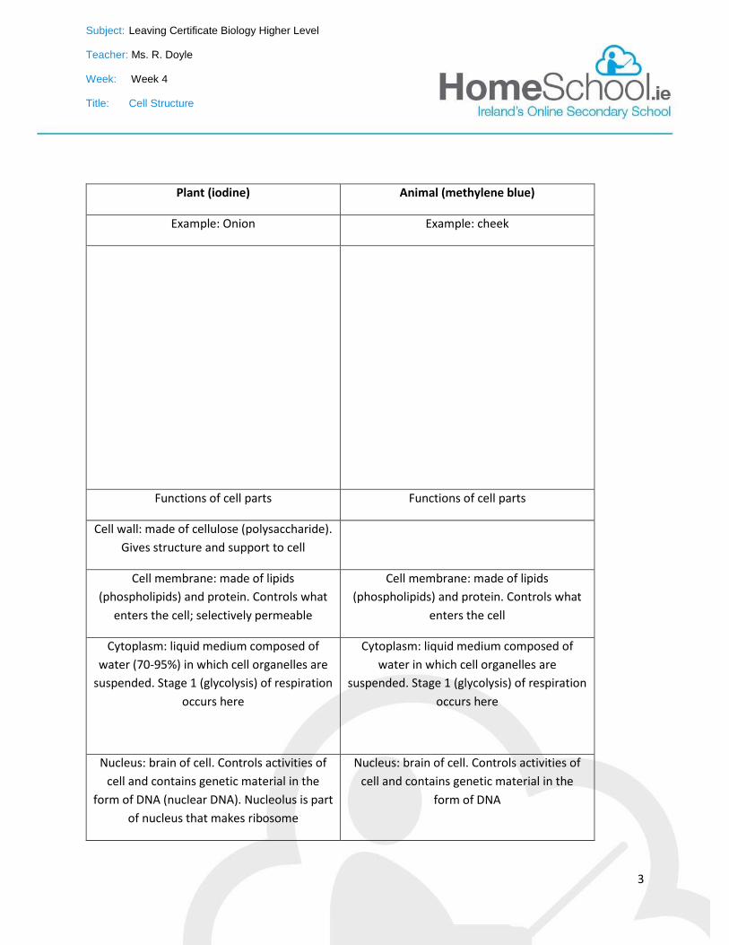
Plant (iodine) Animal (methylene blue)
Example: Onion Example: cheek
Functions of cell parts Functions of cell parts
Cell wall: made of cellulose (polysaccharide).
Gives structure and support to cell
Cell membrane: made of lipids
(phospholipids) and protein. Controls what
enters the cell; selectively permeable
Cell membrane: made of lipids
(phospholipids) and protein. Controls what
enters the cell
Cytoplasm: liquid medium composed of
water (70-95%) in which cell organelles are
suspended. Stage 1 (glycolysis) of respiration
occurs here
Cytoplasm: liquid medium composed of
water in which cell organelles are
suspended. Stage 1 (glycolysis) of respiration
occurs here
Nucleus: brain of cell. Controls activities of
cell and contains genetic material in the
form of DNA (nuclear DNA). Nucleolus is part
of nucleus that makes ribosome
Nucleus: brain of cell. Controls activities of
cell and contains genetic material in the
form of DNA
Subject: Leaving Certificate Biology Higher Level
Teacher: Ms. R. Doyle
Week: Week 4
Title: Cell Structure
4
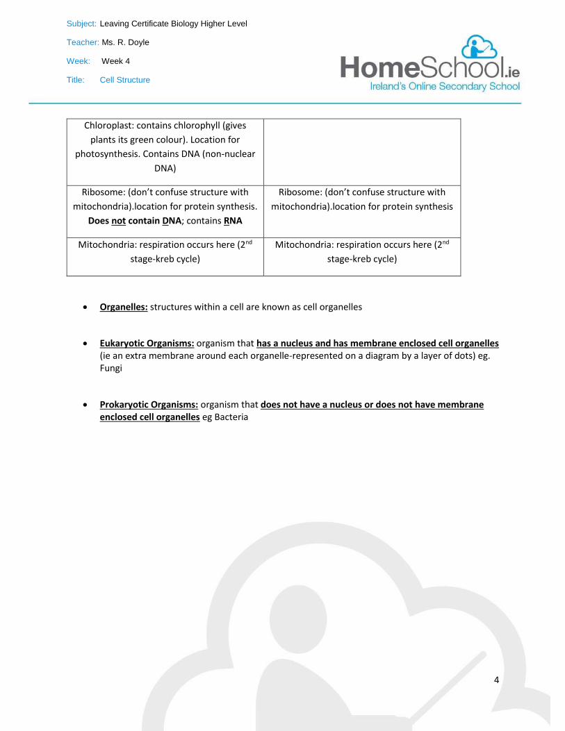
Chloroplast: contains chlorophyll (gives
plants its green colour). Location for
photosynthesis. Contains DNA (non-nuclear
DNA)
Ribosome: (don’t confuse structure with
mitochondria).location for protein synthesis.
Does not contain DNA; contains RNA
Ribosome: (don’t confuse structure with
mitochondria).location for protein synthesis
Mitochondria: respiration occurs here (2nd
stage-kreb cycle)
Mitochondria: respiration occurs here (2nd
stage-kreb cycle)
Organelles: structures within a cell are known as cell organelles
Eukaryotic Organisms: organism that has a nucleus and has membrane enclosed cell organelles (ie an extra membrane around each organelle-represented on a diagram by a layer of dots) eg. Fungi
Prokaryotic Organisms: organism that does not have a nucleus or does not have membrane enclosed cell organelles eg Bacteria
Subject: Leaving Certificate Biology Higher Level
Teacher: Ms. R. Doyle
Week: Week 4
Title: Cell Structure
5
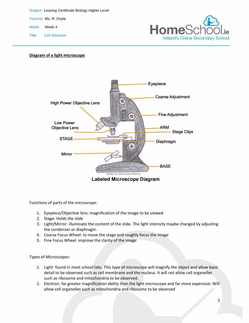
Diagram of a light microscope
Functions of parts of the microscope:
1. Eyepiece/Objective lens: magnification of the image to be viewed 2. Stage: Holds the slide 3. Light/Mirror: illuminate the content of the slide. The light intensity maybe changed by adjusting
the condenser or diaphragm. 4. Coarse Focus Wheel: to move the stage and roughly focus the image 5. Fine Focus Wheel: improve the clarity of the image
Types of Microscopes:
1. Light: found in most school labs. This type of microscope will magnify the object and allow basic detail to be observed such as cell membrane and the nucleus. It will not allow cell organelles such as ribosome and mitochondria to be observed.
2. Electron: far greater magnification ability than the light microscope and far more expensive. Will allow cell organelles such as mitochondria and ribosome to be observed
Subject: Leaving Certificate Biology Higher Level
Teacher: Ms. R. Doyle
Week: Week 4
Title: Cell Structure
6
Revision Questions on Cell Structure
1. what is the role of the cell wall in plants 2. name the polysaccharide found in the cell wall of a plant cell 3. what group of biomolecules does the polysaccharide named in the previous question belong to 4. name two groups of biomolecules found in the cell membrane 5. what type of lipids are found in the cell membrane 6. what is the role of the cell membrane 7. where in the cell does protein synthesis occur 8. where in the cell does photosynthesis take place 9. what activity occurs in the ribosome 10. name the structures found in the nucleus that contain genetic information 11. name the cell organelle involved in respiration 12. explain the term “prokaryotic organism” 13. give an example of a prokaryotic organism 14. give an example of a eukaryotic organism 15. explain the term eukaryotic organism 16. draw and label a diagram of a plant cell marking where photosynthesis (X), respiration (Y) and
protein synthesis (Z) occurs 17. what features will help identify prokaryotic organisms under a microscope 18. give an example of an animal cell
19. give an example of a plant cell
20. name the stain used to examine a plant cell under a microscope
21. name the stain used to examine an animal cell under a microscope
22. what is the function of a stain when examining cells under a microscope
23. list the stages involved in preparing a slide to be examined under a microscope
24. why is water added when preparing a slide
25. what is the function of a cover slip when preparing a slide
26. how is the coverslip added to the glass slide and why is it done in this way
27. if the magnification of the eyepiece is X10 and the magnification of the objective lens is X40,
what is the total magnification
28. give an example of an animal cell
29. give an example of a plant cell
30. name the stain used to examine a plant cell under a microscope
31. name the stain used to examine an animal cell under a microscope
32. what is the function of a stain when examining cells under a microscope
33. list the stages involved in preparing a slide to be examined under a microscope
34. why is water added when preparing a slide
35. what is the function of a cover slip when preparing a slide
Subject: Leaving Certificate Biology Higher Level
Teacher: Ms. R. Doyle
Week: Week 4
Title: Cell Structure
7
36. how is the coverslip added to the glass slide and why is it done in this way
37. if the magnification of the eyepiece is X10 and the magnification of the objective lens is X40,
what is the total magnification
38. what is the function of the coarse focus wheel
39. how are animals cells to be examined under a microscope collected for the experiment
Subject: Leaving Certificate Biology Higher Level
Teacher: Ms. R. Doyle
Week: Week 4
Title: Cell Structure
8
Experiment
Title: Preparation and examination of a plant/animal cell under the microscope
Aim: to prepare and examine both a plant (onion) and animal (cheek) cell under the microscope
List of Apparatus: onion, saliva, glass slide X2, dropper, water, iodine, methylene blue, cover slip,
microscope
Method:
1. Take a thin layer of onion (or rub finger/ swab inside cheek) and place on a glass slide
2. Add two drops of water (to prevent cells drying out)
3. Place a cover slip over the cells; add to the glass slide slowly at a 45o angle using a mounted
needle (cover slip holds cells in place/ protects objective lens of microscope and by adding it at
a 45o angle will prevent the formation of air bubbles)
4. Add drops of iodine (plant) or methylene blue (animal) stain using a dropper around the edges
of the cover slip. The stain will travel towards the cells naturally (stain helps to highlight cell
organelles from cytoplasm especially nucleus)
5. Place glass slide on stage of microscope and turn on light of microscope
6. Observe under low magnification followed by medium and high using coarse and fine focus
wheel to adjust the image
7. Draw an image of what was observed under microscope
Subject: Leaving Certificate Biology Higher Level
Teacher: Ms. R. Doyle
Week: Week 4
Title: Cell Structure
9
Results:
Image of cells
Plant Cell Animal Cell
Subject: Leaving Certificate Biology Higher Level
Teacher: Ms. R. Doyle
Week: Week 4
Title: Cell Structure
10
Questions based on experiment
1. give an example of an animal cell________________
2. give an example of a plant cell_______________________
3. name the stain used to examine a plant cell under a microscope __________________
4. name the stain used to examine an animal cell under a microscope_________________
5. what is the function of a stain when examining cells under a microscope
____________________________________________________________________
6. list the stages involved in preparing a slide to be examined under a microscope
______________________________________________________________________________
______________________________________________________________________________
______________________________________________________________________________
______________________________________________________________________________
____________________________
7. why is water added when preparing a slide ________________________________________
8. what is the function of a cover slip when preparing a slide
__________________________________________________________________
9. how is the coverslip added to the glass slide and why is it done in this way
____________________________________________________________________
10. if the magnification of the eyepiece is X10 and the magnification of the objective lens is X40,
what is the total magnification _____________________________________
11. what is the function of the coarse focus wheel
____________________________________________________________________
12. how are animals cells to be examined under a microscope collected for the experiment
______________________________________________________________________________
_______________________________________________________
13. how are plant cells to be examined under a microscope collected for the experiment
Subject: Leaving Certificate Biology Higher Level
Teacher: Ms. R. Doyle
Week: Week 4
Title: Cell Structure
11
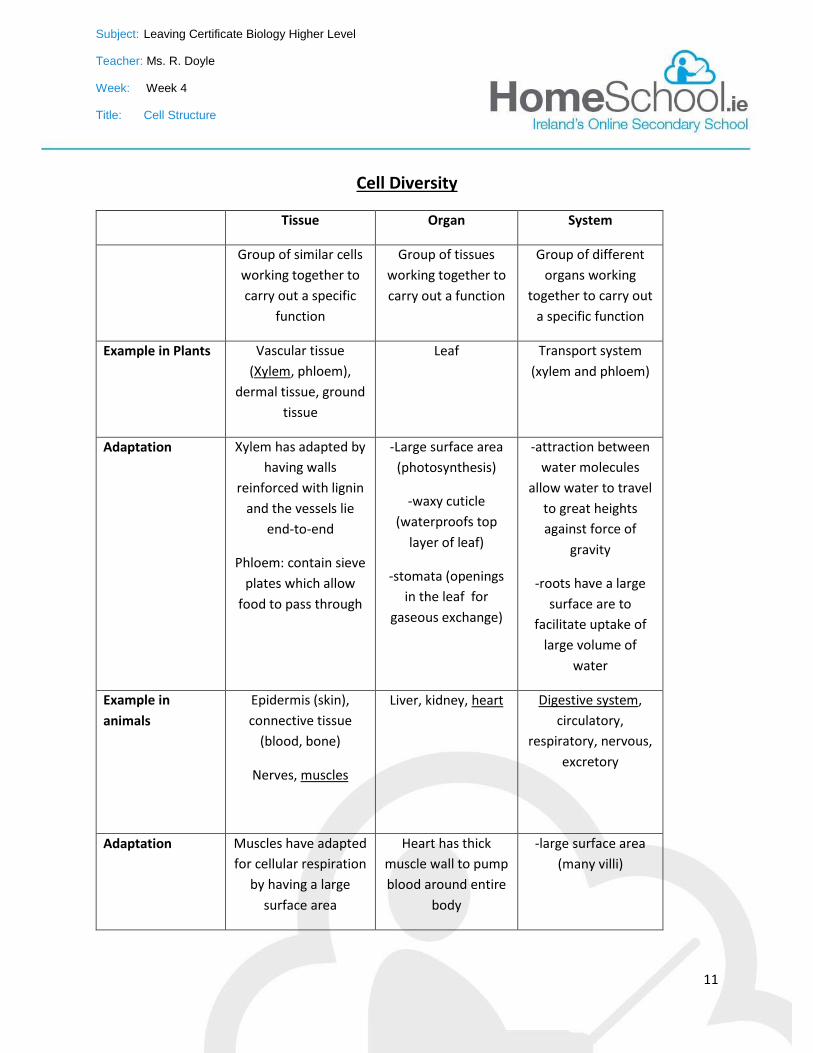
Cell Diversity
Tissue Organ System
Group of similar cells
working together to
carry out a specific
function
Group of tissues
working together to
carry out a function
Group of different
organs working
together to carry out
a specific function
Example in Plants Vascular tissue
(Xylem, phloem),
dermal tissue, ground
tissue
Leaf Transport system
(xylem and phloem)
Adaptation Xylem has adapted by
having walls
reinforced with lignin
and the vessels lie
end-to-end
Phloem: contain sieve
plates which allow
food to pass through
-Large surface area
(photosynthesis)
-waxy cuticle
(waterproofs top
layer of leaf)
-stomata (openings
in the leaf for
gaseous exchange)
-attraction between
water molecules
allow water to travel
to great heights
against force of
gravity
-roots have a large
surface are to
facilitate uptake of
large volume of
water
Example in
animals
Epidermis (skin),
connective tissue
(blood, bone)
Nerves, muscles
Liver, kidney, heart Digestive system,
circulatory,
respiratory, nervous,
excretory
Adaptation Muscles have adapted
for cellular respiration
by having a large
surface area
Heart has thick
muscle wall to pump
blood around entire
body
-large surface area
(many villi)
Subject: Leaving Certificate Biology Higher Level
Teacher: Ms. R. Doyle
Week: Week 4
Title: Cell Structure
12
-one cell thick (rapid
exchange between
cells and blood)
-good blood supply
(transport)
Tissue culture: is the growth of cells on a sterile nutrient medium outside of a living organism-on
glassware.
In-vitro: means grown on glass ware
Uses of tissue culture:
Micropropagation of plants (carrots) o Produces large quantities of carrots very rapidly
Cancer research
Skin graft
Subject: Leaving Certificate Biology Higher Level
Teacher: Ms. R. Doyle
Week: Week 4
Title: Cell Structure
13
Revision Questions on Cell Diversity
1. define the term tissue 2. give an example of a tissue in plants and state how this tissue has adapted for its role 3. give an example of a tissue in animals and state how this tissue has adapted for its role 4. explain the term organ 5. give an example of an organ in plants and state how this tissue has adapted for its role 6. give an example of an organ in animals and state how this tissue has adapted for its role 7. explain the term system 8. give an example of a system in plants and state how this tissue has adapted for its role 9. give an example of a system in plants and state how this tissue has adapted for its role 10. explain the term tissue culture 11. what is meant by “in-vitro” 12. state two uses of tissue cultures 13. give an advantage of tissue cultures
Subject: Leaving Certificate Biology Higher Level
Teacher: Ms. R. Doyle
Week: Week 4
Title: Cell Structure
14
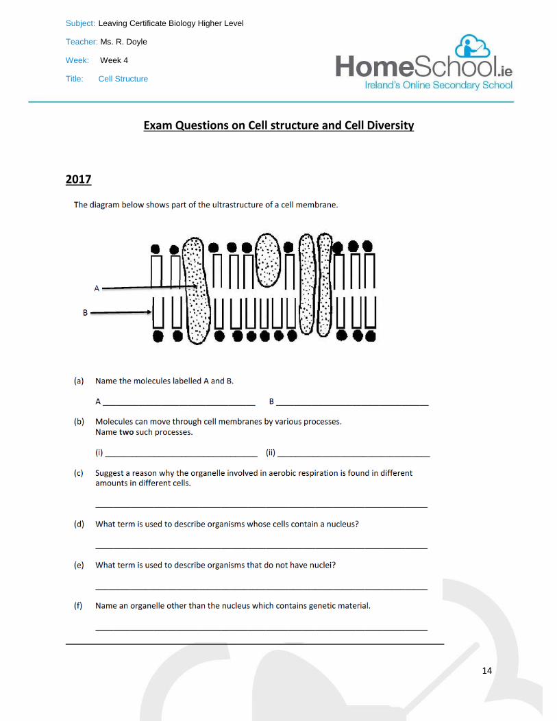
Exam Questions on Cell structure and Cell Diversity
2017
Subject: Leaving Certificate Biology Higher Level
Teacher: Ms. R. Doyle
Week: Week 4
Title: Cell Structure
15
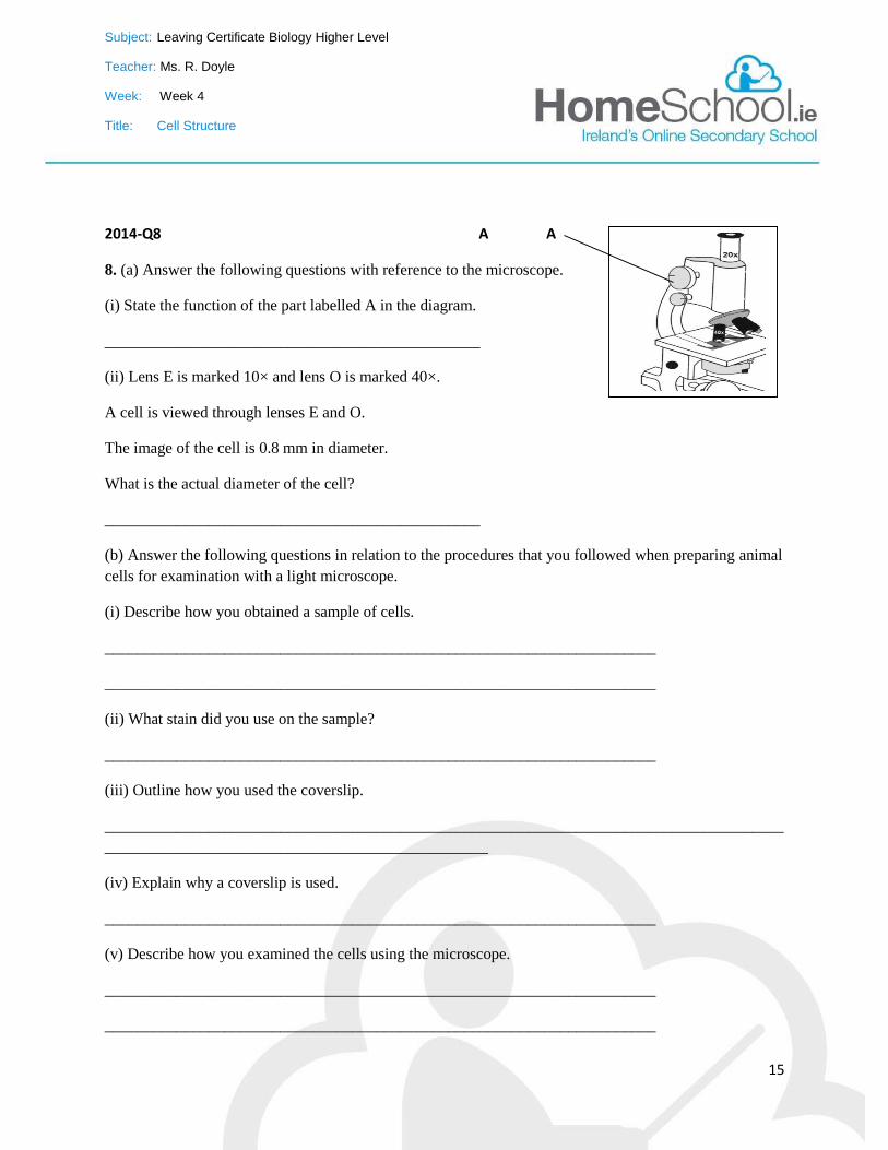
2014-Q8 A A
8. (a) Answer the following questions with reference to the microscope.
(i) State the function of the part labelled A in the diagram.
_______________________________________________
(ii) Lens E is marked 10× and lens O is marked 40×.
A cell is viewed through lenses E and O.
The image of the cell is 0.8 mm in diameter.
What is the actual diameter of the cell?
_______________________________________________
(b) Answer the following questions in relation to the procedures that you followed when preparing animal
cells for examination with a light microscope.
(i) Describe how you obtained a sample of cells.
_____________________________________________________________________
_____________________________________________________________________
(ii) What stain did you use on the sample?
_____________________________________________________________________
(iii) Outline how you used the coverslip.
_____________________________________________________________________________________
________________________________________________
(iv) Explain why a coverslip is used.
_____________________________________________________________________
(v) Describe how you examined the cells using the microscope.
_____________________________________________________________________
_____________________________________________________________________
Subject: Leaving Certificate Biology Higher Level
Teacher: Ms. R. Doyle
Week: Week 4
Title: Cell Structure
16
(vi) Draw a labelled diagram of the cells as seen at high magnification.
2013 Q9 (b)
In the microscopic examination of a plant cell:
1. Name the stain that you used and the colour it imparted to the cell wall.
_____________________________________________________________________
2. How did you apply the stain to the cells on the slide?
_____________________________________________________________________
Subject: Leaving Certificate Biology Higher Level
Teacher: Ms. R. Doyle
Week: Week 4
Title: Cell Structure
17
2012
Q2 (a)
What is a tissue?
_____________________________________________________________________
(ii) Give an example of an animal tissue.
_____________________________________________________________________
(iii) State a role of the animal tissue referred to in (ii).
_____________________________________________________________________
(iv) Give one way in which the tissue referred to in (ii) is adapted to carry out its function(s).
_____________________________________________________________________
Q7
(viii) A microscope has an eyepiece lens marked ×10 and an objective lens marked ×20.What is the total
magnification of the image?
_____________________________________________________________________
Q8
(i) Are fungi prokaryotic or eukaryotic? __________________________________________
(ii) Name one structure in plant cells not found in fungi.
_____________________________________________________________________
Q12 (a)
(i) From the following list, circle any term that describes the nutrition of a
typical plant: give a definition for the type of feeding circled
parasitic; heterotrophic; saprophytic; autotrophic.
Definiton:____________________________________________________________
Subject: Leaving Certificate Biology Higher Level
Teacher: Ms. R. Doyle
Week: Week 4
Title: Cell Structure
18
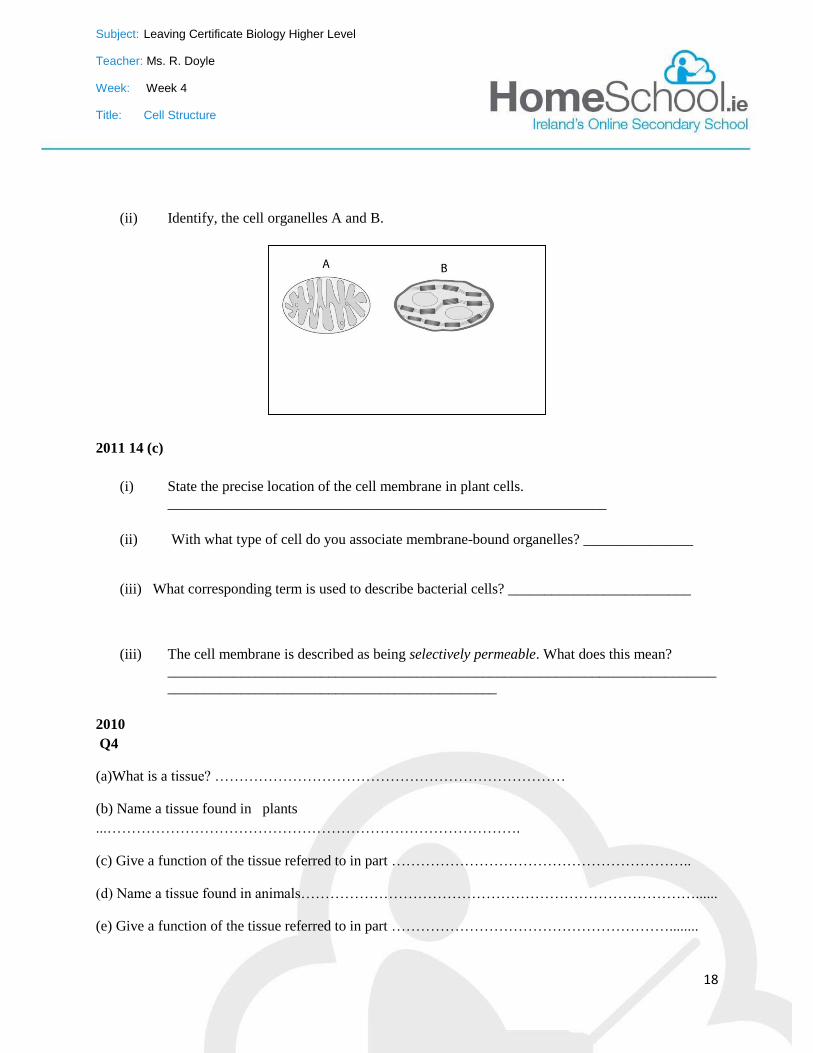
(ii) Identify, the cell organelles A and B.
2011 14 (c)
(i) State the precise location of the cell membrane in plant cells.
____________________________________________________________
(ii) With what type of cell do you associate membrane-bound organelles? _______________
(iii) What corresponding term is used to describe bacterial cells? _________________________
(iii) The cell membrane is described as being selectively permeable. What does this mean?
___________________________________________________________________________
_____________________________________________
2010
Q4
(a)What is a tissue? ………………………………………………………………
(b) Name a tissue found in plants
...………………………………………………………………………….
(c) Give a function of the tissue referred to in part ……………………………………………………..
(d) Name a tissue found in animals………………………………………………………………………......
(e) Give a function of the tissue referred to in part …………………………………………………........
Subject: Leaving Certificate Biology Higher Level
Teacher: Ms. R. Doyle
Week: Week 4
Title: Cell Structure
19
Q8 (a)
(ii) Why is a control normally used when carrying out an experiment?
………………………………………………………………………………………
(b)For which purpose did you use each of the following in the course of your practical studies?
(i) Methylene blue or iodine solution when examining cells with the microscope.
………………………………………………………………………………………
Q14 (c)
(i) In relation to membranes in cells, what is meant by selective permeability
____________________________________________________________
(ii) Give two locations in a cell where there is a selectively permeable membrane
1.____________________________ 2._____________________________
2006
Q8
(a) State a function of each of the following components of a cell.
(i) Ribosome……………………………………………………………………………
(ii) Cell membrane……………………………………………………………………………….
(b) Answer the following questions in relation to the preparation, staining and microscopic observation of
a slide of an animal cell.
(i) What type of animal cell did you use?.......................................................................................
(ii) How did you obtain the cell? ……………………………………………………………..
(iii) Name the stain that you used …………………………………………………………………
Describe how you applied the stain ……………………………………………………..
………………………………………………………………………………………
Subject: Leaving Certificate Biology Higher Level
Teacher: Ms. R. Doyle
Week: Week 4
Title: Cell Structure
20
(iii) After staining, a cover slip is placed on the slide. Give a reason for this
…………………….…………………………………………………………………………………………
…………………………………………………………………
(iv) How did you apply the cover slip?..........................................................................................
Why did you apply it in this way?
………………………………………………………………………………………
(v) Describe the difference in colour or depth of colour, if any, between the nucleus and cytoplasm when
the stained cell was viewed under the microscope.
…………………………………………………………….. …………………..........
SEC Sample Paper
Q1.
(a) State a function of the cell membrane
_______________________________________________________________
(b) State one feature that would allow you to identify an eukaryotic cell
_______________________________________________________________
What is meant by tissue culture? _______________________________________________________
_____________________________________________________________________
List two uses of tissue culturing 1. _______________________ 2.____________________________
List an advantage of tissue culturing_____________________________
Subject: Leaving Certificate Biology Higher Level
Teacher: Ms. R. Doyle
Week: Week 4
Title: Cell Structure
21
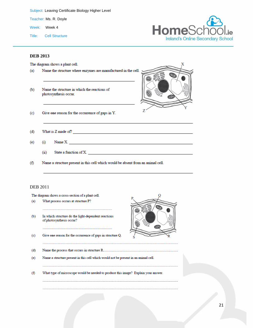
DEB 2013
DEB 2011

Subject: Leaving Certificate Biology Higher Level
Teacher: Ms. R. Doyle
Week: Week 4
Title: Cell Structure
22