Embed Size (px)
Citation preview

J. Cell Sci. 14, 571-585 (1974)
Printed in Great Britain
AN ELECTRON-MICROSCOPE STUDY OF CELL
DELETION IN THE ANURAN TADPOLE TAIL
DURING SPONTANEOUS METAMORPHOSIS
WITH SPECIAL REFERENCE TO APOPTOSIS
OF STRIATED MUSCLE FIBRES
J. F. R. KERR, B. HARMON AND J. SEARLEDepartment of Pathology, University of Queensland Medical School,Herston, Queensland 4006, Australia
SUMMARY
The mechanism of cell deletion responsible for involution of the anuran tadpole tail duringspontaneous metamorphosis was studied by light and electron microscopy, attention beingfocused on epidermis and striated muscle.
The earliest indication of pending dissolution of epidermal cells was found to be aggregationof condensed chromatin beneath the nuclear envelope. This is followed by breaking up of thenucleus, and cytoplasmic condensation and budding with the production of a number of com-pact, membrane-bounded cell fragments with relatively well preserved organelles. These arethen ingested and degraded by nearby viable cells, the majority by distinctive macrophage-likecells, which are scattered throughout the epidermis, and a few by epithelial cells. The morpho-logical changes observed in the dying epidermal cells are the same as those described both in the'programmed cell death' that plays an important role in the normal development of vertebrateembryos and in the type of cell death that has been shown to be involved in regulating the size oftissues in adult mammals under normal as well as pathological conditions; it has been suggestedelsewhere that apoptosis might be a suitable name for the phenomenon.
Deletion of striated muscle fibres in the tadpole tail is accomplished by a process that appearsto be a modification, of classical apoptosis, in which dilatation and confluence of elements of thesarcoplasmic reticulum lead to internal fragmentation, the usual surface budding presumably beingprecluded by the large volume and specialized structure of these cells. The early and late nuclearchanges, and the apparent ultrastructural integrity of organelles in the membrane-boundedmuscle fragments are typical of apoptosis, and subsequent degradation within macrophagesfollows the standard stereotyped pattern. An essentially similar process has been described byothers in the muscles of metamorphosing insect larvae, but whether striated muscle cells inadult higher vertebrates can undergo apoptosis is still uncertain.
INTRODUCTION
The rapid involution of the anuran tadpole tail that takes place during metamorphosisprovides a striking example of decrease in the size of vertebrate tissues under normalconditions. Whilst a number of studies have attested to involvement of lysosomalhydrolases in the regressive process (for review, see Weber, 1969), it seems likely thatthese enzymes are concerned, not with initiation of degeneration of the cells to beeliminated, but with digestion of their remnants within macrophages (Weber, 1963):in fact, there appears to have been relatively little detailed investigation of changes inthe doomed cells prior to their engulfment.
572 J. F. R. Kerr, B. Harmon andj. Searle
The role of focal, precisely controlled, and often massive cell death in the fashioningof organs and limbs during the embryonic development of vertebrates has long beenappreciated (Glucksmann, 1951; Saunders, 1966; Ballard & Holt, 1968; Menkes,Sandor&Ilies, 1970). Electron microscopy of various species (Bellairs, 1961; Saunders& Fallon, 1966; Farbman, 1968; Manasek, 1969; Webster & Gross, 1970; Hammar&Mottet, 1971; Mottet & Hammar, 1972) has disclosed a constant pattern of morpho-logical change in the dying cells, characterized by gross condensation of cytoplasm andnuclear chromatin; the appearances are fundamentally different from those observedin coagulative necrosis (Trump, Goldblatt & Stowell, 1965a, b; Trump & Ginn, 1969),which is found under pathological conditions in tissues damaged by noxious stimuli.It has recently become clear, however, that the distinctive type of cell death that is soconspicuous at certain stages of normal vertebrate ontogeny also accounts for contin-uous, albeit less massive, dropout of cells in the tissues of mature animals (Kerr,Wyllie & Currie, 1972), and, in at least some of the organs of adult mammals, itsoccurrence is augmented and inhibited by hormones (Kerr et al. 1972; Kerr & Searle,1973; Wyllie, Kerr, Macaskill & Currie, 1973). Indeed, it now appears probable thatit constitutes a general mechanism of controlled cell deletion, which complementsmitosis in regulating the size of vertebrate tissues and organs throughout life: the termapoptosis has been suggested to highlight this role in cell population kinetics (Kerret al. 1972).
The present paper reports a light- and electron-microscope study of the mechanismof cell deletion in the anuran tadpole tail during spontaneous metamorphosis, attentionbeing focused on epidermis and striated muscle. It was found that epidermal epithelialcells undergo ultrastructurally typical apoptosis, but that the process in striated musclefibres is modified by the large size and specialized structure of these cells.
MATERIALS AND METHODS
Tadpoles of the dwarf tree frog, Litoria glauerti, were chosen for study because of the relativelysmall amount of pigment in their tails: it was found that abundant pigment granules present inother locally-available species tended to obscure some ultrastructural details. The tadpoles werecollected from their natural habitat and kept at summer room temperatures (25—35 °C) in. alarge water-filled glass tank containing water-plants. Tissue from the distal half of their tailswas processed for light and electron microscopy when they reached the stage of metamorphosischaracterized by advanced forelimb development and regression of the tail to about half itsoriginal dimensions; the length of the tail at this time was approximately equal to that of eachextended hind limb.
For light-microscope studies, 10 tadpoles were killed by decapitation, and transverse slicesof their tails were fixed in 4% formaldehyde solution. Paraffin sections of this tissue werestained with haematoxylin and eosin. For electron microscopy, it was found that preliminaryperfusion of the blood vascular system of intact animals with glutaraldehyde solution and sub-sequent immersion fixation of small blocks resulted in better preservation of the soft connectivetissues than did dicing of fresh tissue in fixative. Each of 10 tadpoles was narcotized, the heartexposed and the ventricle pierced with the tip of a fine pipette, and the vascular system gentlyperfused for 4 min with ice-cold 5 % cacodylate-buffered glutaraldehyde solution (pH 72).This procedure was followed by appreciable hardening of the tail, the distal half of which wasthen amputated, cut into small cubes and fixed for 2 h in the same solution. The blocks werepostfixed for 90 min in 1 % cacodylate-buffered osmium tetroxide solution, stained for 30 minin 5 % uranyl acetate solution, dehydrated in ethanol, and embedded in Epon 812. Thin sections

Cell deletion in tadpole tail 573
were cut, stained with lead citrate, and examined in a Hitachi HS7S electron microscope. One-micrometre sections were also cut from some blocks, stained with toluidine blue, and examinedwith a light microscope.
RESULTS
Examination of histological sections of the epidermis covering both the main bodyof the tail and the tail fins disclosed many clearly delineated spherical and ovoidcytoplasmic fragments scattered amongst the intact epithelial cells. They vary con-siderably in size, and often contain one or more pyknotic remnants of nuclei. Theirprecise topographical relationship to surrounding viable cells was usually difficult todetermine with the light microscope. These structures are identical in appearance withthe dying cells observed in normal embryos (Gliicksmann, 1951); their developmentconstitutes the cardinal histological feature of apoptosis (Kerr et al. 1972), and it isconvenient to refer to them as apoptotic bodies.
Electron microscopy revealed 2 distinctly different cell types in the epidermis.Epithelial cells, which comprise the bulk of the population, are characterized by thepresence of desmosomes, cytoplasmic tonofibrils, and numerous microvilli (Figs. 1,3, 5); mucous vesicles, similar to those illustrated by Michaels, Albright & Patt (1971),were seen in surface cells. Cells of the second type are only sparsely distributed amongstthe epithelial cells, and they never extend to the external surface. Their cytoplasm israther less densely stained than that of the epithelial cells (Figs. 1, 4), they do notcontain tonofibrils, and desmosomes were not detected at their points of contact withadjoining cells. Since these latter cells appear to be largely responsible for the phago-cytosis and degradation of apoptotic bodies in the epidermis (Figs. 1, 2, 4), they will becalled macrophage-like cells.
The earliest indications of apoptosis of epithelial cells are aggregation of chromatinnear the nuclear membrane and cytoplasmic condensation, the appearances being thesame as those described in the regressing external gills oiRanapipiens (fig. 1 in Michaelset al. 1971), in vertebrate embryos (Bellairs, 1961; Manasek, 1969), and in a varietyof adult mammalian tissues (Kerr, 1971; Helminen & Ericsson, 1972; Kerr et al. 1972;Kerr & Searle, 1973). The nuclei then fragment, and the condensing cells bud toproduce extremely compact apoptotic bodies of various sizes, which are probablyrapidly phagocytosed, for they were infrequently observed in the extracellular space(Fig. 1). Though the majority are taken up by macrophage-like cells (Figs. 1, 2, 4), afew are ingested by epithelial cells (Fig. 3). Well preserved organelles and nuclearfragments can still be identified in recently phagocytosed bodies (Figs. 1, 2), butprogressive degradation then ensues (Figs. 1, 3, 4), and they are soon reduced tolysosomal residual bodies (Fig. 4). Some of the macrophage-like cells were found tobe packed with such telolysosomes (Fig. 5), whereas organelles with the ultrastructuralfeatures of secondary lysosomes are relatively uncommon in epithelial cells (Fig. 5).
The earliest histological change indicative of dissolution of muscle fibres is theoccurrence of longitudinal clefts between myofibrils, best seen in toluidine blue-stained sections of Epon-embedded tissue. This is followed by fragmentation of thecytoplasm into a number of so-called sarcolytes with well preserved cross-striations, the
574 J- F. R. Kerr, B. Harmon andj. Searle
appearances being the same as those figured by Weber (1969). The clusters of sarcolytesare then invaded by macrophages, and their phagocytosis is succeeded by progressiveloss of their organized structure. At a late stage of the process, the cylindrical spacepreviously occupied by each fibre was seen to be filled with residual body-ladenmacrophages, in which myofibrils could no longer be identified.
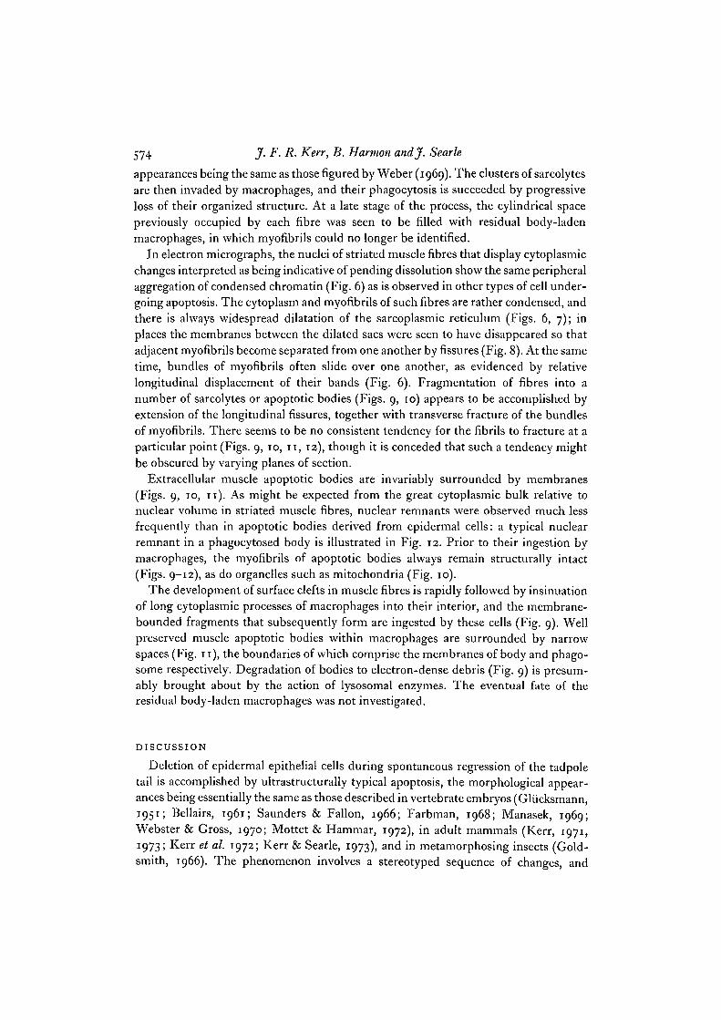
In electron micrographs, the nuclei of striated muscle fibres that display cytoplasmicchanges interpreted as being indicative of pending dissolution show the same peripheralaggregation of condensed chromatin (Fig. 6) as is observed in other types of cell under-going apoptosis. The cytoplasm and myofibrils of such fibres are rather condensed, andthere is always widespread dilatation of the sarcoplasmic reticulum (Figs. 6, 7); inplaces the membranes between the dilated sacs were seen to have disappeared so thatadjacent myofibrils become separated from one another by fissures (Fig. 8). At the sametime, bundles of myofibrils often slide over one another, as evidenced by relativelongitudinal displacement of their bands (Fig. 6). Fragmentation of fibres into anumber of sarcolytes or apoptotic bodies (Figs. 9, 10) appears to be accomplished byextension of the longitudinal fissures, together with transverse fracture of the bundlesof myofibrils. There seems to be no consistent tendency for the fibrils to fracture at aparticular point (Figs. 9, io, 11, 12), though it is conceded that such a tendency mightbe obscured by varying planes of section.
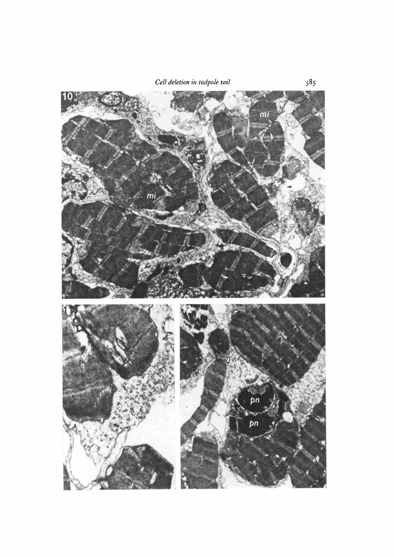
Extracellular muscle apoptotic bodies are invariably surrounded by membranes(Figs. 9, 10, 11). As might be expected from the great cytoplasmic bulk relative tonuclear volume in striated muscle fibres, nuclear remnants were observed much lessfrequently than in apoptotic bodies derived from epidermal cells: a typical nuclearremnant in a phagocytosed body is illustrated in Fig. 12. Prior to their ingestion bymacrophages, the myofibrils of apoptotic bodies always remain structurally intact(Figs. 9-12), as do organelles such as mitochondria (Fig. 10).
The development of surface clefts in muscle fibres is rapidly followed by insinuationof long cytoplasmic processes of macrophages into their interior, and the membrane-bounded fragments that subsequently form are ingested by these cells (Fig. 9). Wellpreserved muscle apoptotic bodies within macrophages are surrounded by narrowspaces (Fig. 11), the boundaries of which comprise the membranes of body and phago-some respectively. Degradation of bodies to electron-dense debris (Fig. 9) is presum-ably brought about by the action of lysosomal enzymes. The eventual fate of theresidual body-laden macrophages was not investigated.
DISCUSSION
Deletion of epidermal epithelial cells during spontaneous regression of the tadpoletail is accomplished by ultrastructurally typical apoptosis, the morphological appear-ances being essentially the same as those described in vertebrate embryos (Gliicksmann,1951; Bellairs, 1961; Saunders & Fallon, 1966; Farbman, 1968; Manasek, 1969;Webster & Gross, 1970; Mottet & Hammar, 1972), in adult mammals (Kerr, 1971,1973; Kerr et al. 1972; Kerr & Searle, 1973), and in metamorphosing insects (Gold-smith, 1966). The phenomenon involves a stereotyped sequence of changes, and

Cell deletion in tadpole tail 575
begins with peripheral aggregation of nuclear chromatin; this is followed by nuclearfragmentation, and cytoplasmic condensation associated with prolific budding toproduce membrane-bounded apoptotic bodies of varying size. As is the case in othersolid tissues (Kerr, 1971, 1973), the actual process of budding is rarely observed,possibly because of the avidity with which viable cells engulf the condensing ones(Kerr, 1973); it is much more frequently seen in apoptosis of mouse ascites tumourcells induced by actinomycin D, where all cells are separated from one another byabundant plasma and phagocytosis is long delayed (authors' unpublished observations).
A few of the apoptotic bodies in the tadpole skin are ingested by epithelial cells.However, the paucity of secondary lysosomes in these cells at a fairly advanced stageof tail regression suggests that they play a relatively minor role in disposing of cellfragments: epithelial cells are sometimes more active in this regard in the embryo(Farbman, 1968), and under pathological conditions in adult mammals (Kerr, 1971,1973; Kerr & Searle, 1972). The majority of the bodies in the tail epidermis are ingestedand degraded by specialized cells, whose cytoplasm becomes progressively laden withtelolysosomes. Very similar cells in the epithelial lining of adult rat prostatic acinidispose of apoptotic bodies both in the normal gland and during the rapid atrophy thatfollows castration (Kerr & Searle, 1973); in grossly atrophic glands they display thesame packing with lysosomal residual bodies as is illustrated in Fig. 5. Further study ofepithelial surfaces is needed to determine whether such cells are of common occurrence.
In an electron-microscope study of epidermal changes in thyroxine-induced tadpoletail involution carried out by Gona (1969), a clear distinction was not made betweenautophagy on the one hand and heterophagy of cell remnants on the other. Autophagicvacuoles may indeed be difficult to distinguish from ingested apoptotic bodies that donot contain nuclear remnants (Kerr, 1973), but differentiation between these twoprocesses is important, for the former merely involves segregation and degradation ofpart of the cytoplasm of a viable cell, whilst the latter is an indication of cell death.Autophagy undoubtedly occurs in the tadpole tail, but our findings do not suggest thatit plays a major part in the regressive process.
It was noted in passing that several other types of cells in the tail undergo typicalapoptosis, but most interest centred on the mode of deletion of striated muscle fibres.A priori it might be expected that the relatively huge mass-to-surface ratio of these cellswould preclude their condensing and budding to produce apoptotic bodies in the usualway, but it would, nevertheless, seem unlikely that a different and unique mechanismshould have evolved for the controlled deletion of muscle. It is thus of interest that thenuclear changes in the early stages of muscle fibre dissolution are typical of apoptosis.Further, the extracellular muscle fragments are membrane-bounded, and show thesame striking preservation of the fine structure of organelles as is apparent in apoptoticbodies of diverse origins (Kerr et al. 1972); the pyknotic nuclear remnants, thoughrare, are morphologically typical of apoptosis (compare Fig. 2 with Fig. 12). Lucentvacuoles, possibly derived from the endoplasmic reticulum, have been observed inapoptotic bodies by several workers (Manasek, 1969; Kerr et al. 1972; Mottet &Hammar, 1972; Kerr, 1973), and the dilatation of the sarcoplasmic reticulum thataccompanies apoptosis-like nuclear change in muscle is probably an analogous process.

576 J. F. R. Kerr, B. Harmon andj. Searle
Just how confluence of the resulting spaces leads to the formation of membrane-bounded apoptotic bodies is not yet clear, but estimates of the surface area of thesarcoplasmic reticulum (Peachey, 1965) indicate that ample membrane is available toencompass the fragments. Indeed vacuoles, possibly representing redundant mem-brane, are seen at the surface of newly formed bodies (Fig. 9). Extension of fissuresbetween myofibrils to the cell surface is presumably associated with fusion of theirbounding membranes with the sarcolemma. The T-system does not appear from themicrographs to be involved, in spite of its known normal communication with theexterior (McCallister & Hadek, 1970). In summary, apoptosis of muscle fibres appearsto represent a modification of the basic pattern, in which internal cell membranesparticipate to a large degree in the formation of membrane-bounded cytoplasmicfragments. Once ingested by macrophages, the muscle apoptotic bodies go through theusual series of degradative changes within phagolysosomes.
Finally, it is significant that the ultrastructural events described in breakdown oflarval intersegmental muscles during metamorphosis in Diptera (Crossley, 1968)appear to be essentially the same as those observed in the tadpole. In particular,Crossley stressed that changes in the doomed muscles precede invasion by phagocytichaemocytes, and described partial breakdown of subdivisions of the sarcoplasmicreticulum with the creation of abnormal elongated vacuoles between myofibrils; heconsidered that fracture of the myofibrils takes place at the level of the I-bands.
This work was supported by a grant from the National Health and Medical Research Councilof Australia. We are grateful to Mrs L. Kerslake and Miss A. Francis for their technical assis-tance.
REFERENCESBALLARD, K. J. & HOLT, S. J. (1968). Cytological and cytochemical studies on cell death and
digestion in the foetal rat foot: the role of macrophages and hydrolytic enzymes. J. Cell Sci.3. 245-262.
BELLAIRS, R. (1961). Cell death in chick embryos as studied by electron microscopy.^. Anat. 95,54-6o.
CROSSLEY, A. C. (1968). The fine structure and mechanism of breakdown of larval interseg-mental muscles in the blowfly Calliphora erythrocephala.J. Insect Physiol. 14, 1389-1407.
FARBMAN, A. I. (1968). Electron microscope study of palate fusion in mouse embryos. DeviBiol. 18, 93-116.
GLUCKSMANN, A. (195 I) . Cell deaths in normal vertebrate ontogeny. Biol. Rev. 26, 59-86.GOLDSMITH, M. (1966). The anatomy of cell death. J. Cell Biol. 31, 41 A.GONA, A. G. (1969). Light and electron microscopic study on thyroxine-induced in vitro re-
sorption of the tadpole tail fin. Z. Zellforsch. mikrosk. Anat. 95, 483-494.HAMMAR, S. P. & MOTTET, N. K. (1971). Tetrazolium salt and electron-microscopic studies of
cellular degeneration and necrosis in the interdigital areas of the developing chick limb. J.Cell Sci. 8, 229-251.
HELMINEN, H. J. & ERICSSON, J. L. E. (1972). Ultrastructural studies on prostatic involution inthe rat. Evidence for focal irreversible damage to epithelium, and heterophagic digestion inmacrophages. J. Ultrastruct. Res. 39, 443-455.
KERR, J. F. R. (1971). Shrinkage necrosis: a distinct mode of cellular death. J. Path. 105, 13-20.KERR, J. F. R. (1973). Some lysosome functions in liver cells reacting to sublethal injury. In
Lysosomes in Biology and Pathology, vol. 3 (ed. J. T. Dingle), pp. 365-394. Amsterdam andLondon: North-Holland.

Cell deletion in tadpole tail 577
KERR, J. F. R. & SEARLE, J. (1972). The digestion of cellular fragments within phagolysosomesin carcinoma cells. J. Path. 108, 55-58.
KERR, J. F. R. & SEARLE, J. (1973). Deletion of cells by apoptosis during castration-inducedinvolution of the rat prostate. Virchows Arch. path. Anat. Physiol., Abt. B Zellpath. 13, 87-102.
KERR, J. F. R., WYLLIE, A. H. &CURRIE, A. R. (1972). Apoptosis: a basic biological phenomenonwith wide-ranging implications in tissue kinetics. Br.J. Cancer 26, 239-257.
MANASEK, F. J. (1969). Myocardial cell death in the embryonic chick ventricle, jf. Embryol. exp.Morph. 21, 271-284.
MCCALLISTER, L. P. & HADEK, R. (1970). Transmission electron microscopy and stereo ultra-structure of the T system in frog skeletal muscle. J. Ultrastruct. Res. 33, 360-368.
MENKES, B., SANDOR, S. &ILIES, A. (1970). Cell death in teratogenesis. In Advances in Teratology,vol. 4 (ed. D. H. M. Woollam), pp. 169-215. London: Logos Press.
MICHAELS, J. E., ALBRIGHT, J. T. & PATT, D. I. (1971). Fine structural observations on celldeath in the epidermis of the external gills of the larval frog, Ranapipiens. Am.J. Anat. 132,301-318.
MOTTET, N. K. & HAMMAR, S. P. (1972). Ribosome crystals in necrotizing cells from the posteriornecrotic zone of the developing chick limb. jf. Cell Sci. 11, 403-414.
PEACHEY, L. D. (1965). The sarcoplasmic reticulum and transverse tubules of the frog's sartorius.J. CellBiol. 25, 209-231.
SAUNDERS, J. W., JR. (1966). Death in embryonic systems. Science, N.Y. 154, 604-612.SAUNDERS, J. W., JR. & FALLON, J. F. (1966). Cell death in morphogenesis. In Major Problems
in Developmental Biology (ed. M. Locke), pp. 289-314. New York and London: AcademicPress.
TRUMP, B. F. & GINN, F. L. (1969). The pathogenesis of subcellular reaction to lethal injury.In Methods and Achievements in Experimental Pathology, vol. 4 (ed. E. Bajusz & G. Jasmin),pp. 1-29. Basel and New York: Karger.
TRUMP, B. F., GOLDBLATT, P. J. & STOWELL, R. E. (1965a). Studies on necrosis of mouse liverin vitro. Ultrastructural alterations in the mitochondria of hepatic parenchymal cells. Lab.Invest. 14, 343-37I-
TRUMP, B. F., GOLDBLATT, P. J. & STOWELL, R. E. (1965 b). Studies of necrosis in vitro of mousehepatic parenchymal cells. Ultrastructural alterations in endoplasmic reticulum, Golgiapparatus, plasma membrane, and lipid droplets. Lab. Invest. 14, 2000-2028.
WEBER, R. (1963). Behaviour and properties of acid hydrolases in regressing tails of tadpolesduring spontaneous and induced metamorphosis in vitro. Ciba Fdn Symp. on Lysosomes(ed. A. V. S. de Reuck& M. P. Cameron), pp. 282-300. London: Churchill.
WEBER, R. (1969). Tissue involution and lysosomal enzymes during anuran metamorphosis. InLysosomes in Biology and Pathology, vol. 2 (ed. J. T. Dingle & H. B. Fell), pp. 437-461.Amsterdam and London: North-Holland.
WEBSTER, D. A. & GROSS, J. (1970). Studies on possible mechanisms of programmed cell deathin the chick embryo. Devi Biol. 22, 157-184.
WYLLIE, A. H., KERR, J. F. R., MACASKILL, I. A. M. & CURRIE, A. R. (1973)- Adrenocorticalcell deletion: the role of ACTH. J. Path, (in Press).
{Received 25 July 1973)

578 J. F. R. Ken, B. Harmon andj. Searle
Figs. 1-3. Apoptosis of epidermal epithelial cells.Fig. 1. The full thickness of the epidermis is included in this micrograph, the external
surface being at top right and the basement lamella (W) at bottom left. A moderatelywell preserved apoptotic body and a partly degraded body are seen within a large macro-phage-like cell (m). Mucous vesicles (v) can be recognized in the former, indicatingthat it is derived from a superficial epithelial cell; both bodies contain pyknotic remnantsof nuclei (pn). A third, extremely compact apoptotic body (arrow) is still extracellular.The wide space between epithelial cells may be a fixation artifact. It was, however, con-sistently observed in all animals, t, tonofibrils in epithelial cell, x 5300.
Fig. 2. A well preserved apoptotic body lies within the cytoplasm of an epidermalmacrophage-like cell (m). It contains multiple nuclear remnants (pn), in several ofwhich both dense chromatin aggregates and relatively lucent areas are evident: theearly nuclear changes of apoptosis that precede such fragmentation are illustrated inFig. 6. Note the cluster of tonofibrils (i) and the marked cytoplasmic condensation. Asecond, partly degraded body is visible at the top right corner of the micrograph.The digestive vacuole at top left contains unrecognizable debris, n, nucleus of themacrophage-like cell, x 7400.
Fig. 3. A degenerate apoptotic body with a nuclear remnant (pn) is seen within aphagosome in the cytoplasm of an epidermal cell, which is clearly identified as epi-thelial by the presence of tonofibrils (t) and desmosomes (d). x 10200.

Cell deletion in tadpole tail 579
C E L 14

580 J. F. R. Kerr, B. Harmon andj. Searle
Figs. 4, 5. Large macrophage-like cells in epidermis.Fig. 4. The macrophage-like cell in the centre of the micrograph contains at least
seven partly degraded apoptotic bodies, and several whorls of membranes. Its cyto-plasm is more electron-lucent than that of the surrounding epithelial cells, x 4900.
Fig. 5. The cytoplasm of this macrophage-like cell is densely packed with membranewhorls, which probably comprise lipid-rich residues of apoptotic bodies withinlysosomes. Lysosomes (/) are, by contrast, relatively sparse in the epithelial cells, whichare characterized by the presence of tonofibrils (t), desmosomes (d), and surfacemicrovilli. x 5600.

Cell deletion in tadpole tail

582 J. F. R. Kerr, B. Harmon andjf. Searle
Figs. 6-9. Apoptosis of striated muscle fibres.Fig. 6. The muscle fibre nucleus shows characteristic peripheral aggregation of con-
densed chromatin. Elements of the sarcoplasmic reticulum are dilated, and there islongitudinal displacement of myofibrils relative to one another. Note the surfacecleft developing between myofibrils near the right-hand edge of the micrograph,x 10400.
Fig. 7. Part of a muscle fibre undergoing early apoptosis. Note the gross dilatationof the sarcoplasmic reticulum. x 5600.
Fig. 8. Part of another fibre displaying slightly more advanced changes. Septa betweendilated elements of the sarcoplasmic reticulum have now disappeared resulting inthe formation of a longitudinal fissure, x 11000.
Fig. 9. Macrophage lying within a muscle fibre undergoing apoptosis. It is insinuatingprocesses of its cytoplasm into clefts that have developed between myofibrils. Apoptoticbodies, which are clearly of muscle origin and vary considerably in size, and whichshow different degrees of digestion, are seen within the macrophage. x 6000.

Cell deletion in tadpole tail

584 J. F. R. Kerr, B. Harmon andj. Searle
Figs. 10-12. Apoptosis of striated muscle fibres.Fig. 10. Cluster of muscle apoptotic bodies. Some lie within macrophages; others
are still extracellular. Note the well preserved mitochondria {mi) in 2 of the bodies.Even at this magnification, it is clear that the muscle fragments are bounded by mem-branes, x 6000.
Fig. 11. The extracellular apoptotic body at the bottom of the micrograph is boundedby a membrane; the ingested body within the macrophage is surrounded by a narrow,membrane-enclosed space, the outer limit of which comprises the phagosome mem-brane, x 16800.
Fig. 12. Extracellular and phagocytosed muscle apoptotic bodies, one with densenuclear remnants (pn). x 5300.
Cell deletion in tadpole tail