Embed Size (px)
Citation preview
Cell Cycle
Cell Cycle•describes the life cycle of a cell•differs fundamentally between prokaryotes & eukaryotes
•shares four features among all cell types–reproductive signal–DNA replication–genome segregation–cytokinesis
•produces a new individual or new parts
Prokaryotic Cell Cycle•one circular chromosome–packed on a protein frame in the nucleoid
–anchored to the plasma membrane•chromosome replication yields two anchored circles
•cell growth separates anchored circles
•annular pinching separates daughter cells
Binary Fission
binary fission in a
bacteriumFigure 9.2
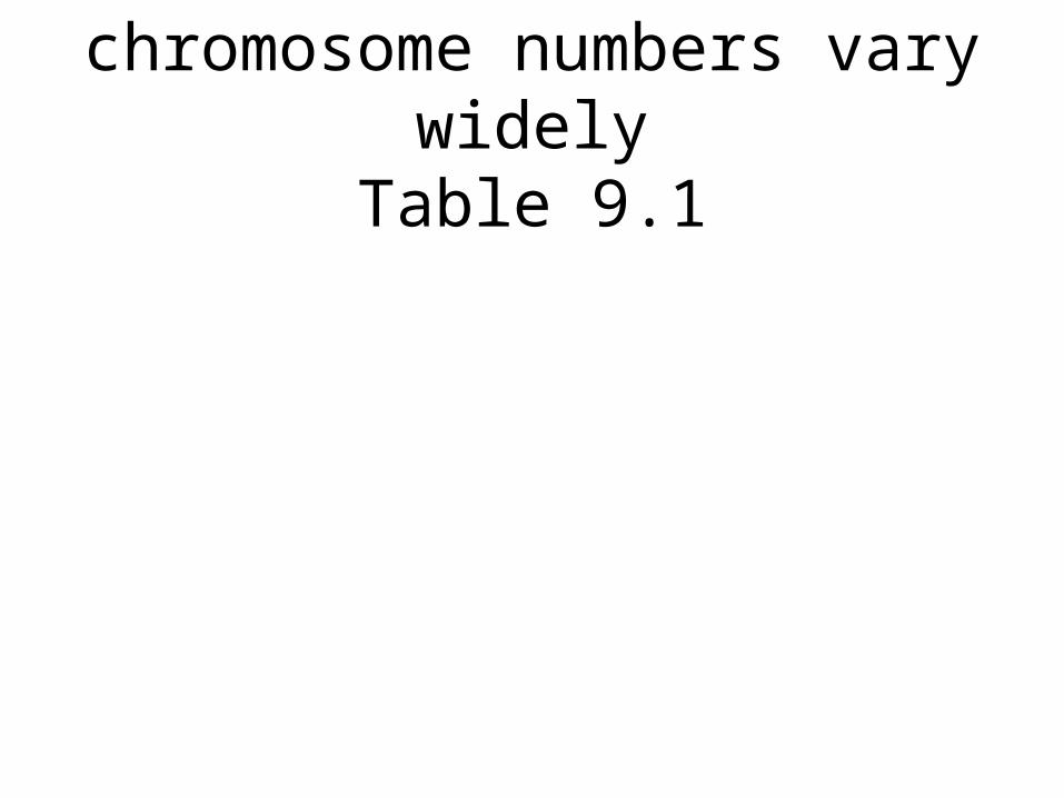
chromosome numbers vary widely
Table 9.1
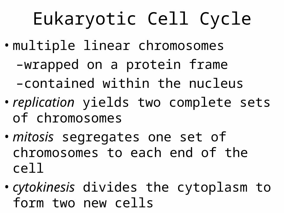
Eukaryotic Cell Cycle•multiple linear chromosomes–wrapped on a protein frame–contained within the nucleus
•replication yields two complete sets of chromosomes
•mitosis segregates one set of chromosomes to each end of the cell
•cytokinesis divides the cytoplasm to form two new cells
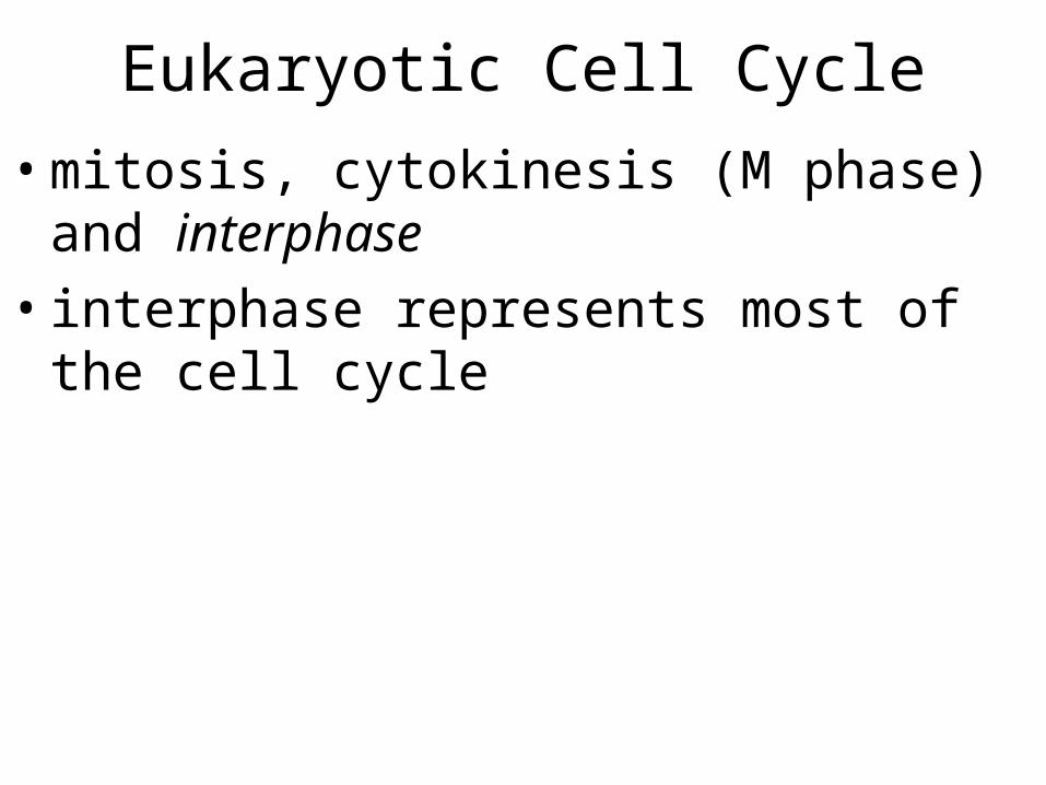
Eukaryotic Cell Cycle
•mitosis, cytokinesis (M phase) and interphase
•interphase represents most of the cell cycle
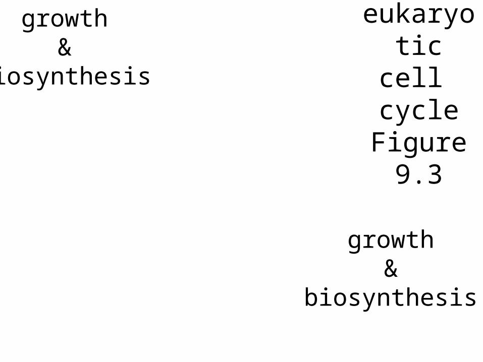
eukaryotic cell cycleFigure 9.3
growth &
biosynthesis
growth &
biosynthesis
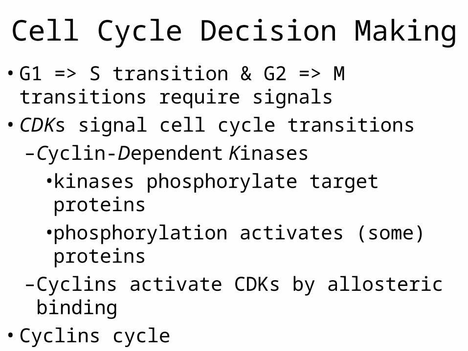
Cell Cycle Decision Making•G1 => S transition & G2 => M transitions require signals
•CDKs signal cell cycle transitions–Cyclin-Dependent Kinases•kinases phosphorylate target proteins•phosphorylation activates (some) proteins
–Cyclins activate CDKs by allosteric binding
•Cyclins cycle
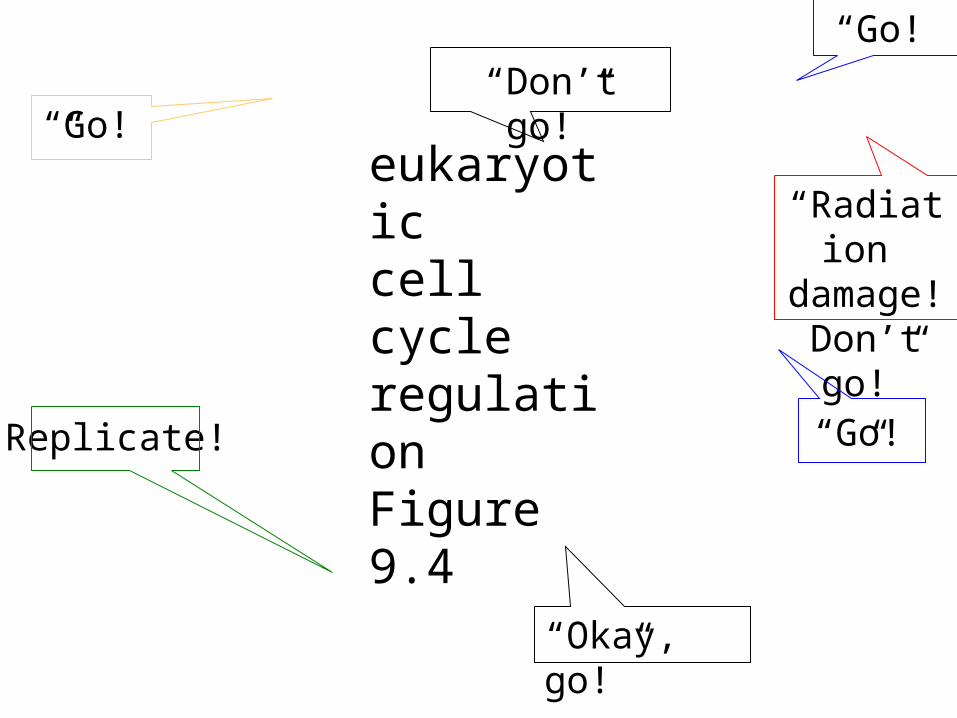
eukaryotic cell cycleregulationFigure 9.4
“Go!”“Don’t go!”
“Radiation
damage!Don’t go!”“Go!”
“Okay, go!”
“Go!”
Replicate!
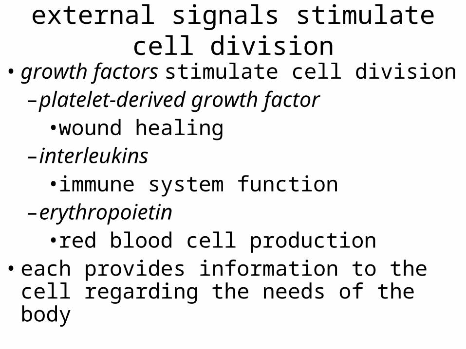
external signals stimulate cell division
•growth factors stimulate cell division–platelet-derived growth factor•wound healing
–interleukins•immune system function
–erythropoietin•red blood cell production
•each provides information to the cell regarding the needs of the body
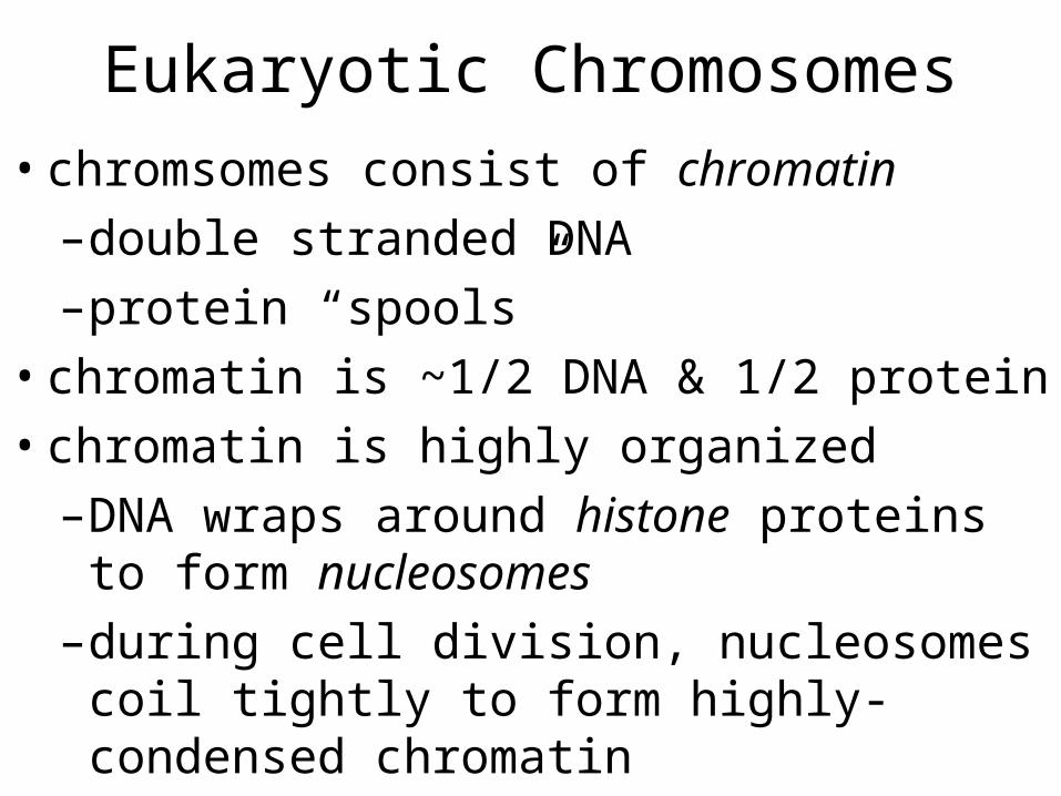
Eukaryotic Chromosomes•chromsomes consist of chromatin–double stranded DNA–protein “spools”
•chromatin is ~1/2 DNA & 1/2 protein•chromatin is highly organized–DNA wraps around histone proteins to form nucleosomes
–during cell division, nucleosomes coil tightly to form highly-condensed chromatin
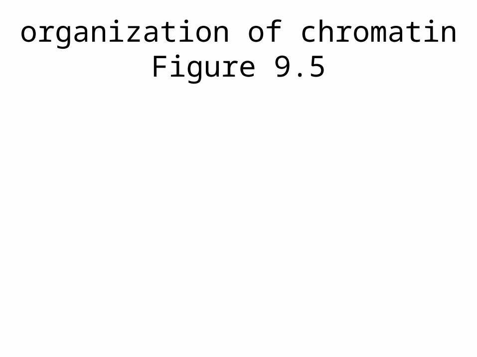
organization of chromatinFigure 9.5
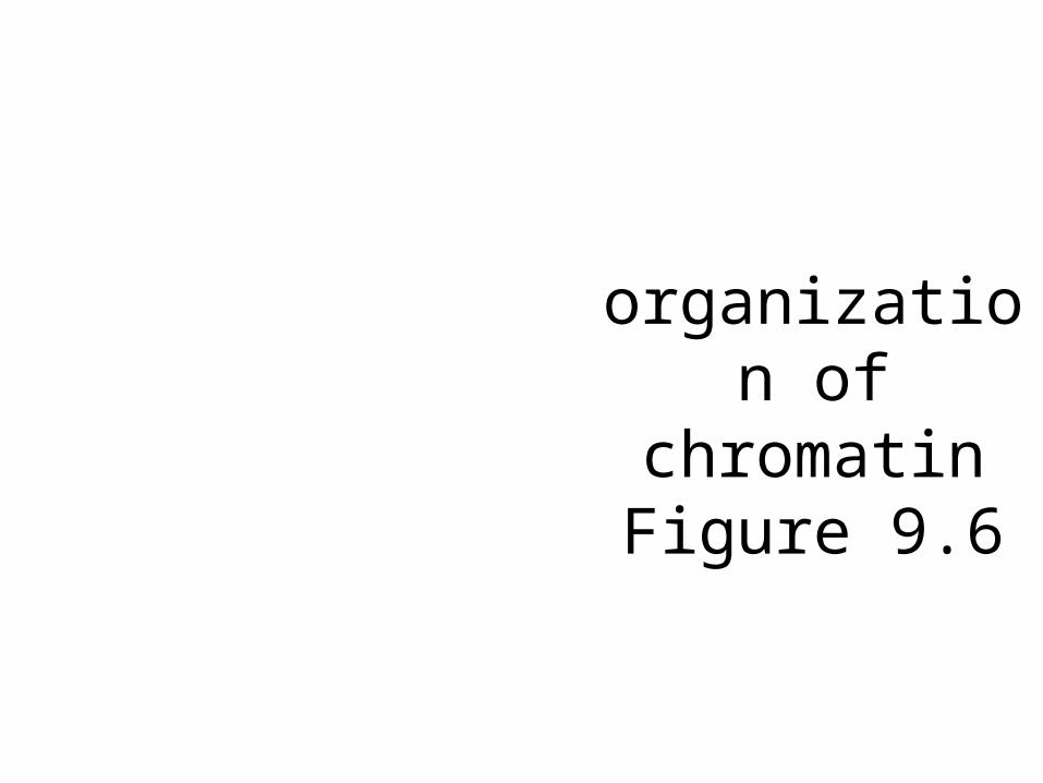
organization of
chromatinFigure 9.6
Replication•during S phase–partially wound DNA is replicated to form two identical copies of each chromosome
–two sister chromatids remain attached at the centromere
•each chromosome enters M phase as two linked sister chromatids
•mitosis separates the chromatids and distributes one to each daughter cell
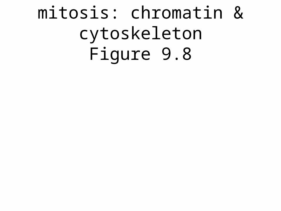
mitosis: chromatin & cytoskeletonFigure 9.8
events of mitotic phases
•Prophase–chromosomes condense–nucleolus disorganizes–spindle apparatus begins to organize
–kinetochores forms
mitosis: cartoon versionFigure 9.8
Mitotic spindle apparatusFigure 9.7
events of mitotic phases
•Prometaphase–nuclear envelope is fragmented
–spindle fibers bind kinetochores
–chromosomes begin to migrate to equatorial plate
events of mitotic phases•Metaphase–chromosomes are aligned at equatorial plate
•Anaphase–sister chromatids separate–daughter chromosomes migrate to poles
•Telophase–prophase is reversed
Animal Cytokinesis Plant
Figure 9.10
Cytokinesis: Division of Cytoplasm
•Animals–annular pinching by actin & myosin ring
•Plants–deposition of cell plate by Golgi vesicles
•Organelles are distributed to daughter cells ~randomly
Modes of Reproduction•asexual reproduction–production of genetic clones through mitotic cell divisions
–common among plants (vegetative propagation) and unicellular eukaryotes
–eliminates costs & risks associated with sexual reproduction
–offspring lack genetic variability
Modes of Reproduction•sexual reproduction–offspring exhibit genetic variability•each bears a unique combination of parental genetic contributions
–requires•meiosis - reduction of chromosome number from 2n (diploid) to 1n (haploid)•fertilization - combination of 1n parental contributions to produce 2n offspring
Fungal/animal life cyclesFigure 9.12
Modes of Reproduction•meiosis produces–gametes - animals, some protists–spores - fungi, plants, some protists•produce 1n adults•produce gametes
•fertilization (gamete fusion) produces–zygotes•produce 2n adults and/or•undergo meiosis
Modes of Reproduction•meiosis–two divisions–reduces 2n parent cell to 1n products–always produces 4 haploid products•begins with 4 homologous chromatids
–recombination produces novel chromatids
–phases resemble mitotic phases, except•meiosis I - homologs pair at prophase•meiosis I - homologs separate at anaphase
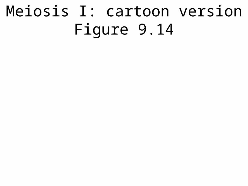
Meiosis I: cartoon versionFigure 9.14
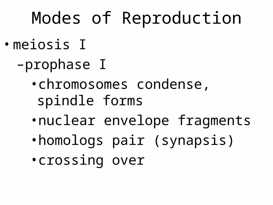
Modes of Reproduction
•meiosis I–prophase I•chromosomes condense, spindle forms•nuclear envelope fragments•homologs pair (synapsis)•crossing over
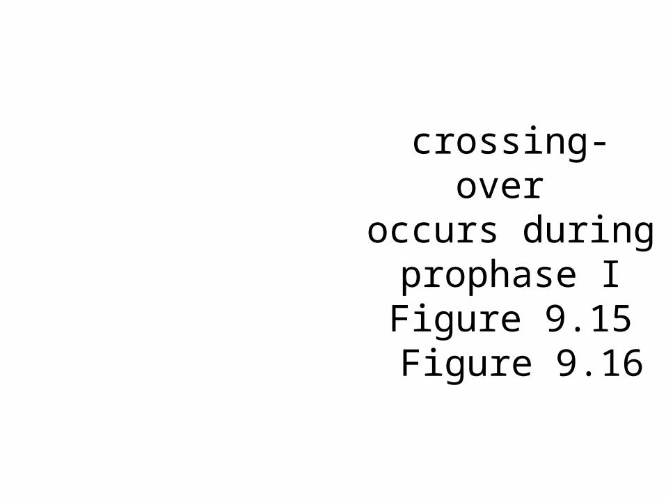
crossing- over
occurs during prophase IFigure 9.15 Figure 9.16
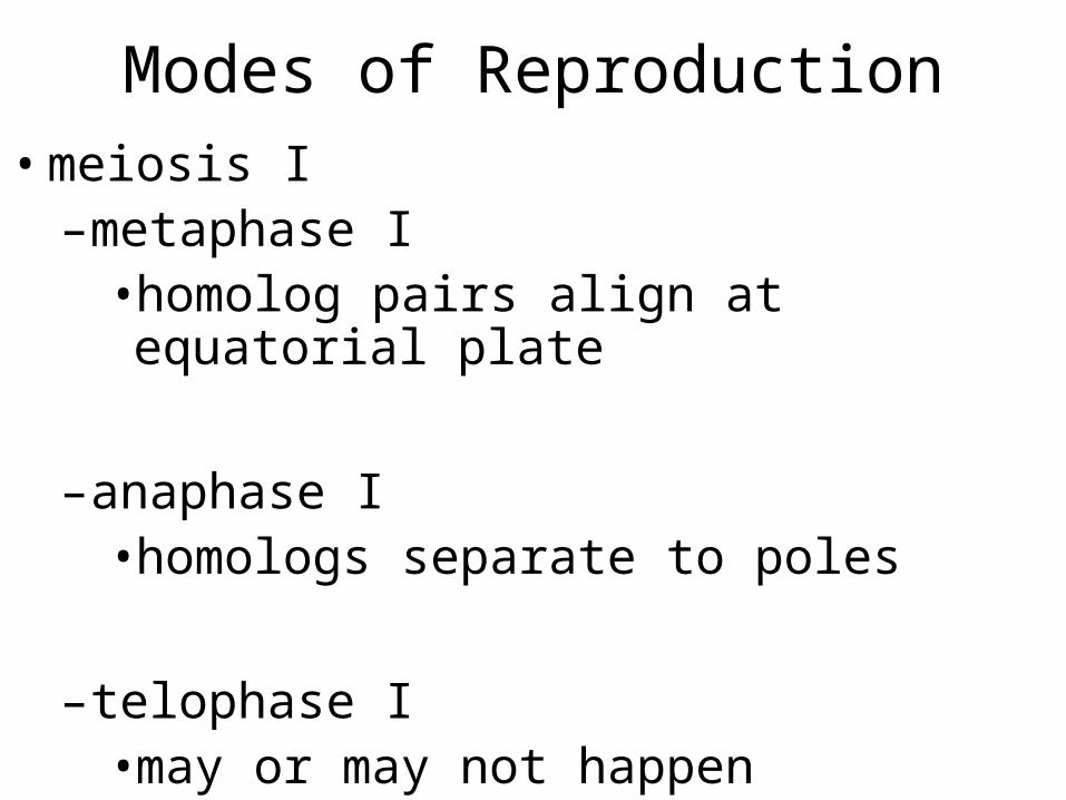
Modes of Reproduction•meiosis I–metaphase I•homolog pairs align at equatorial plate
–anaphase I•homologs separate to poles
–telophase I•may or may not happen
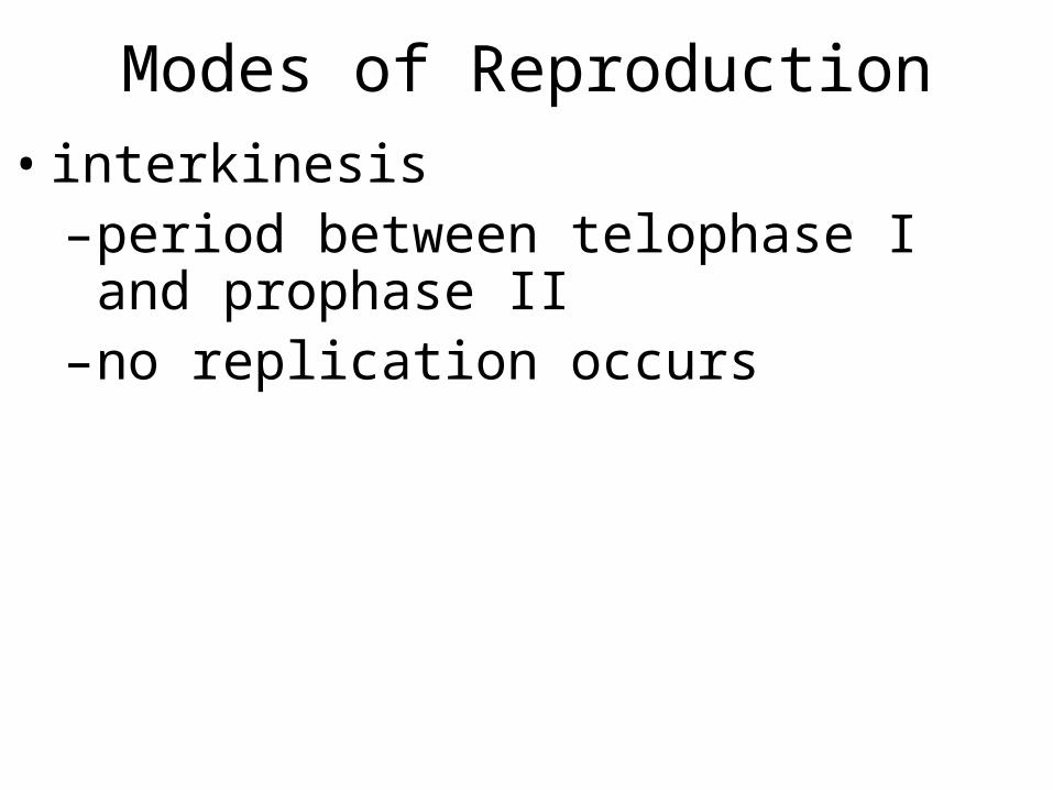
Modes of Reproduction•interkinesis–period between telophase I and prophase II
–no replication occurs
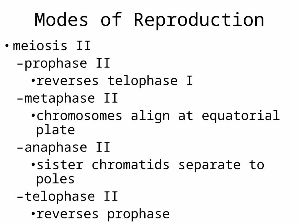
Modes of Reproduction•meiosis II–prophase II•reverses telophase I
–metaphase II•chromosomes align at equatorial plate
–anaphase II•sister chromatids separate to poles
–telophase II•reverses prophase
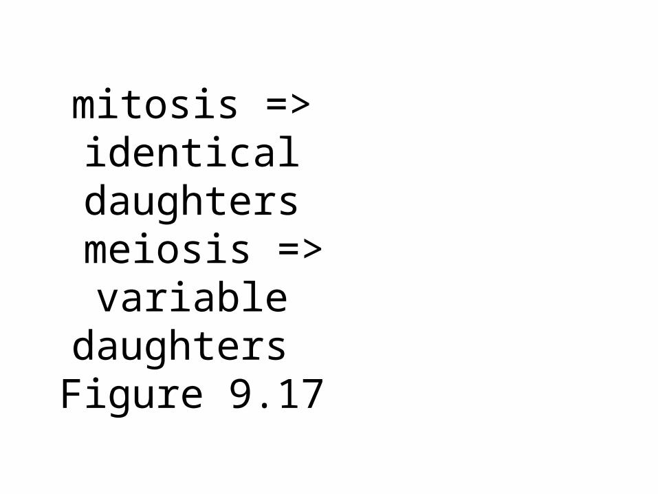
mitosis => identical daughters
meiosis => variable
daughters Figure 9.17
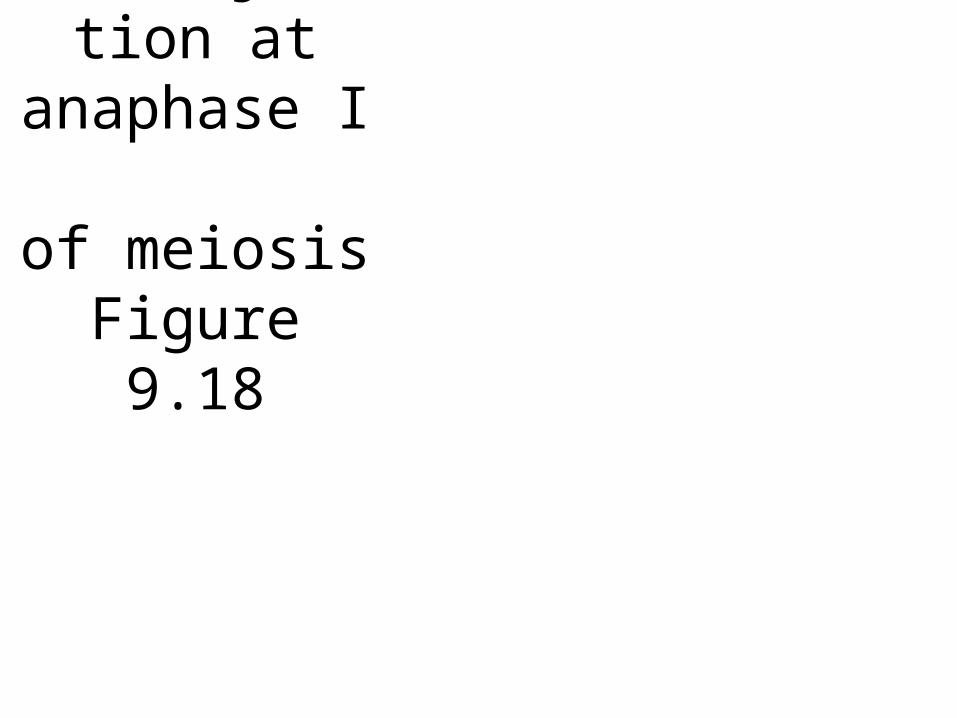
nondisjunction at
anaphase I
of meiosisFigure 9.18
Errors of Reproduction
•meiotic errors may result in chromosomal disorders–aneuploidy - incorrect genetic complement•nondisjuction•translocation
Errors of Reproduction
•meiotic errors may result in chromosomal disorders–polyploidy•3, 4, or more sets of chromosomes•can perform mitosis (reproduce asexually)•1, 3,5, etc. cannot perform meiosis
Cell Death•death by necrosis–damage, poison, starvation
•death by apoptosis (programmed cell death)–discards un-needed or old cells
–signals are common in many organisms
–many cancers result from failed apoptosis
apoptosis: programmed cell death
Figure 9.18
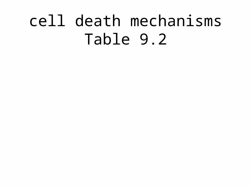
cell death mechanismsTable 9.2