Embed Size (px)
Citation preview
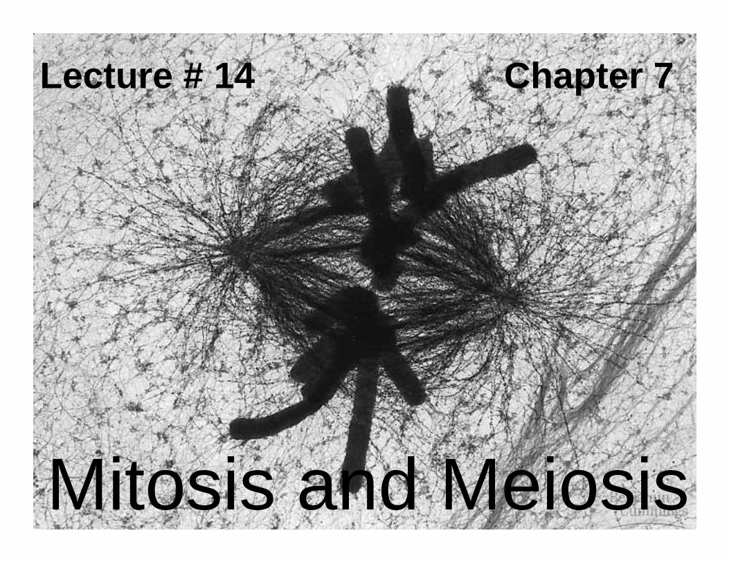
Lecture # 14 Chapter 7
Mitosis and Meiosis
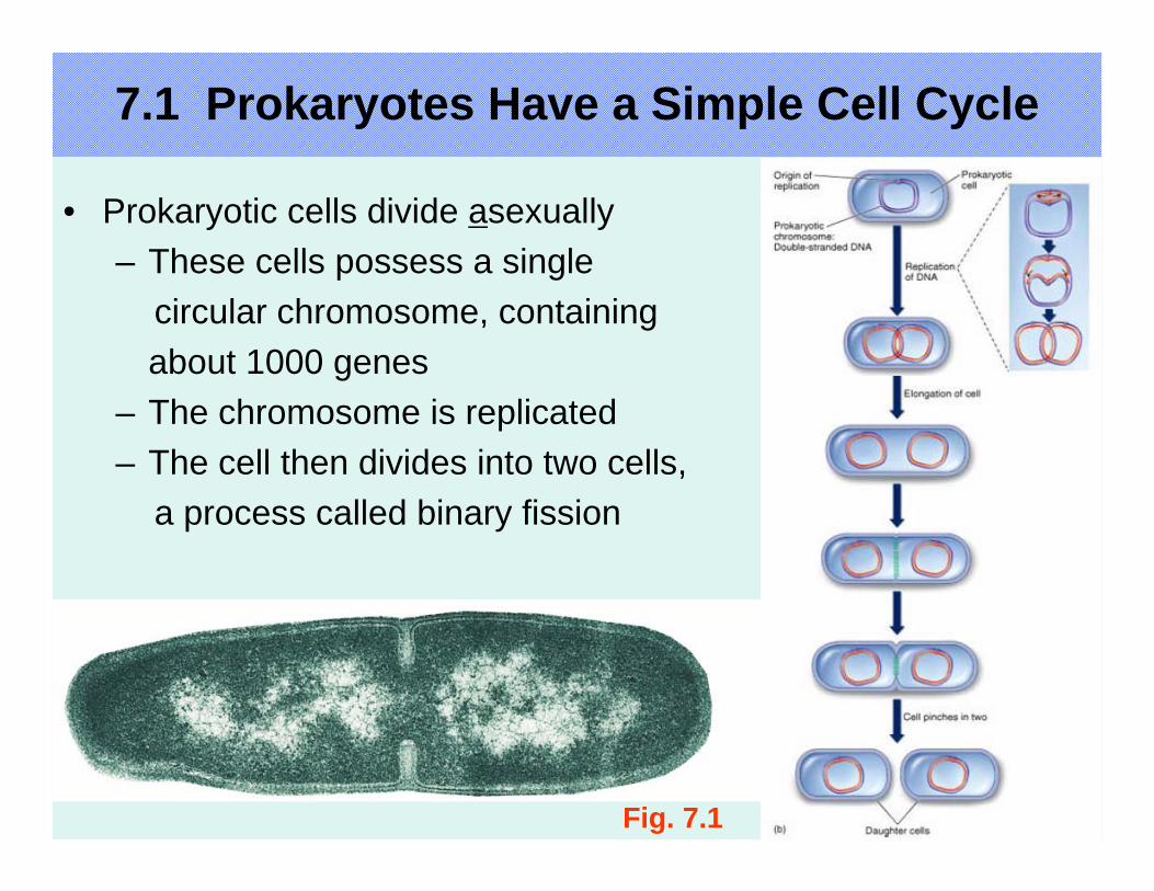
Fig. 7.1
7.1 Prokaryotes Have a Simple Cell Cycle
• Prokaryotic cells divide asexually– These cells possess a single
circular chromosome, containingabout 1000 genes
– The chromosome is replicated– The cell then divides into two cells,
a process called binary fission
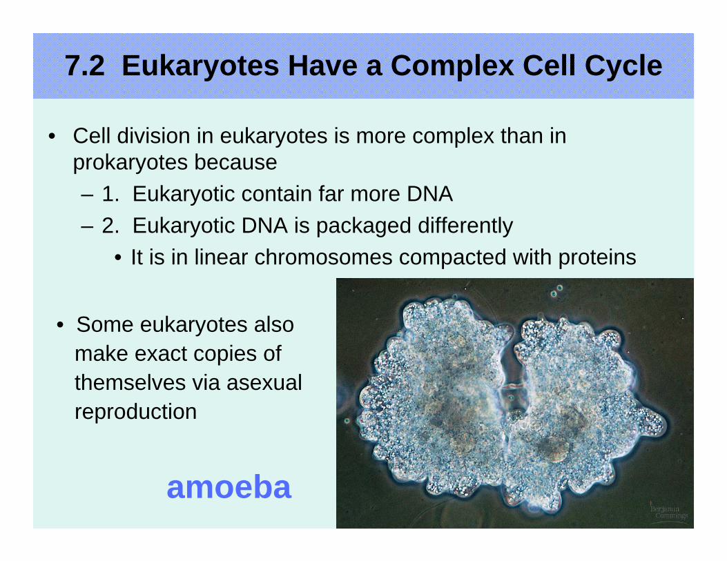
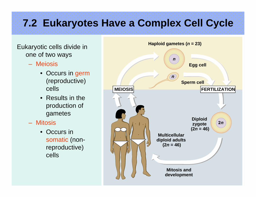
7.2 Eukaryotes Have a Complex Cell Cycle
• Cell division in eukaryotes is more complex than in prokaryotes because– 1. Eukaryotic contain far more DNA– 2. Eukaryotic DNA is packaged differently
• It is in linear chromosomes compacted with proteins
• Some eukaryotes alsomake exact copies ofthemselves via asexualreproduction
amoeba
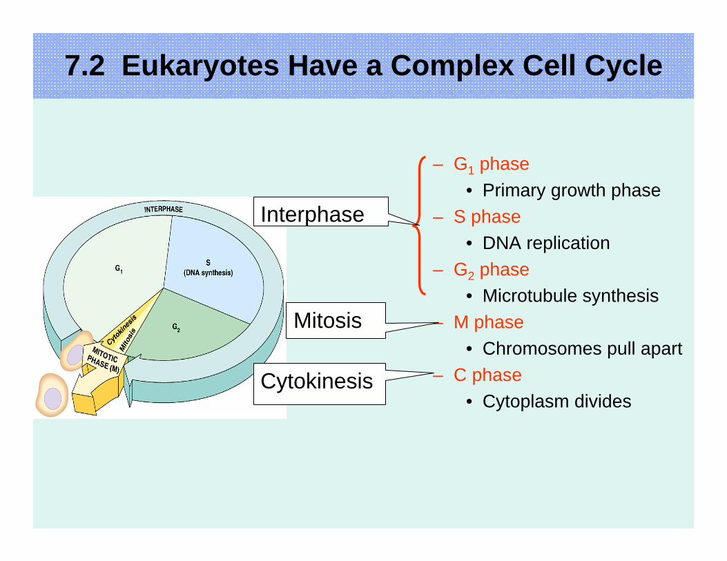
– G1 phase• Primary growth phase
– S phase• DNA replication
– G2 phase• Microtubule synthesis
– M phase• Chromosomes pull apart
– C phase• Cytoplasm divides
Interphase
Mitosis
Cytokinesis
7.2 Eukaryotes Have a Complex Cell Cycle
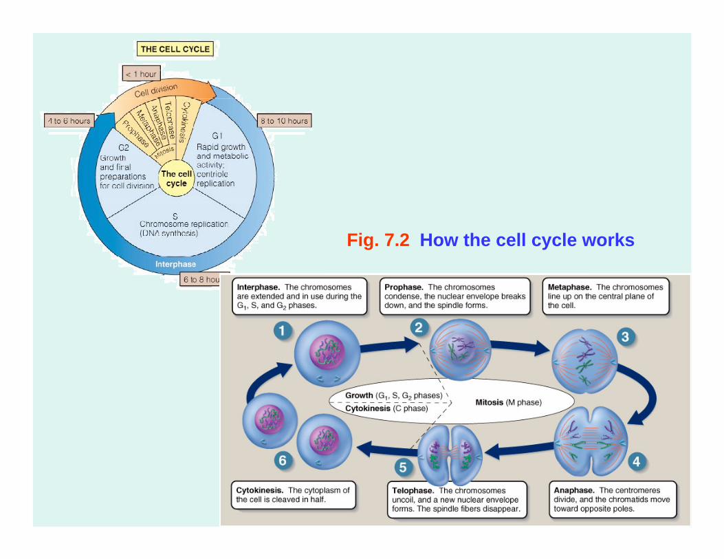
Fig. 7.2 How the cell cycle works
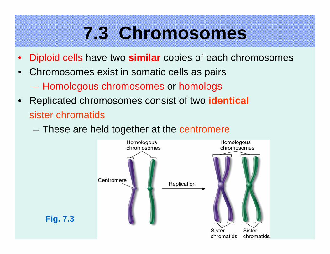
Fig. 7.3
• Diploid cells have two similar copies of each chromosomes• Chromosomes exist in somatic cells as pairs
– Homologous chromosomes or homologs• Replicated chromosomes consist of two identical
sister chromatids– These are held together at the centromere
7.3 Chromosomes
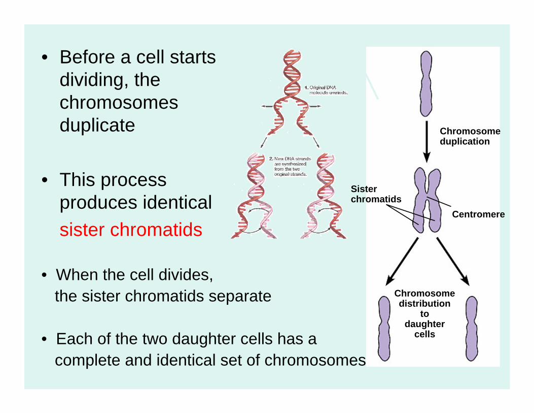
• Before a cell starts dividing, the chromosomes duplicate
• This process produces identicalsister chromatids
Sister chromatids
Chromosomeduplication
Chromosomedistribution
todaughter
cells
Centromere
• When the cell divides,the sister chromatids separate
• Each of the two daughter cells has acomplete and identical set of chromosomes
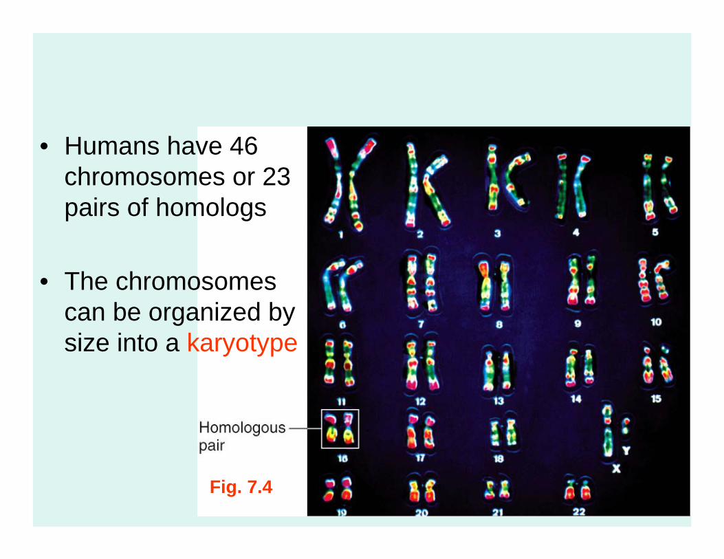
Fig. 7.4
• Humans have 46 chromosomes or 23 pairs of homologs
• The chromosomes can be organized by size into a karyotype
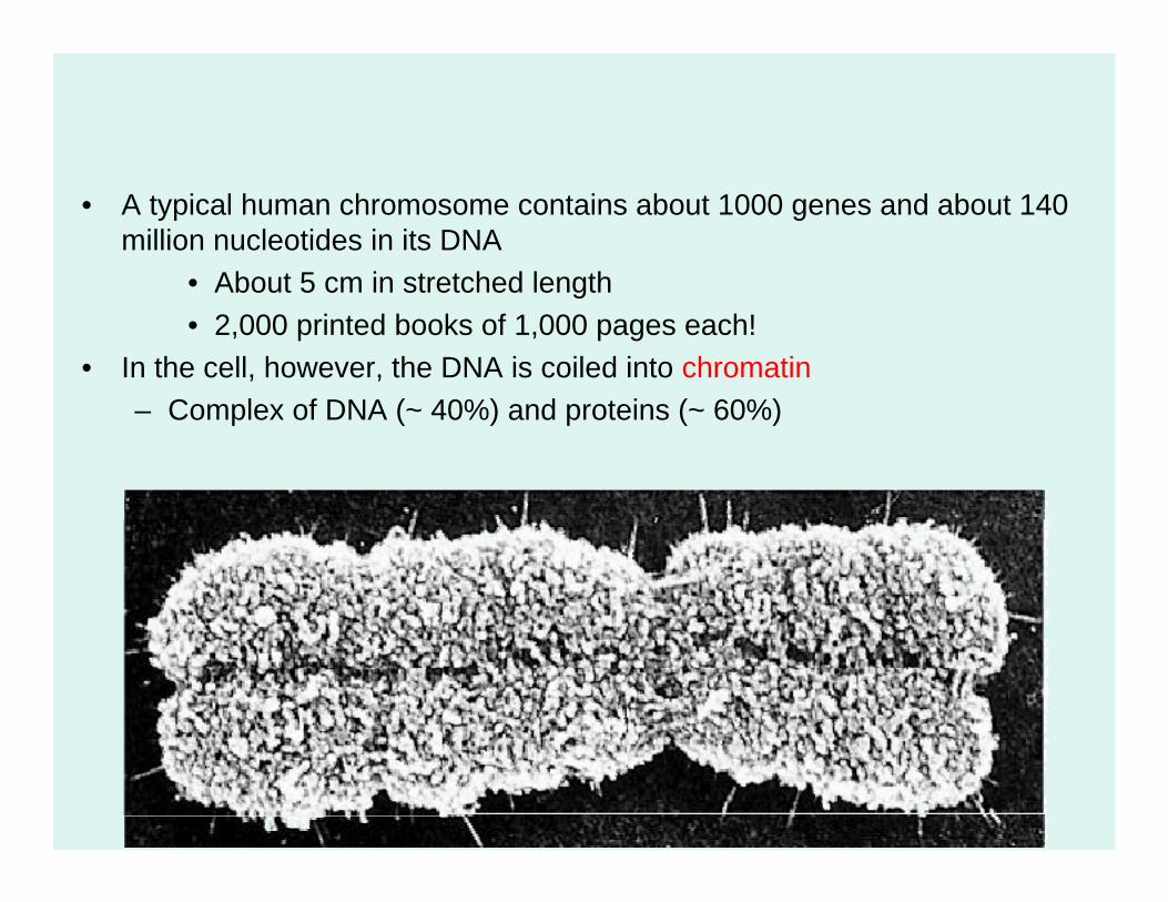
• A typical human chromosome contains about 1000 genes and about 140 million nucleotides in its DNA
• About 5 cm in stretched length• 2,000 printed books of 1,000 pages each!
• In the cell, however, the DNA is coiled into chromatin– Complex of DNA (~ 40%) and proteins (~ 60%)
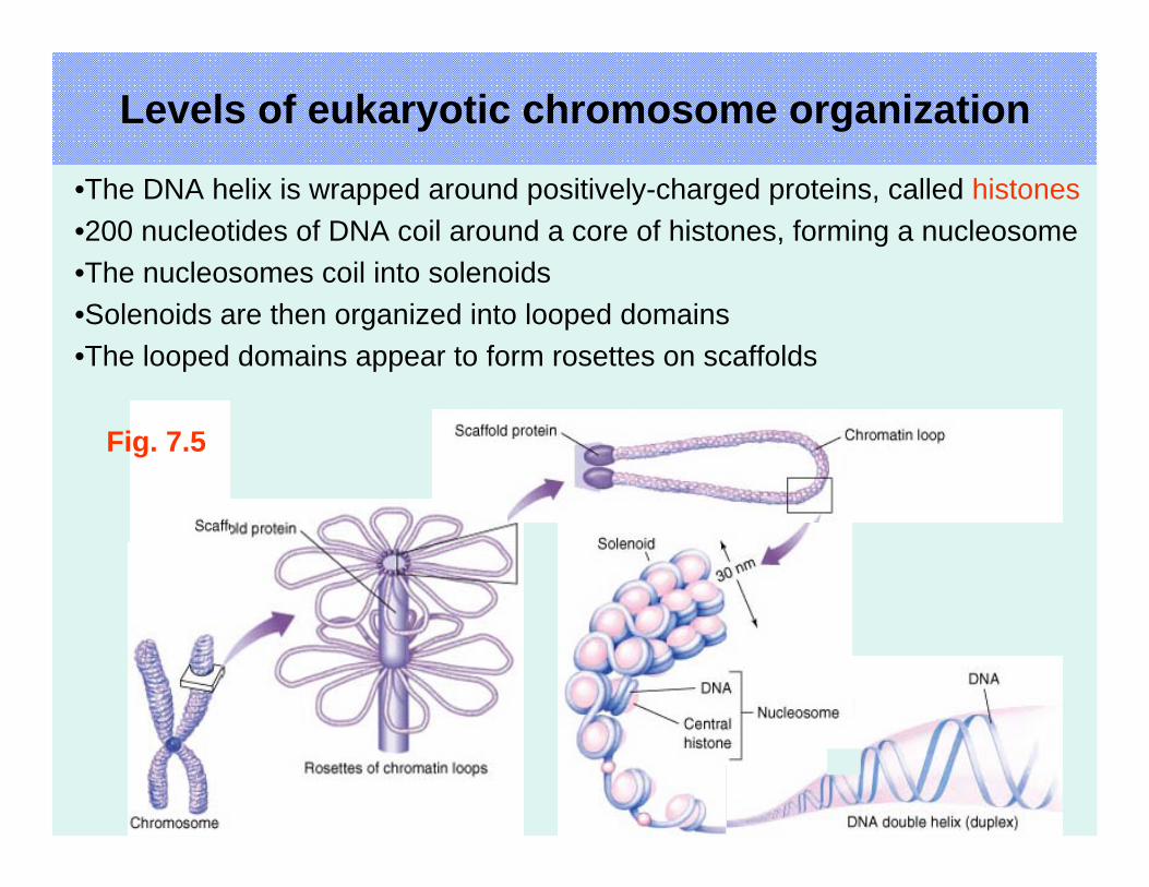
Levels of eukaryotic chromosome organization
•The DNA helix is wrapped around positively-charged proteins, called histones•200 nucleotides of DNA coil around a core of histones, forming a nucleosome•The nucleosomes coil into solenoids•Solenoids are then organized into looped domains•The looped domains appear to form rosettes on scaffolds
Fig. 7.5
7.2 Eukaryotes Have a Complex Cell Cycle
Eukaryotic cells divide in one of two ways– Meiosis
• Occurs in germ(reproductive) cells
• Results in the production of gametes
– Mitosis• Occurs in
somatic (non-reproductive) cells
MEIOSIS FERTILIZATION
Haploid gametes (n = 23)
Egg cell
Sperm cell
Diploidzygote
(2n = 46)Multicellular
diploid adults(2n = 46)
Mitosis anddevelopment
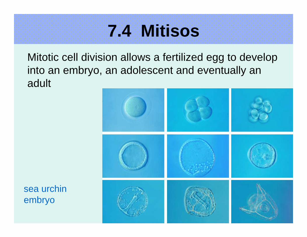
7.4 MitisosMitotic cell division allows a fertilized egg to develop into an embryo, an adolescent and eventually an adult
sea urchinembryo
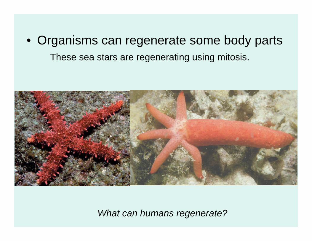
• Organisms can regenerate some body partsThese sea stars are regenerating using mitosis.
What can humans regenerate?

- Asexual reproduction, development, growth and cell replacement are mitotic divisions
Deadcells
Dividingcells
Epidermis, the outer layer of the skin
Dermis
skin
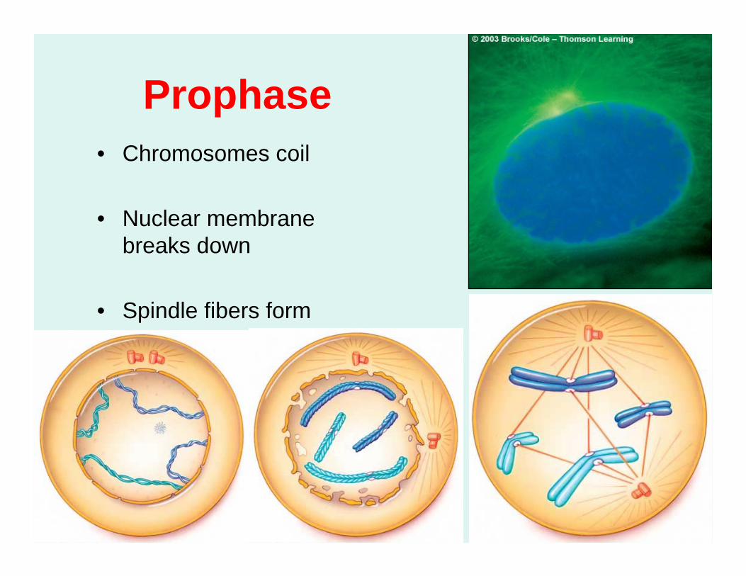
Prophase• Chromosomes coil
• Nuclear membrane breaks down
• Spindle fibers form
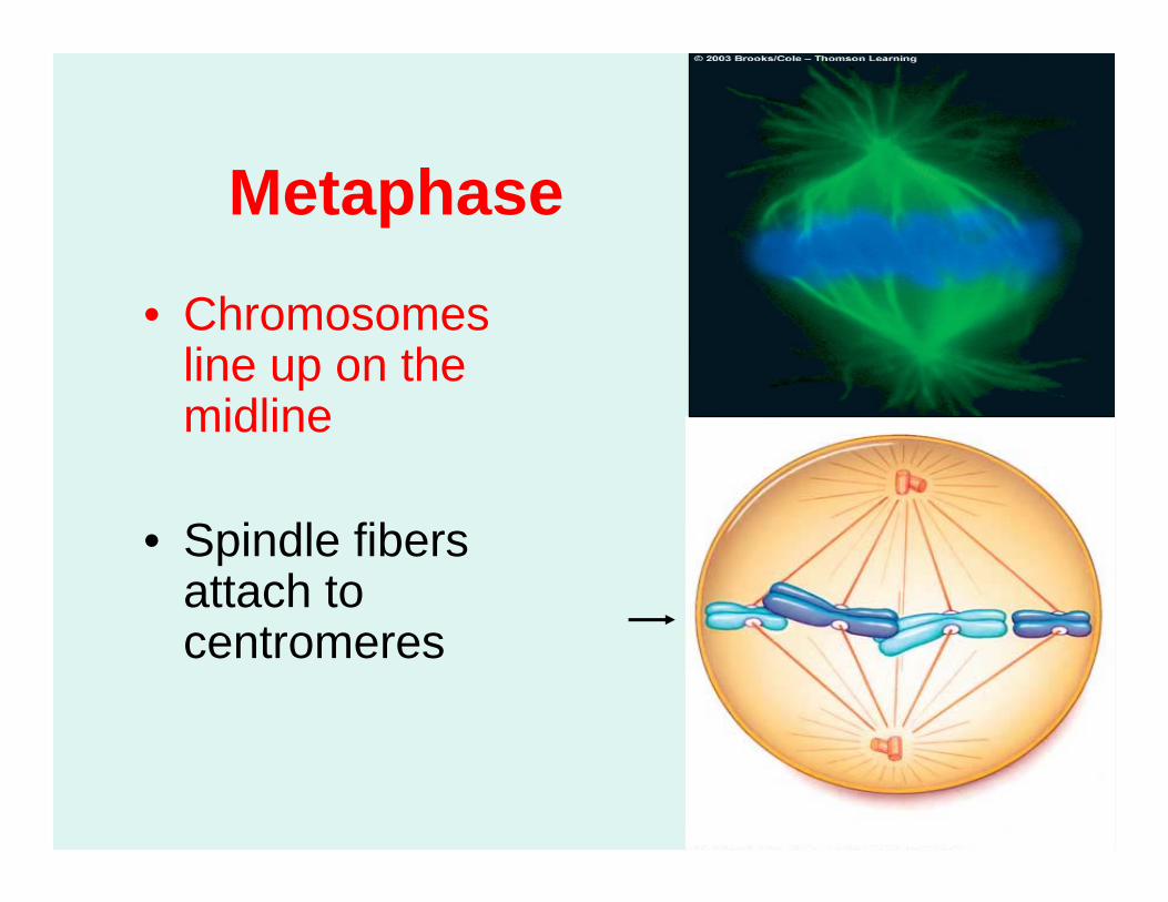
Metaphase
• Chromosomes line up on the midline
• Spindle fibers attach to centromeres
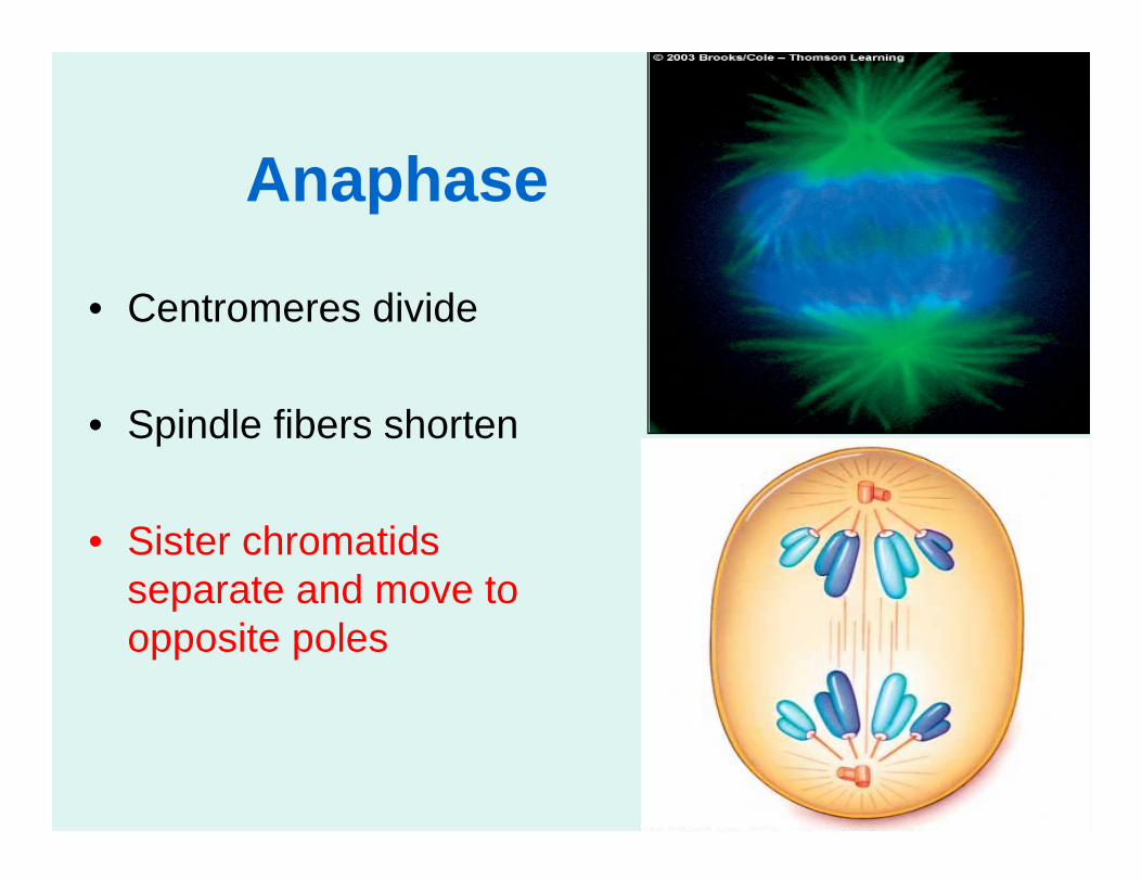
Anaphase
• Centromeres divide
• Spindle fibers shorten
• Sister chromatids separate and move to opposite poles
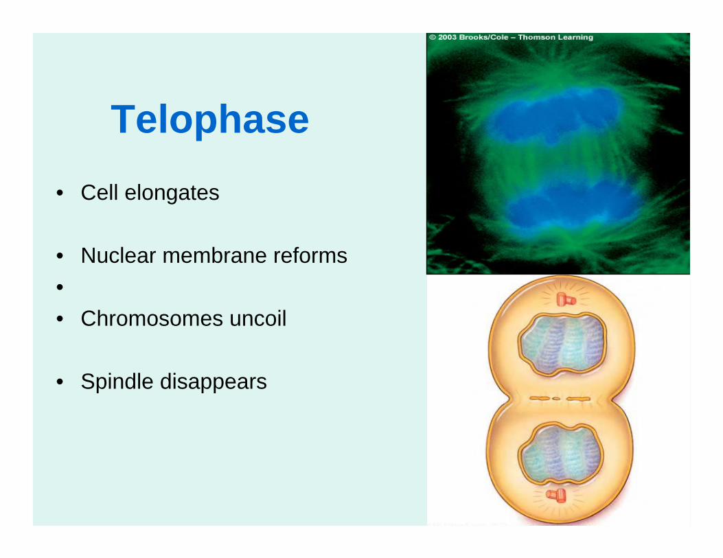
Telophase• Cell elongates
• Nuclear membrane reforms•• Chromosomes uncoil
• Spindle disappears

Cytokinesis
• Division of the cytoplasm
• Cleave furrow forms at equator of the cell
• Constriction tightens by contraction of filaments
• Cell is divided into two identical cells
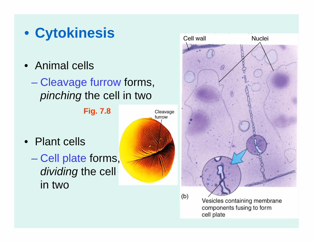
• Cytokinesis
• Animal cells
• Plant cells
Fig. 7.8
– Cleavage furrow forms, pinching the cell in two
– Cell plate forms, dividing the cell in two