Embed Size (px)
Citation preview
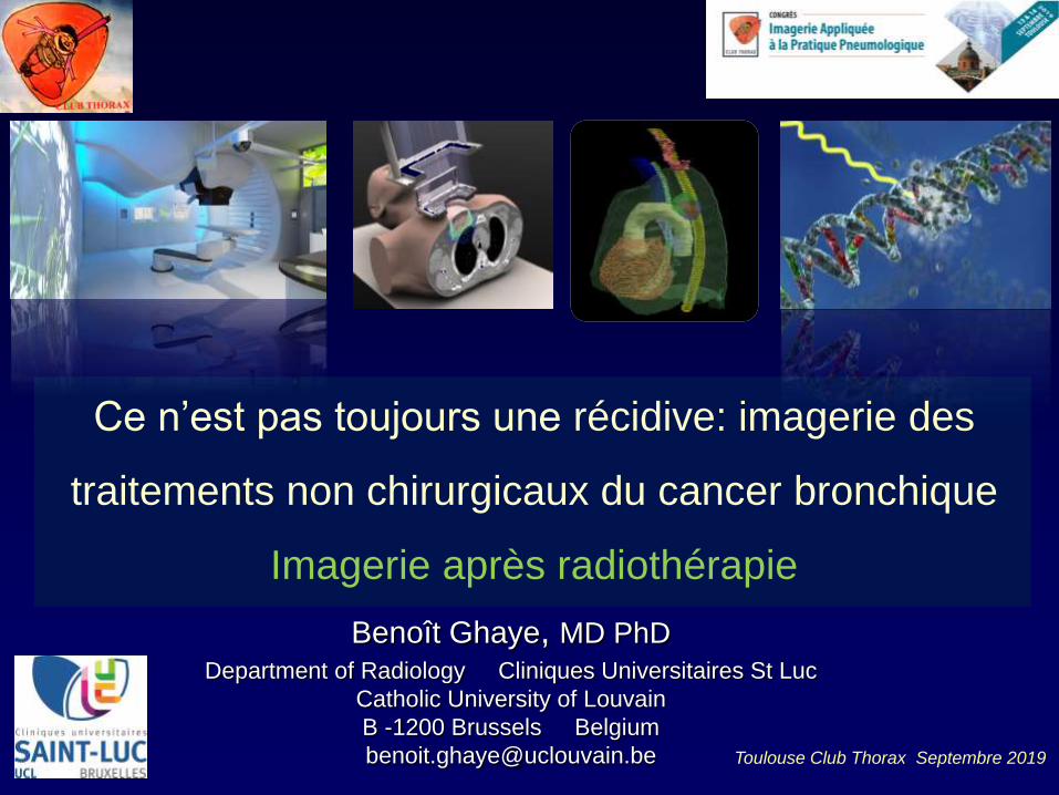
Benoît Ghaye, MD PhD
Department of Radiology Cliniques Universitaires St Luc
Catholic University of Louvain
B -1200 Brussels Belgium
[email protected] Toulouse Club Thorax Septembre 2019
Ce n’est pas toujours une récidive: imagerie des
traitements non chirurgicaux du cancer bronchique
Imagerie après radiothérapie
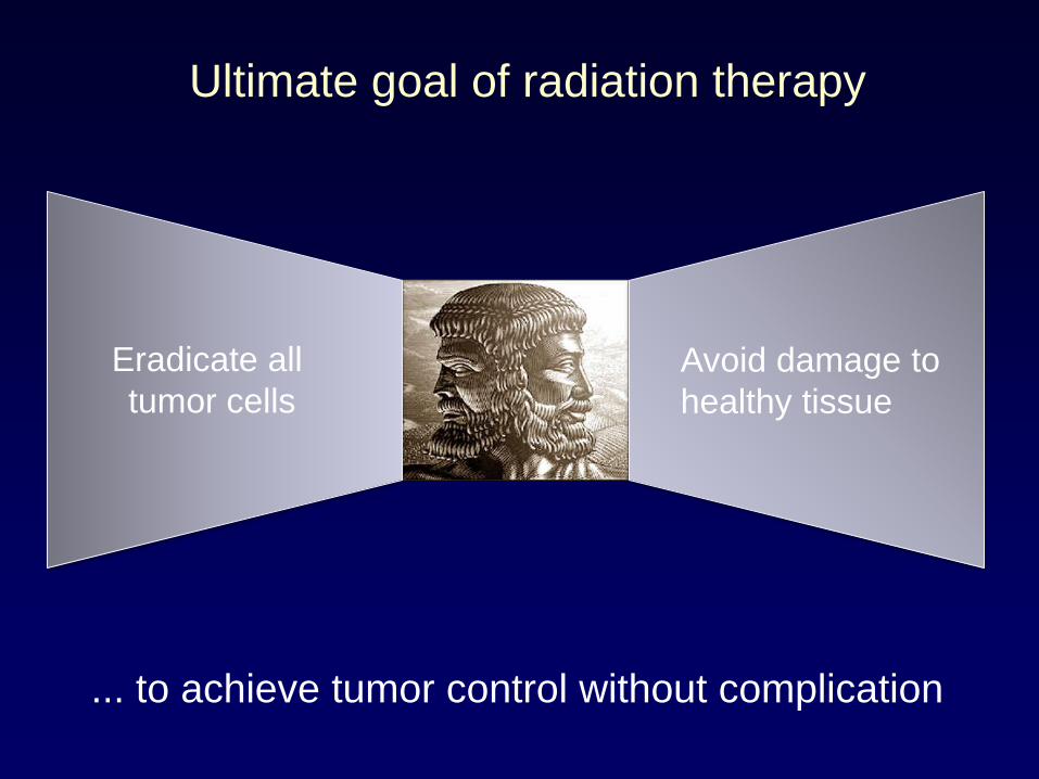
Ultimate goal of radiation therapy
Eradicate all
tumor cells
Avoid damage to
healthy tissue
... to achieve tumor control without complication

Radiation therapyHow does it work
2D-conventional RT
• Two parallel beams with opposed orientations
• 2 Gy per field combination per day
• (≤) 60 Gy total
• Limited beam orientation → large areas of normal tissue irradiated
Park RadioGraphics 2000;20:83

Radiation-induced lung disease
Reference point : day of completion of radiation therapy
Early phase : transient radiation pneumonitis
appears between 1 – 3 months
lasts up to 6 months
dyspnea, cough, low-grade fever, discomfort
steroids
Late phase : chronic radiation fibrosis
unresolved radiation pneumonitis
6 - 12 months (up to 24 months)
stable after 2 years
mostly asymptomatic
progressive dyspnea, dry cough, cor pulmonale
Davis AJR 1992;159:1157
Park RadioGraphics 2000;20:83

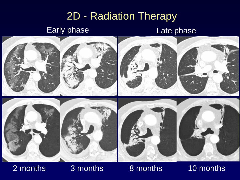
2D - Radiation Therapy
3 months2 months
Early phasePathology : acute exsudative phase followed by organizing phase with interstitial infiltration by mononuclear and other inflammatory cells
Lung injury (diffuse / patchy / nodular)
• Ground-glass
• Consolidation
Generally confined to field of irradiation
Do not conform to anatomic boundaries
Pleural effusion
Atelectasis
Usually regresses over 6 months
No sequellae
Choi RadioGraphics 2004;24:985

2D - Radiation Therapy
8 months
Late phase
10 months
Lung volume loss (mediastinal shift)
Architectural distortion
Consolidation
Well-defined
Shrinkage
Sharper demarcation
Shape/location may change →12 mths
Air bronchogram
Traction bronchectasis
May stabilize
Evolve up to 24 mths
(Small pleural effusion or thickening)
Park RadioGraphics 2000;20:83
Choi RadioGraphics 2004;24:985

Benveniste RadioGraphics 2019;39:344
2D - Radiation Therapy
2D - Radiation Therapy
3 months 8 months2 months
Early phase Late phase
10 months
Radiation-induced lung disease
Technique
• Conventional vs. conformational
• Portals and beam arrangement
• Dose : total (rarely < 20, commonly 20-40, almost always > 40 Gy)
fractionation and dose rate
• Irradiated volume (V20Gy)
• Physical characteristics of irradiation
Treatment
• Prior irradiation
• Chemo, Immunotherapy
• Steroids (rebound)
Tumor
• Tumor location
Patient
• Age
• Lung performance status
• Preexisting lung disease Park RadioGraphics 2000;20:83
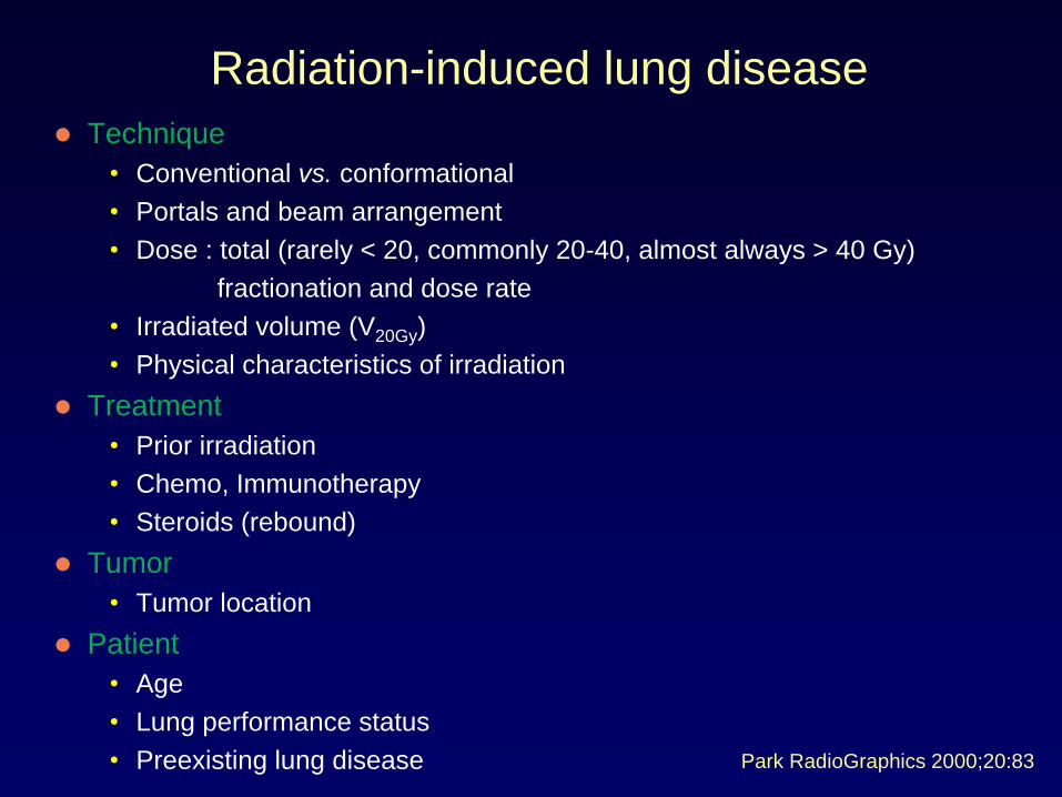

Radiation-induced lung disease
Technique
• Conventional vs. conformational
• Portals and beam arrangement
• Dose : total (rarely < 20, commonly 20-40, almost always > 40 Gy)
fractionation and dose rate
• Irradiated volume (V20Gy)
• Physical characteristics of irradiation
Treatment
• Prior irradiation
• Chemo, Immunotherapy
• Steroids (rebound)
Tumor
• Tumor location
Patient
• Age
• Lung performance status
• Preexisting lung disease
Aoki Radiology 2004;230:101
Park RadioGraphics 2000;20:83
Radiation-induced lung disease
Technique
• Conventional vs. conformational
• Portals and beam arrangement
• Dose : total (rarely < 20, commonly 20-40, almost always > 40 Gy)
fractionation and dose rate
• Irradiated volume (V20Gy)
• Physical characteristics of irradiation
Treatment
• Prior irradiation
• Chemo, immunotherapy
• Steroids (rebound)
Tumor
• Tumor location
Patient
• Age
• Lung performance status
• Preexisting lung disease Benveniste Clin Radiol 2013;68:e275

Radiation therapy New techniques
3D-CRT
IMRT
SBRT
SABR4D-RT4D-PET-IGRT
BOOST
PROTON
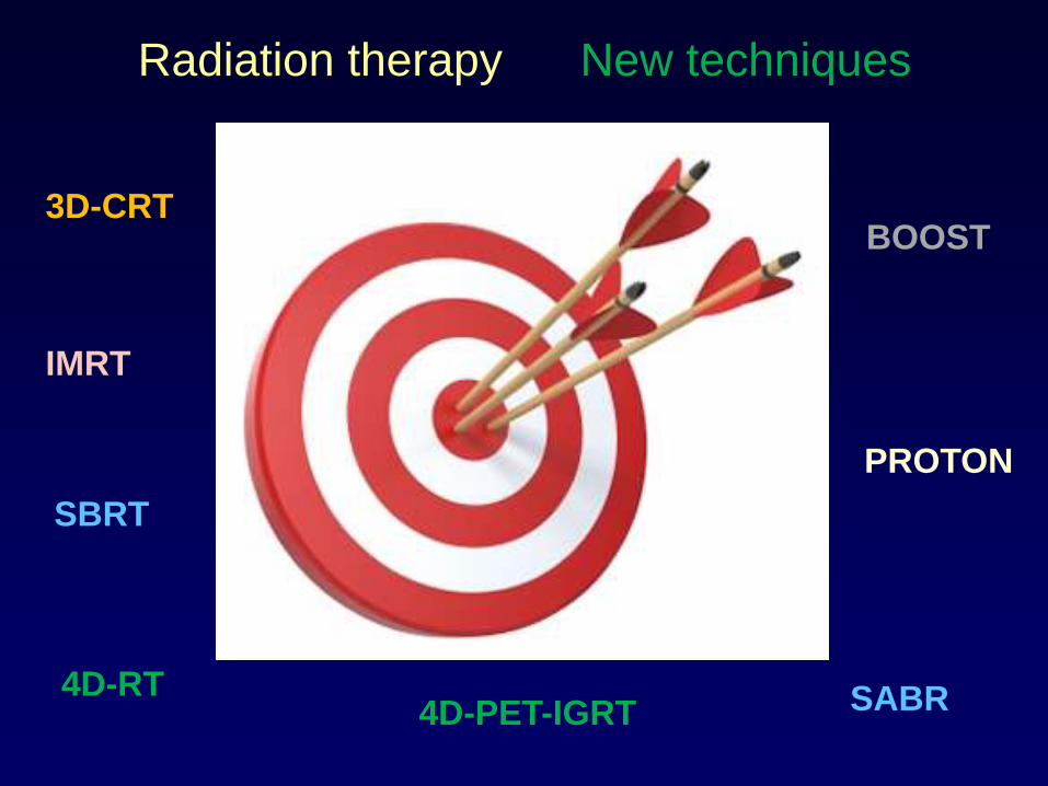
3D-CRT
IMRT
SBRT
SABR4D-RT4D-PET-IGRT
BOOST
PROTON
Radiation therapy New techniques
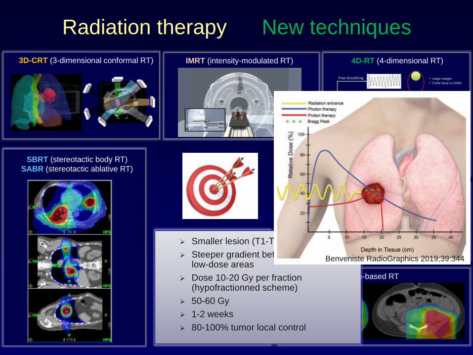
3D-CRT (3-dimensional conformal RT) IMRT (intensity-modulated RT)
SBRT (stereotactic body RT)
SABR (stereotactic ablative RT)
4D-RT (4-dimensional RT)
4D-PET-IGRT 4-D - PET - image-guided RT
Dose escalation
(Boost dose)
Proton-based RT
Smaller lesion (T1-T2), ≤ 5 cm
Steeper gradient between high- and low-dose areas
Dose 10-20 Gy per fraction (hypofractionned scheme)
50-60 Gy
1-2 weeks
80-100% tumor local control
Radiation therapy New techniques
Benveniste RadioGraphics 2019;39:344

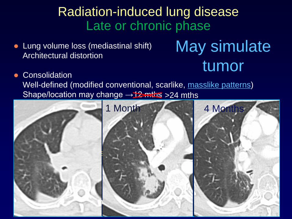
Lung volume loss (mediastinal shift)
Architectural distortion
Consolidation
Well-defined (modified conventional, scarlike, masslike patterns)
Shape/location may change →12 mths
Shrinkage
Sharper demarcation
Air bronchogram
Traction bronchectasis
May stabilize
Evolve up to 24 mths
(Small pleural effusion or thickening)
Radiation-induced lung diseaseLate or chronic phase
Koenig AJR 2002;178:1383-88
Larici RadioGraphics 2011;31:771-89

Radiation-induced lung diseaseLate or chronic phase
Lung volume loss (mediastinal shift)
Architectural distortion
Consolidation
Well-defined (modified conventional, scarlike, masslike patterns)
Shape/location may change →12 mths
Shrinkage
Sharper demarcation
Air bronchogram
Traction bronchectasis
May stabilize
Evolve up to 24 mths
(Small pleural effusion or thickening)
Less extensive
>24 mths
Koenig AJR 2002;178:1383-88
Larici RadioGraphics 2011;31:771-89

Lung volume loss (mediastinal shift)
Architectural distortion
Consolidation
Well-defined (modified conventional, scarlike, masslike patterns)
Shape/location may change →12 mths
Shrinkage
Sharper demarcation
Air bronchogram
Traction bronchectasis
May stabilize
Evolve up to 24 mths
(Small pleural effusion or thickening)
Radiation-induced lung diseaseLate or chronic phase
Less extensive
6 Months 9 Months 3 Years
Koenig AJR 2002;178:1383-88
Larici RadioGraphics 2011;31:771-89
>24 mths

Lung volume loss (mediastinal shift)
Architectural distortion
Consolidation
Well-defined (modified conventional, scarlike, masslike patterns)
Shape/location may change →12 mths
Shrinkage
Sharper demarcation
Air bronchogram
Traction bronchectasis
May stabilize
Evolve up to 24 mths
(Small pleural effusion or thickening)
Radiation-induced lung diseaseLate or chronic phase
3 Months 6 Months 12 Months 18 Months
>24 mths

Lung volume loss (mediastinal shift)
Architectural distortion
Consolidation
Well-defined (modified conventional, scarlike, masslike patterns)
Shape/location may change →12 mths
Shrinkage
Sharper demarcation
Air bronchogram
Traction bronchectasis
May stabilize
Evolve up to 24 mths
(Small pleural effusion or thickening)
Radiation-induced lung diseaseLate or chronic phase
PRE POST Larici RadioGraphics 2011;31:771-89
>24 mths
Radiation-induced lung diseaseLate or chronic phase
Lung volume loss (mediastinal shift)
Architectural distortion
Consolidation
Well-defined (modified conventional, scarlike, masslike patterns)
Shape/location may change →12 mths
Shrinkage
Sharper demarcation
Air bronchogram
Traction bronchectasis
May stabilize
Evolve up to 24 mths
(Small pleural effusion or thickening)
May simulate
tumor
1 Month 4 Months
>24 mths

Radiation-induced lung diseaseLate or chronic phase
Lung volume loss (mediastinal shift)
Architectural distortion
Consolidation
Well-defined (modified conventional, scarlike, masslike patterns)
Shape/location may change →12 mths
Shrinkage
Sharper demarcation
Air bronchogram
Traction bronchectasis
May stabilize
Evolve up to 24 mths
(Small pleural effusion or thickening)
May simulate
tumor
9 Months2 Months
>24 mths
Dahele J Thorac Oncol 2011;6:1221-8
Ronden Int J Radiat Oncol Biol Phys 2018;100:115-21
Infections
Locally recurrent tumor
Radiation-induced tumor
Drug-induced lung disease
Radiation-induced lung diseaseDifferential diagnosis

Infections
• Abrupt onset
• Pulmonary opacities appearing before completion of RT
outside radiation portals
• Respect anatomic boudaries
• Diffuse
• Bilateral
• Centrilobular, tree-in-bud opacities
• Cavitation
• Filling-in of bronchi
Locally recurrent tumor
Radiation-induced tumorChoi RadioGraphics 2004;24:985
Radiation-induced lung diseaseDifferential diagnosis

Infections
Locally recurrent tumor
• Usually within 2 years
• Increase in size of radiation fibrosis area
• Homogeneous opacification
• Absence of air bronchogram
• Convex border of irradiated lung
• Filling-in of bronchi 2
• Others: LK, enlarging LN or pleural effusion
• PET/CT >> CT : sensitivity 100% vs. 71%, specificity 92 vs 95% 1
• No PET before 3-6 months (PET uptake occasionnally up to 24 months)
• Pathological proof required
Radiation-induced tumor1Kim RadioGraphics 1992;12:269
1Bury ERJ 1999;14:13762Libshitz Radiology 1999;210:25
Choi RadioGraphics 2004;24:985
5 M
onth
s9 M
onth
sRadiation-induced lung disease
Differential diagnosis

Radiation-induced lung diseaseDifferential diagnosis
Infections
Locally recurrent tumor
• Usually within 2 years
• Increase in size of radiation fibrosis area
• Homogeneous opacification
• Absence of air bronchogram
• Convex border of irradiated lung
• Filling-in of bronchi 2
• Others: LK, enlarging LN or pleural effusion
• PET/CT >> CT : sensitivity 100% vs. 71%, specificity 92 vs 95% 1
• No PET before 3-6 months (PET uptake occasionnally up to 24months)
• Pathological proof required
Radiation-induced tumor1Kim RadioGraphics 1992;12:269
1Bury ERJ 1999;14:13762Libshitz Radiology 1999;210:25
Choi RadioGraphics 2004;24:985

Infections
Locally recurrent tumor
• Usually within 2 years (! SBRT Takeda)
• Increase in size of radiation fibrosis area
• Homogeneous opacification
• Absence of air bronchogram
• Filling-in of bronchi 2
• Convex border of irradiated lung
• Others: LK, enlarging LN or pleural effusion
• PET/CT >> CT : sensitivity 100% vs. 71%, specificity 92 vs. 95% 1
• No PET before 3-6 months (PET uptake occasionnally up to 24 mths)
• Pathological proof required
Radiation-induced tumor
9 Months5 Months
Radiation-induced lung diseaseDifferential diagnosis
1Kim RadioGraphics 1992;12:2691Bury ERJ 1999;14:1376
2Libshitz Radiology 1999;210:25
Choi RadioGraphics 2004;24:985
Infections
Locally recurrent tumor
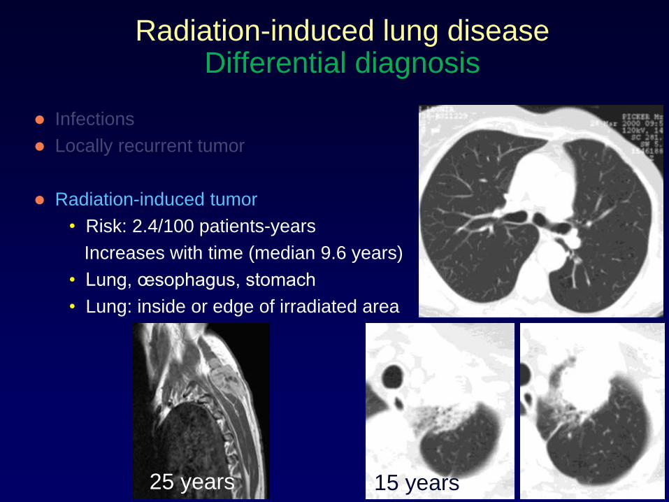
Radiation-induced tumor
• Risk: 2.4/100 patients-years
Increases with time (median 9.6 years)
• Lung, œsophagus, stomach
• Lung: inside or edge of irradiated area
15 years25 years
Radiation-induced lung diseaseDifferential diagnosis
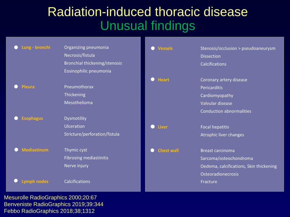
Radiation-induced thoracic diseaseUnusual findings
Lung - bronchi Organizing pneumonia
Necrosis/fistula
Bronchial thickening/stenosis
Eosinophilic pneumonia
Pleura Pneumothorax
Thickening
Mesothelioma
Esophagus Dysmotility
Ulceration
Stricture/perforation/fistula
Mediastinum Thymic cyst
Fibrosing mediastinitis
Nerve injury
Lymph nodes Calcifications
Vessels Stenosis/occlusion > pseudoaneurysm
Dissection
Calcifications
Heart Coronary artery disease
Pericarditis
Cardiomyopathy
Valvular disease
Conduction abnormalities
Liver Focal hepatitis
Atrophic liver changes
Chest wall Breast carcinoma
Sarcoma/osteochondroma
Oedema, calcifications, Skin thickening
Osteoradionecrosis
Mesurolle RadioGraphics 2000;20:67
Benveniste RadioGraphics 2019;39:344
Febbo RadioGraphics 2018;38;1312
Fracture
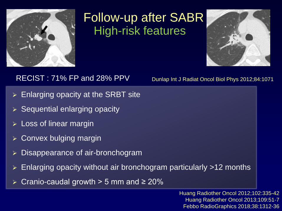
Enlarging opacity at the SRBT site
Sequential enlarging opacity
Loss of linear margin
Convex bulging margin
Disappearance of air-bronchogram
Enlarging opacity without air bronchogram particularly >12 months
Cranio-caudal growth > 5 mm and ≥ 20%
Follow-up after SABR
Huang Radiother Oncol 2012;102:335-42
Huang Radiother Oncol 2013;109:51-7
Febbo RadioGraphics 2018;38:1312-36
RECIST : 71% FP and 28% PPV Dunlap Int J Radiat Oncol Biol Phys 2012;84:1071
High-risk features

Vansteenkiste J Ann Oncol 2014;25:1462-74
Ghaye Diagn Interv Imaging 2016;97:1037-52
Febbo RadioGraphics 2018;38;1312-36
Follow-up after SABRHigh-risk features
Sensitivity
and specificity
> 90 %
Low risk Intermediate risk High risk

≥4 HRF
Sens 92%
Spec 85%
2 HRF
Sens 85%
Spec 100%
Peulen Int J Radiat Oncol Biol Phys 2016;96:134-4113 pts with local recurrence vs. 26 non recurrence
88 patients without local recurrence
50% have HRF
≥ 3 HRF in 25%
increased rate of FU CT, (PET and biopsy)
large interreader variability
Ronden Int J Radiat Oncol Biol Phys 2018;100:115-21
Dahele J Thorac Oncol 2011;6:1221-8
High-risk featuresFollow-up after SABR

Follow-up after SABR
5/201911/20183/201812/201711/2017
10/20174/201711/201611/201512/2014

Radiomics
1Li Med Phys 2017;44:4341-43492Mattonen SA Int J Radiat Oncol Biol Phys 2016;94:1121-8
3Seabra and Ghaye, ECR 2018
Imaging features from pre-treatment or post-treatment CT are
associated with clinical outcomes in NSCLC treated with SABR1,2
3
Follow-up after SABR
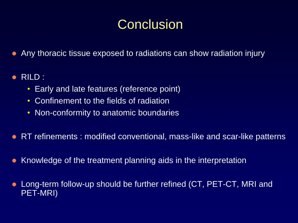
Any thoracic tissue exposed to radiations can show radiation injury
RILD :
• Early and late features (reference point)
• Confinement to the fields of radiation
• Non-conformity to anatomic boundaries
RT refinements : modified conventional, mass-like and scar-like patterns
Knowledge of the treatment planning aids in the interpretation
Long-term follow-up should be further refined (CT, PET-CT, MRI and PET-MRI)
Conclusion
Acknowledgements
Members of Club Thorax, France
• Mostafa El Hajjam, Paris
• Jacques Giron, Toulouse
• Gilbert Ferretti, Grenoble
• Anne-Sophie Claes, Nîmes
Radiation Therapy Dpt, UCL, Brussels
• Marie Wanet
• Xavier Geets
Radiology Dpt, UCL, Brussels
• Jacques Malghem
• Rita Seabra
• Nicolas Michoux