Embed Size (px)
Citation preview

CASE STUDY “Arnold Chiari type 1” Jan Camus, Antwerp, Belgium Fully certified craniosacral therapist January 14th 2015
1
INTRODUCTION
Craniosacral Therapy is a gentle, hands‐on form of manual therapy which addresses dysfunction within
the craniosacral system, which follows the movement and flow of cerebrospinal fluid within the
ventricles of the brain, the dural tube, and the central nervous system surrounding the spinal and cranial
nerves. Of course, a human body is more than that.
Water is the main raw material we are made of. Depending on the organ or the function more or less
water will constitute the building material. For example brain material is 90% water. Bones only 22%.
Teeth a mere 10%. "Water in our bodies has different physical properties from ordinary bulk water,
because of the presence of proteins and other biomolecules. Proteins change the properties of water to
perform particular tasks in different parts of our cells. Water can be viewed as a 'designer fluid' in living
cells." (Dr. Martin Gruebele, University of Illinois). Interfacial water exhibits structural organizations that
differ from what is termed "bulk" water. This "vicinal" water seems to be influenced by structural
properties that make up the cell. An example of this, and in relation to the water in a tempero
mandibular joint, : "The combined data from three different methods lead to the conclusion that all or
almost all of the water in the intact disc is bound water and does not have properties consistent with
free or bulk water. … Water is the carrier of the most important molecules of life, like proteins and
DNA.” (Dr. Gerald Pollack).
In other words, water in our cells is an information carrier. Water is also ‘movement’. As in vortexing. A
vortex occurs naturally in nature, as in streams, rivers, waterfalls, etc. The vortex is a kind of mechanical
perturbation or agitation. Vortexing is a very powerful way of increasing structure.
Now when certain information is to be held still in our system, ‐ I am taught to withhold my tears, strong
people do not show emotions, I can not express my anger because he/she is my boss/my parent/my
lover… , and so on – we, unconsciously, spend energy to suppress the energy of that emotion, to hold
still the water in which this emotional information is stored. So grows an energy cyste. Cells in which the
water is not allowed to move anymore or no way is known to release it. The longer it lasts, the more
energy is needed to keep the blocked energy in that cyste under control. This is very, very tiring,
depleting.
The therapist uses specific techniques not only to help restore physiological balance of the nervous
system, the brain and cranial bones, but to invite the water in the cells holding the blocked energy to
move again, to restore natural movement. Homeostasis is restored to the client through restoring
optimal functioning of these areas. When restriction of natural movement or dysfunction in these areas
has been corrected or relieved, a client may also experience an improvement of their physical, mental,
emotional and spiritual well–being. Within these texts Dr. Upledger’s research and clinical experience
has described numerous case examples from his osteopathic practice about how Craniosacral Therapy
treats not only the physiological symptoms of disease but also the emotional components that
contribute to and aggravate an injury, symptom or illness. Thus he and other practitioners of this
method recognize there can be a strong correlation between the physical and emotional aspects of
disease. They stipulate that therapists need to consider the emotional implications of recurring chronic
pain, injury or illness and explore what may underline any resistance to the healing process.

CASE STUDY “Arnold Chiari type 1” Jan Camus, Antwerp, Belgium Fully certified craniosacral therapist January 14th 2015
2
Scientific research studies of this genre continue to be challenging. Emotional energy is unique to each
individual situation and experience, therefore it is difficult to measure and quantify. It is made clear here
that emotion may not always play a direct part in the healing of a physical injury, as many physical
injuries are able to resolve with a prescribed form of treatment on a physical level. However this case
study seeks to describe and explain what occurred with a client who had a congenital anomaly that did
not stabilize after following the prescribed physical therapy technique.
What is chiari malformation?
Chiari malformation, also known as an Arnold‐Chiari malformation, is a congenital (present at birth)
defect occurring in the back of the head where the brain and spinal cord connect.
There are four types of Chiari malformations:
Type 1 – Occurring when the base of the skull and upper spinal area do not form properly, a type 1
Chiari malformation commonly goes unnoticed until problems arise in the adolescent or adult years of
life. The headaches most typical of Chiari I malformations are usually located at the back of the head,
and are often made worse by exertion.
Type 2 – The most common of all Chiari malformations, type 2 is caused by part of the back of the brain
shifting downward through the bottom of the skull. Type 2 Chiari malformations are typically seen in
infants who are born with spina bifida, a neurological condition that causes a portion of the spinal cord
and the surrounding structures to develop outside, instead of inside, the body. Type 2 Chiari
malformations can also be associated with hydrocephalus, a condition in which there is an
overproduction or lack of absorption of the cerebral spinal fluid (CSF) that is found inside of the
ventricles (fluid‐filled areas) inside of the brain. The increased fluid causes the pressure inside of the
head to increase and the skull bones to expand to a larger‐than‐normal appearance.
Type 3 – Type 3 Chiari malformations occur when the back of the brain protrudes out of an opening in
the back of the skull area.
Type 4 – Type 4 Chiari malformations occur when the back of the brain fails to develop normally.
What causes chiari malformation?
The exact cause of Chiari malformation is unknown; however, it is believed that problems during fetal
development may cause abnormal brain function.
Theories suggest that the following may predispose the fetus to problems that affect the normal
development of the head during pregnancy:
exposure to hazardous chemicals/substances
lack of proper vitamins and nutrients in the diet
infection
prescription or illegal drug and alcohol consumption

CASE STUDY “Arnold Chiari type 1” Jan Camus, Antwerp, Belgium Fully certified craniosacral therapist January 14th 2015
3
In craniosacral therapy is also considered the possibility of an emotional impact in the human cells,
wether at conception, during pregnancy, at birthing or inn early childhood, as being a possible cause.
What are the symptoms of a Chiari malformation?
The following are the most common symptoms of a Chiari malformation. However, each person may
experience symptoms differently. Symptoms may include:
headaches
stiffness or pain in the neck or back of the head area
poor feeding and swallowing (mainly children)
decreased strength in the arms (mainly children)
decreased sensation in the arms and legs (mainly children)
rapid, back and forth, eye movement
developmental delays(mainly children)
weak cry (mainly children)
breathing problems (mainly children)
The symptoms of Chiari malformation may resemble other conditions or medical problems. Always
consult your physician for a diagnosis.
When can a Chiari malformation be diagnosed?
If a Chiari malformation occurs with other congenital (present at birth) defects, the diagnosis may be
made at birth. Other times, the diagnosis is made after the onset of specific signs and symptoms, and
after diagnostic testing.
Diagnostic tests that may be performed to confirm the diagnosis of a Chiari malformation include:
magnetic resonance imaging (MRI) – a diagnostic procedure that uses a combination of large
magnets, radiofrequencies, and a computer to produce detailed images of organs and structures
within the body.
Typical treatment for a Chiari malformation:
Surgery. The basic operation is one of uncrowning the area at the base of the cerebellum where it is
pushing against the brainstem and spinal cord. This is done by removing a small portion of bone at the
base of the skull deep to the neck muscles as well as often removing a part of the back of the first and
occasionally additional spinal column segments.
At the Children’s Hospital of New York, they have been collecting information since 1998 using
intraoperative electrophysiological monitoring to help determine whether opening of the dura mater is

CASE STUDY “Arnold Chiari type 1” Jan Camus, Antwerp, Belgium Fully certified craniosacral therapist January 14th 2015
4
a necessary component of surgery on Chiari I malformation. They discovered that most of the
improvement in nerve impulses through the brain and spinal cord occurs after removal of the bone.
However, they did not see any further improvement after opening the dura, suggesting that children
may not require this additional step of surgery. Accordingly, for the past four years they have performed
a less invasive operation where the dura is not opened during surgery. At this time, they have seen
excellent clinical and radiographic results without any significant operative complications after bony
decompression without dural opening. This is important because the complication rate after surgery has
been reported to be nearly 4 times higher if the dura is opened during surgery.
In addition, the use of endoscopes has allowed for this procedure to be performed through smaller
incisions, which helps in the reduction of post operative pain and speech recovery.
Typical surgery treatment for a Chiari malformation will be determined based on:
age, overall health, and medical history
the extent of the condition
the type of condition
tolerance for specific medications, procedures, or therapies
expectations for the course of the condition
personal opinion or preference
Medical management consists of frequent physical examinations and diagnostic testing to monitor the
growth and development of the brain, spinal cord, skull, and backbones.
Some types of Chiari malformations may require surgery to relieve increased pressure inside the head or
neck area, or to help drain excess cerebral spinal fluid from the brain. Very severe Chiari malformations
may be life threatening.
The full extent of the problems associated with a Chiari malformation are usually not completely
understood immediately at birth, but may be revealed as the child grows and develops.
The decision whether to have surgery is up to each individual and their doctor. Some of the factors that
are considered are the severity of symptoms, whether the symptoms are getting worse, whether the
nervous system is being compromised, whether there are any complicating issues, and the surgeon's
own experience and judgment. Unfortunately, there is no single, objective measure to say whether
someone should have surgery and many clients will find that different doctors may have different
opinions. Some doctors are more aggressive in their treatment approach and some are more
conservative. A recent survey about when to recommend surgery showed that there was general
agreement among surgeons in the extreme cases ‐ no or mild symptoms, don't operate; severe,
progressive symptoms or syringomyelia, operate ‐ but there was little agreement in the middle. In one
of the survey's hypothetical cases, the surgeons were split almost evenly down the middle on whether
to operate or not.

CASE STUDY “Arnold Chiari type 1” Jan Camus, Antwerp, Belgium Fully certified craniosacral therapist January 14th 2015
5
As with any surgery, the chance of success depends on the individual case, so each person should ask
their doctor what their chance of having a successful surgery is. It should be noted that success can
mean different things to different people, so it is best to ask specific questions such as what are the
odds I will be symptom free; what are the odds I will be mostly better; and what are the odds I will get
worse.
Unfortunately, there is not a lot of strong surgical outcome research. For clients with just Chiari (no
syringomyelia), up to 50% become symptom free after surgery, with another 10%‐30% improving
significantly. On the flip side, for 10%‐20%, the surgery will be a failure and they will likely require
additional surgeries. Keep in mind these are not scientific numbers and each client should discuss their
own chance of success with their doctor.
What are the possible complications of surgery? This is another question that is important for every
client to ask their doctor so that they fully understand the risks and potential outcomes of surgery.
Many of the complications of decompression surgery have to do with opening the dura and research has
shown that opening the dura (duraplasty) does increase the complication rate. There is a risk of
infection and sometimes the patch that is sewn in leaks or becomes scarred. A more serious
complication ‐ not necessarily related to opening the dura ‐ occurs when the brain slumps further into
the spinal area after the surgery.
What will happen without surgery? The natural progression of Chiari ‐ as doctors call it ‐ varies from
person to person and is not well understood. For example, why do some people develop symptoms in
their 30's while others have symptoms their whole life? For many people with no or mild symptoms, the
symptoms will not get worse and surgery will not be necessary. However, there are also anecdotal
reports of symptoms becoming rapidly worse, sometimes after a sneeze or a fall. If a client does not
have surgery, many doctors will recommend monitoring the situation with routine MRI's and
neurological exams.
HISTORY
This case involves a 55 year old adult female who presented, to this author and therapist, with a
congenital anomaly, Arnold Chiari type 1. This client is a professional caretaker and educator of disabled
young people for over twenty years. She is a divorced (domestic violence) mother of one son (28, drug
addict, no contact). Her job is emotionally heavy and tiring. She has undergone a hysterectomy at the
age of 29 because of endometriosis. The client has remarried. As a young child she had an episode of
cervicalgia (neck pain) and headache which regressed spontaneously. Since summer 2011 she has
headaches situated at the level of the occiput and high cervical. Additional complaints about vertigo,
general ailment, difficulty swallowing and thereby easily choking.
She suffers bladder infections two to three times a month. Sleep pattern is irregular and quality poor.
When pain becomes excruciating (pain chart 7/8), every 6 weeks to three months, she takes a 3 to 4
days sick leave from the job to recover the best she can. Medication are mainly muscle relaxants
(Tetrazepam, variable doses).

CASE STUDY “Arnold Chiari type 1” Jan Camus, Antwerp, Belgium Fully certified craniosacral therapist January 14th 2015
6
In February 2013 she has her first neurosurgical medical consultation. Images are made by CT (computer
tomograpghy) and by MRI (magnetic resonance imaging) scanning of the head. A Chiari malformation
type 1 is confirmed. There is no syringomyelia. The cerebellar tonsillar descent is not graded in the
report, but is said to reach beyond the skull base. Uncartrosis (wear on intervertebral discs) from C4C5
till C6C7. Contact with spinal cord at level C4C5 and C6C7. Discus hernia at the right at level C6C7 with
probable radicular conflict (pression on the nerve root). The images (pages 10 ‐ 12) show a spinal
canalstenosis (a narrowing causing a restriction) C4C5, C5C6 without any indication of medulopathy
(nerve root compression).
(In Dorland's Medical Dictionary herniation is defined as an abnormal protrusion of an organ or other
body structure through a defect or natural opening in a covering, membrane, muscle, or bone.)
Possible treatment is posterior fossa decompression (PFD) by enlarging the foramen magnum and
duraplasty (opening of the dura).
Our first session, March 2013, following her friend’s advice, is a desperate try to avoid brain surgery. She
has seen at that time four different neurosurgeons. The last one confirming her that surgery is not yet
urgent, but will be inevitable in future. This neurosurgeon puts her on a yearly control by MRI scan.
TESTS AND MEASUREMENTS
As an introduction she immediately states she heeded her friend’s advice only because of the said “soft
touch” treatment. She absolutely refuses any other form of testing or measuring. First impression, later
frequently confirmed, is hypersensitivity.
Till today I have conscientiously respected her choice in this matter.
TREATMENT
First session with the client has been limited to a 45 minutes lasting first (cranial) vault hold. During this
period three spontaneous still points will occur lasting up to 12 minutes. Next session, three weeks later,
the client reports she slept that same night for 14 hours non‐interrupted. The bladder infection
gradually disappeared in a three days period.
After 1 month, 3 sessions, a frontal lift and a release of the ethmoid results in the client’s subjective
feeling of opening and space.
At the end of the second month, 5 sessions, the client can already tolerate the third (occipital) vault.
Major spontaneous unwinding in the neck and right side of the head. The client expresses rage, sadness
and helplessness. First somato emotional releases.
In the following two months the client, during a first (cranial) vault hold will go into SER and relive her
birth. She testifies afterwards about the lightness in her chest, no gasping for air anymore and the
weight in her head has disappeared. Her craniosacral rhythm, end of that particular session, is balanced,
equal to both sides, quality feels strong, the pumping goes deep at 7 to 8 movements a minute. One

CASE STUDY “Arnold Chiari type 1” Jan Camus, Antwerp, Belgium Fully certified craniosacral therapist January 14th 2015
7
session an exceptionally large energetic cyste in the pericardium releases, another on the occiput lateral
right.
Months 5th till 8th the first (cranial) vault has become relaxing standard procedure. As from the 6th
month onwards the client allows mouthwork. During an ‘Avenue of Expression’, after having invited to
release all diaphragma’s and the dural tube, first mobilizing the ossa zygomatica (cheek‐bones), testing
the vomer’s synchronicity with the sphenoid, softly inviting the hard palate into torsion and shear while
at all time stabilizing the sphenoid and having spent five to six minutes inviting the ethmoid to unwind,
the left TemporoMandibularJoint releases together with the maxillary and the mandible.
After 15 sessions over an 8 month period the client has an appointment for her yearly control. The
report states (translated from Dutch) “In comparison to the previous control the hernia C6C7 has clearly
regressed in volume. … As at this moment there is not a clear indication of a severe medullary
compression there is no meaningful reason for her to have this Arnold Chiari treated.”
There are no images available of this between control visit.
The client’s next 22 sessions the treatments are basically cranial (first) and occipital (third) holds during
twenty to forty minutes.
Subsequent session include SER around fear and powerlessness associated with a pregnancy, the client
releases anger and sadness.
Last summer one session is dedicated to contact each and every vertebra starting at the coccyx all the
way up till the occiput. During the contact with T4T5 the client testifies of a sequence of important
movements inside her head, without any sensation of pain or discomfort. Six sessions later, during a
0
1
2
3
4
5
Sessions per month

CASE STUDY “Arnold Chiari type 1” Jan Camus, Antwerp, Belgium Fully certified craniosacral therapist January 14th 2015
8
relaxing cranial vault hold, the tentorium spontaneously starts to unwind for some twenty minutes,
again without any feeling of discomfort for the client.
The frequency and the length of the sessions are at all times decided in consultation with the client.
RESULTS
Last MRI scan check (November 2014), after more than thirty CS treatments during 23 months, the
neurosurgeon has described the client’s state as “stable, not descended any further, even slightly
improvement”.
The neurosurgeon asked the client what she had done. She explained briefly what CST is about (she said
he first pretended not to know!).
He now has encouraged her to continue the CST treatment!
The client has not taken any sick leaves since December 2013.
CASE STUDY “Arnold Chiari type 1” Jan Camus, Antwerp, Belgium Fully certified craniosacral therapist January 14th 2015
9
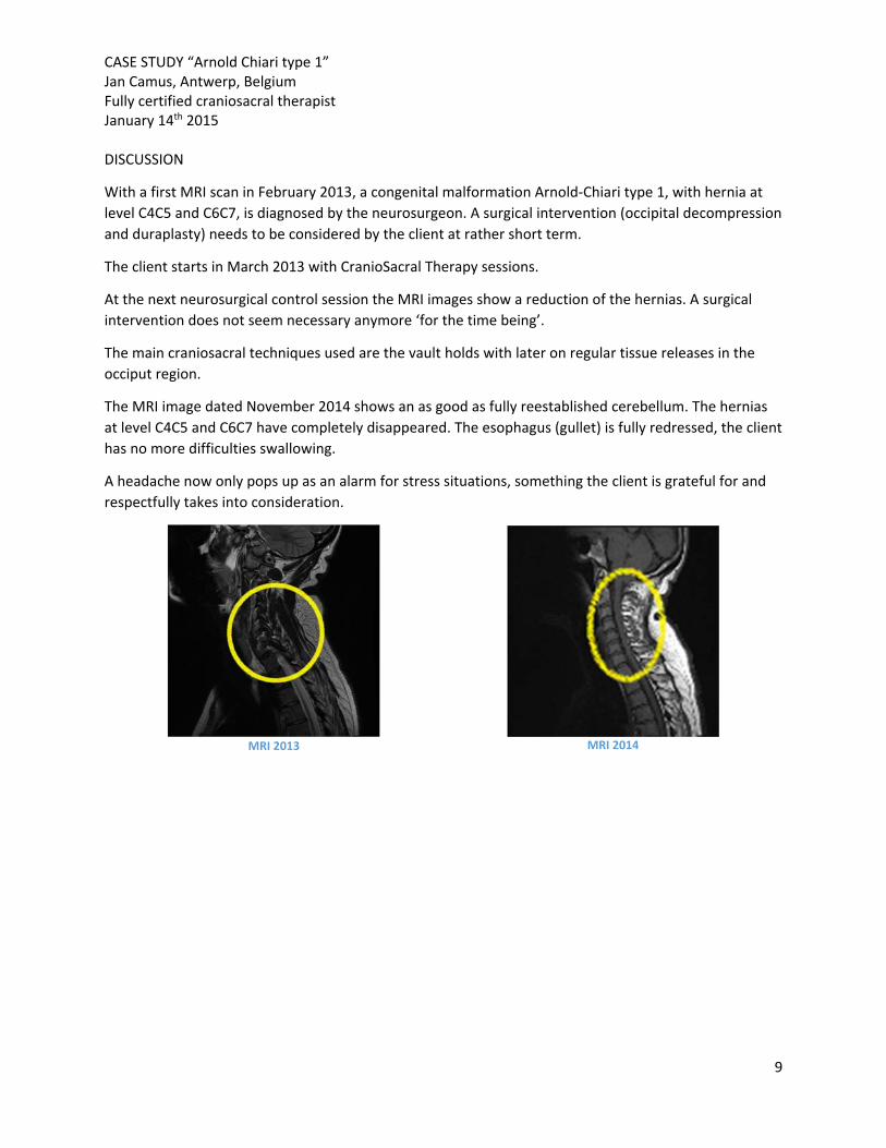
DISCUSSION
With a first MRI scan in February 2013, a congenital malformation Arnold‐Chiari type 1, with hernia at
level C4C5 and C6C7, is diagnosed by the neurosurgeon. A surgical intervention (occipital decompression
and duraplasty) needs to be considered by the client at rather short term.
The client starts in March 2013 with CranioSacral Therapy sessions.
At the next neurosurgical control session the MRI images show a reduction of the hernias. A surgical
intervention does not seem necessary anymore ‘for the time being’.
The main craniosacral techniques used are the vault holds with later on regular tissue releases in the
occiput region.
The MRI image dated November 2014 shows an as good as fully reestablished cerebellum. The hernias
at level C4C5 and C6C7 have completely disappeared. The esophagus (gullet) is fully redressed, the client
has no more difficulties swallowing.
A headache now only pops up as an alarm for stress situations, something the client is grateful for and
respectfully takes into consideration.
MRI 2013
MRI 2014

CASE STUDY “Arnold Chiari type 1” Jan Camus, Antwerp, Belgium Fully certified craniosacral therapist January 14th 2015
10
CT 2013‐1 CT 2013‐2
CT 2013‐3 CT 2013‐4

CASE STUDY “Arnold Chiari type 1” Jan Camus, Antwerp, Belgium Fully certified craniosacral therapist January 14th 2015
11
MRI 2013 1 MRI 2013 2
MRI 2013 3
MRI 2013 4
CASE STUDY “Arnold Chiari type 1” Jan Camus, Antwerp, Belgium Fully certified craniosacral therapist January 14th 2015
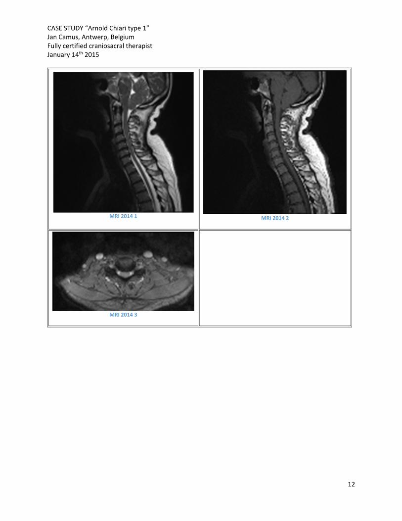
12
MRI 2014 1
MRI 2014 2
MRI 2014 3

CASE STUDY “Arnold Chiari type 1” Jan Camus, Antwerp, Belgium Fully certified craniosacral therapist January 14th 2015
13
REFERENCES
Upledger, John E., D.O., F.A.A.O. (1983). Craniosacral Therapy. Seattle, WA: Eastland Press. Upledger,
John E., D.O., F.A.A.O. (1987). Craniosacral Therapy II Beyond the Dura. Seattle, WA: Eastland Press.
Upledger, John E., D.O., O.M.M. (1990). SomatoEmotional Release and Beyond. Palm Beach Gardens, FL:
UI Publishing, Inc. Upledger, John E., D.O., O.M.M. (1996). A Brain Is Born: Exploring the Birth and
Development of the Central Nervous System. Berkeley, CA: North Atlantic Books
Upledger, John E., D.O., O.M.M. (2003). Cell Talk: Transmitting mind into DNA. Berkeley, CA: North
Atlantic Books
http://www.hopkinsmedicine.org/neurology_neurosurgery/centers_clinics/pediatric_neurosurgery/con
ditions/chiari‐malformation/
http://www.columbianeurosurgery.org/conditions/chiari‐malformation/
http://www.conquerchiari.org/education /chiari‐faqs.html
Pollack, Gerald H., PhD. The Fourth Phase of Water, Beyond Solid, Liquid, and Vapor. Seattle. Ebner and
Sons. Publishers. 2013.


![Rx161 Arnold-Chiari Malformationfinalcopy0048502.netsolhost.com/.../pdfs/RXforms/Arnold_Chiari_Malformation.pdfArnold-Chiari malformation [Chiari malformation (CM)] is a congenital](https://img.dokumen.tips/doc/110x75/5ab9a8f17f8b9ac60e8e5491/rx161-arnold-chiari-malforma-malformation-chiari-malformation-cm-is-a-congenital.jpg)