Embed Size (px)
Citation preview

Int J Clin Exp Med 2015;8(3):3270-3278www.ijcem.com /ISSN:1940-5901/IJCEM0005171
Case ReportSarcomatous transformation of EGFR and TP53 mutation-positive metastatic adenocarcinoma of the lungs, masquerading as a primary pleomorphic sarcoma of the proximal femur
Midori Toda-Ishii1,2, Keisuke Akaike1,2, Aiko Kurisaki-Arakawa1, Atsushi Arakawa1, Kenta Mukaihara1,2, Yoshiyuki Suehara2, Tatsuya Takagi2, Kazuo Kaneko2, Takashi Yao1, Tsuyoshi Saito1
1Department of Human Pathology, Juntendo University School of Medicine, Hongo 2-1-1, Bunkyo-ku, Tokyo, 113-8421 Japan; 2Department of Orthopaedic Surgery, Juntendo University School of Medicine, Hongo 2-1-1, Bunkyo-ku, Tokyo 113-8421, Japan
Received December 23, 2014; Accepted February 22, 2015; Epub March 1, 2015; Published March 15, 2015
Abstract: We investigated a case of metastatic adenocarcinoma of the lungs at the left proximal femur, masquerad-ing as a primary pleomorphic sarcoma. A 72-year-old woman presented with pain in her left thigh in conjunction with a mass that had been gradually growing over a few months. She was being treated with gefitinib for lung adenocarci-noma positive for the epidermal growth factor receptor (EGFR) mutation L858R, and had multiple bone metastases. The lung adenocarcinoma and metastases had stabilized with the treatment. The metastatic lesions in the bone had also received radiation; however, a tumor in the proximal femur kept growing despite treatment. A biopsy speci-men from the proximal femur revealed the proliferation of spindle-shaped cells without an epithelial glandular com-ponent. The patient underwent en bloc resection of the proximal femur that was replaced by prosthesis. Histologi-cally, the resected tumor was entirely composed of pleomorphic cells and tumor giant cells exhibiting no apparent glandular structures. Tumor cells were diffusely positive for p53 and focally positive for epithelial markers and EGFR, but were negative for thyroid transcription factor-1, suggesting an initial diagnosis of primary pleomorphic sarcoma. Genetic examination revealed mutations in EGFR and p53 that were of the same type as the lung tumor, leading to the final diagnosis of the femoral mass as a sarcomatous transformation of metastatic lung adenocarcinoma. However, secondary genetic alterations that might explain the acquired resistance to gefitinib could not be found in the proximal femoral tumor. The patient remains alive and the remaining lesions are well controlled.
Keywords: Lung adenocarcinoma, EGFR, TP53, metastasis
Introduction
Non-small cell lung cancer (NSCLC) accounts for a significant number of cancer-related deaths in the world. Recently, epidermal growth factor receptor (EGFR) tyrosine kinase inhibi-tors (TKIs), such as gefitinib and erlotinib, have provided a marked benefit for patients with NSCLC tumors harboring specific genetic alter-ations [1-3]. However, in spite of remarkable initial response, EGFR-mutated NSCLCs even-tually acquire secondary resistance to these agents [4]. Several mechanisms of acquired resistance to EGFR-TKIs have been described, the most common of which is a secondary EGFR T790M point mutation in exon 20 [4, 5]. Other molecular mechanisms of resistance are
the upregulation of MET/HGF, HER2 mutations, HER3 overexpression, persistent activation of IGF-1R, mutation of PIK3CA/AKT, loss or down-regulation of PTEN, and abnormal dimerization of STAT3 [5-8]. Additionally, epithelial to mesen-chymal transition (EMT) has been reported as a cause of acquired resistance to EGFR-TKIs [5, 9]. There were several studies and case reports describing changes in EMT status and transfor-mation to small cell lung cancer (SCLC) [10-12]. However, we could not find a case that described a bone metastasis undergoing sarcomatous transformation after treatment with gefitinib. Herein, we describe a case of sarcomatous overgrowth of metastatic adenocarcinoma of the lungs at the left proximal femur, masquer-ading as a primary pleomorphic sarcoma.

Sarcomatous transformation of lung adenocarcinoma
3271 Int J Clin Exp Med 2015;8(3):3270-3278
Detection of the same mutations of EGFR and TP53 in the both lung and femoral lesions led us to the final diagnosis of the latter as meta-static carcinoma.
Materials and methods
Immunohistochemistry
Immunohistochemical staining was performed with the following antibodies: thyroid transcrip-
Table 1. Primer sequences used in this study
Gene Re-gion
Forward/Reverse Primer sequence Size
(bp)EGFR Ex 18 F 5’-CAA GTG CCG TGT CCT GGC ACC CAA GC-3’ 381
R 5’-CCA AAC ACT CAG TGA AAC AAA GAG-3’Ex 19 F 5’-GCA CCA TCT CAC AAT TGC CAG TTA-3’ 207
R 5’-AAA AGG TGG GCC TGA GGT TCA-3’Ex 20 F 5’-GAA ACT CAA GAT CGC ATT CAT GC-3’ 379
R 5’-GCA AAC TCT TGC TAT CCC AGG AG-3’Ex21 F 5’-CAG CCA TAA GTC CTC GAC GTG G-3’ 374
R 5’-CAT CCT CCC CTG CAT GTG TTA AAC-3’p53 Ex 5 F 5’-CTC TTC CTG CAG TAC TCC CCT GC-3’ 211
R 5’-GCC CCA GCT GCT CAC CAT CGC TA-3’Ex 6 F 5’-GAT TGC TCT TAG GTC TGG CCC CTC-3’ 182
R 5’-GGC CAC TGA CAA CCA CCC TTA ACC-3’Ex 7 F 5’-GCT TGC CAC AGG TCT CCC CAA G-3’ 192
R 5’-AGG CTG GCA AGT GGC TCC TGA C-3’Ex 8 F 5’-TGG TAA TCT ACT GGG ACG GA-3’ 134
R 5’-GCT TAG TGC TCC CTG GGG GC-3’Ex 9 F 5’-GCC TCT TTC CTA GCA CTG CCC AAC-3’ 101
R 5’-CCC AAG ACT TAG TAC CTG AAG GGT G-3’Kras Ex 2 F 5’-AAG GCC TGC TGA AAA TGA C-3’ 166
R 5’-TGG TCC TGC ACC AGT AAT ATG-3’Ex 3 F 5’-GAG ACT GTG TTC TCC CTT CTC A-3’ 131
R 5’-CTC ATG TAC TGG TCC CTC ATT G-3’ Ex 4 F 5’-TGG ACA GGT TTT GAA AGA TAT TTG-3’ 381
R 5’-ATT AAG AAG CAA TGC CCT CTC AAG-3’Nras Ex 2 F 5’-GAA CCA AAT GGA AGG TCA CA-3’ 301
R 5’-TGG GTA AAG ATG ATC CGA CA-3’Ex 3 F 5’-GGT GAA ACC TGT TTG TTG GA-3’ 272
R 5’-AAC CTA AAA CCA ACT CTT CCC A-3’ Hras Ex 2 F 5’-AGG AGA CCC TGT AGG AGG A-3’ 169
R 5’-CGC TAG GCT CAC CTC TAT AGT G-3’ Ex 3 F 5’-CTG CAG GAT TCC TAC CGG A-3’ 160
R 5’-ACT TGG TGT TGT TGA TGG CA-3’ PIK3CA Ex 9 F 5’-GCT AGA GAC AAT GAA TTA AGG GAA A-3’ 122
R 5’-AGC ACT TAC CTG TGA CTC CA-3’Ex 20 F 5’-AAC TGA GCA AGA GGC TTT GG-3’ 122
R 5’-CTT TTC AGT TCA ATG CAT GCT G-3’
tion factor-1 (TTF-1) (DAKO, clonal; 8G7G3/1), p53 (Leica Biosystems, clonal; PAb 1801), CA- M5.2 (Becton, Dickinson and Company, clonal; CAM5.2), epithelial mem-brane antigen (EMA) (Lei- ca Biosystems, clonal; GP1.4), AE1/3 (Leica Bio- systems, clonal; AE1 and AE3, mixed to a ratio of 20:1), desmin (Leica Bio- systems, clonal; DE-R-11), SMA (DAKO, clonal; 1A4), M-actin (DAKO, clonal; HHF35), and EGFR (DAKO, clonal; 2-18C9).
Mutation analysis of the EGFR, TP53, KRAS, NRAS, HRAS, and PIK3CA
Genome DNA was extract-ed from formalin-fixed, paraffin-embedded bloc- ks from which the tumor-bearing areas were dis-sected manually with a scalpel. DNA samples from the primary lung tumor as well as tumor-ous and non-tumorous tissue from the left femo-ral tumor were prepared for mutation analysis. Bidirectional sequencing of EGFR, TP53 KRAS, NRAS, HRAS, and PIK3CA were performed. The primer sequences used in this study are listed in Table 1. PCR cycling con-ditions were as follows: 94°C for 2 minutes fol-
lowed by 40 cycles of 94°C for 30 seconds, 55°C for 30 seconds, and 72°C for 30 seconds, and a final hold at 72°C for 2 minutes.
Case presentation
A 72-year-old woman presented with pain in her left thigh that had persisted over a few months concomitant with a gradually growing mass. The patient was receiving gefitinib to treat lung

Sarcomatous transformation of lung adenocarcinoma
3272 Int J Clin Exp Med 2015;8(3):3270-3278

Sarcomatous transformation of lung adenocarcinoma
3273 Int J Clin Exp Med 2015;8(3):3270-3278
adenocarcinoma that had multiple bone metastases and was positive for the EGFR mutation L858R (Figure 1A-D). The patient had no respiratory symp-toms, and she had never smoked. The primary lung adenocarcinoma and all metastatic lesions except one on the left proximal femur had remained sta-ble during treatment (Figure 1E). The bone met-astatic lesions had also received radiation; howev-er, the proximal femoral tumor continued to grow despite treatment. A phys-ical examination revealed swelling and pain at the left proximal thigh. La- boratory tests detected high levels of carcinoem-bryonic antigen, although gefitinib treatment decr- eased the levels from 446.2 ng/mL to 23.5 ng/mL prior to surgery. Radiography revealed an osteosclerotic lesion with osteolytic change in the left proximal femur (Figure 1F). Computed tomogra-phy revealed that the tumor inside of the left femur gradually enlarged, penetrated the cortex, and invaded the surrounding soft tissue (Figure 1G, 1H). Magnetic resonance imaging uncovered a mass with isointensity on T2- weighted images and with heterogenous intensity on fat-suppressed T2-weight-
Figure 1. Presurgical imaging of the primary tumor and femoral metastasis. (A) Computed tomography (CT) of the chest showed a high-density nodular mass before gefitinib treatment. (B) Plain radiography revealed an osteolytic lesion in the left proximal femur before gefitinib treatment. (C, D) Contrast enhanced CT of the femurs and pelvis showed a thin cortex in the left femur with no soft tissue mass before gefitinib treatment. (E) Chest CT taken during gefitinib treatment and irradiation revealed marked shrinkage of the tumor. (F) Plain radiograph taken before sur-gery showed an osteosclerotic lesion with osteolytic change in the left proximal femur and decreased permeability into the surrounding soft tissue. (G, H) Enhanced CT taken before surgery revealed an isodense mass within the bone marrow and surrounding soft tissue. (I, J) Magnetic resonance imaging performed before surgery revealed a mass with isointensity on T2-weighted images (I) and with heterogenous intensity on fat-suppressed T2-weighted images (J) around the left femur.
Figure 2. Histologies of the primary lung adenocarcinoma and femoral tumor. (A) Biopsy specimen shows adenocarcinoma without an apparent sarcomatous component. (B, C) Tumor cells in the lung adenocarcinoma are negative for EGFR staining (B), but positive for thyroid transcription factor-1 (TTF-1) staining (C). (D) Tumor cells show diffuse expression of p53. (E) The resected femoral tumor shows a proliferation of spindle-shaped pleomorphic cells. (F) The tumor contains myxoid area with lower cellularity. (G) The tumor includes a necrotic area. (H) Mitoses are frequently observed.

Sarcomatous transformation of lung adenocarcinoma
3274 Int J Clin Exp Med 2015;8(3):3270-3278
ed images (Figure 1I, 1J). A biopsy specimen from the proximal femur revealed the prolifera-tion of spindle-shaped cells without an epithe-lial glandular component. With an initial diagno-sis of pleomorphic sarcoma, the patient underwent en bloc resection of the left proxi-mal femur where tumorous tissue was replaced with an artificial joint.
Pathological examination
The biopsy specimen from the lung tumor revealed adenocarcinoma without a sarcoma-tous component (Figure 2A). Immunohisto- chemically, EGFR was not detected (Figure 2B) but diffuse TTF-1 (Figure 2C) and focal p53 (Figure 2D) staining were observed. The resect-
Figure 3. Immunohistochemistry of the left proximal femoral tumor. (A) The tumor cells show focal expression of EGFR. (B) The tumor cells are negative for TTF-1. (C) The tumor cells show diffuse expression of p53. (D-F) The tumor cells show focal expressions of CAM5.2 (D), epithelial membrane antigen (E), and AE1/AE3 (F).
Sarcomatous transformation of lung adenocarcinoma
3275 Int J Clin Exp Med 2015;8(3):3270-3278
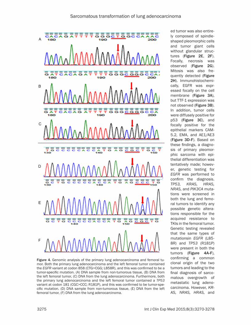
ed tumor was also entire-ly composed of spindle-shaped pleomorphic cells and tumor giant cells without glandular struc-tures (Figure 2E, 2F). Focally, necrosis was observed (Figure 2G). Mitosis was also fre-quently detected (Figure 2H). Immunohistochemi- cally, EGFR was expr- essed focally on the cell membrane (Figure 3A), but TTF-1 expression was not observed (Figure 3B). In addition, tumor cells were diffusely positive for p53 (Figure 3C), and focally positive for the epithelial markers CAM- 5.2, EMA, and AE1/AE3 (Figure 3D-F). Based on these findings, a diagno-sis of primary pleomor-phic sarcoma with epi-thelial differentiation was tentatively made; howev-er, genetic testing for EGFR was performed to confirm the diagnosis. TP53, KRAS, HRAS, NRAS, and PIK3CA muta-tions were screened in both the lung and femo-ral tumors to identify any possible genetic altera-tions responsible for the acquired resistance to TKIs in the femoral tumor. Genetic testing revealed that the same types of mutationsin EGFR (L85- 8R) and TP53 (R181P) were present in both the tumors (Figure 4A-F), confirming a common clonal origin of the two tumors and leading to the final diagnosis of sarco-matous overgrowth of metastatic lung adeno-carcinoma. However, KR- AS, NRAS, HRAS, and
Figure 4. Genomic analysis of the primary lung adenocarcinoma and femoral tu-mor. Both the primary lung adenocarcinoma and the left femoral tumor contained the EGFR variant at codon 858 (CTG>CGG; L858R), and this was confirmed to be a tumor-specific mutation. (A) DNA sample from non-tumorous tissue, (B) DNA from the left femoral tumor, (C) DNA from the lung adenocarcinoma. Furthermore, both the primary lung adenocarcinoma and the left femoral tumor contained a TP53 variant at codon 181 (CGC>CCC; R181P), and this was confirmed to be tumor-spe-cific mutation. (D) DNA sample from non-tumorous tissue, (E) DNA from the left femoral tumor, (F) DNA from the lung adenocarcinoma.

Sarcomatous transformation of lung adenocarcinoma
3276 Int J Clin Exp Med 2015;8(3):3270-3278
PIK3CA mutations were not detected. The patient remains alive and walks with the assis-tance of crutches; the remaining lesions are well controlled.
Discussion
When the patient was first admitted, two pos-sibilities were considered regarding the femoral tumor. One was that it was a metastasis from the primary lung adenocarcinoma. A primary bone tumor was the other differential diagnosis because it was puzzling that only the femoral tumor grew progressively among the multiple metastatic lesions that were being identically treated. Biopsy from the proximal femur revealed that the tumor was entirely composed of a proliferation of sarcomatous pleomorphic cells without glandular structure, thus support-ing the diagnosis of a primary bone tumor. Furthermore, histological examination of the surgical specimen showed features consistent with a diagnosis of primary pleomorphic sarco-ma of the proximal femur. However, genetic testing of EGFR and TP53 was performed for further confirmation, and this analysis led us to revise the diagnosis to that of a metastasis due to identical genetic patterns in both the lung and femoral tumors. TTF-1 is used as a sensi-tive marker for lung adenocarcinoma, although loss of its expression has been reported to cor-relate with increased tumor aggressiveness [13]. In this case, TTF-1 expression was absent in the metastatic tumor despite its strong expression in the lung biopsy specimen. Hence, this patient illustrates the importance of genet-ic testing for resolving ambiguous cases.
As the patient had received radiation therapy (24 Gy) for metastases in the lumbar vertebrae and left femur 1 year previously, radiation-induced sarcoma or dedifferentiation of meta-static lung adenocarcinoma could also explain the femoral tumor. Several studies describe that radiation-related sarcoma commonly occurs after radiation therapy, although the interval between irradiation and detection of the second malignancy is at least 3-5 years [14-18]. Thus, the possibility of the radiation-related sarcoma was disregarded. Nakanishi et al. reported that TP53 mutations were one of the causative factors of radiation-induced sar-coma [18]. In the present case, the TP53 muta-tion R181P was detected in both the lung biop-
sy specimen and the left femoral tumor; thus, this alteration in TP53 was deemed not related to the radiation.
Various studies show that almost all EGFR-mutation-positive NSCLCs acquire resistance to EGFR-TKIs despite remarkably good respons-es initially. Recently, several mechanisms of acquired resistance to EGFR-TKIs in NSCLC have been described, the most common of which is a secondary EGFR T790M mutation [4, 5]. Other molecular mechanisms of resistance include upregulation of HGF/MET, HER2 muta-tions, HER3 overexpression, persistent activa-tion of IGF-1R, mutations of PIK3CA/AKT, loss or downregulation of PTEN, and abnormal dimerization of STAT3 [5-8]. Some secondary genetic alterations or histological changes occurred in all 11 cases of lung adenocarcino-ma with acquired resistance to EGFR-TKIs in a study by Uramoto et al. [10]. Sequist et al. described that histological change were observed in 8 of 37 cases with drug-resistant NSCLCs carrying EGFR mutations, and EMT was described as the cause of drug resistance in 3 of these 8 cases [11]. The molecular mech-anisms and the associated mesenchymal phe-notypes underlying drug resistance to TKIs remain unknown, although it has been shown that cell lines undergoing EMT are intrinsically resistant to EGFR inhibitors [19-21]. In our case, secondary genetic alterations such as T790M in EGFR, or additional mutations in Kras, Nras, Hras, and PIK3CA, were not found. The femoral tumor in this case was entirely composed of pleomorphic cells and tumor giant cells with bizarre nuclei, and no apparent glan-dular structures were observed. It is not clear whether the lung tumor was pure adenocarci-noma or pleomorphic carcinoma from the small biopsy specimen. According to the WHO classi-fication, a carcinoma is defined as pleomorphic if the sarcomatous component is greater than 10% [22, 23]. EGFR mutation was reported to occur in 15-20% of pleomorphic carcinomas of the lungs, and EGFR-TKIs such as gefitinib and erlotinib have high efficiency against EGFR-mutated tumors including pleomorphic carci-noma [24-26]. Furthermore, metastases often arise from poorly differentiated components of tumors with pleomorphic features. However, the fact that the primary tumor as well as the remaining metastatic lesions remained stable due to TKI treatment suggests that the lung
Sarcomatous transformation of lung adenocarcinoma
3277 Int J Clin Exp Med 2015;8(3):3270-3278
adenocarcinoma was a pure adenocarcinoma while EMT occurred only in the proximal femo-ral tumor. Thus, EMT could be the cause of acquired drug resistance to TKI in this case.
In conclusion, we investigated a metastatic adenocarcinoma of the lungs on the left proxi-mal femur, masquerading as a primary pleo-morphic sarcoma. Our results show that genet-ic testing is highly recommended in such cases when the histologic features of suspected metastases are markedly different from the pri-mary lesion.
After acceptance of the manuscript, a new lesion of the right lung was noticed by the chest-CT and it has been gradually enlarged. The biopsy from this lesion revealed sarcoma-tous feature without apparent glandular struc-tures. Because the original lesion of the right lung remained stable, the newly established lesion was considered to be metastasized from the femoral lesion.
Acknowledgements
This work was supported in part by a Grant-in-Aid for General Scientific Research from the Ministry of Education, Culture, Sports, Science and Technology (#26670286 to Tsuyoshi Saito and #25861342 to Yoshiyuki Suehara), Tokyo, Japan.
Disclosure of conflict of interest
None.
Address correspondence to: Dr. Tsuyoshi Saito, De-partment of Human Pathology, Juntendo University School of Medicine, Hongo 2-1-1, Bunkyo-ku, Tokyo 113-8421, Japan. Tel: +81-3-3813-3111; Fax: +81-3-3813-3428; E-mail: [email protected]
References
[1] Mok TS, Wu YL, Thongprasert S, Yang CH, Chu DT, Saijo N, Sunpaweravong P, Han B, Margono B, Ichinose Y, Nishiwaki Y, Ohe Y, Yang JJ, Chewaskulyong B, Jiang H, Duffield EL, Wat-kins CL, Armour AA, Fukuoka M. Gefitinib or carboplatin-paclitaxel in pulmonary adenocar-cinoma. N Engl J Med 2009; 361: 947-957.
[2] Mitsudomi T, Morita S, Yatabe Y, Negoro S, Okamoto I, Tsurutani J, Seto T, Satouchi M, Tada H, Hirashima T, Asami K, Katakami N, Takada M, Yoshioka H, Shibata K, Kudoh S, Shimizu E, Saito H, Toyooka S, Nakagawa K,
Fukuoka M. Gefitinib versus cisplatin plus docetaxel in patients with non-small-cell lung cancer harbouring mutations of the epidermal growth factor receptor (WJTOG3405): an open label, randomised phase 3 trial. Lancet Oncol 2010; 11: 121-128.
[3] Maemondo M, Inoue A, Kobayashi K, Suga-wara S, Oizumi S, Isobe H, Gemma A, Harada M, Yoshizawa H, Kinoshita I, Fujita Y, Okinaga S, Hirano H, Yoshimori K, Harada T, Ogura T, Ando M, Miyazawa H, Tanaka T, Saijo Y, Hagi-wara K, Morita S, Nukiwa T. North-East Japan Study Group Gefitinib or chemotherapy for non-small-cell lung cancer with mutated EGFR. N Engl J Med 2010; 362: 2380-2388.
[4] Kobayashi S, Boggon TJ, Dayaram T, Jänne PA, Kocher O, Meyerson M, Johnson BE. EGFR mu-tation and resistance of non-small-cell lung cancer to gefitinib. N Engl 2005; 352: 786-792.
[5] Lin Y, Wang X, Jin H. EGFR-TKI resistance in NSCLC patients: mechanisms and strategies. Am J Cancer 2014; 4: 411-435
[6] Engelman JA, Zejnullahu K, Mitsudomi T, Song Y, Hyland C, Park JO, Lindeman N, Gale C M, Zhao X, Christensen J, Kosaka T, Holmes AJ, Rogers AM, Cappuzzo F, Mok T, Lee C, John-son, Cantley LC, Jänne PA. MET amplification leads to gefitinib resistance in lung cancer by activating ERBB3 signaling. Science 2007; 316: 1039-1043.
[7] Yano S, Wang W, Li Q, Matsumoto K, Sakura-ma H, Nakamura T, Ogino H, Kakiuchi S, Hani-buchi M, NIshioka Y, Uehara H, Mitsudomi T, Yatabe Y, Nakamura T, Sone S. Hepatocyte growth factor induces gefitinib resistance of lung adenocarcinoma with epidermal growth factor receptor-activating mutations. Cancer Res 2008; 68: 9479-9487.
[8] Yamamoto C, Basaki Y, Kawahara A, Nakashi-ma K, Kage M, Izumi H, Kohno K, Uramoto H, Yasumoto K, Kuwano M, Ono M. Loss of PTEN expression by blocking nuclear translocation of EGR1 in gefitinib-resistant lung cancer cells harboring epidermal growth factor receptor-activating mutations. Cancer Res 2010; 70: 8715-8725.
[9] Suda K, Tomizawa K, Fujii M, Murakami H, Osada H, Maehara Y, Yatabe Y, Sekido Y, Mitsu-domi T. Epithelial to mesenchymal transition in an epidermal growth factor receptor-mutant lung cancer cell line with acquired resistance to erlotinib. J Thorac Oncol 2011; 6: 1152-1161.
[10] Uramoto H, Shimokawa H, Hanagiri T, Kuwano M, Ono M. Expression of selected gene for ac-quired drug resistance to EGFR-TKI in lung ade-nocarcinoma. Lung Cancer 2011; 73: 361-365.

Sarcomatous transformation of lung adenocarcinoma
3278 Int J Clin Exp Med 2015;8(3):3270-3278
[11] Sequist LV, Waltman BA, Dias-Santagata D, Digumarthy S, Turke AB, Fidias P, Bergethon K, Shaw AT, Gettinger S, Cosper AK, Akhavanfard S, Heist RS, Temel J, Christensen JG, Wain JC, Lynch TJ, Vernovsky K, Mark EJ, Lanuti M, Iafrate AJ, Mino-Kenudson M, Engelman JA. Genotypic and histological evolution of lung cancers acquiring resistance to EGFR inhibi-tors. Sci Transl Med 2011; 3: 75-26.
[12] Watanabe S, Sone T, Matsui T, Yamamura K, Tani M, Okazaki A, Kurokawa K, Tambo Y, Taka-to H, Ohkura N, Waseda Y, Katayama N, Kasa-hara K. Transformation to small-cell lung can-cer following treatment with EGFR tyrosine kinase inhibitors in a patient with lung adeno-carcinoma. Lung Cancer 2013; 82: 370-372.
[13] Barletta JA, Perner S, Iafrate AJ, Yeap BY, Weir BA, Johnson LA, Johnson BE, Meyerson M, Ru-bin MA, Travis WD, Loda M, Chirieac LR. Clini-cal significance of TTF-1 protein expression and TTF-1 gene amplification in lung adenocar-cinoma. J Cell Mol Med 2009; 13: 1977-1986.
[14] Patel SR. Radiation-induced sarcoma. Curr Treat Options Oncol 2000; 1: 258-261.
[15] Sabanas AO, Dahlin DC, Childs DS Jr, Ivins JC. Postradiation sarcoma of bone. Cancer 1956; 9: 528-542.
[16] Cruz M, Coley BL, Stewart FW. Postradiation bone sarcoma; report of eleven cases. Cancer 1957; 10: 72-88.
[17] Arlen M, Higinbotham NL, Huvos AG, Marcove RC, Miller T, Shah IC. Radiation-induced sarco-ma of bone. Cancer 1971; 28: 1087-1099.
[18] Nakanishi H, Tomita Y, Myoui A, Yoshikawa H, Sakai K, Kato Y, Ochi T, Aozasa K. Mutation of the p53 gene in postradiation sarcoma. Lab Invest 1998; 78: 727-733.
[19] Thomson S, Buck E, Petti F, Griffin G, Brown E, Ramnarine N, Iwata KK, Gibson N, Haley JD. Epithelial to mesenchymal transition is a deter-minant of sensitivity of non-small-cell lung car-cinoma cell lines and xenografts to epidermal growth factor receptor inhibition. Cancer Res 2005; 65: 9455-9462.
[20] Frederick BA, Helfrich BA, Coldren CD, Zheng D, Chan D, Bunn PA, Jr, Raben D. Epithelial to mesenchymal transition predicts gefitinib re-sistance in cell lines of head and neck squa-mous cell carcinoma and non-small cell lung carcinoma. Mol Cancer Ther 2007; 6: 1683-1691.
[21] Fuchs BC, Fujii T, Dorfman JD, Goodwin JM, Zhu AX, Lanuti M, Tanabe KK. Epithelial-to-mesenchymal transition and integrin-linked ki-nase mediate sensitivity to epidermal growth factor receptor inhibition in human hepatoma cells. Cancer Res 2008; 68: 2391-2399.
[22] WHO Classification of Tumours Pathology and Genetics of Tumours of the Lung, Thymus and Heart. In: Travis WD, Brambilla E, Muller-Her-melink HK, Harris CC, editors. World Health Organization International Agency for Re-search on Cancer. IARC Press; 2004. pp. 53-62.
[23] Travis WD. Sarcomatoid neoplasms of the lung and pleura. Arch Pathol Lab Med 2010; 134: 1645-1658.
[24] Kaira K, Horie Y, Ayabe E, Murakami H, Taka-hashi T, Tsuya A, Nakamura Y, Naito T, Endo M, Kondo H, Nakajima T, Yamamoto N. Pulmonary pleomorphic carcinoma: a clinicopathological study including EGFR mutation analysis. J Tho-rac Oncol 2010; 5: 460-465.
[25] Lee S, Kim Y, Sun JM, Choi YL, Kim JG, Shim YM, Park YH, Ahn JS, Park K, Han JH, Ahn MJ. Molecular profiles of EGFR, K-ras, c-met, and FGFR in pulmonary pleomorphic carcinoma, a rare lung malignancy. J Cancer Res Clin Oncol 2011; 137: 1203-1211.
[26] Chang YL, Wu CT, Shih JY, Lee YC. EGFR and p53 status of pulmonary pleomorphic carcino-ma: implications for EGFR tyrosine kinase in-hibitors therapy of an aggressive lung malig-nancy. Ann Surg Oncol 2011; 18: 2952-2960.


![New Trends In Internal Medicine2009hocc.medicine.psu.ac.th/files/acadamic/New_Trends... · cytopenia EGF-R profile EGFR FISH EGFR FISH docetaxel gefitinib ñu EGFR FISH Lf-]utnn EGF-R](https://img.dokumen.tips/doc/110x75/60098f15be7b15544f1b652e/new-trends-in-internal-cytopenia-egf-r-profile-egfr-fish-egfr-fish-docetaxel-gefitinib.jpg)