Embed Size (px)
Citation preview
Case ReportMounier-Kuhn Syndrome in an Elderly Female withPulmonary Fibrosis
Panagiotis Boglou,1 Nikolaos Papanas,2 Anastasia Oikonomou,3
Stamatia Bakali,4 and Paschalis Steiropoulos1
1Department of Pneumonology, Medical School, Democritus University of Thrace, 68100 Alexandroupolis, Greece2Second Department of Internal Medicine, Medical School, Democritus University of Thrace, 68100 Alexandroupolis, Greece3Department of Radiology, Medical School, Democritus University of Thrace, 68100 Alexandroupolis, Greece4Department of Microbiology, Medical School, Democritus University of Thrace, 68100 Alexandroupolis, Greece
Correspondence should be addressed to Paschalis Steiropoulos; [email protected]
Received 9 May 2016; Accepted 20 July 2016
Academic Editor: Stephen P. Peters
Copyright © 2016 Panagiotis Boglou et al. This is an open access article distributed under the Creative Commons AttributionLicense, which permits unrestricted use, distribution, and reproduction in any medium, provided the original work is properlycited.
Mounier-Kuhn syndrome (MKS), or tracheobronchomegaly, is a rare clinical and radiologic condition characterized by pronouncedtracheobronchial dilation and recurrent lower respiratory tract infections. Tracheobronchomegaly presents when the defect extendsto the central bronchi. MKS can be diagnosed in adult women when the transverse and sagittal diameters of the trachea, rightmainstem bronchus, and left mainstem bronchus exceed 21, 23, 19.8, and 17.4mm, respectively. Its diagnosis is based on chestradiograph and chest computed tomography (CT). Patients, usuallymiddle-agedmen,may be asymptomatic or presentwith clinicalmanifestations ranging from minimal symptoms with preserved lung function to severe respiratory failure. Pulmonary functiontests (PFTs) typically reveal a restrictive pattern. This report presents an elderly woman with previously diagnosed pulmonaryfibrosis with symptoms of increased sputum production and haemoptysis. High-resolution chest CT showed tracheal and mainstem bronchi dilatation along with bronchial diverticulosis. PFTs indicated a restrictive pattern characteristic of the underlyingpulmonary fibrosis. The patient is the oldest, referred to the female gender, at presentation of MKS hitherto reported. This casehighlights the need to includeMKS in the differential diagnosis of recurrent lower respiratory tract infections, even in older subjects.
1. Introduction
Mounier-Kuhn syndrome (MKS) is an infrequent congen-ital syndrome, whose hallmark is airway enlargement. Onhistological examination, absence or atrophy of the elasticfibres within the tracheal wall is typically found [1–7]. Thiscondition results in airway dilatation in the trachea andbronchi. Tracheal diverticula may also occur, mainly in theposterior trachea [8]. The typical features of this entity werefirst described by Mounier-Kuhn in 1932 [9].
Patients usually complain of recurrent respiratory tractinfections along with various functional perturbations: fromminimal involvement with preservation of lung function,through severe disease with bronchiectasis to overt respira-tory failure [1–7, 10]. Patients are typically middle-agedmales[2–4]. Its diagnosis is based on chest radiograph and chest
computed tomography (CT). MKS is frequently overlooked,but should be considered in the investigation of recurrentlower respiratory tract infections [1–9].
This report presents a female patient, the oldest hithertodescribed, with MKS who had been previously diagnosedwith pulmonary fibrosis.
2. Case Presentation
An 83-year-old woman was referred to the pneumonologydepartment of our hospital due to increasing productivecough and haemoptysis. During the last 30 years, she hadexperienced dyspnoea on exertion and increased expec-toration of mucoid sputum that became purulent duringinfectious exacerbations, often with bloody streaked sputum.She denied fever, wheezes, chest pain, and weight loss, or any
Hindawi Publishing CorporationCase Reports in MedicineVolume 2016, Article ID 8708251, 4 pageshttp://dx.doi.org/10.1155/2016/8708251
2 Case Reports in Medicine
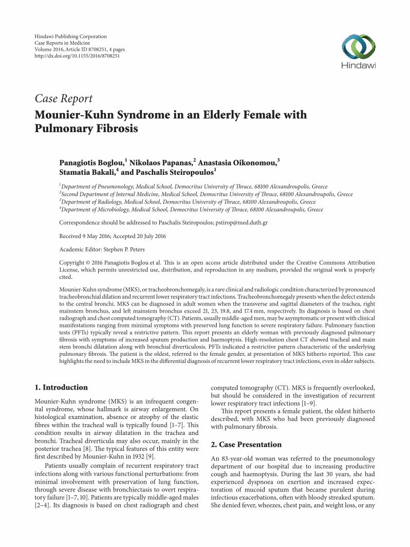
Figure 1: Chest X-ray conducted 24 years ago, displaying a tracheaenlargement without any signs of fibrosis.
other symptom indicative of gastroesophageal reflux disease(GERD).
Her past medical history included pulmonary fibrosisdiagnosed 6 years before in another country; left ovariancancer treated with hysterosalpingo-oophorectomy 20 yearsbefore; arterial hypertension; diabetes mellitus and osteo-porosis.Therewas no family history of any respiratory diseaseand she had never smoked. No history of GERDwas noted inher medical records.
On physical examination, her vital signs were normal.Finger clubbing was not present. Mild inspiratory crackles atthe lower third of both lung fields were revealed. Laboratoryinvestigations were as follows: erythrocyte sedimentationrate: 15mm/h; C-reactive protein: 1.03mg/L with oxygensaturation at 96% on room air and arterial blood gasesanalysis with PaO
2: 77.3mmHg, PCO
2: 41.5mmHg, and PH:
7.45 (FiO2: 21%). Immunologic tests (rheumatoid factor, anti-
CCP (anticyclic citrullinated peptide), C3, C4, p-ANCA,
and c-ANCA) were within normal range while antinuclearantibody (ANA) levels were mildly raised (1/160) but therewas no characteristic immunofluorescence pattern.
Pulmonary function tests (PFTs), conducted 1 monthbefore her admission, revealed a forced expiratory volumein 1 sec (FEV
1) of 1.83 L (58% predicted), a forced vital
capacity (FVC) of 1.95 L (55% predicted), and FEV1/FVC
of 105%. We were not able to perform static lung volumesmeasurement and diffusion test, due to the patient’s inabilityto cooperate. Sputum results were negative for mycobacteria.Bronchoscopy was not performed because the patient did notconsent.
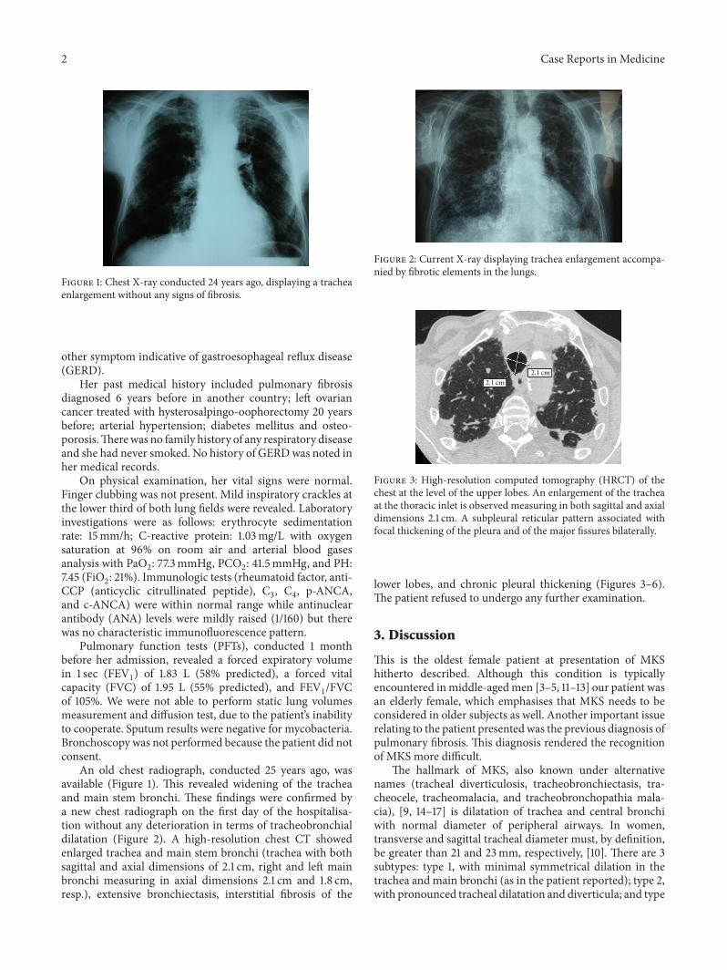
An old chest radiograph, conducted 25 years ago, wasavailable (Figure 1). This revealed widening of the tracheaand main stem bronchi. These findings were confirmed bya new chest radiograph on the first day of the hospitalisa-tion without any deterioration in terms of tracheobronchialdilatation (Figure 2). A high-resolution chest CT showedenlarged trachea and main stem bronchi (trachea with bothsagittal and axial dimensions of 2.1 cm, right and left mainbronchi measuring in axial dimensions 2.1 cm and 1.8 cm,resp.), extensive bronchiectasis, interstitial fibrosis of the
Figure 2: Current X-ray displaying trachea enlargement accompa-nied by fibrotic elements in the lungs.
2.1 cm2.1 cm
Figure 3: High-resolution computed tomography (HRCT) of thechest at the level of the upper lobes. An enlargement of the tracheaat the thoracic inlet is observed measuring in both sagittal and axialdimensions 2.1 cm. A subpleural reticular pattern associated withfocal thickening of the pleura and of the major fissures bilaterally.
lower lobes, and chronic pleural thickening (Figures 3–6).The patient refused to undergo any further examination.
3. Discussion
This is the oldest female patient at presentation of MKShitherto described. Although this condition is typicallyencountered inmiddle-agedmen [3–5, 11–13] our patient wasan elderly female, which emphasises that MKS needs to beconsidered in older subjects as well. Another important issuerelating to the patient presented was the previous diagnosis ofpulmonary fibrosis. This diagnosis rendered the recognitionof MKS more difficult.
The hallmark of MKS, also known under alternativenames (tracheal diverticulosis, tracheobronchiectasis, tra-cheocele, tracheomalacia, and tracheobronchopathia mala-cia), [9, 14–17] is dilatation of trachea and central bronchiwith normal diameter of peripheral airways. In women,transverse and sagittal tracheal diameter must, by definition,be greater than 21 and 23mm, respectively, [10]. There are 3subtypes: type 1, with minimal symmetrical dilation in thetrachea and main bronchi (as in the patient reported); type 2,with pronounced tracheal dilatation and diverticula; and type

Case Reports in Medicine 3
2.1 cm
1.8 cm
Figure 4: HRCT of the chest at the level of the carina. An enlarge-ment of the right and left main bronchi is observed measuringin axial dimensions 2.1 cm and 1.8 cm, respectively. A subpleuralreticular pattern associated with focal thickening of the pleura andtraction bronchiectasis and bronchiolectasis is also noted.
1.6 cm2.1 cm
Figure 5: HRCT of the chest at level below carina shows dilatedright and left main bronchi and undulating wall of left mainbronchus, indicating bronchial diverticulosis. A subpleural reticularpattern associated with focal thickening of the pleura and tractionbronchiectasis and bronchiolectasis is also noted.
3, whereby marked tracheal and bronchial dilatation extendsfurther until the distal bronchi bilaterally [18].
Aetiology of MKS is unclear. There may be a primarydefect or atrophy of elastic and smoothmuscle tissue [19, 20].MKS may also present in association with miscellaneousconditions, for example, Ehlers-Danlos syndrome, Marfansyndrome, connective tissue diseases, ataxia telangiectasia,and ankylosing spondylitis [21–24]. Diseases resulting insevere upper lobe fibrosis, such as sarcoidosis, cystic fibrosis,or diffuse pulmonary fibrosis, and airway inflammatoryconditions, notably allergic bronchopulmonary aspergillosis,are also implicated in its pathogenesis [21–24]. Pulmonaryfibrosis of the patient was considered idiopathic, due to thefollowing reasons. First, serological findings were negative.Additionally any occupational exposure was excluded.
There are no pathognomonic symptoms present in MKS.Patients are usually asymptomatic while excessive sputumproductionmay occur secondary to bronchiectasis and lower
Figure 6: HRCT of the chest at the level of the lower lobes. Anextended and coarse subpleural reticular pattern is noted associatedwith traction bronchiectasis and bronchiolectasis as well as withhoneycombing.
respiratory tract infection [18]. Occasional haemoptysis anddyspnoea may be seen as well [22]. In our case, the patientpresented with productive cough and haemoptysis.
Its diagnosis rests on imaging studies. In the rare case ofgross tracheal enlargement, chest X-rays may be diagnostic.More frequently, however, chest CT is required to reliablyascertain tracheal dimensions and to investigate any devel-opment of complications, for example, bronchiectasis [25]. Atthe same time, PFTs reveal a restrictive pattern.
The effect of enlarged airways on spirometry derives fromthe weakness of the tracheobronchial walls and hypotoniain the myoelastic elements, resulting in dynamic airwaycompression (expiratory collapse during forced exhalation)and dynamic restriction.The restrictive pattern in our patientis magnified from the underlying fibrosis and possibly bythe associated retention of secretions. Nevertheless, thesefindings are not always met in MKS, because cases withnormal spirometric values have been also reported [5].Diffusion test could have been helpful; however this was notperformed, due to the inability of the patient to cooperate.
Asymptomatic patients require no treatment. Therapyincludes respiratory physiotherapy and antibiotics duringinfectious exacerbations, [26–32], while tracheal stenting isvery rarely employed [2, 33].
In conclusion, this case highlights that a chest CT scanshould be performed in patients reporting chronic recurrentlower respiratory tract infections to investigate underlyingconditions, including MKS. Indeed, this condition, despitelong-term follow-up for repeated lower respiratory infectionsand chronic cough, had long been left undiagnosed in ourpatient, until a high-resolution chest CT was performed. Ourcase is the oldest female patient described in literature withMKS, indicating that appropriate diagnostic workup may berequired in elderly subjects as well.
Competing Interests
The authors declare that there are no competing interests.

4 Case Reports in Medicine
Authors’ Contributions
Paschalis Steiropoulos conceived and wrote the paper andacquired data; Panagiotis Boglou provided material supportand reviewed literature; AnastasiaOikonomou acquired data;Stamatia Bakali acquired data and providedmaterial support;Nikolaos Papanas made critical revisions of the paper.
References
[1] G. L. Adani, J. Baccarani, D. Lorenzin et al., “Renal trans-plantation in a patient affected by Mounier-Kuhn syndrome,”Transplantation Proceedings, vol. 37, no. 10, pp. 4215–4217, 2005.
[2] C. O. Randak and M. Weinberger, “A child with progressivemultiple tracheal diverticulae: a variation of theMounier-Kuhnsyndrome,” Pediatric Pulmonology, vol. 48, no. 8, pp. 841–843,2013.
[3] D. D. Odell, A. Shah, S. P. Gangadharan et al., “Airway stentingand tracheobronchoplasty improve respiratory symptoms inMounier-Kuhn syndrome,” Chest, vol. 140, no. 4, pp. 867–873,2011.
[4] D. S. Ushakumari, N. Grewal, and M. Green, “Mounier-Kuhnsyndrome: anesthetic experience,” Case Reports in Anesthesiol-ogy, vol. 2012, Article ID 674918, 2 pages, 2012.
[5] E. Krustins, Z. Kravale, and A. Buls, “Mounier-Kuhn syndromeor congenital tracheobronchomegaly: a literature review,” Res-piratory Medicine, vol. 107, no. 12, pp. 1822–1828, 2013.
[6] G. Enriquez, L. Cadavid, E. Garces-Inigo et al., “Tracheo-bronchomegaly following intrauterine tracheal occlusion forcongenital diaphragmatic hernia,” Pediatric Radiology, vol. 42,no. 8, pp. 916–922, 2012.
[7] J. B. Ng and E. A. Bittner, “Tracheobronchomegaly: a rare causeof endotracheal tube cuff leak,” Anesthesiology, vol. 114, no. 5,article 1211, 2011.
[8] M.R.Himalstein and J. C.Gallagher, “Tracheobronchiomegaly,”Annals of Otology, Rhinology & Laryngology, vol. 82, no. 2, pp.223–227, 1973.
[9] P. Mounier-Kuhn, “Dilatation de la trachee: constatations,radiographiques et bronchoscopies,” Lyon Medical, vol. 150, pp.106–109, 1932.
[10] R. S. Fraser, P. D. Pare, N. L. Muller, and N. Colman,“Bronchiectasis and other bronchial abnormalities,” in Diagno-sis of Diseases of Chest, pp. 2285–2287,W.B. Saunders Company,Philadelphia, Pa, USA, 1999.
[11] A. K. Jaiswal, S. Munjal, R. Singla, V. Jain, and D. Behera, “A 46-year-old man with tracheomegaly, tracheal diverticulosis, andbronchiectasis: Mounier-Kuhn syndrome,” Lung India, vol. 29,no. 2, pp. 176–178, 2012.
[12] M. Arroyo-Cozar, M. Ruiz-Garcıa, E. M. Merlos, D. Vielba,and E. Macıas, “Case report: respiratory infection due to alcali-genes xylosoxidans in a patient with mounier-kuhn syndrome,”Revista Chilena de Infectologia, vol. 29, no. 5, pp. 570–571, 2012.
[13] A. D. L. Bastos and I. L. A. Brito, “Mounier-kuhn syndrome:radiological findings and clinical presentation,” RadiologiaBrasileira, vol. 44, no. 3, pp. 198–2000, 2011.
[14] E. R. Czyhlarz, “Uber Pulsionsdivertikel der Trachea mitBemerkungen uber das Verhalten der elastischen Fasern annormalen Tracheen undBronchien,”Zentralblatt fur AllgemeinePathologie und Pathologische Anatomie, vol. 18, pp. 721–728,1897.
[15] E. M. Bateson and M. Woo-Ming, “Tracheo-bronchomegaly,”Clinical Radiology, vol. 24, no. 3, pp. 354–358, 1973.
[16] S. Gay and P. Dee, “Tracheobronchomegaly-the Mounier-Kuhnsyndrome,”British Journal of Radiology, vol. 57, no. 679, pp. 640–644, 1984.
[17] W. A. Engle, M. D. Cohen, W. H. McAlister, and N. T.Griscom, “Neonatal tracheobronchomegaly,” American Journalof Perinatology, vol. 4, no. 2, pp. 81–85, 1987.
[18] M. Schwartz and L. Rossoff, “Tracheobronchomegaly,” Chest,vol. 106, no. 5, pp. 1589–1590, 1994.
[19] I. Katz, M. Levine, and P. Hermam, “Tracheobronchomegaly(Mounier-Kuhn Syndrome): CT diagnosis,” American Journalof Roentgenology, vol. 88, pp. 1084–1094, 1962.
[20] H. Spencer, “Congenital abnormalities of the lung: congenitaltracheobronchomegaly,” in Pathology of the Lung, H. Spencer,Ed., pp. 129–130, PergamonPress,Oxford,UK, 4th edition, 1985.
[21] M. A. Blake, P. D. Clarke, and H. M. Fenlon, “Thoracic caseof the day:Mounier-Kuhn syndrome (tracheobronchomegaly),”American Journal of Roentgenology, vol. 173, no. 3, pp. 822–825,1999.
[22] J. Van Schoor, G. Joos, and R. Pauwels, “Tracheobronchome-galy: the Mounier-Kuhn syndrome: report of two cases andreview of the literature,” European Respiratory Journal, vol. 4,no. 10, pp. 1303–1306, 1991.
[23] A. C. Sane, E. L. Effmann, and S. D. Brown, “TheMounier-Kuhnsyndrome in a patient with the Kenny-Caffey syndrome,”Chest,vol. 102, no. 2, pp. 618–619, 1992.
[24] J. H. Woodring, P. A. Barrett, S. R. Rehm, and P. Nurenberg,“Acquired tracheomegaly in adults as a complication of diffusepulmonary fibrosis,” American Journal of Roentgenology, vol.152, no. 4, pp. 743–747, 1989.
[25] P. H. Lakshminarayana and M. E. Woodske, “Mounier-Kuhnsyndrome: imaging in recurrent pulmonary infections,” Amer-ican Journal of Respiratory and Critical Care Medicine, vol. 185,no. 2, p. 225, 2012.
[26] F. Noori, S. Abduljawad, D. M. Suffin et al., “Mounier-Kuhnsyndrome: a case report,” Lung, vol. 188, no. 4, pp. 353–354, 2010.
[27] J. J. Ip, P. K. Hui, S. H. Lam, W. W. Lam, and M. T. Chau,“Mounier-Kuhn syndrome: an unusual underlying cause forchronic coughs and recurrent pneumonias,”Hong KongMedicalJournal, vol. 19, no. 4, pp. 365.e3–365.e4, 2013.
[28] F. A. Khasawneh and A. J. Jou-Tindo, “A 30-year-old womanwith recurrent lower respiratory tract infections,”Chest, vol. 143,no. 5, pp. 1500–1503, 2013.
[29] M. C. Pacheco, J. N. Sancho-Chust, and E. Chiner, “Mounier-Kuhn syndrome diagnosed in an adult,”Archivos de Bronconeu-mologia, vol. 46, no. 12, pp. 665–666, 2010.
[30] L. Dalar, E. Eryuksel, F. Kosar et al., “Central airway obstructiondue to malignant fibrous histiocytoma metastasis in a case withMounier-Kuhn syndrome,” Tuberkuloz ve Toraks, vol. 60, no. 2,pp. 167–171, 2012.
[31] B. D. Kent, I. Sulaiman, N. B. Akasheh, P. Nadarajan, E.Moloney, and S. J. Lane, “An unusual cause of spontaneouspneumothorax: the Mounier-Kuhn syndrome,” Irish MedicalJournal, vol. 104, no. 5, pp. 152–153, 2011.
[32] O. D. Lyons and J. J. Gilmartin, “A grossly abnormal trachea-severe tracheal diverticulosis and Mounier-Kuhn syndrome,”Irish Medical Journal, vol. 107, no. 3, pp. 82–83, 2014.
[33] H. Dutau, F. Maldonado, D. P. Breen, and A. Colchen, “Endo-scopic successful management of tracheobronchomalacia withlaser: apropos of aMounier-Kuhn syndrome,” European Journalof Cardio-Thoracic Surgery, vol. 39, no. 6, pp. e186–e188, 2011.

Submit your manuscripts athttp://www.hindawi.com
Stem CellsInternational
Hindawi Publishing Corporationhttp://www.hindawi.com Volume 2014
Hindawi Publishing Corporationhttp://www.hindawi.com Volume 2014
MEDIATORSINFLAMMATION
of
Hindawi Publishing Corporationhttp://www.hindawi.com Volume 2014
Behavioural Neurology
EndocrinologyInternational Journal of
Hindawi Publishing Corporationhttp://www.hindawi.com Volume 2014
Hindawi Publishing Corporationhttp://www.hindawi.com Volume 2014
Disease Markers
Hindawi Publishing Corporationhttp://www.hindawi.com Volume 2014
BioMed Research International
OncologyJournal of
Hindawi Publishing Corporationhttp://www.hindawi.com Volume 2014
Hindawi Publishing Corporationhttp://www.hindawi.com Volume 2014
Oxidative Medicine and Cellular Longevity
Hindawi Publishing Corporationhttp://www.hindawi.com Volume 2014
PPAR Research
The Scientific World JournalHindawi Publishing Corporation http://www.hindawi.com Volume 2014
Immunology ResearchHindawi Publishing Corporationhttp://www.hindawi.com Volume 2014
Journal of
ObesityJournal of
Hindawi Publishing Corporationhttp://www.hindawi.com Volume 2014
Hindawi Publishing Corporationhttp://www.hindawi.com Volume 2014
Computational and Mathematical Methods in Medicine
OphthalmologyJournal of
Hindawi Publishing Corporationhttp://www.hindawi.com Volume 2014
Diabetes ResearchJournal of
Hindawi Publishing Corporationhttp://www.hindawi.com Volume 2014
Hindawi Publishing Corporationhttp://www.hindawi.com Volume 2014
Research and TreatmentAIDS
Hindawi Publishing Corporationhttp://www.hindawi.com Volume 2014
Gastroenterology Research and Practice
Hindawi Publishing Corporationhttp://www.hindawi.com Volume 2014
Parkinson’s Disease
Evidence-Based Complementary and Alternative Medicine
Volume 2014Hindawi Publishing Corporationhttp://www.hindawi.com