Embed Size (px)
Citation preview
Case ReportIsolated Splenic Vein Thrombosis: 8-Year-Old Boy with MassiveUpper Gastrointestinal Bleeding and Hypersplenism
Mohammad Ali Kiani,1 Arash Forouzan,2 Kambiz Masoumi,2 Behnaz Mazdaee,2
Mohammad Bahadoram,3 Hamid Reza Kianifar,1 and Hassan Ravari4
1Pediatric Gastroenterology Ward, Department of Pediatrics, Mashhad University of Medical Sciences, Mashhad 9177948564, Iran2Department of Emergency Medicine, Imam Khomeini General Hospital, Ahvaz Jundishapur University of Medical Sciences,Ahvaz 6193673166, Iran3Medical Student Research Committee and Social Determinant of Health Research Center,Ahvaz Jundishapur University of Medical Sciences, Ahvaz 6193673166, Iran4Vascular and Endovascular Surgery Research Center, Imam Reza Hospital, Faculty of Medicine,Mashhad University of Medical Sciences, Mashhad 9177948564, Iran
Correspondence should be addressed to Kambiz Masoumi; [email protected]
Received 12 May 2015; Revised 9 July 2015; Accepted 21 July 2015
Academic Editor: Denis A. Cozzi
Copyright © 2015 Mohammad Ali Kiani et al. This is an open access article distributed under the Creative Commons AttributionLicense, which permits unrestricted use, distribution, and reproduction in any medium, provided the original work is properlycited.
We present an 8-year-old boy who was referred to our center with the complaint of upper gastrointestinal bleeding and wasdiagnosed with hypersplenism and progressive esophageal varices. Performing a computerized tomography (CT) scan, wediscovered a suspicious finding in the venography phase in favor of thrombosis in the splenic vein. Once complementaryexaminations were done and due to recurrent bleeding and band ligation failure, the patient underwent splenectomy. And duringthe one-year follow-up obvious improvement of the esophageal varices was observed in endoscopy.
1. Introduction
Splenic vein thrombosis (SVT) is known as one of the rarecauses of upper gastrointestinal bleeding and is mostly seenin the fifth decade of life and male sex [1, 2]. So far, 37different etiologies have been reported for SVT [3]. Thetypical manifestations are bleeding from gastric varices char-acterized by anemia, hematemesis, melena, or hematocheziaseen in 15%–50% of patients. Splenomegaly is observed in allpatients with SVT whether it be in physical examination orimaging evaluation or during surgery [3]. Sometimes othersymptoms of splenomegaly such as thrombocytopenia orpancytopenia and abdominal pain are also manifestations ofthe disease [4]. It must be noted that since liver cirrhosisis absent in SVT, these patients do not have chronic liverdisease [4]. Being asymptomatic, in most cases, makes thediagnosis of SVT difficult. However reports of SVT haveincreased in recent years which could be due to advancement
in imaging techniques [5]. Previously, though, most casesof SVT were distinguished in postmortem autopsy butnow with the developments such as celiac angiography andsplenoportography most cases are readily detectable. Sinceabdominal CT is used for patients with acute pancreatitis aswell as preoperative patients of chronic pancreatitis, SVT isoften an incidental finding in the computerized tomography(CT) scan [5]. The diagnosis test of choice for the assessmentof SVT is venous-phase celiac angiography [5].This conditionresults in increased localized sinistral portal pressure, whichis also known as sinistral portal hypertension. The major-ity of patients with SVT and the resulting sinistral portalhypertension, in contrast to patients with generalized portalhypertension, are asymptomatic and have normal hepaticfunction. Gastrointestinal bleeding secondary to esophagealor gastric varices may occur in these patients (Figure 1). Mostpatients with SVT have peripheral arteries other than gastricvarices which never go bleeding. As a result asymptomatic
Hindawi Publishing CorporationCase Reports in PediatricsVolume 2015, Article ID 480507, 4 pageshttp://dx.doi.org/10.1155/2015/480507
2 Case Reports in Pediatrics
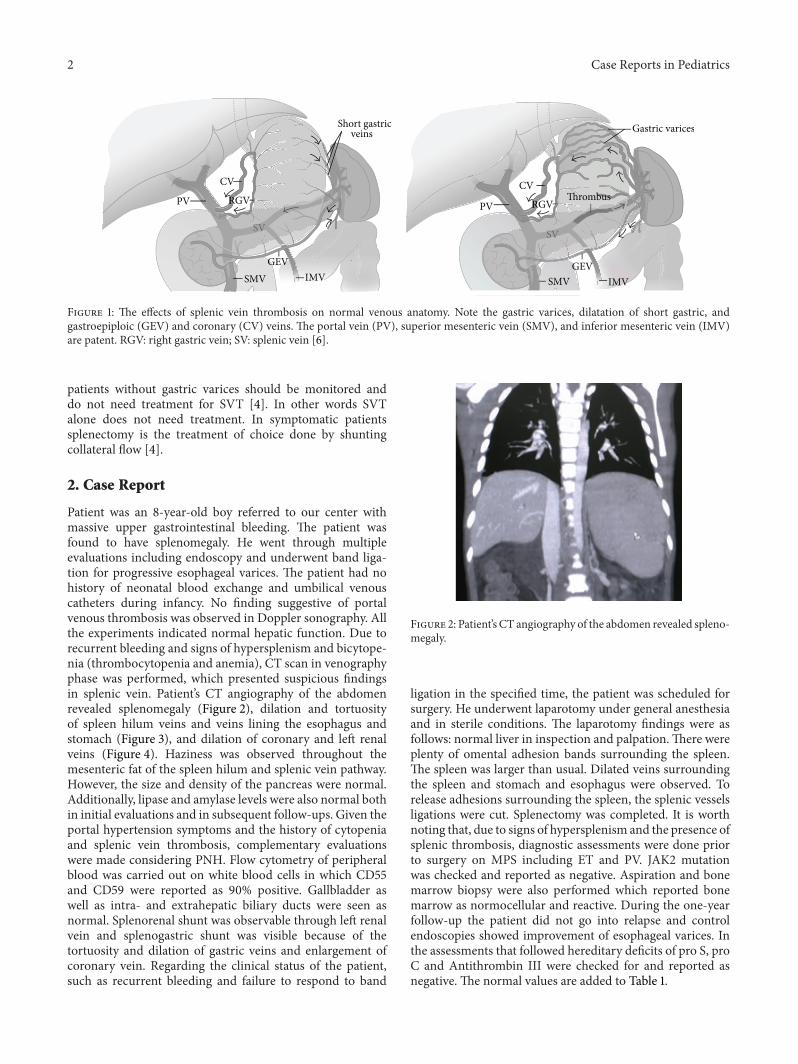
Short gastric Gastric varices
Thrombus
veins
CV CVRGV RGV
GEV GEV
PV PV
SV SV
IMV IMVSMV SMV
Figure 1: The effects of splenic vein thrombosis on normal venous anatomy. Note the gastric varices, dilatation of short gastric, andgastroepiploic (GEV) and coronary (CV) veins. The portal vein (PV), superior mesenteric vein (SMV), and inferior mesenteric vein (IMV)are patent. RGV: right gastric vein; SV: splenic vein [6].
patients without gastric varices should be monitored anddo not need treatment for SVT [4]. In other words SVTalone does not need treatment. In symptomatic patientssplenectomy is the treatment of choice done by shuntingcollateral flow [4].
2. Case Report
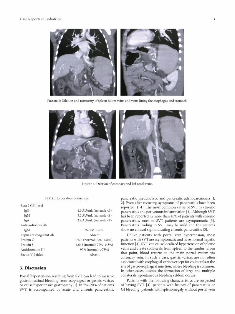
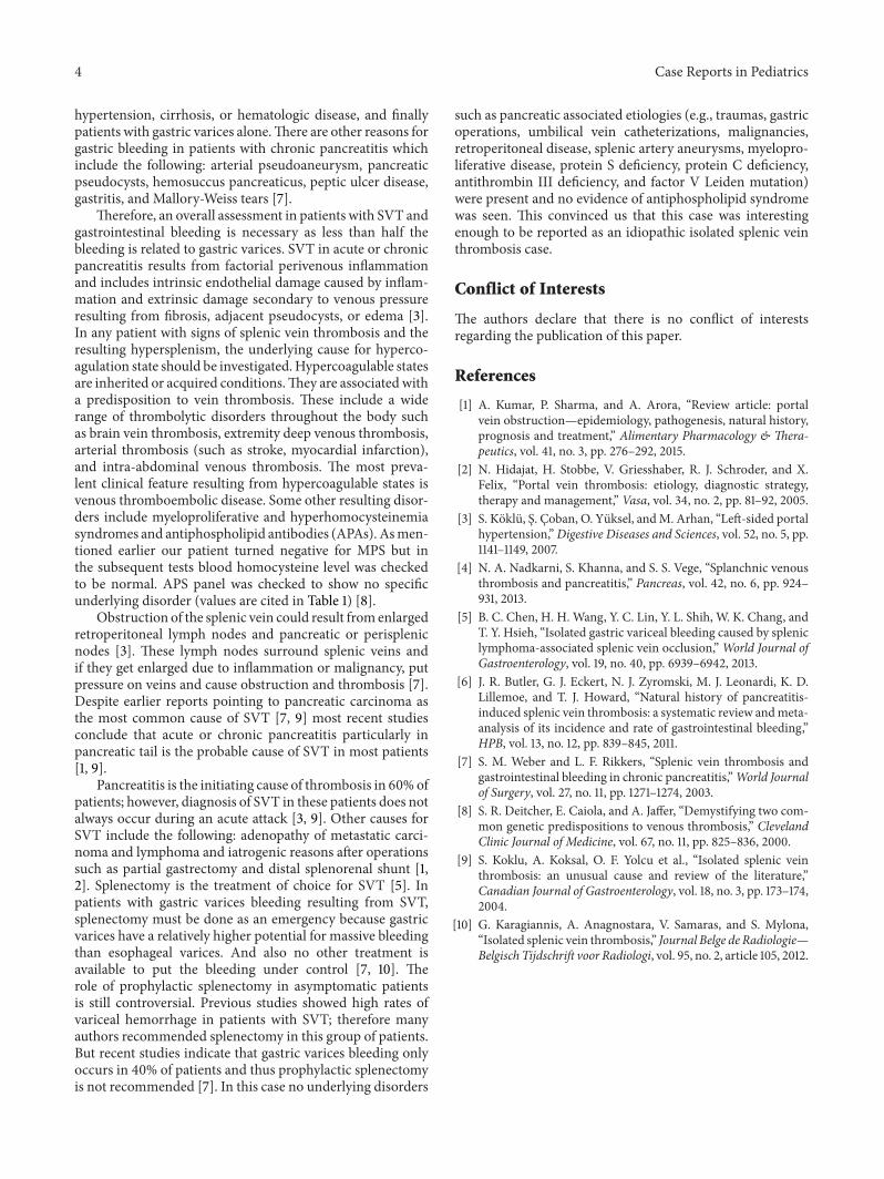
Patient was an 8-year-old boy referred to our center withmassive upper gastrointestinal bleeding. The patient wasfound to have splenomegaly. He went through multipleevaluations including endoscopy and underwent band liga-tion for progressive esophageal varices. The patient had nohistory of neonatal blood exchange and umbilical venouscatheters during infancy. No finding suggestive of portalvenous thrombosis was observed in Doppler sonography. Allthe experiments indicated normal hepatic function. Due torecurrent bleeding and signs of hypersplenism and bicytope-nia (thrombocytopenia and anemia), CT scan in venographyphase was performed, which presented suspicious findingsin splenic vein. Patient’s CT angiography of the abdomenrevealed splenomegaly (Figure 2), dilation and tortuosityof spleen hilum veins and veins lining the esophagus andstomach (Figure 3), and dilation of coronary and left renalveins (Figure 4). Haziness was observed throughout themesenteric fat of the spleen hilum and splenic vein pathway.However, the size and density of the pancreas were normal.Additionally, lipase and amylase levels were also normal bothin initial evaluations and in subsequent follow-ups. Given theportal hypertension symptoms and the history of cytopeniaand splenic vein thrombosis, complementary evaluationswere made considering PNH. Flow cytometry of peripheralblood was carried out on white blood cells in which CD55and CD59 were reported as 90% positive. Gallbladder aswell as intra- and extrahepatic biliary ducts were seen asnormal. Splenorenal shunt was observable through left renalvein and splenogastric shunt was visible because of thetortuosity and dilation of gastric veins and enlargement ofcoronary vein. Regarding the clinical status of the patient,such as recurrent bleeding and failure to respond to band
Figure 2: Patient’s CT angiography of the abdomen revealed spleno-megaly.
ligation in the specified time, the patient was scheduled forsurgery. He underwent laparotomy under general anesthesiaand in sterile conditions. The laparotomy findings were asfollows: normal liver in inspection and palpation.There wereplenty of omental adhesion bands surrounding the spleen.The spleen was larger than usual. Dilated veins surroundingthe spleen and stomach and esophagus were observed. Torelease adhesions surrounding the spleen, the splenic vesselsligations were cut. Splenectomy was completed. It is worthnoting that, due to signs of hypersplenism and the presence ofsplenic thrombosis, diagnostic assessments were done priorto surgery on MPS including ET and PV. JAK2 mutationwas checked and reported as negative. Aspiration and bonemarrow biopsy were also performed which reported bonemarrow as normocellular and reactive. During the one-yearfollow-up the patient did not go into relapse and controlendoscopies showed improvement of esophageal varices. Inthe assessments that followed hereditary deficits of pro S, proC and Antithrombin III were checked for and reported asnegative. The normal values are added to Table 1.
Case Reports in Pediatrics 3
Figure 3: Dilation and tortuosity of spleen hilum veins and veins lining the esophagus and stomach.
Figure 4: Dilation of coronary and left renal veins.
Table 1: Laboratory evaluation.
Beta 2 GP1 levelIgG 4.3 AU/mL (normal: <5)IgM 3.2AU/mL (normal: <8)IgA 2.4AU/mL (normal: <8)
Anticardiolipin AbIgM 9.63MPL/mL
Lupus anticoagulant Ab AbsentProtein C 85.6 (normal: 70%–130%)Protein S 120.2 (normal: 77%–143%)Antithrombin III 97% (normal: >75%)Factor V Leiden Absent
3. Discussion
Portal hypertension resulting from SVT can lead to massivegastrointestinal bleeding from esophageal or gastric varicesor cause hypertensive gastropathy [1]. In 7%–20% of patientsSVT is accompanied by acute and chronic pancreatitis,
pancreatic pseudocysts, and pancreatic adenocarcinoma [1,2]. Even after recovery, symptoms of pancreatitis have beenreported [1, 4]. The most common cause of SVT is chronicpancreatitis and perivenous inflammation [4]. Although SVThas been reported in more than 45% of patients with chronicpancreatitis, most of SVT patients are asymptomatic [3].Pancreatitis leading to SVT may be mild and the patientsshow no clinical sign indicating chronic pancreatitis [3].
Unlike patients with portal vein hypertension, mostpatients with SVT are asymptomatic and have normal hepaticfunction [4]. SVT can cause localized hypertension of splenicveins and create collaterals from spleen to the fundus. Fromthat point, blood returns to the main portal system viacoronary vein. In such a case, gastric varices are not oftenassociated with esophageal varices except for collaterals at thesite of gastroesophageal junction, where bleeding is common.In other cases, despite the formation of large and multiplecollaterals, spontaneous bleeding seldom occurs.
Patients with the following characteristics are suspectedof having SVT [4]: patients with history of pancreatitis orGI bleeding, patients with splenomegaly without portal vein
4 Case Reports in Pediatrics
hypertension, cirrhosis, or hematologic disease, and finallypatients with gastric varices alone.There are other reasons forgastric bleeding in patients with chronic pancreatitis whichinclude the following: arterial pseudoaneurysm, pancreaticpseudocysts, hemosuccus pancreaticus, peptic ulcer disease,gastritis, and Mallory-Weiss tears [7].
Therefore, an overall assessment in patients with SVT andgastrointestinal bleeding is necessary as less than half thebleeding is related to gastric varices. SVT in acute or chronicpancreatitis results from factorial perivenous inflammationand includes intrinsic endothelial damage caused by inflam-mation and extrinsic damage secondary to venous pressureresulting from fibrosis, adjacent pseudocysts, or edema [3].In any patient with signs of splenic vein thrombosis and theresulting hypersplenism, the underlying cause for hyperco-agulation state should be investigated.Hypercoagulable statesare inherited or acquired conditions.They are associated witha predisposition to vein thrombosis. These include a widerange of thrombolytic disorders throughout the body suchas brain vein thrombosis, extremity deep venous thrombosis,arterial thrombosis (such as stroke, myocardial infarction),and intra-abdominal venous thrombosis. The most preva-lent clinical feature resulting from hypercoagulable states isvenous thromboembolic disease. Some other resulting disor-ders include myeloproliferative and hyperhomocysteinemiasyndromes and antiphospholipid antibodies (APAs). Asmen-tioned earlier our patient turned negative for MPS but inthe subsequent tests blood homocysteine level was checkedto be normal. APS panel was checked to show no specificunderlying disorder (values are cited in Table 1) [8].
Obstruction of the splenic vein could result from enlargedretroperitoneal lymph nodes and pancreatic or perisplenicnodes [3]. These lymph nodes surround splenic veins andif they get enlarged due to inflammation or malignancy, putpressure on veins and cause obstruction and thrombosis [7].Despite earlier reports pointing to pancreatic carcinoma asthe most common cause of SVT [7, 9] most recent studiesconclude that acute or chronic pancreatitis particularly inpancreatic tail is the probable cause of SVT in most patients[1, 9].
Pancreatitis is the initiating cause of thrombosis in 60% ofpatients; however, diagnosis of SVT in these patients does notalways occur during an acute attack [3, 9]. Other causes forSVT include the following: adenopathy of metastatic carci-noma and lymphoma and iatrogenic reasons after operationssuch as partial gastrectomy and distal splenorenal shunt [1,2]. Splenectomy is the treatment of choice for SVT [5]. Inpatients with gastric varices bleeding resulting from SVT,splenectomy must be done as an emergency because gastricvarices have a relatively higher potential for massive bleedingthan esophageal varices. And also no other treatment isavailable to put the bleeding under control [7, 10]. Therole of prophylactic splenectomy in asymptomatic patientsis still controversial. Previous studies showed high rates ofvariceal hemorrhage in patients with SVT; therefore manyauthors recommended splenectomy in this group of patients.But recent studies indicate that gastric varices bleeding onlyoccurs in 40% of patients and thus prophylactic splenectomyis not recommended [7]. In this case no underlying disorders
such as pancreatic associated etiologies (e.g., traumas, gastricoperations, umbilical vein catheterizations, malignancies,retroperitoneal disease, splenic artery aneurysms, myelopro-liferative disease, protein S deficiency, protein C deficiency,antithrombin III deficiency, and factor V Leiden mutation)were present and no evidence of antiphospholipid syndromewas seen. This convinced us that this case was interestingenough to be reported as an idiopathic isolated splenic veinthrombosis case.
Conflict of Interests
The authors declare that there is no conflict of interestsregarding the publication of this paper.
References
[1] A. Kumar, P. Sharma, and A. Arora, “Review article: portalvein obstruction—epidemiology, pathogenesis, natural history,prognosis and treatment,” Alimentary Pharmacology & Thera-peutics, vol. 41, no. 3, pp. 276–292, 2015.
[2] N. Hidajat, H. Stobbe, V. Griesshaber, R. J. Schroder, and X.Felix, “Portal vein thrombosis: etiology, diagnostic strategy,therapy and management,” Vasa, vol. 34, no. 2, pp. 81–92, 2005.
[3] S. Koklu, S. Coban, O. Yuksel, andM. Arhan, “Left-sided portalhypertension,”Digestive Diseases and Sciences, vol. 52, no. 5, pp.1141–1149, 2007.
[4] N. A. Nadkarni, S. Khanna, and S. S. Vege, “Splanchnic venousthrombosis and pancreatitis,” Pancreas, vol. 42, no. 6, pp. 924–931, 2013.
[5] B. C. Chen, H. H. Wang, Y. C. Lin, Y. L. Shih, W. K. Chang, andT. Y. Hsieh, “Isolated gastric variceal bleeding caused by spleniclymphoma-associated splenic vein occlusion,”World Journal ofGastroenterology, vol. 19, no. 40, pp. 6939–6942, 2013.
[6] J. R. Butler, G. J. Eckert, N. J. Zyromski, M. J. Leonardi, K. D.Lillemoe, and T. J. Howard, “Natural history of pancreatitis-induced splenic vein thrombosis: a systematic review andmeta-analysis of its incidence and rate of gastrointestinal bleeding,”HPB, vol. 13, no. 12, pp. 839–845, 2011.
[7] S. M. Weber and L. F. Rikkers, “Splenic vein thrombosis andgastrointestinal bleeding in chronic pancreatitis,”World Journalof Surgery, vol. 27, no. 11, pp. 1271–1274, 2003.
[8] S. R. Deitcher, E. Caiola, and A. Jaffer, “Demystifying two com-mon genetic predispositions to venous thrombosis,” ClevelandClinic Journal of Medicine, vol. 67, no. 11, pp. 825–836, 2000.
[9] S. Koklu, A. Koksal, O. F. Yolcu et al., “Isolated splenic veinthrombosis: an unusual cause and review of the literature,”Canadian Journal of Gastroenterology, vol. 18, no. 3, pp. 173–174,2004.
[10] G. Karagiannis, A. Anagnostara, V. Samaras, and S. Mylona,“Isolated splenic vein thrombosis,” Journal Belge de Radiologie—Belgisch Tijdschrift voor Radiologi, vol. 95, no. 2, article 105, 2012.

Submit your manuscripts athttp://www.hindawi.com
Stem CellsInternational
Hindawi Publishing Corporationhttp://www.hindawi.com Volume 2014
Hindawi Publishing Corporationhttp://www.hindawi.com Volume 2014
MEDIATORSINFLAMMATION
of
Hindawi Publishing Corporationhttp://www.hindawi.com Volume 2014
Behavioural Neurology
EndocrinologyInternational Journal of
Hindawi Publishing Corporationhttp://www.hindawi.com Volume 2014
Hindawi Publishing Corporationhttp://www.hindawi.com Volume 2014
Disease Markers
Hindawi Publishing Corporationhttp://www.hindawi.com Volume 2014
BioMed Research International
OncologyJournal of
Hindawi Publishing Corporationhttp://www.hindawi.com Volume 2014
Hindawi Publishing Corporationhttp://www.hindawi.com Volume 2014
Oxidative Medicine and Cellular Longevity
Hindawi Publishing Corporationhttp://www.hindawi.com Volume 2014
PPAR Research
The Scientific World JournalHindawi Publishing Corporation http://www.hindawi.com Volume 2014
Immunology ResearchHindawi Publishing Corporationhttp://www.hindawi.com Volume 2014
Journal of
ObesityJournal of
Hindawi Publishing Corporationhttp://www.hindawi.com Volume 2014
Hindawi Publishing Corporationhttp://www.hindawi.com Volume 2014
Computational and Mathematical Methods in Medicine
OphthalmologyJournal of
Hindawi Publishing Corporationhttp://www.hindawi.com Volume 2014
Diabetes ResearchJournal of
Hindawi Publishing Corporationhttp://www.hindawi.com Volume 2014
Hindawi Publishing Corporationhttp://www.hindawi.com Volume 2014
Research and TreatmentAIDS
Hindawi Publishing Corporationhttp://www.hindawi.com Volume 2014
Gastroenterology Research and Practice
Hindawi Publishing Corporationhttp://www.hindawi.com Volume 2014
Parkinson’s Disease
Evidence-Based Complementary and Alternative Medicine
Volume 2014Hindawi Publishing Corporationhttp://www.hindawi.com