Embed Size (px)
Citation preview

Bulgarian Journal of Veterinary Medicine, 2014, 17, No 3, 231238 ISSN 1311-1477; online at http://tru.uni-sz.bg/bjvm/bjvm.htm
Case report
A CASE OF MALIGNANT TRICHOEPITHELIOMA (MATRICAL CARCINOMA) IN A CAT: PATHOMORPHOLOGICAL AND
IMMUNOHISTOCHEMICAL FINDINGS
M. E. ALCIGIR & S. A.VURAL
Department of Pathology, Faculty of Veterinary Medicine, Ankara University, Ankara, Turkey
Summary
Alcigir, M. E. & S. A. Vural, 2014. A case of malignant trichoepithelioma (matrical carci-noma) in a cat: Pathomorphological and immunohistochemical findings. Bulg. J. Vet. Med., 17, No 3, 231238.
Malignant trichoepithelioma (MTE) or matrical carcinoma is a neoplastic change of matrical and inner root sheet cells. It is generally described in dogs, with no sex predisposition. In this case, a glassy skin mass was taken from a 8-year-old, female mixed breed cat. Histopathologically, it was presented with the typical matrical cells islands, central keratinisation and necrosis, hyalinated collagens, ghost cells and trichohyaline granules. Masson’s trichrome stain differentiated epithelial cells from connective tissue in the subcutis. Immunohistochemistry (ABC immunperoxidase method) revealed the malignancy of matrical cells. The Ki67 marker was especially helpful to show the malignancy potential of cells. The Her2neu marker, one of epidermal growth factors, reacted to invasive cells located at the periphery of islands. Connexin 43 was usefull to show loss of connection in malignant cells. Due to the less frequent occurrence and unusual localisation of MTE, the described case aimed at revealing microscopical and immunohistochemical findings by using potential markers of MTE tumours in the skin of cats.
Key words: cat, immunohistochemistry, malignant trichoepithelioma, pathomorphology
Malignant trichoepithelioma (MTE), also known as matrical carcinoma, originates from hair follicle matrical or inner root sheet (Goldschmidt & Hendrick, 2002). The tumour is a not frequently seen skin tumour and uncommon in cats. MTE is not identified in other species except in a mouse. It develops most commonly in dogs between 1 and 15 years of age in spi-
te of being observed between 4 and 11 years of age in cats (Goldschmidt & Hen-drick, 2002; Mulas et al., 2007). The oc-currence of the tumour is documented in dogs although its clinical features are not mentioned, but it was suspected on the basis of ulcerative and large plaque-like appearence with rough edges (Gross et al., 2005). The tumour grows rapidly in the

A case of malignant trichoepithelioma (matrical carcinoma) in a cat ...
BJVM, 17, No 3 232
dermis and subcutaneous tissue and can metastasise to regional lymph nodes and lungs. It generally shows recurrence after operation. The skin appears “glassy” and ulceration is common. Its predilection sites are the dorsal trunk and the neck, thoracic region and tail (Goldschmidt & Hendrick, 2002). Microscopically, inva-sive anaplastic matrical cells or follicular infundibulum epithelial cells constitute trabecules, cord and nests of different si-zes. Some cells can include eosinophilic intracytoplasmic trichohyaline granules. In some areas, distinct matrical keratinisa-tion formation and necrotic cells which are composed of ghost or shadow cells in larger islands can be found (Goldschmidt & Hendrick, 2002).
In terms of immunohistochemistry, nuclear positivities are encountered using Ki67, PCNA, p21 and p27 in epithelial cells of malignant hair follicles of dogs (Inoue et al., 2006; Souza et al., 2008). β-catenin and pancytokeratin expressions in nuclei and cytoplasms of hair follicle epithelial cells are reported as benign co-unterpart in a cat (Tavasoli et al., 2013). Another reports positive reactions obtai-ned by using different types of cytoke-ratins in epithelial cells of hair follicles in a mouse. In the same study, some positive reactions are also obtained by using pro-filaggrin and involucrin on the inner sur-face of squamous epithelium of hair folli-cles (Martin de las Mulas et al., 2008).
In this case, a circumscript large tu-mour located at the subcutis of left gluteal region was described as malignant tricho-epithelioma. The presentation was deemed valuable when compared to benign coun-terparts because of the rare occurence in cats and the unusual localisation. There are no reports about connexin-43, Her2-neu and CEA expressions in malignant trichoepithelioma except for Ki67 expres-
sion. Ki67 is a poweful marker of malig-nant activity in the nuclei of all types of anaplastic cells (Hazan et al., 2002; Iqbal et al., 2002). It was believed that the im-munohistochemical features of the case will throw a diferent light on skin tumors.
A 8-year-old female mixed breed cat was submitted to the Department of Sur-gery, Ankara University, Faculty of Vete-rinary Medicine with complaints of exces-sive mass under the skin of the left gluteal region. After clinical examination, the mass was removed surgically and sent to Department of Pathology for diagnosis. After macroscopical examination, tissue samples were taken from lesions for histopathology and fixed in 10% formalin. Then, they were processed routinely and embedded in paraffin. Sections were cut at 5 μm thickness from paraffin blocks and stained with haematoxylin-eosin (H&E) and Masson’s trichrome methods. After histochemical stainings, indirect immu-noperoxidase method (ABC-P) was ap-plied using connexin 43 (Cx43), carci-noembryogenic antigen (CEA), Her2 neu, and Ki67 markers. To this end, after de-paraffinization and dehydration, the per-oxidase activity was blocked. Trypsinisa-tion was performed by using 0.1% trypsin solution. Non-specific proteins were blocked with protein blocking sera (Per-oxidase Detection System, Novocastra, RE7110-K, Leica Biosystems). The sec-tions were incubated with primary anti-bodies (polyclonal rabbit anti connexin 43, monoclonal mouse CEA, monoclonal mouse Her2neu, monoclonal rabbit Ki67– Table 1). Then, biotinylated link antibody and horse radish peroxidase (HRP) anti-bodies were applied, respectively (Peroxi-dase Detection System, RE7110-K, No-vocastra, Leica Biosystems). Control sec-tions were treated with PBS instead of primary antibodies. As chromogen, 3-ami-

M. E. Alcigir & S. A. Vural
BJVM, 17, No 3 233
no-9-ethylcarbazole (AEC) (Santa Cruz Biotechnology Inc.) was selected. Coun-
Fig. 1. Macroscopic appearance of the mass.
terstaining were performed with Gill’s haematoxylin. Sections were mounted with glycergel and examined on light mic-roscope (Leica Microsystems, DM4000B).
The mass covered with skin weighed 20 g and its size was 5×4×1 cm. Its cut
surface was homogeneous and of white colour (Fig. 1). Histopathologically, ana-plastic hair follicle epithelium, having large and hypochromatic nuclei was ob-served. Prominent nucleoli, some of them with hyperchromatic nuclei, constituted islands such as cords and trabecules. The-re were keratohyalin formation and nec-rosis composed of cellular debris at the centre of large islands (Fig. 2 and 3). In addition, ghost or shadow cells were ob-served towards the centre of necrotic deb-ris (Fig. 4). Eosinophilic trichohyaline granules were also visible in the cyto-plasm of anaplastic cells (Fig. 5).
After Masson’s trichrome staining, malignant epithelial cell islands were dif-ferentiated from the stroma and the dermal connective tissue (Fig. 6). Immunohisto-chemically, the Ki67 and Her2neu posi-tive reactions exhibited granular or diffuse patterns of brownish-red color localised in
Table 1. Panel of used antibodies
Antibody Features Clone Trade Dilution
Ki67 Monoclonal rabbit Clone SP6 Labvision 1:50
Her2neu Monoclonal mouse c-ErbB-2 oncoprotein Novocastra 1:40
CEA Monoclonal mouse COL-1 Neomarkers 1:100
Cx43 Polyclonal rabbit GJA-1 Abcam 1:100
Fig. 2. MTE cells (arrow) and central necrosis (asterix). Bar=55 µm, H&E.
Fig. 3. MTE cells (arrows). Bar=40 µm, H&E.

A case of malignant trichoepithelioma (matrical carcinoma) in a cat ...
BJVM, 17, No 3 234
cytoplasms of epithelial cells. However, CEA positive reactions were observed in
Fig. 6. MTE islands differentiating connective tissue (arrows). Bar=40 µm, Masson’s
trichrome stain.
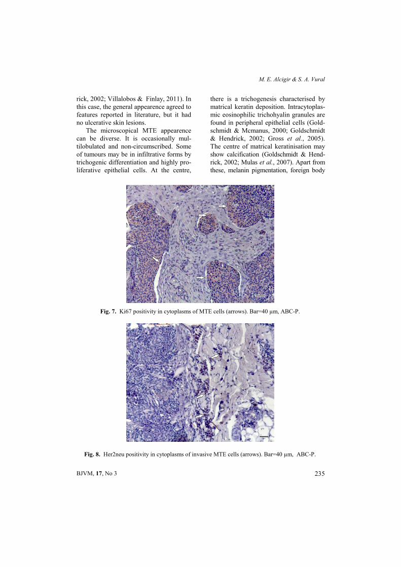
cytoplasms and nuclei. Localisation of Cx43 positivities in cytoplasms and mem-branes of anaplastic epithelial cells was observed. Positive reactions were ob-tained by Ki67 in all anaplastic epithelial cells and by Her2neu – in invasive highly malignant cells (Fig. 7 and 8). However, all anaplastic matrical cells and hair folli-cle epithelia reacted slightly with Cx43 and CEA with (Fig. 9 and 10).
Malignant trichoepitheliomas (MTE) are rarely seen skin tumours in cats (Gold-schmidt & Hendrick, 2002). In dogs, most
affected are animals of late middle age. Although there is no defined sex predilec-tion, a breed predisposition exists in Bas-set Hounds, Bull Mastiffs, Irish Setters, Poodles, English Spaniels, and Golden Retrievers. Tumours can develop on the trunk in dogs, but are encountered on the head, tail, and extremities in cats. The tumour originates from the hair follicle (infundibulum, isthmus, and inferior por-tions) (Weiss & Frese, 1974; Villalobos & Finlay, 2011). The mass in the subcutis in our 8-year-old mixed breed cat was local-ised in left gluteal region. For cats, this localisation is different from others re-ported in the literature. Metastasis is not common, possible sites are the regional lymph nodes and the lungs. Surgical exci-sion is considered as the treatment of choice (Goldschmidt & Hendrick, 2002; Villalobos & Finlay, 2011). In the present case, the health condition of the patient was followed but we were not informed about any recurrency after the surgical excision. On the other hand, MTE gene-rally develop exophytically and grow as intradermal masses. The masses appear as multiple grey-white foci of keratinous matrix. For many of them, the skin is ge-nerally ulcerated (Goldschmidt & Hend-
Fig. 4. Ghost or shadow cells at the periphery of necrotic debris (arrows). Bar=40 µm, H&E.
Fig. 5. Intracytoplasmic eosinophilic tricho-hyalin granules (arrows). Bar=40 µm, H&E.
M. E. Alcigir & S. A. Vural
BJVM, 17, No 3 235
rick, 2002; Villalobos & Finlay, 2011). In this case, the general appearence agreed to features reported in literature, but it had no ulcerative skin lesions.
The microscopical MTE appearence can be diverse. It is occasionally mul-tilobulated and non-circumscribed. Some of tumours may be in infiltrative forms by trichogenic differentiation and highly pro-liferative epithelial cells. At the centre,
there is a trichogenesis characterised by matrical keratin deposition. Intracytoplas-mic eosinophilic trichohyalin granules are found in peripheral epithelial cells (Gold-schmidt & Mcmanus, 2000; Goldschmidt & Hendrick, 2002; Gross et al., 2005). The centre of matrical keratinisation may show calcification (Goldschmidt & Hend-rick, 2002; Mulas et al., 2007). Apart from these, melanin pigmentation, foreign body
Fig. 7. Ki67 positivity in cytoplasms of MTE cells (arrows). Bar=40 µm, ABC-P.
Fig. 8. Her2neu positivity in cytoplasms of invasive MTE cells (arrows). Bar=40 µm, ABC-P.

A case of malignant trichoepithelioma (matrical carcinoma) in a cat ...
BJVM, 17, No 3 236
reaction or purulent inflammation due to secondary bacterial complication can be found (Weiss & Frese, 1974).
Malignant trichoepithelial cells are generally found as islands, chords or tra-becules composed of basaloid cells with differentiation to internal and external root sheath. At the periphery, there are malignant cells with pleomorphic hyper-chromatic nuclei and scanty eosinophilic
cytoplasms and thickened basal lamina (Goldschmidt & Mcmanus, 2000; Gold-schmidt & Hendrick, 2002, Villalobos & Finlay, 2011). These cells have usually moderate or high mitotic activity (Weiss & Fresse, 1974; Gross et al., 2005). On the other hand, a matrical differentiation is developed with participation of ghost or shadow cells at the centre of epithelial islands. Also they can include fibrotic or
Fig. 9. Slightly positive Cx43 cells (arrows). Bar=35 µm, ABC-P.
Fig. 10. Slightly positive CEA cells (arrows). Bar=50 µm, ABC-P.

M. E. Alcigir & S. A. Vural
BJVM, 17, No 3 237
mucinous stroma. In higher malignant forms, central cystic degeneration and lar-ge necrotic areas are found (Goldschmidt & Mcmanus, 2000; Villalobos & Finlay, 2011). In our case, similar results were obtained, but there were no melanin pig-mentation, foreign body reaction, secon-dary bacterial complication and cystic de-generation.
There is little information about im-munohistological features of trichoepithe-lioma. They were investigated in only few studies on hair follicle tumours with p21, p27, PCNA, Ki67 and cytokeratin anti-bodies (Inoue et al., 2006, Mulas et al., 2007, Souza et al., 2008). Mild or mode-rate reactions are obtained with markers. However, in this case, the carcinoembryo-genic antigen (CEA), Her2neu (cerbB on-coprotein) and connexin 43 (Cx43) mar-kers apart from Ki67 marker were used. In this context, useful results from Her2neu and Ki67 were obtained. Her2neu is one of epithelial growth factor receptors that is generally utilised for showing invasive cells especially in the breast, endo-metrium, gastric cancer prognosis (Tan & Yu, 2007; Santin et al., 2008). Up to now, it is demonstrated in cutaneous melanoma in terms of its expression in human skin (El-Sheikh et al., 2009). It is not used for detecting invasive cells and prognosis evaluation in skin tumours. In this case report, the Her2neu and Ki67 markers were preferred for showing malignancy and were found useful. The Cx43 marker is gap junction or transmembrane protein and normally plays a critical role in cellu-lary adhesion and coordination (Cameron et al., 2003). It is reported that Cx43 has a tumour suppresive role (Langlois et al., 2010). In the present case, there was a high malignancy due to loss of cell to cell adhesion between malignant cells. Hence, slight Cx43 positivity is interpreted as a
result of abnormal proliferation. CEA is also a glycoprotein providing cellular ad-hesion. It is used to show malignancy es-pecially in gastric, colonic, breast and lung adenocarcinoma (Thomas et al., 2008). The marker is a cell surface glyco-protein and is utilised to detect high anaplastic features of malignant skin tu-mors cells in of different origin (Heyder-man et al., 1984; Thomas et al., 2008). In this patient, the marker was applied to MTE cells to show high anaplasia, how-ever, less positive reactions were obtained when compared to other marker positivi-ties. The slight positive reactions sug-gested that CEA is not as effective as Cx43 to indicate loss of cellulary adhe-sion. Therefore, the Her2neu and Cx43 markers should be selected by patholo-gists to show malignancy of skin tumours apart Ki67 and other markers documented in the literature.
REFERENCES
Cameron, S. J., S. Malik, M. Akaike, N. Lerner-Marmarosh, C. Yan, J. D. Lee, J. Abe & J. Yang, 2003. Regulation of epi-dermal growth factor-induced connexin 43 gap junction communication by big mito-gen-activated protein kinase1/ERK5 but not ERK1/2 kinase activation. Journal of Biological Chemistry, 278, 18682–18688.
El-Sheikh, S. M., S. M. El-Sheikh & I. M. El-Morsy, 2009. Detection of c-Kit (CD117) and Her2/neu in oral and cutaneous malig-nant melanomas. Journal of Egyptian Women's Dermatology Society, 6, 66–73.
Goldschmidt, M. H. & P. Mcmanus, 2000. Dermatopathology. Skin Tumors of the Dog and Cat. http://cal.vet.upenn.edu/ derm2epi/epidex.html (25 January 2013, date last accessed).
Goldschmidt, M. H. & M. J. Hendrick, 2002. Tumors of the skin and soft tissues. Chap-
A case of malignant trichoepithelioma (matrical carcinoma) in a cat ...
BJVM, 17, No 3 238
ter 2. In: Tumors of Domestic Animals, ed D. J. Meuten, Iowa State Press, USA, p. 60.
Gross, T. L., P. J. Ihrke, E. J. Walder & V. K. Affolter, 2005. Skin Disease of Dog and Cat. In: Clinical and Histopathologic Di-agnosis, Blackwell Publishing, Iowa, USA, pp. 634–637.
Hazan, C., K. Melzer, K. S. Panageas, E. Li, H. Kamino, A. Kopf, C. Cordon-Cardo, I. Osman & D. Polsky, 2002. Evaluation of the proliferation marker MIB-1 in the prognosis of cutaneous malignant mela-noma. Cancer, 95, 634–640.
Heyderman, E., R. M. Graham, D. V. Chap-man, T. C. Richardson & P. H. McKee, 1984. Epithelial markers in primary skin cancer: An immunoperoxidase study of the distribution of epithelial membrane anti-gen (EMA) and carcinoembryogenic anti-gen (CEA) in 65 primary skin carcinomas. Histopathology, 8, 423–434.
Inoue, M., H. Wu & S. Une, 2006. Pathomor-phological and ımmunohistochemical find-ings of cystic mucinous hyperplasia of gall bladder. Journal of Veterinary Medical Science, 68, 779–782.
Iqbal, S., T. J. Anderson & L. P. Marson, 2002. MIB-1 assessments in breast can-cers. Breast, 11, 252–256.
Langlois, S., K. N. Cowan, Q. Shao, B. J. Cowan & D. W. Laird, 2010. The tumor-suppresive function of connexin43 in kera-tinocytes is mediated in part via interaction with caveolin-1. Cancer, 70, 4222–4232.
Martin de las Mulas, J., A. M. Molina, Y. Mil-lan, L. Carraco & R. Moyano, 2007. Spon-taneous trichoepithelioma in a laboratory mouse: gross, microscopic and immuno-histochemical findings. Laboratory Ani-mals, 41, 136–140.
Santin, A. D., S. Bellone, J. J. Roman, J. K. McKenney & S. Pecorelli, 2008. Trastu-zumab treatment in patients with advanced or recurrent endometrial carcinoma over-expressing HER2/neu. International Jour-nal of Gynaecology & Obstetrics, 102, 128–131.
Souza, P. C., N. M. Ocarino, W. L. F. Tavares, J. N. Boeloni, G. D. Cassali & R. Serakides, 2008. Association of nucleolar organizing regions and Ki-67 expression with recurrence rate of hair follicle tumor in dogs. Arquivo Brasileiro de Medicina Veterinária e Zootecnia, 60, 1075–1083.
Tan, M. & D. Yu, 2007. Molecular mecha-nisms of erbB2-mediated breast cancer chemoresistance. Advances in Experimen-tal Medicine and Biology, 608, 119–129.
Tavasoli, A., H. Golhahi, M. N. Rad & A. Tay-mouri, 2013. Occurence of trichoepithe-lioma in a cat: Histopathologic and immu-nohistochemical study. Asian Pacific Jour-nal of Tropical Biomedicine, 3, 413–415.
Thomas, S. N., F. Zhu, R. L. Schnaar, C. S. Alves & K. Konstantopoulos, 2008. Carcinoembryonic antigen and CD44 vari-ant isoforms cooperate to mediate colon carcinoma cell adhesion to E- and L-selectin in shear flow. Journal of Biologi-cal Chemistry, 283, 15647–15655.
Villalobos, A. & M. Finlay, 2011. Epidermal and Hair Follicle Tumors. The Merck Vet-erinary Manual, http://www.merckmanu als.com/vet/integumentary_system/tumors _of_the_skin_and_soft_tissues/epidermal_and_hair_follicle_tumors.html (25 January 2013, date last accessed).
Weiss, E. & K. Frese, 1974. Tumors of the skin. Bulletin of World Health Organiza-tion, 50, 79–100.
Paper received 22.10.2013; accepted for publication 20.02.2014
Correspondence:
Mehmet Eray Alcigir Department of Pathology, Ankara University, Faculty of Veterinary Medicine, 06110, Diskapi, Ankara/Turkey, Phone: +90-312-3170315/4382 e-mail: [email protected], [email protected]