Embed Size (px)
Citation preview

Bulgarian Journal of Veterinary Medicine, 2016, 19, No 4, 334–339 ISSN 1311-1477; DOI: 10.15547/bjvm.924
Case report
CLINICAL AND MORPHOLOGICAL INVESTIGATIONS IN A SPONTANEOUS CRYPTOSPORIDIUM ENTERITIS
OUTBREAK IN CALVES
I. KALKANOV1, I. DINEV1, K. DIMITROV1 & P. ILIEV2
1Department of General and Clinical Pathology, 2Department of Veterinary Micro-biology, Infectious and Parasitic Diseases, Faculty of Veterinary Medicine,
Trakia University, Stara Zagora, Bulgaria
Summary
Kalkanov, I., I. Dinev, K. Dimitrov & P. Iliev, 2016. Clinical and morphological investiga-tions in a spontaneous Cryptosporidium enteritis outbreak in calves. Bulg. J. Vet. Med., 19, No 4, 334–339. The purpose of this report was to present the results of histopathological examination during the course of a natural Cryptosporidium parvum outbreak, as well as the morphology of cells infiltrating affected gastrointestinal tract areas in newborn calves. The study included 18 calves exhibiting a marked diarrhoeic syndrome, between 1 and 8 days of age. Sporulated C. parvum oocysts were de-monstrated in faecal smears stained by the method of Henriksen. Coproantigens of C. parvum were detected by the rapid Rainbow calf scour 5 BIO K 306 test. Gross lesions were mainly present in the gastrointestinal tract, together with inflammation in regional mesenteric lymph nodes. Microscopic histopathological lesions consisted mainly in desquamative catarrh of intestinal mucosa and intestinal villous atrophy. The affected intestinal epithelium contained multiple Cryptosporidium spp. forms at different stage of the life cycle. The results from histopathological studies of the outbreak allowed confirming some main morphogenetic features of the disease caused by C. parvum in newborn and growing calves.
Key words: calves, Cryptosporidium parvum, cryptosporidiosis, pathology
Gastrointestinal diseases in newborn and juvenile calves are the commonest prob-lems in this livestock category. The losses are due to high mortality and morbidity rates, reduced weight gain after remission from the disease and disease treatment costs. Neonatal diarrhoea in calves is a multifactorial disease influenced by the
breed, feeding regimen and farming con-ditions. The immunological status of the dams from the herd, farm management and the occurrence of different infectious agents are also important (Bendali et al., 1999; Scott et al., 2004). The risk for oc-currence of the disease is the highest du-ring the first month, then decreases as age

I. Kalkanov, I. Dinev, K. Dimitrov & P. Iliev
BJVM, 19, No 4 335
advances (Frank & Kaneene, 1993; Ben-dali et al., 1999). At a global scale, neona-tal diarrhoea is associated with five pri-mary enteropathogens: Cryptosporidium parvum, rotaviruses, bovine coronavirus (BCV), enterotoxigaenic Escherichia coli (Е. coli K99) (Garcia et al., 2000), and Salmonella, all causing diarrhoea. Out-breaks at farms are often observed, espe-cially when the animals originate from different farms with unknown immu-nological status (Naylor, 2002).
Cryptosporidium spp. (phylum Api-complexa, family Cryptosporidiidae) are coccidian protozoa affecting the gastroin-testinal tract in several mammalian and amphibian species and only the lungs in birds (Helmy et al., 2013). Some of the members of this family possess also well expressed zoo-anthropogenic potential (Castro-Hermida et al., 2002). The possi-bility for subclinical course and parasite shedding explain the broad geographic spread of cryptosporidiosis. According to Joachim et al. (2003), the prevalence of Cryptosporidium spp. in calves in Euro-pean countries varies between 20% and 40%. Together with some bacterial and viral pathogens, Cryptosporidium parvum is considered as one of the most frequent agents of neonatal gastroenterites in calves up to 3 months of age (Chartier et al., 2013). The relative death rate is low, but morbidity within the general popula-tion in the contact environment is high (Castro-Hermida et al., 2002). The most important clinical sign reported by Joachim et al. (2003) is profuse diarrhoea. Its duration and intensity correspond to the localisation of the infection and the extent of morphological aberrations of the intestinal mucosa. The histological find-ings of colonised distal small intestine compartments consist in villous atrophy, metaplasia and desquamation of the sur-
face epithelium (Fayer & Xiao, 2007). The authors explained that similar lesions could be also observed in the duodenum, caecum and the colon. In their view, the cell infiltration in lamina propria con-sisted mainly of neutrophil leukocytes.
The purpose of this report was to es-tablish the histopathological alterations during the course of a natural C. parvum outbreak, as well as the morphology of cells infiltrating affected gastrointestinal tract areas in newborn calves.
The study was conducted on 18 calves with clearly manifested diarrhoeic syn-drome aged between 1 and 8 days.
Case history. A sudden increase in mortality rates in calves from the 24th h of life to 6–8 days of age was reported at a cattle farm housing 200 animals from the Limousin and Hereford breeds, grazing freely on pasture. Clinically, bloody diar-rhoea, rapid dehydration and death, with-out fever, were observed. The palliative general fluid therapy and parenteral treat-ment with macrolide antibiotics were of no use. At that time, no preventive vacci-nation in cows have been performed du-ring the dry period.
Coprological samples were obtained manually from ampula recti of each ani-mal and stored at 4 oC until the analysis. Sporulated oocysts were detected on fae-cal smears stained by the method of Hen-riksen (Emmonya Biotech, Chelopech) – modified Ziehl-Neelsen staining for acid-fast microorganisms. The presence of C. parvum coproantigens was confirmed through Rainbow calf scour 5 BIO K 306 (BIOX Diagnostics) rapid test, designed for detection of rotavirus, coronavirus, E. coli – F5, Cryptosporidium parvum and Clostridium perfringens type А.
Gross and histological examinations. Eight carcasses were submitted to routine necropsy using the standard protocol. Tis-
Clinical and morphological investigations in a spontaneous Cryptosporidium enteritis outbreak in calves
BJVM, 19, No 4 336
sue samples (size 2.5 cm) were collected from the affected gastrointestinal tract areas (abomasum, duodenum, jejunum with mes-enteric lymph nodes, ileum, caecum, co-lon, rectum) for histopathological exami-nation. Samples (2.5×2.5×1 cm) were also obtained from parenchymal organs – liver, lungs, kidneys, spleen and heart. Speci-mens for histological examination were fixed in 10% neutral buffered formalin and embedded in paraffin. From paraffin blocks, 4 μm cross sections were cut on a Leica RM 2235 microtome and conven-tionally stained with haematoxylin-eosin.
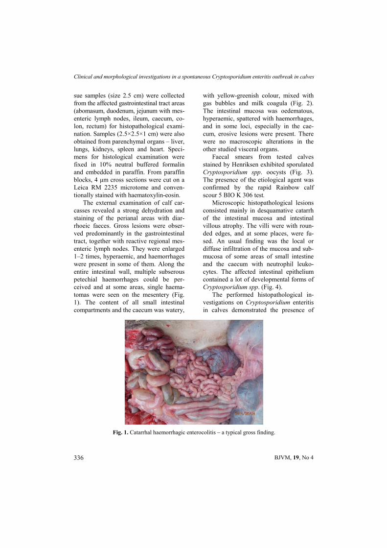
The external examination of calf car-casses revealed a strong dehydration and staining of the perianal areas with diar-rhoeic faeces. Gross lesions were obser-ved predominantly in the gastrointestinal tract, together with reactive regional mes-enteric lymph nodes. They were enlarged 1–2 times, hyperaemic, and haemorrhages were present in some of them. Along the entire intestinal wall, multiple subserous petechial haemorrhages could be per-ceived and at some areas, single haema-tomas were seen on the mesentery (Fig. 1). The content of all small intestinal compartments and the caecum was watery,
with yellow-greenish colour, mixed with gas bubbles and milk coagula (Fig. 2). The intestinal mucosa was oedematous, hyperaemic, spattered with haemorrhages, and in some loci, especially in the cae-cum, erosive lesions were present. There were no macroscopic alterations in the other studied visceral organs.
Faecal smears from tested calves stained by Henriksen exhibited sporulated Cryptosporidium spp. oocysts (Fig. 3). The presence of the etiological agent was confirmed by the rapid Rainbow calf scour 5 BIO K 306 test.
Microscopic histopathological lesions consisted mainly in desquamative catarrh of the intestinal mucosa and intestinal villous atrophy. The villi were with roun-ded edges, and at some places, were fu-sed. An usual finding was the local or diffuse infiltration of the mucosa and sub-mucosa of some areas of small intestine and the caecum with neutrophil leuko-cytes. The affected intestinal epithelium contained a lot of developmental forms of Cryptosporidium spp. (Fig. 4).
The performed histopathological in-vestigations on Cryptosporidium enteritis in calves demonstrated the presence of
Fig. 1. Catarrhal haemorrhagic enterocolitis – a typical gross finding.

I. Kalkanov, I. Dinev, K. Dimitrov & P. Iliev
BJVM, 19, No 4 337
Cryptosporidium spp. in 100% of animals along the entire intestinal tract. The ob-served intensive loss of intestinal villi, especially in the distal small intestinal compartment, is associated exactly with established extensive Cryptosporidium in fection (Rosales et al., 1998). At the same time, we believe that the observed signs of regeneration and repair, mainly under the form of crypt hyperplasia in about one quarter of cases (5 calves) are an attempt for restoration of mucosal cell barrier, although at an insufficient extent. This fact, as well as the reported high-degree atrophy of intestinal villi predispose to secondary complications, facilitating the colonisation of other pathogens (Argenzio et al., 1990). The hypothesis is confirmed by the results from a number of animal experimentations (Tzipori et al., 1982; Heine et al., 1984; Argenzio et al., 1990; Moore et al., 1995; Gookin et al., 2002).
The intensive loss of enterocytes is at-tributed to the pathogenetic mechanism of Cryptosporidium infection, namely forma-tion of parasitic vacuole after penetration of the agent into the cytoplasm (de Graaf et al., 1990; O’Handley & Olson, 2006).
Cryptosporidium infection and neutro-philic infiltration in described case report was established not only in the distal small intestinal compartment, but also in lamina propria of the proximal colon. We agree with the thesis of some researchers that these lesions are at the background of bloody diarrhoea (Jewis et al., 1966) and that in cases when only small intestine is affected and the colon is intact, faeces were free of blood (Jewis et al., 1966; Kovatch & White, 1972).
The observed outbreak of neonatal calf diarrhoea associated with Crypto-sporidium spp. confirms that the pathogen is responsible for a considerable share of gastrointestinal tract diseases in newborn and juvenile calves. We also affirm that the susceptibility to this infection is the highest until the 3rd week of life. Our stu-dies gave us reason to acknowledge, simi-larly to other research teams, that at this age, the etiology of enterites in calves could be complicated with other agents, mainly of bacterial or viral origin (Char-tier et al., 2013). The histological results allowed confirming some main morpho-genetic features of Cryptosporidium par-
Fig. 2. Specific yellow-greenish content mixed with milk coagula, oedema and hyperaemia of caecal mucosa.

Clinical and morphological investigations in a spontaneous Cryptosporidium enteritis outbreak in calves
BJVM, 19, No 4 338
vum attachment to the surface of entero-cytes in the distal small intestine and pro-ximal colon (Gay et al., 2012). We also believe that poor hygienic conditions and production system deficiencies, as well as the insufficient amount of colostrum could be factors for appearance of the disease (Castro-Hermida et al., 2002).
REFERENCES
Argenzio, A., A. Liacos & M. Levy, 1990. Villous atrophy, crypt hyperplasia, cellular infiltration, and impaired glucose-Na ab-
sorption in enteric cryptosporidiosis of pigs. Gastroenterology, 98, 1129–1140.
Bendali, F., M. Sanaa, H. Bichet, & F. Schel-cher, 1999. Risk factors associated with diarrhoea in newborn calves. Veterinary Research, 30, 509–522.
Castro-Hermida, A., Y. Gonzales-Losada, M. Mezo-Menendez, E. Aresmazas, 2002. A study of cryptosporidiosis in a cohort of neonatal calves. Veterinary Parasitology, 106, 11–17.
Chartier, C., A. Rieux, A. Delafosse, A. Le-hebel & C. Paraud, 2013. Detection of Cryptosporidium oocysts in fresh calf fae-
Fig. 3. Cryptosporidium spp. oocysts, faecal smear, Ziehl-Neelsen, ×1000.
Fig. 4. Multiple developmental forms of Cryptosporidium spp. in mucous ileal crypts, H&E, ×300.

I. Kalkanov, I. Dinev, K. Dimitrov & P. Iliev
BJVM, 19, No 4 339
ces: Characteristics of two simple tests and evaluation of a semi-quantitative approach. The Veterinary Journal, 198, 148–152.
de Graaf, A., J.van Dijk & W. Bovee, 1990. QUALITY: Quantification improvement by converting lineshapes to the Lorentzian type. Magnetic Resonance in Medicine, 13, 343–357.
Fayer, R. & L. Xiao, 2007. Cryptosporidium and Cryptosporidiosis, 2nd edn, CRC Press, Boca Raton.
Frank, N. & J. Kaneene, 1993. Management risk factors associated with calf diarrhea in Michigan dairy herds. Journal of Dairy Science, 76, 1313–1323.
Garcia, A., J. Ruiz-Santa-Quiteria, J. Orden, D. Cid, R. Sanz, M. Gomez-Bautista & R. De La Fuente, 2000. Rotavirus and con-current infections with other enteropatho-gens in neonatal diarrheic calves in Spain. Comparative Immunology, Microbiology and Infectious Diseases, 23, 175–183.
Gay, G., T. Courtheoux, C. Reyes, S. Tournier & Y. Gachet, 2012. A stochastic model of kinetochore–microtubule attachment accu-rately describes fission yeast chromosome segregation. The Journal of Cell Biology, 196, 757–774.
Gookin, J., S. Nordone & R. Argenzio, 2002. Host responses to Cryptosporidium infec-tion. Journal of Veterinary Internal Medi-cine, 16, 12–21.
Heine, J., J. Pohlenz & H. Moon, 1984. En-teric lesions and diarrhea in gnotobiotic calves monoinfected with Cryptosporid-ium species. Journal of Infectious Dis-ease, 150, 768–775.
Helmy, M., C. Ballhaus, R. Fonseca, R. Wirth, T. Nagel & M. Tredoux, 2013. Noble metal nanoclusters and nanoparticles pre-cede mineral formation in magmatic sulfide melts. Nature Communications, 4, 2405.
Joachim, A., T. Krull, J. Schwarzkopf & A. Daugshies, 2003. Prevalence and control of bovine cryptosporidiosis in German dairy herds. Veterinary Parasitology, 112, 277–288.
Jewis, H., T. Merril & H. Sprinz, 1966. Coc-cidiosis in the guinea pig small intestine due to a cryptosporidium. American Jour-nal of Veterinary Research, 27, 408–414.
Kovatch, R. & J. White, 1972. Cryptospori-diosis in two juvenile rhesus monkeys. Veterinary Pathology, 9, 426–440.
Moore, R., S. Tzipori & J. Griffiths, 1995. Temporal changes in permeability and structure of piglet ileum after site-specific infection by Cryptosporidium parvum. Gastroenterology, 108, 1030–1039.
Naylor, M., 2002. Neonatal ruminant diarrhea. In: Large Animal Internal Medicine, 3rd edn, ed B. P. Smith, Mosby, pp. 352–366.
O’Handley, R. & M. Olson, 2006. Giardiasis and cryptosporidiosis in ruminants. Vete-rinary Clinics of North America: Food Animal practice, 22, 623–643.
Rosales, M., T. Arrnedo & C. Mascaro, 1998. Ultrastructural details of Cryptosporidium parvum development in calf intestine. Memórias do Instituto Oswaldo Cruz, 93, 847–850.
Scott, C., O. Schuldiner & P. Neufeld, 2004. Role and regulation of starvation-induced autophagy in the Drosophila fat body. De-velopmental Cell, 7, 167–178.
Tzipori, S., K. Angus & I. Campbell, 1982. Experimental infection of lambs with Cryptosporidium isolated from a human patient with diarrhoea. Gut, 23, 71–74.
Paper received 22.04.2015; accepted for publication 15.06.2015
Correspondence: Ismet Kalkanov, DVM Department of General and Clinical Pathology Faculty of Veterinary Medicine, 6000 Stara Zagora, Bulgaria, e-mail: [email protected]