Embed Size (px)
Citation preview
Bulgarian Journal of Veterinary Medicine, 2016, 19, No 4, 308–316 ISSN 1311-1477; DOI: 10.15547/bjvm.933
Original article
STUDIES ON CULTURAL CHARACTERISTICS OF CONTAGIOUS ECTHYMA (ORF) VIRUS
L. IVANOV, M. HRISTOV & R. PESHEV
National Diagnostic and Research Veterinary Medical Institute, Sofia, Bulgaria
Summary
Ivanov, L., M. Hristov & R. Peshev, 2016. Studies on cultural characteristics of contagious ecthyma (Orf) virus. Bulg. J. Vet. Med., 19, No 4, 308–316. Ecthyma contagiosum is a viral disease of sheep and goats, which is caused by Parapoxvirus. Primary cell cultures of lamb testis, lamb kidney, rabbit kidney and permanent cell lines: foetal sheep kidney (FSK), sheep foetal thymus (SFT), sheep foetal muscle (SFM), sheep plexus choroideus (SPC), bo-vine trachea (BTR), embryonic bovine trachea (EBTR), Madin Darby bovine kidney (MDBK) and baby hamster kidney (BHK 21) were used. For inoculation of the cell cultures, 10% suspensions of scabs in PBS or nasal swabs from Orf infected sheep and goats were used. The vaccinal strain Phy-laxia was used as reference. Reproduction of isolates was controlled daily by assessment of the cyto-pathic effect and dynamics was monitored on 5 types of cell cultures after 10 consecutive passages. Intracellular, extracellular and total virus titres were determined. The viral replication in cell cultures was confirmed by PCR. Most sensitive to the virus were the primary and permanent cell cultures of sheep and rabbit origin, the least sensitive were the permanent cell cultures. Over the permanent cell cultures monolayer changes appeared on 3rd–4th day, then increased, and between the 5th and 7th days covered most space until the 7th–12th day when complete lost of monolayer was observed. Viral yield of EBTR and MDBK varied from log103.33 to log 103.76 TCID50/mL. Cytopathic effect after stationary culturing was found by hour 12–24: later then the roller cultivation and the viral titres were by 0.5 to 1.0 log10 lower. DNA from 045 gene, 392 bp of size, was amplified successfully by PCR. The pri-mary and permanent cell cultures of sheep origin were the most suitable for cultivation of Orf viruses, because the viruses were multiplied in highest quantity. Upon use of primary and permanent cell cul-tures of sheep origin, the cytophatic effect was the strongest and depended on the type of cultivation: roller or stationary. The used PCR variant was able to confirm the Orf virus in samples as well as in cell cultures.
Key words: cell culture, Orf virus, PCR
INTRODUCTION
The causal agent of ecthyma conta-giousum is the Orf virus from the genus Parapoxvirus of family Poxviridae. The infection is often spread among small ru-
minants – sheep and goats in the countries with developed sheep farming, causing pustular rashes on the lips, genital area, legs and udder. In newborn and growing
L. Ivanov, M. Hristov & R. Peshev
BJVM, 19, No 4 309
sheep and goats, the virus causes pustular and papular rashes around the mouth, in-ability for suckling from their mothers, resulting in growth retardation or death. Contagious ecthyma is a zoonotic skin disease which affects the hands of people in contact with infected sheep (Paiba et al. 1999). The infection can cause lymphade-nitis, generalised malaise or bullous pem-phigoid (Leavell et al., 1968; Wilkinson, 1977; Murphy & Ralphs, 1996).
The Orf virus is cultivated successfully in primary cell cultures derived from em-bryonic sheep skin (Greig, 1957) and sheep testis (Plowright et al., 1959; Said 2013), rabbit kidney (Kuyumdziev & To-dorov, 1962). Using primary cell cultures from bovine testis cells, Balassu & Robin-son (1987) have ascertained new DNA replication 4–6 hours post infection and replication plateau between the 25th and 30th h. Accumulation of polypeptides is observed 10 h after inoculation with peak between post infection hours 14 and 16, with up to 35 polypeptides. Infectious virus has been ascertained between hours 16 and 18, which were produced until post inoculation hour 40.
Attempts for adaptation of Orf virus on the different primary cell cultures – lamb testicle (Lt), lamb kidney (Lk), calf testicle (Ct), calf kidney (Ck) and perma-nent cell cultures from monkey kidney (MK – Vero), swine kidney (SK), rabbit kidney (RK), diploid fibroblasts, HELA, BHK 21, HEP have been performed (Traykova, 1982). The cited author suc-ceeded to cultivate the virus in primary cell of sheep and bovine origin and to reproduce the disease with cell culture virus on lambs. In MK permanent cell cultures, the cytopathic effect was ob-served after 2–3 passages and disappeared after the fifth passage. Howsawi & Abu Elsein (2000) infected primary sheep tes-
tis cell cultures and adapted the Orf virus on Vero cells. The development of the vi-rus was demonstrated by neutralisation with positive serum.
For development of diagnostic methods and immunoprophylaxis, it is necessary to detect the most suitable cell line, provid-ing accumulation of sufficient quantity of virus. This is accompanied with difficul-ties, because different virus isolates have different ability to adapt to the cell cul-tures. The purpose of the current study was to study the cultural characteristics of Orf virus, isolated from sheep and goats in Bulgaria.
MATERIALS AND METHODS
Cell cultures
Different primary and permanent cell cul-tures were used for determination of sensi-tivity for Orf virus reproduction. Primary cell cultures – sheep testis (ST), sheep kidney (SK) and rabbit kidney cells (RK) and permanent cell lines – foetal sheep kidney (FSK), sheep foetal thymus (SFT), bovine trachea (BTR), embryonic bovine trachea (EBTR), Madin Darby bovine kidney (MDBK) and baby hamster kidney (BHK 21) were used.
As growing media MEM Eagle with Hank’s or Earle salts, Media 199 with addition of antibiotics (penicillin 100 UI/mL, streptomycin 100 µg/mL), 0.2 M L-glutamine, non-essential amino acids, 0.075% sodium bicarbonate and 10% foe-tal bovine serum (FBS) were used. The same media and additives were used as maintenance medium, but at lower quan-tity – 2% FBS. Inoculation of cell cultures was performed with and without adsorp-tion for 2 h at 37 oC, or by direct dispens-ing of inocula into the growth media. Mul-tiplications of the viruses were controlled daily for presence of cytopathic effect
Studies on cultural characteristics of contagious ecthyma (Orf) virus
BJVM, 19, No 4 310
over the monolayer. During the attempts for primary isolation, cell cultures were tracked until the 15th day post inoculation and viral development was evaluated mi-croscopically. In the presence of cyto-pathic changes over the monolayer, the viruses were cultivated on the same cell culture for 3 and more consecutive pas-sages. Non-inoculated cell cultures were used as controls, and evaluated in parallel to inoculated ones.
Dynamics of Orf virus multiplication was traced in more than five cell cultures after performing 10 consecutive passages. Cytopathic effect strength was evaluated as 1+ when 25% of the monolayer was affected and 4+ (100% affected mono-layer). The serial passages were titrated additionally for determination of most suitable cell cultures for virus growth.
Two types of viral cultivations: sta-tionary and roller were used for all Orf virus isolates on ST primary cell culture.
For determination of intracellular, ex-tracellular and total Orf virus titres, cell cultures (ST, RK, SFT) were inoculated with 0.2 mL viral suspension containing 0.01 TCID50/mL. After two-hour adsorp-tion the inoculum was poured out, the infected monolayer was rinsed and was dispensed onto maintenance media. At defined intervals after inoculation (24, 48, 72, 96, and 120th h) two tubes with in-fected cell cultures were taken out and the titres of the intracellular, extracellular and total virus counts were determined. Viral titres in TCID50/mL were calculated as per Reed & Muench (1938).
Viral samples
Crusts or nasal swabs from 14 infected sheep and 16 goats with signs of ecthyma contagiosum from Pasarel, Troyan, Kyus-tendil, Bankya, Rakitnitsa, Stara Zagora and Avren were used for cell cultures in-oculation. Additionally crusts from two tars from a national hunting farm in Smo-lyan region were investigated. The Phy-laxia vaccinal strain was used as refe-rence. Sterile minced crusts were used for preparation of 10% suspensions in a mor-tar with quartz sand and saline or phos-phate buffered saline (PBS) with pH 7.2. Antibiotics – penicillin 500 UI/mL and streptomycin 500 µg/mL were added to the suspensions. Samples were centrifuged at 4,000 rpm, 15 min and the supernatants were used for inoculation of cell cultures.
Polymerase chain reaction for confirma-tion of virus presence
Field samples from sheep and goats with signs of ecthyma contagiosum were exa-mined by polymerase chain reaction (PCR) for demonstration of 045 gene. Viral DNA was extracted by DNA ge-nomic kit (Qiagen, Germany) according to the manufacturer’s instruction. Presence of DNA was checked after electrophoresis in agarose gel, and the quantity of viral DNA was determined by spectrophotome-ter Gene Quant II. Primers, multiplying the highly conservative gene 045, enco-ding the late transcription factor VLTF-1 of Orf virus and the parameters described by Kottaridi et al., (2006) were used for performing the PCR (Table 1). As a posi-
Table 1. Nucleotide sequencing of used primers and the size of obtained products
Primers Nucleotide sequencing Product size, bp
Forward 5’-CCTACTTCTCGGAGTTCAGC-3’ 392
Reverse 5’-GCAGCACTTCTCCTCGTAG-3’ 392
L. Ivanov, M. Hristov & R. Peshev
BJVM, 19, No 4 311
tive control Phylaxia vaccinal strain was used. In the same reaction six Orf isolates, adapted on permanent SFT cell cultures were investigated.
RESULTS
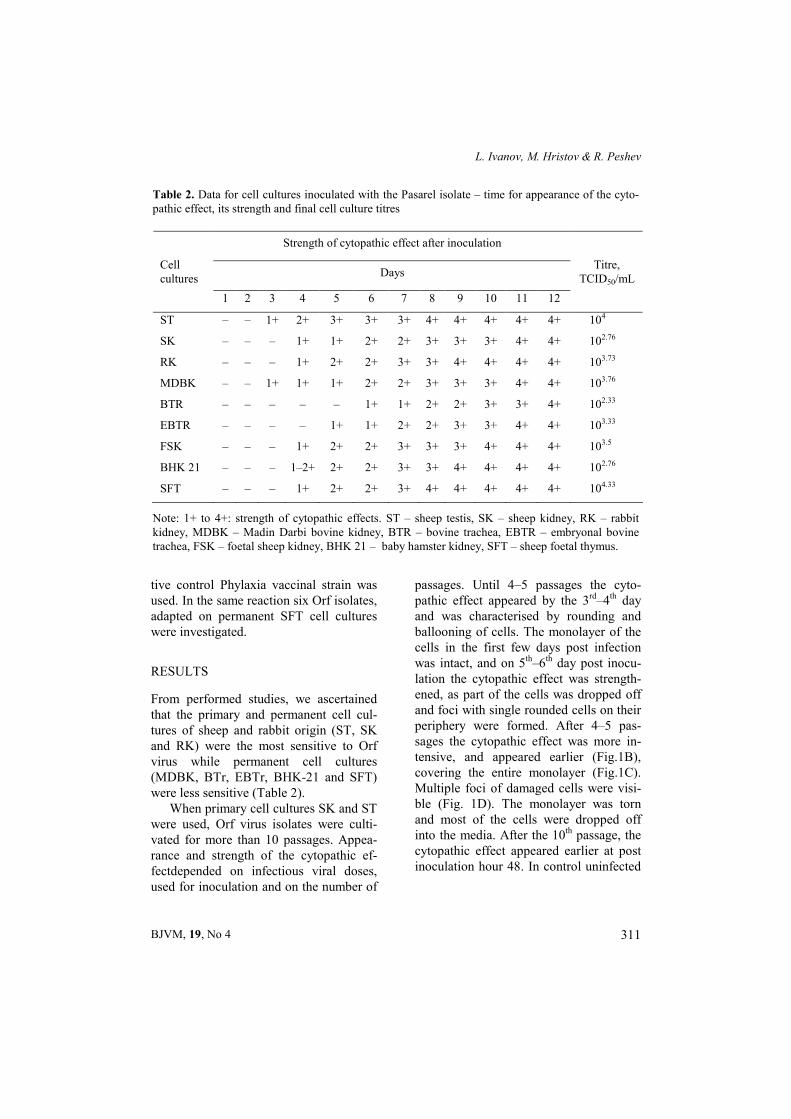
From performed studies, we ascertained that the primary and permanent cell cul-tures of sheep and rabbit origin (ST, SK and RK) were the most sensitive to Orf virus while permanent cell cultures (MDBK, BTr, EBTr, BHK-21 and SFT) were less sensitive (Table 2).
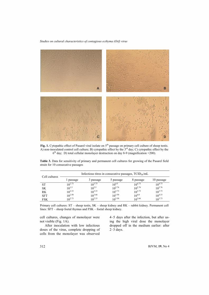
When primary cell cultures SK and ST were used, Orf virus isolates were culti-vated for more than 10 passages. Appea-rance and strength of the cytopathic ef-fectdepended on infectious viral doses, used for inoculation and on the number of
passages. Until 4–5 passages the cyto-pathic effect appeared by the 3rd–4th day and was characterised by rounding and ballooning of cells. The monolayer of the cells in the first few days post infection was intact, and on 5th–6th day post inocu-lation the cytopathic effect was strength-ened, as part of the cells was dropped off and foci with single rounded cells on their periphery were formed. After 4–5 pas-sages the cytopathic effect was more in-tensive, and appeared earlier (Fig.1B), covering the entire monolayer (Fig.1C). Multiple foci of damaged cells were visi-ble (Fig. 1D). The monolayer was torn and most of the cells were dropped off into the media. After the 10th passage, the cytopathic effect appeared earlier at post inoculation hour 48. In control uninfected
Table 2. Data for cell cultures inoculated with the Pasarel isolate – time for appearance of the cyto-pathic effect, its strength and final cell culture titres
Strength of cytopathic effect after inoculation
Days Cell cultures
1 2 3 4 5 6 7 8 9 10 11 12
Titre, TCID50/mL
SТ – – 1+ 2+ 3+ 3+ 3+ 4+ 4+ 4+ 4+ 4+ 104
SK – – – 1+ 1+ 2+ 2+ 3+ 3+ 3+ 4+ 4+ 102.76
RK – – – 1+ 2+ 2+ 3+ 3+ 4+ 4+ 4+ 4+ 103.73
MDBK – – 1+ 1+ 1+ 2+ 2+ 3+ 3+ 3+ 4+ 4+ 103.76
BTR – – – – – 1+ 1+ 2+ 2+ 3+ 3+ 4+ 102.33
EBTR – – – – 1+ 1+ 2+ 2+ 3+ 3+ 4+ 4+ 103.33
FSK – – – 1+ 2+ 2+ 3+ 3+ 3+ 4+ 4+ 4+ 103.5
BHK 21 – – – 1–2+ 2+ 2+ 3+ 3+ 4+ 4+ 4+ 4+ 102.76
SFT – – – 1+ 2+ 2+ 3+ 4+ 4+ 4+ 4+ 4+ 104.33
Note: 1+ to 4+: strength of cytopathic effects. ST – sheep testis, SK – sheep kidney, RK – rabbit kidney, MDBK – Madin Darbi bovine kidney, BTR – bovine trachea, EBTR – embryonal bovine trachea, FSK – foetal sheep kidney, BHK 21 – baby hamster kidney, SFT – sheep foetal thymus.
Studies on cultural characteristics of contagious ecthyma (Orf) virus
BJVM, 19, No 4 312
cell cultures, changes of monolayer were not visible (Fig. 1A).
After inoculation with low infectious doses of the virus, complete dropping of cells from the monolayer was observed
4–5 days after the infection, but after us-ing the high viral dose the monolayer dropped off in the medium earlier: after 2–3 days.
C
A B
D
Fig. 1. Cytopathic effect of Pasarel viral isolate on 5th passage on primary cell culture of sheep testis. A) non-inoculated control cell culture; B) cytopathic effect by the 3rd day; C) cytopathic effect by the
6th day; D) total cellular monolayer destruction on day 8-9 (magnification ×200).
Table 3. Data for sensitivity of primary and permanent cell cultures for growing of the Pasarel field strain for 10 consecutive passages
Infectious titres in consecutive passages, TCID50/mL Cell cultures
1 passage 3 passage 5 passage 8 passage 10 passage
ST 102.73 103.73 104.5 104.73 104.73 SK 101.5 102.5 102.76 102.76 102.76 RK 102.33 103.33 103.73 103.73 103.73 SFT 102.50 103.66 103.50 104.0 104.33 FSK 102.73 102.33 103.50 103.66 103.73
Primary cell cultures: ST – sheep testis, SK – sheep kidney and RK – rabbit kidney. Permanent cell lines: SFT – sheep foetal thymus and FSK – foetal sheep kidney.
L. Ivanov, M. Hristov & R. Peshev
BJVM, 19, No 4 313
Upon titration of the virus on ST cell culture, the extra cellular virus titre was 103.66 TCID50 /mL, the intracellular one – 102.66 TCID50/mL and the total virus titre was 104.0 TCID50/mL. After using SK cell culture, the cytopathic effect appeared later with lower titres (by 0.5 to 1.5 log10) compared to the ST cell culture.
In using RK cell cultures, the cyto-pathic effect appeared by the 2nd–3rd day, as cells were rounded and lost connection with neighbouring cells. After appearance of the cytopathic effect, after 4-5 days almost all cells were affected, a great part of them dropped off to the maintainance media. The viral titres ranged from log 102.33 to log 103.66 TCID50/mL.
By using permanent cell cultures of sheep (SFT, SFK) and bovine origin (BTR, EBTR and MDBK) the changes of monolayer appeared by the 3rd–4th day, and after 5 to 7 days they covered a larger area of cell monolayer. In SFT and bovine cell cultures, dropping off of a great part of monolayer cells into the maintainance median was observed by 7th–9th and 10th–12th day, respectively. Viral titres on EBTR and MDBK cell cultures ranged from log103.33 to log103.76 TCID50/mL.
The investigation of the dynamics of multiplication of Pasarel strain on primary and permanent cell cultures for 10 con-secutive passages, the viral growth ini-tially increased up to the 5th passage. After
that, in permanent cell cultures of sheep and primary cell cultures of rabbit origin, a constant level of infectious titres was ascertained (Table 3).
The cytopathic effect in primary ST cell cultures of investigated Orf virus iso-lates in roller cultivation appeared earlier (after the 12th h), compared to the statio-nary cultivation (after the 24th h). Depen-ding on cell cultures (sheep or bovine) the viral titres were by 0.5 to 1.0 log10 lower vs stationary cultivation.
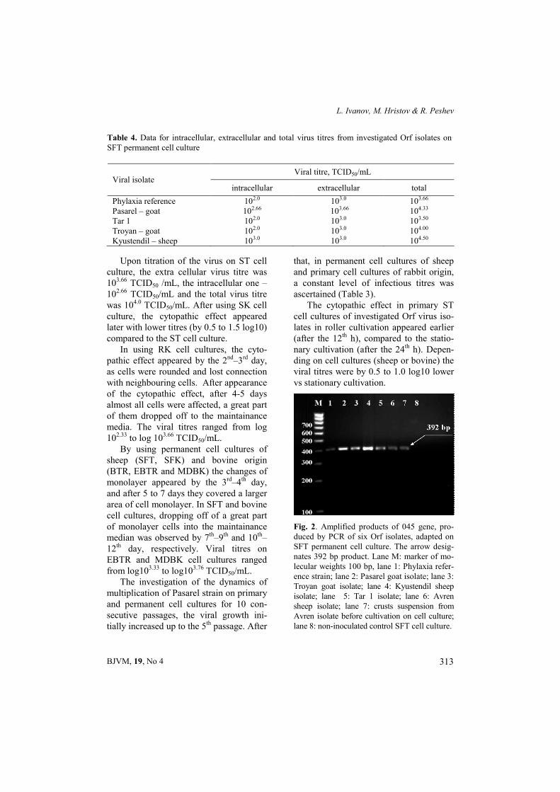
Fig. 2. Amplified products of 045 gene, pro-duced by PCR of six Orf isolates, adapted on SFT permanent cell culture. The arrow desig-nates 392 bp product. Lane M: marker of mo-lecular weights 100 bp, lane 1: Phylaxia refer-ence strain; lane 2: Pasarel goat isolate; lane 3: Troyan goat isolate; lane 4: Kyustendil sheep isolate; lane 5: Tar 1 isolate; lane 6: Avren sheep isolate; lane 7: crusts suspension from Avren isolate before cultivation on cell culture; lane 8: non-inoculated control SFT cell culture.
Table 4. Data for intracellular, extracellular and total virus titres from investigated Orf isolates on SFT permanent cell culture
Viral titre, TCID50/mL Viral isolate
intracellular extracellular total
Phylaxia reference 102.0 103.0 103.66 Pasarel – goat 102.66 103.66 104.33 Tar 1 102.0 103.0 103.50 Troyan – goat 102.0 103.0 104.00 Kyustendil – sheep 103.0 103.0 104.50
Studies on cultural characteristics of contagious ecthyma (Orf) virus
BJVM, 19, No 4 314
After determination of intracellular, extracellular and total virus counts of five viral Orf isolates, the quantity of the intra-cellular virus was the lowest, while the quantity of extracellular and total viruses were the highest when permanent SFT cell culture was used (Table 4).
The PCR used primers, multiplying a fragment from the highly conservative 045 gene of the Orf virus, encoding the late transcription factor VLTF-1. With the used primers and parameters of reaction, DNA of 045 gene (size 392 bp) was am-plified from the six tested isolates and from the Phylaxia control reference strain (Fig. 2).
DISCUSSION
For studies of the Orf virus cultural char-acteristics, we used primary and perma-nent cell cultures of different origin. Stud-ies on the Orf virus replication were per-formed by Balassu & Robinson (1987) on primary cell cultures. The authors studied the kinetics of accumulation of the viral DNA, analysed the viral polypeptides and the production of infectious virus. They have found that the virus yield on the pri-mary cell cultures from sheep testicles and rabbit kidneys were the highest and have concluded that they were the most suitable cells for multiplication of the virus, which was confirmed by the current investiga-tion. Most probably the receptors on this type of cells were most suitable for at-tachment and growth of the virus.
With increasing the number of pas-sages, the titres of tested isolates also in-creased, demonstrating the adaptation of the agents to the cells with increasing the number of passages. The obtained data are similar to the results of Greig (1957) for primary cell cultures from sheep embry-onic skin after 10 serial passages. The
observed cytopathic effect is characterised by rounding of many cells, followed by increased granularity of the cellular cyto-plasm and fragmentation. Most of the cells affected by the Orf virus are de-tached from the walls and floated in the maintainance medium with advancing of the viral development. Most of the cells that remained on the walls were accumu-lated as plaques with long cytoplasmic shoots. With increasing the number of passages, the cytopathic effect appeared earlier. Houssawi & Abu Elsein (2000) have made two passages of Orf virus on primary sheep testicles and after that the virus was adapted on permanent Vero CC. Pyknosis and cell dropping off at 5–8 days post inoculation have been observed by Said (2013). After infection of chicken embryo with positive cell culture isolates, over the chorioallantoic membrane the author found thickening of the membrane in nine samples and seven of them were confirmed as Orf viruses by agar gel im-mune diffusion test.
In the present study the titres of virus cultivated in the permanent cell cultures of bovine origin were lower, but these cell cultures may also be used for cultivation. Most probably the reason for lower titres was the incomplete correspondence be-tween cellular and viral receptors. Viral multiplication after the 3rd subcultivation after using the SPEV cell line was not ascertained. This indicates discrepancy of viral and cellular receptors, which did not allow optimal viral development resulting in progressive decrease in the number of the viral infectious particles. Another probable reason is the formation of in-complete or defective viral particles that interfere with the infectious virus. A lot of experiments for Orf virus cultivation on the primary or permanent cell cultures have not been successfull. Using a wide
L. Ivanov, M. Hristov & R. Peshev
BJVM, 19, No 4 315
variety of permanent cell cultures, Trayk-ova (1982) found cytopathic effect only in MK cell cultures, that appeared on 4th–5th day post inoculation and resulted in rounding and ballooning of the cells, without foci formation. Cytopathic effect was observed only on 2nd and 3rd passage and in 4–5th passage it was completely lost and the culture stayed unchanged. In other permanent cell cultures, the titres were lower, in permanent RK the cytopathic effect disappeared after the second passage, while in SPEV – after the third one.
Nagington & Whittle (1961) have ino-culated amniotic cells with material from man with contagious ecthyma and 18 h post infection they found rounding, scat-tering and granulation of the cells and absence of other changes till the 15th day. After that they have observed few empty spaces on the edge of the monolayer and have attempted to cultivate the virus in permanent cell cultures. In Hela, MK 2, Am9 cell cultures they did not observe development of the virus for 5 passages.
In the present study Orf viruses, ini-tially adapted on the primary cell cultures from ST were used on permanent cell cul-tures of sheep and bovine origin. Cultiva-tion on the permanent cell cultures of sheep origin demonstrated the adaptation of the virus on this type of cell cultures. On cell cultures of bovine origin clear cytopathic effect, which later weakened was observed after the first 2–3 passages. Sheep parapox virus was cultivated and was titred by plaque method on the per-manent MDBK cell culture by Kruse & Weber (2001), and was used for studying the apoptosis. The authors proved selec-tive induction of apoptosis in antigen-presenting cells of mice by sheep parapoxvirus.
The used PCR variant successfully multiplied part of DNA both from field
samples as well as from viral isolates adapted on cell culture. This is an evi-dence that isolates obtained and adapted on cell cultures were Orf viruses and con-firmed their successful cultivation.
CONCLUSIONS
Primary and permanent cell cultures of sheep origin were found as most suitable for cultivation of Orf viruses because the viruses were multiplied with highest quan-tity.
The cytopathic effect was the strongest in primary and permanent cell cultures of sheep origin and depended on the type of cultivation: roller or stationary.
The used variant of PCR was capable to confirm the Orf virus in samples and in cell cultures.
REFERENCES
Balassu, T. C. & A. J. Robinson, 1987. Orf virus replication in bovine testis cells: ki-netics of viral DNA, polypeptide, and in-fectious virus production and analysis of virion polypeptides. Archives of Virology, 97, 267–281.
Greig, A., 1957. Contagious ecthyma of sheep. II - In vitro cultivation of the virus, Cana-dian Journal of Comparative Medicine, 21, 304–308.
Housawi, F. & E. Abu-Elzein, 2000. Conta-gious ecthyma associated with myasis in sheep. Revue scientifique et technique (In-ternational Office of Epizootics), 19, 863–866.
Kottaridi, C., K. Nomikou, R. Lelli, P. Markoulatos & O. Mangana, 2006. Labo-ratory diagnosis of contagious ecthyma: Comparison of different PCR protocols with virus isolation in cell culture. Journal of Virological Methods, 134, 119–124.
Kruse, N. & O. Weber, 2001. Selective induc-tion of apoptosis in antigen presenting
Studies on cultural characteristics of contagious ecthyma (Orf) virus
BJVM, 19, No 4 316
cells in mice by parapoxvirus ovis. Journal of Virological Methods, 75, 4699–4704.
Kuyumdziev, I. & T. Todorov, 1962. Tissue cultural and electronic microscopy studies on virus of Contagious Ecthyma. Microbi-ology Institute 13, 21–28.
Leavell, U. W., M. J. McNamara, G. R. Muel-lin, W. M. Talbert, R. C. Rucker & A. J. Dalton, 1968. Orf. Report of 19 human cases with clinical and pathological obser-vations. Journal of the American Veteri-nary Medical Association, 204, 109–116.
Murphy, J. K. & I. G. Ralphs, 1996. Bullous pemphigoid complicating human orf. Brit-ish Journal of Dermatology, 134, 929–930.
Nagington, J. & C. Whittle, 1961. Human orf. Isolation of the virus by tissue culture. British Medical Journal, 18, 1324–1327.
Paiba, G. A. D., R. H. Thomas, K. L. Morgan, M. Bennett, R. L. Salmon, R. Chalmers, S. M. Kench, T. J. Coleman, D. Meadows, P. Morgan-Capner, P. Softley & M. Sillis, 1999. Orf (contagious pustular dermatitis) in farmworkers: Prevalence and risk fac-tors in three areas of England. The Veteri-nary Record, 145, 7–11.
Plowright, W., M. A. Witcomb & R. D. Ferris, 1959. Studies with a strain of contagious pustular dermatitis virus in tissue culture. Archives für die gesamte Virusforschung, 9, 214–231.
Reed, L. J. & H. Muench, 1938. A simple method of estimating fifty percent end-points. American Journal of Hygiene, 27, 493–497.
Said, A., 2013. Trials for isolation of conta-gious pustular dermatitis virus (CPDV) from sheep in Ismailia Governorate. Re-search in Zoology, 3, 10–14.
Traykova, M., 1982. Attempts for adapring virus of contagious ecthyma on cell cul-tures. Veterinary Medicine Sciences (Sofia), 10, 18–25 (BG).
Wilkinson, J. D., 1977. Orf: A family with unusual complications. British Journal of Dermatology, 97, 447–450.
Paper received 12.06.2015; accepted for publication 13.11.2015
Correspondence: Lyudmil Ivanov National Diagnostic and Research Veterinary Medical Institute 15 P. Slaveikov Blvd 1000, Sofia, tel. +359 889 95 96 95 e-mail: [email protected]