Embed Size (px)
Citation preview

119
Animal Science Papers and Reports vol. 36 (2018), no. 2, 119-148 Institute of Genetics and Animal Breeding, Jastrzębiec, Poland
Canis lupus familiaris as relevant animal model for breast cancer - a comparative oncology review
Lajos Raduly1,2, Roxana Cojocneanu-Petric1*, Orsolya Sarpataki2, Sergiu Chira1, Atanas G. Atanasov5,6, Cornelia Braicu1, Ioana Berindan-Neagoe1,3,4,#, Ioan Marcus2,# 1 Research Center for Functional Genomics, Biomedicine and Translational Medicine,
Iuliu Hatieganu University of Medicine and Pharmacy, Cluj-Napoca, Romania2 University of Agricultural Sciences and Veterinary Medicine, Faculty of Veterinary Medicine,
Cluj-Napoca, Romania 3 MEDFUTURE - Research Center for Advanced Medicine, Iuliu-Hatieganu University
of Medicine and Pharmacy, Cluj-Napoca, Romania 4 Department of Functional Genomics, Proteomics and Experimental Pathology,
The Oncology Institute Prof. Dr. Ion Chiricuta, Cluj-Napoca, Romania 5 Institute of Genetics and Animal Breeding, Polish Academy of Sciences, 05-552 Jastrzębiec6 Department of Pharmacognosy, University of Vienna, Althanstrasse 14, 1090 Vienna, Austria*Co-First author (Equal contribution): Roxana Cojocneanu-Petric#Authors with equal contribution
(Accepted March 26, 2018)
Comparative oncology represents an important tool in cancer research and drug development. After the sequencing of the canine genome, many similarities between canine and human tumors have been noticed in what concerns physiological and pathological conditions, including tumorigenesis and many clinical aspects. Breast cancer is a complex and heterogeneous group of mammary neoplastic diseases, thus, it is important to find suitable animal models for new biomarker discovery and therapeutic strategies. From histopathological to molecular level, many similarities between canine and human breast cancers show that dogs can be reliable models for this pathology, and can improve the therapeutic options. In this review, we synthesized the most recent studies that demonstrate the homologies between dogs and humans in terms of mammary cancer development
*Corresponding author: [email protected]
120
and progression. These findings have the potential to bring important contributions to human and veterinary medicine, under the concept of “One Health”.
KEYWORDS: animal model / biomarker / breast cancer / comparative oncology / therapy
In the last years, a new global concept was developed in the interdisciplinary collaboration between human, animal and environment health sciences. This strategy called One Health represents a combined effort of multiple disciplines to improve health in humans, animals and plants in the global ecosystem, using an integrated research approach [Lerner and Berg 2015]. Human cancers lack multiple models for experimental research. Respectfully, after the accomplishment of Human Genome Project, other genomes – including canine – were sequenced, and showed common features with the human genome. The Human Genome Project provides us much important information especially in clinical medicine to understanding of human diseases in terms of human biology and pathology. Moreover, canine cancers have certain characteristics among which are a conservatory immune system, specific tumor microenvironment, and a high degree of tumor heterogeneity. Canine spontaneous tumors have additional unique features, some of which are being absent in mouse models, such as maintaining tumor microenvironment conditions necessary for the development and progression of malignancies, including specific oxygen levels in the body and the growth of new blood vessels.
The field of comparative oncology has experienced an exceptional advancement over the past years and has progressively captivated the interest of specialists in cancer research through the development of new study areas such as comparative genomics. This research field could be the key to a better understanding of the relevant challenges involving cancer, starting from epidemiological issues, prevention or novel targeted therapies, to personalized health care [Huminiecki et al. 2017, Huminiecki and Horbańczuk 2018, Nunney et al. 2015].
Comparative genomics represents an important field of biology which focuses on comparing the genomes of different species. In cancer research, these comparative studies are very significant, since they lay the grounds for finding molecular similarities between species such as human, mouse and dog, and promote them as study models for a better understanding of pathogenesis processes in all species. After the sequencing of the first human genome as a result of the Human Genome Project, researchers started to sequence the genomes of various important animal models, such as the mouse genome sequenced (Mouse Genome Sequencing Consortium) by Waterston [Mouse Genome Sequencing, Waterston et al. 2002], and the canine genome sequenced by Linblad-Toh and published in 2005 [Lindblad-Toh et al. 2005]. Time and again, sequencing has proven itself to be an important tool for finding similarities between species in terms of DNA, RNA, miRNAs, and lncRNAs sequences, to be used for emerging studies of mutations, common genes and their role in new cancer therapeutic approaches [Pop et al. 2014, Irimie et al. 2015]. The identification and use of animal models which develop spontaneous tumors with similar molecular profiles to human cancers can provide important information, essential for making advances
L. Raduly et al.

121
in discovering new and improved oncological drugs.Several murine models have been developed and used to investigate human cancer
mechanisms, starting with the identification of factors correlated with malignant transformation, invasion or metastasis, and, more recently, for the evaluation of novel prognostic or diagnostic biomarkers [Richmond and Su 2008, Brown et al. 2015]. These models were used to investigate the factors involved in malignant development and progression, as well as to examine the response to therapy. Nowadays, mouse cancer models are used to investigate signaling pathways involved in major mechanisms like carcinogenesis, cancer promotion, progression and metastasis. Many studies demonstrated that several murine models also showed heterogeneity and genomic instability, and did not present any of the key features that define cancer in humans, like long periods of latency, complexity of cancer recurrence and metastases, and the response to new generation therapies [Seok et al. 2013, Schiffman and Breen 2015].
Another issue related to mouse models is represented by their incapacity to predict tumor response to therapy, this being translated into lack of success for many phase I or II clinical trials [Seok et al. 2013, Mak et al. 2014]. The most important aspect influenced by this condition is the fact that a fraction of the patients do not respond to first-line therapy, due to tumor heterogeneity and to the multiclonal development of the disease. Murine models do not develop similar cancer subtypes as humans, and, consequently, only limited drug response tests can be conducted. The newly developed avatar mice are extremely expensive and their use is limited to a few research groups. In this particular situation, more attempts are necessary in order to administer the suitable drugs. Thus, the quality of life of these patients is severely affected, as well as their overall survival [Sparano et al. 2004]. Due to this, it is necessary to find other cancer models for an improved, more efficient investigation of the biology of human malignancies. Larger animal species like dogs or cats have higher chances of developing some form of cancer during their lifetime than smaller animals, since each cell division brings a subsequent risk of spontaneous mutations for a tumor lineage, like in the case of humans [Brown et al. 2015, Fazekas et al. 2016] – Figure 1.
Studies demonstrate that cancers occurring in dogs share many characteristics with human malignancies [Ranieri et al. 2013, Gardner et al. 2016]. The size of the haploid dog genome is estimated to be 2445 Mb, and the diploid dog genome is organized in 38 pairs of autosomes and two sex chromosomes. Studies show that over ~650 Mb of ancestral dog DNA sequence is common with humans and absent in mice [Alvarez 2014]. A comparative analysis of 13,816 protein-coding genes with 1:1:1 orthology in human, mouse and dog, showed that the numbers of lineage-specific non-synonymous substitutions (i.e., amino acid changing; KA) are 0.017, 0.038, and 0.021 [Lindblad-Toh et al. 2005, Rowell et al. 2011]. Dogs are susceptible to inherited diseases that are also common in humans, like cancer and other diseases, such as hemophilia and neuropathologies [Sargan 2004, Schiffman and Breen 2015]. Many studies conducted on canine models showed that they are suitable models for human cancer research and translational medicine [Lindblad-Toh et al. 2005, Rowell et al. 2011, Alvarez 2014].
Canis lupus familiaris as relevant animal model for breast cancer

122
Canine as models for breast cancer research
Recent studies demonstrated high similarities between human breast cancer (HBC) and canine mammary tumors (CMT), from histopathological to molecular level. At the same time, the same course of the disease was described in both species [Grosse et al. 2014, Rasotto et al. 2014]. In female dogs, like in women, there can be found similar shared characteristics of breast cancer, such as spontaneous tumors, hormonal etiology, identical course of the disease, and age of onset. Tumor size evolution, clinical staging and lymph node metastases represent clinical similarities between HBC and CMT which are encountered both at macroscopic and at molecular level [Klopfleisch et al. 2011]. These similarities include the overexpression of estrogen and progesterone receptors, similar proliferation markers, epidermal growth factor resemblance, TP53 mutations, comparable features of metalloproteinases and
L. Raduly et al.
Fig. 1. Advantages of Canine models for New Cancer Therapy development. A) In vitro preclinical studies in cell cultures. B) and C) Preclinical in vivo studies in murine or small animal and human primate and Beagle dog models. D) Phase I human clinical trials: medication safety studies in 20-80 cases – several weeks and months. E) Phase II human clinical trials: safety and efficacy studies, side effect identifying in 100-300 cases – up to two years F) Phase III human clinical trials: safety, efficacy, dosing studies, side effect monitoring 1000-3000 cases – one to four years G) Preclinical comparative oncology studies, better animal model for drug discovery. H) Comparative oncology studies in dogs can eliminate drugs with an unfavorable therapeutic index and can focus in pharmacokinetics, pharmacodynamics in Phase I and II clinical trials. I) Comparative studies can optimize the design of the clinical trials and help to identify most effective drugs in Phase III clinical trials.

123
cyclooxygenases [Queiroga et al. 2011, Queiroga et al. 2015, Spoerri et al. 2015, Hegde et al. 2016].
With the common purpose of increasing survival rates in patients with mammary neoplasms, these studies can help the development of new therapeutic strategies in both human and veterinary medicine. CMT can represent a good model to study HBC, from the epidemiological studies to the histopathological patterns of the tumor [Vascellari et al. 2016].
Incidence and risk factors
Breast cancer represents one of the most frequent cancer in both women and female dogs, with incidence levels growing more and more in the last years. Globally, in 2012, human breast cancer (HBC) represented 25% of all cancers in the female population. After skin cancer, canine mammary tumors (CMT) are the second most common cancers in female dogs. According to statistical studies from different countries, the incidence of CMT in the last years is 25-47.5% [Gupta et al. 2012, Salas et al. 2015]. According to Dobson’s studies (2013), in the UK 27% of all deaths in pure-bred dogs were caused by cancer. Statistical data show that HBC, similar to CMT, represents approximately 50% of malignant tumors in human and dog patients [Queiroga et al. 2011, Dobson 2013].
As companion pets, dogs live in the same environment as their owners and they are exposed to similar carcinogenic factors. These factors, as well as age and obesity, and shared genomes, bring mammary cancer development pattern in dogs closer to the human breast cancer than the murine models [Lim et al. 2015]. Annual incidence of CMT estimated at 198/100,000, is comparable to the rate of 125/100,000 for HBC. A risk for malignant tumor development of approximately 26% in spayed or non-spayed dogs after the second estrus was observed [Radha et al. 2014, Salas et al. 2015]. These studies revealed similar hormonal etiology between CMT and HBC. Several clinical and histopathological similarities between CMT and HBC were observed for the same molecular types and subtypes of mammary cancer. Canine mammary simple carcinomas histologically match human breast carcinomas, presenting large genomic aberrations and showing corresponding key features of human breast cancer [Liu et al. 2014]. In humans, associated risks involve the relation between obesity and mammary tumors, and the influence of hormone-related variables, such as puberty onset, existence and number of pregnancies, history of lactation, and menopause debut. Moreover in canines, studies observed a lower age for the development of CMT in overweight or obese dogs (9.0 years) than in normal bodyweight dogs (10.2 years). At the same time, lymphatic invasion of carcinoma cells was found to be more frequent in overweight dogs than in optimal weight dogs [Liu et al. 2014, Lim et al. 2015].
According to Globocan 2012, human breast cancer presented a high incidence and a lower survival rate in patients above the age of 50, while for the age-wise distribution of CMT, the highest incidence was observed in the age group of 10-12,
Canis lupus familiaris as relevant animal model for breast cancer

124
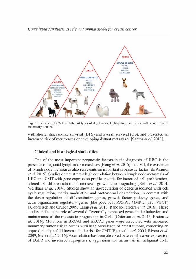
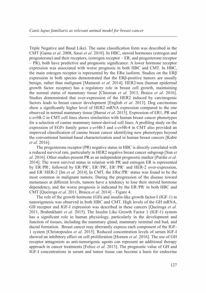
taking into account the relation between human and canine ages [Gupta et al. 2012, Radha et al. 2014, Ferlay et al. 2015, Salas et al. 2015] – Figure 2. High incidence of CMT was reported in purebreds like German Shepherds, Labradors and Spitz [Gupta et al. 2012]. Other studies confirmed that, when looking at CMT frequency according to the breed, it presents a higher percentage among purebred animals in comparison to mixed-breed animals (80% vs. 20%, respectively). The frequency of CMT is also higher in small breeds than in larger breed female dogs. According to size, larger breeds like German Shepherd or Labradors present an incidence of 23%, medium size breeds such as mixed breeds, Boxers or Beagles have 29%, while smaller breeds like Cocker, Maltese, or Schnauzer present a 48.8% incidence of CMT [Salas et al. 2015] – Figure 3. Another study described overall median survival time in 126 dogs to be 1113 days. According to Cox regression analysis, some variables such as histological grade (p=0.029, HR 4.31, 95% CI 1.3-14.31) and age (p=0.009, HR 1.31, 95% CI 1.1-1.62) are important factors in defining the overall survival (OS) [Betz et al. 2012]. Another study showed a ±SD survival time for dogs with recurrence/distant metastases of 11.96±7.58 (range 2-24 months) and the mean time for the detection of recurrence/metastasis of 5.29±5.68 (range 1-21 months), the animals presenting a 48% overall 2-years survival. Large tumors (≥3 cm) and invasive tumors were associated
L. Raduly et al.
Fig. 2. Comparative incidence of CMT and HBC according to age distribution for humans and dogs emphasizes the relevance of dog as suitable animal models for studying breast cancer. CMT development is mostly reported between 8 and 12 years correlated with the 54-64 year in women.
125
with shorter disease-free survival (DFS) and overall survival (OS), and presented an increased risk of recurrences or developing distant metastases [Santos et al. 2013].
Clinical and histological similarities
One of the most important prognostic factors in the diagnosis of HBC is the presence of regional lymph node metastases [Hong et al. 2015]. In CMT, the existence of lymph node metastases also represents an important prognostic factor [de Araujo, et al. 2015]. Studies demonstrate a high correlation between lymph node metastasis of HBC and CMT with gene expression profile specific for increased cell proliferation, altered cell differentiation and increased growth factor signaling [Beha et al. 2014, Weishaar et al. 2014]. Studies show an up-regulation of genes associated with cell cycle regulation, matrix modulation and proteasomal degradation, in contrast with the down-regulation of differentiation genes, growth factor pathway genes, and actin organization regulatory genes (like p53, p21, RXFP1, MMP-2, p27, VEGF) [Klopfleisch and Gruber 2009, Lamp et al. 2013, Raposo-Ferreira et al. 2016]. These studies indicate the role of several differentially expressed genes in the induction and maintenance of the metastatic progression in CMT [Chiorean et al. 2013, Braicu et al. 2016]. Mutations in BRCA1 and BRCA2 genes were associated with increased mammary tumor risk in breeds with high prevalence of breast tumors, conferring an approximately 4-fold increase in the risk for CMT [Egenvall et al. 2005, Rivera et al. 2009, Melin et al. 2016]. A correlation has been observed between the over-expression of EGFR and increased angiogenesis, aggression and metastasis in malignant CMT
Canis lupus familiaris as relevant animal model for breast cancer
Fig. 3. Incidence of CMT in different types of dog breeds, highlighting the breeds with a high risk of mammary tumors.
126
[Carvalho et al. 2013]. Taking all these into account, it is foreseeable that CMT has the potential to become a useful and more commonly used translational model for further studies of HBC [Klopfleisch et al. 2011, Weishaar et al. 2014].
A high histological similarity between human and dog mammary tumors was described in several studies. According to Radmehr Shafiee [2013], the Elston and Ellis method of histological grading used in human medicine can be a reliable prognostic factor in veterinary medicine as well [Shafiee et al. 2013]. The most common tumor types that affect canine mammary glands are complex carcinomas and simple carcinomas. Anaplastic carcinoma subtypes were associated with grade III tumors and carcinoma-tubular subtypes, while carcinoma arising in a complex adenoma/mixed-tumor subtype is associated with grade I tumors. Invasion into the lymphatic system was observed in comedocarcinoma, anaplastic carcinoma, and inflammatory carcinoma subtype of CMT. In addition, the most frequently occurring molecular subtype was luminal A, while the basal-like subtype was the most malignant form associated with grade III tumors and lymphatic invasion [Goldschmidt et al. 2011, Im et al. 2014]. Morphology and immunohistochemical studies demonstrate that canine invasive micropapillary carcinoma features are similar with the ones in humans and present a poor prognosis, aggressive development and high degree of metastases to regional lymph nodes [Goldschmidt et al. 2011, Gamba et al. 2013, Alvarez 2014, Beha et al. 2014] – Table 1. These similarities between the histopathological
L. Raduly et al.
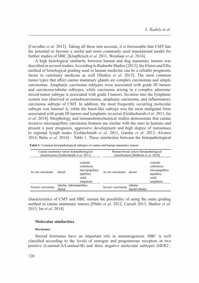
Tabel 1. Common histopathological subtypes of canine and human mammary tumors
Canine mammary tumor histopathological classification [Goldschmidt et al. 2011]
Human breast cancer histopathological classification [Malhotra et al. 2010]
In situ carcinoma ductal
comedo
In situ carcinoma ductal
comedo cribriform cribriform micropapillary micropapillary papillary papillary solid solid anaplastic anaplastic
Invasiv carcinoma tubular, tubulopapillary Invasiv carcinoma tubular ductal ductal lobular
characteristics of CMT and HBC sustain the possibility of using the same grading
method in canine mammary tumors [Pinho et al. 2012, Cassali 2013, Shafiee et al. 2013, Im et al. 2014].
Molecular similarities
Hormones
Steroid hormones have an important role in mammogenesis. HBC is well classified according to the levels of estrogen and progesterone receptors in two positive (Luminal-A/Luminal-B) and three negative molecular subtypes (HER2+,
127
Triple Negative and Basal Like). The same classification form was described in the CMT [Gama et al. 2008, Sassi et al. 2010]. In HBC, steroid hormones (estrogen and progesterone) and their receptors, (estrogen receptor – ER, and progesterone receptor – PR), both have predictive and prognostic significance. A lower hormone receptor expression was associated with worse prognosis in both HBC and CMT. In HBC, the main estrogen receptor is represented by the ERα isoform. Studies on the ERβ expression in both species demonstrated that the ERβ-positive tumors are usually benign, rather than malignant [Mainenti et al. 2014]. HER2/neu (human epidermal growth factor receptor) has a regulatory role in breast cell growth, maintaining the normal status of mammary tissue [Chiorean et al. 2013, Braicu et al. 2016]. Studies demonstrated that over-expression of the HER2 induced by carcinogenic factors leads to breast cancer development [English et al. 2013]. Dog carcinomas show a significantly higher level of HER2 mRNA expression compared to the one observed in normal mammary tissue [Burrai et al. 2015]. Expression of ER1, PR and c-erbB-2 in CMT cell lines shows similarities with human breast cancer phenotypes for a selection of canine mammary tumor-derived cell lines. A profiling study on the expression of EGFr family genes c-erbB-3 and c-erbB-4 in CMT also provided an improved classification of canine breast cancer identifying new phenotypes beyond the conventional luminal-basal characterization used in human breast cancers [Kabir et al. 2016].
The progesterone receptor (PR) negative status in HBC is directly correlated with a reduced survival rate, particularly in HER2 negative breast cancer subgroup [Sun et al. 2016]. Other studies present PR as an independent prognostic marker [Purdie et al. 2014]. The worst survival status in relation with PR and estrogen ER is represented by ER-/PR-, followed by ER-/PR+, ER+/PR-, ER+/PR+ and HER-2 over-expression, and ER+/HER-2- [Im et al. 2014]. In CMT, the ERα-/PR+ status was found to be the most common in malignant tumors. During the progression of the disease toward metastases at different levels, tumors have a tendency to lose their steroid hormone dependency, and the worse prognosis is indicated by the ER-/PR- in both HBC and CMT [Queiroga et al. 2011, Braicu et al. 2014] – Figure 4.
The role of the growth hormone (GH) and insulin-like growth factor-I (IGF-1) in tumorigenesis was observed in both HBC and CMT. High levels of the GH mRNA, GH receptor and IGF-I expression was described in these cancers [Queiroga et al. 2011, Brahmkhatri et al. 2015]. The Insulin Like Growth Factor 1 (IGF-1) system has a significant role in human physiology, particularly in the development and function of tissues, including the mammary gland, mammary terminal end bud, and ductal formation. Breast cancer may aberrantly express each component of the IGF-1 system [Christopoulos et al. 2015]. Reduced concentration levels of serum IGF-I showed an inhibitory effect on cell proliferation [Hornen et al. 2016]. The use of GH receptor antagonists as anti-tumorigenic agents can represent an additional therapy approach in cancer treatments [Felice et al. 2013]. The prognostic value of GH and IGF-I concentrations in serum and tumor tissue can become a basis for endocrine
Canis lupus familiaris as relevant animal model for breast cancer

128
L. Raduly et al.
Fig. 4. Breast cancer molecular subtypes/histological grade/prognosis and therapy similarities for HBC and CMT.
Fig. 5. Schematic GH/IGF axis in breast cancer.

129
therapies development [Christopoulos et al. 2015, Horne et al. 2016]. In addition, a high association was observed between increased IGF-I tumor tissue levels and clinical follow-up of the cancer patients, such as rate of growth, tumor size, skin ulcerations and adherence of tumor cells [Christopoulos et al. 2015, Horne et al. 2016]. This positive correlation between tumorigenesis, malignancy and the GH and IGF-I increased level in malignant tissues can be an indication that this GH/IGF-I axis could have prognostic value [Queiroga et al. 2011, Felice et al. 2013, Christopoulos et al. 2015, Matos and Santos 2015] – Figure 5.
The role of prolactin (PRL) in the development and progression of malignant tumors was described by many recent studies [Wennbo and Tornell 2000, Zemmoura et al. 2013, Shemanko 2016]. The prolactin hormone, which is synthesized by the anterior pituitary gland, has multiple biological actions [Queiroga et al. 2005]. The first role of PRL is the lactogenic action, but in recent years many studies demonstrated that PRL might also act as a growth factor [Michel et al. 2012, Shemanko 2016]. PRL has an important role in mammary epithelial development, and studies show that PRL uptake can increase mammary tumor development. Some studies demonstrate a strong correlation between PRL and sex steroid hormones, especially between progesterone and PRL on the one hand, and the development of the mammary tissue, on the other [Huang et al. 2015, Spoerri et al. 2015]. The malignant phenotype of CMT was associated with loss of ERα (ESR1), PGR, GHR, and prolactin receptor (PRLR) gene expressions [Spoerri et al. 2015, Mohr et al. 2016]. High levels of serum PRL were associated with a decrease in the gene expression of the respective receptors only in malignant mammary neoplasms. Studies show that an increased level of prolactin correlated with steroid hormone levels can represent a prognostic factor in mammary neoplasms [Queiroga et al. 2005, Spoerri et al. 2015].
Molecular markers
Recent studies in human and veterinary medicine are focused on the discovery of prognostic factors that could lead to the identification of therapeutic targets, and on the development of new methods to increase the survival rate in cancer. There is a high similarity between HBC and CMT molecular prognostic markers, with potential impact on comparative oncology [Schiffman and Breen 2015].
Hereditary
BRCA1, BRCA2 and RAD51 genes are expressed normally in breast tissue cells and have important roles in DNA damage repair. BRCA1 and BRCA2 gene mutations can affect the DNA damage repair process and can contribute to an increased tumorigenic potential. HBC displays a high correlation between the aggressive tumor phenotype and low expression of the BRCA1, BRCA2 and RAD51 complex. The protein encoded by the RAD51 gene has an important role in the repair of DNA double strand breaks. In CMT, as in HBC, the role of RAD51 in DNA repair processes was demonstrated.
Canis lupus familiaris as relevant animal model for breast cancer
130
Studies showed the importance of these gene mutations in the development of the aggressive phenotypes of both HBC and CMT, and their role as potential prognostic biomarkers [Im et al. 2013]. The ER negative (P=0.004), PR negative (P=0.046), and triple negative (ER, PR, and HER-2 negative; P=0.016) phenotypes, as well as the basal-like molecular subtype (P=0.019) in Shih Tzu dogs were directly correlated with cytoplasmic and membranous expression of BRCA1 [Im et al. 2013]. Germline mutations in BRCA1/2 were associated with increased risk in mammary cancer development in some breeds with known high prevalence of CMT [Rivera et al. 2009].
Somatic-genetic
Some genes, like Ki-67, PCNA, P53, P63, HER2, EGFR, BRCA etc., involved in human breast cancer, are also active players is CMT carcinogenesis.
Encoded by the MKI-67 gene, antigen Ki-67 is a nuclear protein necessary for cellular proliferation. Ki-67 was identified in many human and dog malignant tissues, and has a role in ribosomal RNA transcription. Antigen Ki-67 inactivation leads to ribosomal RNA (rRNA) synthesis inhibition [Perez et al. 2015, Rossi et al. 2015, Sun et al. 2015]. PCNA, which has a role in the replication process, is a DNA clamp acting as a processor factor for DNA polymerase δ. Antigens Ki-67 and PCNA represent markers to determine cell proliferation, and their expression in human and canine malignant tissues was associated with metastases and poor prognosis [Inwald et al. 2013, Carvalho et al. 2016].
P53 is a protein, also named the guardian of the genome, encoded by the TP53 gene with a controller role in the cell cycle. It has both a DNA repair and tumor suppressor role. Studies demonstrated that mutations in the TP53 gene produce an immunohistochemical expression of accumulated p53 nuclear protein, deregulate cell proliferation and induce or sustain tumorigenesis. This over-expression of the mutant p53 genes in HBC and CMT was correlated with poor survival rates and can represent an important prognostic tool in both species [Queiroga et al. 2011, Dolka et al. 2016]. A positive correlation was described between cleaved caspase-3 (CC3) and Bcl-2 expression; CC3 and higher mitotic index (MI), ERα and p53 expression. In the longer-survival group (>18 months), CC3 expression was negatively correlated with ERα, whereas p53 expression was positively correlated with reduced tumor differentiation, higher mitotic index, invasive growth, and necrosis [Dolka et al. 2016]. There is a high similarity between the organization of the canine p53 coding exons and gene products, and the human p53 gene, and it is a promising therapy target in both human and canine breast cancers [Klopfleisch and Gruber 2009, Dobes et al. 2014, Silwal-Pandit et al. 2014].
Epidermal growth factor receptor (EGFR; ErbB-1; HER1 in humans) and HER-2/neu are members of the ErbB family of receptors, and subfamily of four closely related tyrosine kinases. The overexpression of these receptors has an important role in the development and progression of both HBC and CMT. Comparative studies at biological and molecular levels showed a high degree of homology between HBC
L. Raduly et al.

131
and CMT associated antigens ErbB-1 (91%) and ErbB-2 (92%) [Singer et al. 2012]. Immunohistochemistry studies revealed ErbB-1 over-expression in 3/10 and ErbB-2 in 4/10 patients with CMT. This demonstrates the role in canine tumorigenesis of ErbB-1 and ErbB-2, similar to human carcinomas. The homology of human and canine ErbB-1 and ErbB-2 tumor associated antigens also shows that they can serve as targets for anti-ErbB-1 and anti-ErbB-2 drugs, and can help the further development of new targeted therapies for both species [Singer et al. 2012, Burrai et al. 2015].
Extracellular proteins
Matrix metalloproteinases (MMPs) are part of the zinc-dependent endopeptidases from the metzincin superfamily [Klopfleisch et al. 2011]. They are involved in processes like cleavage of cell surface receptors, as well as in apoptotic processes, playing a major role in cell proliferation, cell migration, differentiation, and angiogenesis. In HBC, studies showed a correlation between MMP-2, MMP-9 and the lymph node metastatic processes [Aresu et al. 2011]. Also, in the aggressive phenotype of CMT, a high MMP-9 expression was observed [Santos et al. 2013]. These studies suggest that MMPs may be target molecules for the switch mechanism that leads to the progression of carcinomas from adenomas [Aresu et al. 2011, Santos et al. 2013].
Phosphatase and tensin homolog (PTEN) protein is encoded by the PTEN gene. With a regulatory activity in cell cycle, the PTEN gene has an important tumor suppressor role. In HBC, studies demonstrated a high correlation between the low expression of the PTEN protein and the aggressive ER-/PR- tumor phenotype, metastasis and low survival rates [Lebok et al. 2015]. Also, a low expression of the PTEN protein in CMT was observed [Qiu et al. 2008, Ressel et al. 2013].
During a normal cell cycle, heat-shock proteins (HSPs) have roles in stabilizing and assisting the trafficking of proteins. Several stress conditions induce an increased expression of the HSPs to protect cells via stabilization of the unfolded or misfolded proteins and restore the balance after the activation of signaling pathways in the event of acute or chronic stress factors. It was demonstrated that HSPs play a double role [Seigneuric et al. 2011]. First, in the cancer cell they contribute to tumor survival, and second, they have an extracellular immunological function to induce an anti-tumor response [Queiroga et al. 2011]. Extracellular heat shock proteins have a cytostimulatory role, inducing immune responses to control microbial infection and eliminate transformed cells [Asea 2006]. Recent studies showed that the expression levels of several HSP in human and canine tumor tissues have a prognostic value [Kumaraguruparan et al. 2006, Romanucci et al. 2008, Santagata et al. 2011]. HSP27 and HSP110 level were increased in human breast cancer, with poor prognostic value present also in other pathologies, like uterine, cervical, and bladder carcinomas [Ciocca et al. 2013]. Studies showed an increased expression of Bcl-2, Bcl-XL, HSP70 and HSP90 in apoptotic processes in both HBC and CMT [Kumaraguruparan et al. 2006]. Other studies revealed the implication of HSP70 and HSP27 in resistance to chemotherapy in HBC [Ma et al. 2013, Nadin et al. 2014, Davidson et al. 2016].
Canis lupus familiaris as relevant animal model for breast cancer

132
These similarities of HSPs in HBC and CMT and their implication in carcinogenesis, deregulations of apoptotic processes and response to therapy, could have a promising target therapy value, on the one hand by inducing pharmacological modification of HSPs expression or activity, or by using the HSPs in anticancer vaccines, on the other [Kumaraguruparan et al. 2006].
Mucins are transmembrane proteins which are glycosylated at the level of proline, threonine, and serine domains, with a role as physical barriers [Zelasko-Leon et al. 2015]. In stress conditions, a mucous barrier formed by secreted and transmembrane mucins protects the epithelium. These types of proteins have important roles in inflammatory processes and cancer [Nicolini et al. 2015, Zelasko-Leon et al. 2015]. Mucins are also active in tumor progression, being present not only as non-invasive markers for HBC, but also as possible therapeutic targets [Nicolini et al. 2015]. In HBC, an important correlation was described between mucin1 (MUC1) over-expression and well-differentiated tumors. MUC1 expression in malignant CMT was also observed. Over-expression of MUC1 was correlated with distant metastasis in mammary tumors in both dogs and humans. This over-expression of mucins in malignant HBC and CMT tissues offers them a good biomarker value, with the potential of representing a molecular marker for prognostic in these malignancies [de Oliveira et al. 2009]. Maspin is a serine proteinase inhibitor with suppressor activity in tumor invasion and metastasis in HBC and CMT. Studies showed that Maspin can represent a very sensitive marker for normal and neoplastic myoepithelium in mammary neoplasia, and can be correlated with the aggressiveness of HBC and CMT [Espinosa de los Monteros et al. 2005]. The tetrasaccharide carbohydrate Sialyl Lewis x (sLx) antigen is usually attached to O-glycans on the cell surface and has an important role in cell-to-cell recognition processes. In HBC and CMT, the sLx antigen facilitates the adhesion of carcinomas to the endothelium, with role in the activation of the metastatic cascade. In both HBC and CMT, studies demonstrated a high correlation between the expression of sLx and local lymph node metastasis [Pinho et al. 2007, Sozzani et al. 2008]. Prostaglandin endoperoxid synthase (PTGS) – is an enzyme with an important role in the formation of prostanoid biological mediators like prostaglandin, prostacyclin and thromboxane. COX-2 is a controlling enzyme with an important role in the conversion of the AA/ARA arachidonic acid to prostaglandin PGH2. It was observed that there is a high correlation between increased levels of COX-2 in HBC [Park et al. 2016] and CMT malignant tissues and biological processes, such as tumorigenesis, low tumor apoptosis, high metastatic processes, tumor angiogenesis, and tumor related inflammation [Park et al. 2014, Park et al. 2016]. Studies also showed an increased level of COX-2 in inflammatory breast cancers, a very rare type of breast cancer [Belevych et al. 2013, Esbona et al. 2016, Park et al. 2016]. These studies show that these COX enzymes are potential targets of non-steroidal anti-inflammatory drugs, which could increase the survival rates in these aggressive diseases [Guimaraes et al. 2014, Park et al. 2014, Park et al. 2016].
L. Raduly et al.
133
Ta
ble
2. B
reas
t can
cer b
iom
arke
rs fo
r dia
gnos
tic a
nd p
rogn
ostic
use
Gen
e sy
mbo
l G
ene
nam
e B
reas
t can
cer t
ype
Expr
e-ss
ion
Prog
no-
stic
B
iolo
gica
l sig
nific
ance
Th
erap
y R
efer
ence
MK
I-67
mar
ker o
f pr
olife
ratio
n K
i-67
her-2
, ER
+, P
R, lu
min
al A
, lu
min
al B
, bas
al-li
ke a
nd "n
ot
clas
sifie
d"
high
po
or
enco
ding
Ki-6
7 nu
clea
r pro
tein
, rol
e in
cel
lula
r pr
olife
ratio
n
Taxa
nes,
Fluo
rour
acil,
Epi
rubi
cin,
C
yclo
phos
pham
ide,
Tra
stuzu
mab
, Doc
etax
el,
Ant
hrac
yclin
e
Che
n et
al.
2015
, Lau
rinav
iciu
s et a
l. 20
15,
Pere
z et
al.
2015
, Ros
si et
al.
2015
, So
nnen
blic
k et
al.
2015
, Sun
et a
l. 20
15,
Elka
blaw
y et
al.
2016
, Lau
rinav
iciu
s et a
l. 20
16
PCN
A
prol
ifera
ting
cell
nucl
ear
antig
en
ERα
med
iate
s pro
lifer
atio
n of
br
east
can
cer
high
po
or
incr
ease
the
proc
essi
vity
of l
eadi
ng st
rand
sy
nthe
sis d
urin
g D
NA
repl
icat
ion
- C
ampb
ell e
t al.
2013
, Yu
et a
l. 20
13, L
iao
et
al. 2
014
TP53
tu
mor
pro
tein
p5
3
lum
inal
B, H
ER2-
enric
hed,
no
rmal
-like
tum
ors,
lum
inal
A
, ER
, PR
mut
atio
n po
or,
high
er
pCR
enco
de a
tum
or su
ppre
ssor
pro
tein
indu
cing
ce
ll cy
cle
arre
st, a
popt
osis,
sene
scen
ce, D
NA
re
pair,
or c
hang
es in
met
abol
ism
Ant
hrac
yclin
e, C
yclo
phos
pham
ide,
Pac
litax
el,
Rad
ioth
erap
y
Deb
et a
l. 20
14, D
obes
et a
l. 20
14, S
ilwal
-Pa
ndit
et a
l. 20
14, Q
uigl
ey e
t al.
2015
, V
ymet
alko
va e
t al.
2015
, Wat
anab
e et
al.
2015
, Wan
g X
u et
al.
2016
EG
FR
epid
erm
al
grow
th fa
ctor
re
cept
or
EGFR
-/HER
2-po
sitiv
e,
lum
inal
bre
ast c
ance
r, tri
ple-
nega
tive
brea
st ca
ncer
hi
gh
poor
re
cept
or fo
r mem
bers
of t
he e
pide
rmal
gro
wth
fa
ctor
fam
ily
Taiw
an c
obra
car
diot
oxin
III,
Van
deta
nib,
Tr
astu
zum
ab, a
nti-E
GFR
vIII
antib
ody
CH
12,
KU
004,
Pan
itum
umab
-Mod
ified
Gol
d N
anop
artic
les C
ompl
exed
to th
e β-
Parti
cle-
Emitt
er, (
177)
Lu, M
eso-
dihy
drog
uaia
retic
aci
d,
Ner
atin
ib
Cho
i et a
l. 20
15, T
ai e
t al.
2015
, Tia
n et
al.
2015
, Xu
et a
l. 20
15, Y
ook
et a
l. 20
15, A
lam
et
al.
2016
, De
And
rade
et a
l. 20
16, K
i et a
l. 20
16, L
im e
t al.
2016
, Tsa
i et a
l. 20
16
BRC
A1
brea
st c
ance
r r1
ER
(-), P
R(-)
, trip
le-n
egat
ive
phen
otyp
e lo
w
poor
en
code
s a n
ucle
ar p
hosp
hopr
otei
n th
at p
lays
a
role
in m
aint
aini
ng g
enom
ic st
abili
ty, a
nd it
al
so a
cts a
s a tu
mor
supp
ress
or
Rad
ioth
erap
y, ru
then
ium
-bas
ed c
ompo
unds
A
min
et a
l. 20
15, D
roog
er e
t al.
2015
, Qiu
et
al. 2
015,
Zha
ng a
nd L
ong
2015
, Hon
gtho
ng
and
Rat
anap
han
2016
B
RCA
2 br
east
can
cer
r2
Trip
le n
egat
ive
low
po
or
geno
me
stab
ility
, spe
cific
ally
the
hom
olog
ous
reco
mbi
natio
n pa
thw
ay fo
r dou
ble-
stra
nd D
NA
re
pair
A
taei
-Kac
houe
i et a
l. 20
15, H
edau
et a
l. 20
15,
Shao
et a
l. 20
15, W
ong-
Bro
wn
et a
l. 20
15,
Yos
hika
wa
et a
l. 20
15, M
eeks
et a
l. 20
16
PTEN
ph
osph
atas
e an
d te
nsin
ho
mol
og
Cen
trally
HER
2-po
sitiv
e pa
tient
s, ER
+ lo
w/
mut
atio
n po
or
nega
tivel
y re
gula
tes i
ntra
cellu
lar l
evel
s of
phos
phat
idyl
inos
itol-3
,4,5
-trisp
hosp
hate
in
cells
and
func
tions
as a
tum
or su
ppre
ssor
by
nega
tivel
y re
gula
ting
AK
T/PK
B si
gnal
ing
path
way
EC/T
rastu
zum
ab, D
ocet
axel
/Tra
stuz
umab
, C
apec
itabi
ne, L
apat
inib
e, C
uO N
anow
ire
fabr
icat
ed w
ith F
olic
aci
d (C
uO-N
w-F
A)
Bur
nett
et a
l. 20
15, D
u et
al.
2015
, Leb
ok e
t al
. 201
5, N
ing
et a
l. 20
15, A
hir e
t al.
2016
, Lo
ibl e
t al.
2016
MU
C1
muc
in 1
cel
l su
rface
as
soci
ated
ER+,
lum
inal
A-li
ke tu
mor
s, lu
min
al B
-like
tu
mor
s(H
ER2)
, MU
C1+
high
po
or
enco
des a
mem
bran
e-bo
und
prot
ein,
role
in
form
ing
Prot
ectiv
e m
ucou
s bar
riers
on
epith
elia
l sur
face
s, ro
le in
intra
cellu
lar
sign
alin
g
MU
C1-C
inhi
bito
r GO
-203
, Din
ucle
ar
plat
inum
(II) c
ompl
ex (P
t12)
use
d w
ith a
nti-
MU
C1 in
hum
an b
reas
t can
cer c
ells,
oxi
dize
d m
anna
n-M
UC1
(M-F
P)
Vas
sila
ros e
t al.
2013
, Ala
m e
t al.
2014
, G
orno
wic
z et
al.
2014
, Bea
tson
et a
l. 20
15,
Had
don
and
Hug
h 20
15, I
izuk
a et
al.
2015
SER
PIN
A6
SER
PIN
B3
serp
in
pept
idas
e in
hibi
tor
Her
-2 n
egat
iv
high
po
or
prot
eina
se in
hibi
tor,
role
in in
vasi
on a
nd
met
asta
sis in
hibi
tion
Dox
orub
icin
, Cyc
loph
osph
amid
e C
ollie
-Dug
uid
et a
l. 20
12, d
e R
onde
et a
l. 20
13
CO
X2
cyto
chro
me
C
oxid
ase
subu
nit I
I
HER
-2, E
R, P
R, l
umin
al A
, lu
min
al B
, trip
le n
egat
ive
high
po
or
Chi
mal
-Ram
irez
et a
l. 20
13, K
arav
itis a
nd
Zhan
g 20
13, A
ggar
wal
et a
l. 20
14, C
hikm
an
et a
l. 20
14, H
an e
t al.
2014
, Ser
ra e
t al.
2016
M
MP-
9 m
atrix
m
etal
lopr
otei
nas
es-9
hum
an b
asal
-like
and
trip
le
nega
tive
tum
ors
high
po
or
a fa
mily
of z
inc-
depe
nden
t end
opep
tidas
es w
ith
impo
rtant
func
tions
in e
xtra
cellu
lar m
atrix
re
mod
elin
g du
ring
deve
lopm
ent a
nd in
in
flam
mat
ion
and
wou
nd re
pair
proc
esse
s
Dox
orub
icin
, Tax
anes
, Mar
imas
tat
Spar
ano
et a
l. 20
04, M
ehne
r et a
l. 20
14
Canis lupus familiaris as relevant animal model for breast cancer
134
Treatment of canine mammary cancer
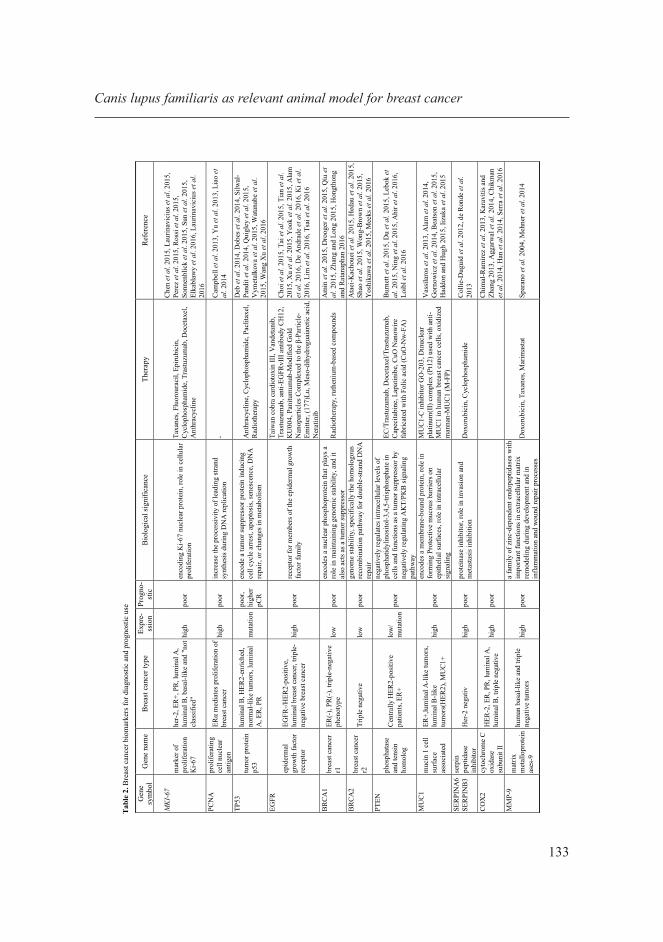
Surgery represents the golden-standard method for breast cancer therapy in CMT. Different types of surgery techniques are applied in CMT according to the tumor type [Tran et al. 2016]. These types of surgery techniques are: lumpectomy in the case of small non-invasive tumors; regional mastectomy in grade I and II tumors with resection of associated glands; unilateral mastectomy in multiple tumors; and bilateral mastectomy in sever multiple tumors in both mammary chains [Sleeckx et al. 2011, Karayannopoulou 2016]. At the time of surgical intervention, like in HBC, canine patients present metastases of mammary tumors, which conduct to a high relapse rate of the malignancy. In HBC, after the surgical treatment, a well-established postoperative adjuvant therapy is applied for best survival rates (Tab. 2). In canine patients, this post-surgical therapy is not routinely used, and the majority of these treatments are still in experimental phases. Some studies described several chemotherapeutics used in veterinary medicine for mammary cancer treatment. Chemotherapy represents an adjuvant treatment in high grade and metastatic CMT [Simon et al. 2006, Karayannopoulou 2016, Tran et al. 2016]. Lack of severe side effects and increased survival rate were observed in the case of chemotherapy using Cyclophosphamide, 5-Fluorouracil and combination with Vincristine or Mitoxantrone in CMT [Clemente et al. 2009, Karayannopoulou 2016]. Canine mammary tumors treated with a lower dose of Docetaxel also showed good results [Simon et al. 2006]. A combination of Gemcitabine and Carboplatin treatment in canine carcinomas showed a moderate toxicity and a 13% response rate [Dominguez et al. 2009].
Conclusions and remarks
The One Health Strategy becomes a worldwide, combined effort of multiple disciplines to develop an integrated research approach in human, animal and plant health for a better understanding and management of several pathologies including cancer. The progress in the field of comparative oncology can be sustained by preclinical studies, which implies the use of relevant animal models, and dogs can be a suitable alternative. The spontaneous mammary tumor development in dogs and the similarity in clinical and biological aspects with mammary tumors in humans demonstrate that dogs can be a valid model for future researches in mammary cancer pathologies. Studies on dogs will help identify the safety and activity of new anticancer drugs and the discovery of relevant biomarkers associated with response or exposure to these treatments. Dog clinical trials may be useful for the development of anticancer agents currently in early human clinical trials. Investigations made in CMT can contribute to the development of research directions in HBC focused on finding new therapeutic strategies and identifying new prognostic factors. The field of comparative oncology, particularly the study of mammary cancer, will lead to important benefits in the context of personalized healthcare and an improved quality of life in both humans and their canine companions.
L. Raduly et al.

135
Conflict of interest Statement
The authors certify that they have NO affiliations with or involvement in any organization or entity with any financial interest in the subject matter or materials discussed in this manuscript.
REFERENCES
1. AGGARWAL A.R.N., AL-ROHIL A., BATRA P.J., FEUSTEL D.M., JONES D., DIPERSIO C.M., 2014 – Expression of integrin alpha3beta1 and cyclooxygenase-2 (COX2) are positively correlated in human breast cancer. BMC Cancer 14, 459.
2. AHIR M., BHATTACHARYA S., KARMAKAR S., MUKHOPADHYAY A., MUKHERJEE S., GHOSH S., CHATTOPADHYAY S., PATRA P., ADHIKARY A., 2016 – Tailored-CuO-nanowire decorated with folic acid mediated coupling of the mitochondrial-ROS generation and miR425-PTEN axis in furnishing potent anti-cancer activity in human triple negative breast carcinoma cells. Biomaterials 76, 115-132.
3. ALAM M., RAJABI H., AHMAD R., JIN C., KUFE D., 2014 - Targeting the MUC1-C oncoprotein inhibits self-renewal capacity of breast cancer cells. Oncotarget 5, (9), 2622-2634.
4. ALAM M.W., PERSSON C.U., REINBOTHE S., KAZI J.U., RONNSTRAND L., WIGERUP C., DITZEL H.J., LYKKESFELDT A.E., PAHLMAN S. and JOGI A., 2016 - HIF2alpha contributes to antiestrogen resistance via positive bilateral crosstalk with EGFR in breast cancer cells. Oncotarget 7(10),11238-11250.
5. ALVAREZ C.E., 2014 - Naturally occurring cancers in dogs: insights for translational genetics and medicine. ILAR Journal 55, (1), 16-45.
6. AMIN R., MORITA-FUJIMURA Y., TAWARAYAMA H., SEMBA K., CHIBA N., FUKUMOTO M., IKAWA S., 2015 - DeltaNp63alpha induces quiescence and downregulates the BRCA1 pathway in estrogen receptor-positive luminal breast cancer cell line MCF7 but not in other breast cancer cell lines. Molecular Oncology 10(4):575-93.
7. ARESU L., GIANTIN M., MORELLO E., VASCELLARI M., CASTAGNARO M., LOPPARELLI R., ZANCANELLA V., GRANATO A., GARBISA S., ARICO A., BRADASCHIA A., MUTINELLI F., DACASTO M., 2011 - Matrix metalloproteinases and their inhibitors in canine mammary tumors. BMC Veterinary Research 7, 33.
8. ASEA A., 2006 - Initiation of the Immune Response by Extracellular Hsp72: Chaperokine Activity of Hsp72. Current Immunology Reviews 2, (3), 209-215.
9. ATAEI-KACHOUEI M., NADAF J., AKBARI M.T., ATRI M., MAJEWSKI J., RIAZALHOSSEINI Y., GARSHASBI M., 2015 - Double Heterozygosity of BRCA2 and STK11 in Familial Breast Cancer Detected by Exome Sequencing. Iranian Journal of Public Health 44, (10), 1348-1352.
10. BEATSON R., MAURSTAD G., PICCO G., ARULAPPU A., COLEMAN J., WANDELL H.H., CLAUSEN H., MANDEL U., TAYLOR-PAPADIMITRIOU J., SLETMOEN M., BURCHELL J.M., 2015 - The Breast Cancer-Associated Glycoforms of MUC1, MUC1-Tn and sialyl-Tn, Are Expressed in COSMC Wild-Type Cells and Bind the C-Type Lectin MGL. PLoS One 10, (5), e0125994.
11. BEHA G., MUSCATELLO L.V., BRUNETTI B., ASPRONI P., MILLANTA F., POLI A., BENAZZI C., SARLI G., 2014 - Molecular phenotype of primary mammary tumours and distant metastases in female dogs and cats. Journal of Comparative Pathology 150, (2-3), 194-197.
12. BELEVYCH A.E., HO H.T., TERENTYEVA R., BONILLA I.M., TERENTYEV D., CARNES C.A., GYORKE S., BILLMAN G.E., 2013 - Dietary omega-3 fatty acids promote arrhythmogenic remodeling of cellular Ca2+ handling in a postinfarction model of sudden cardiac death. PLoS One 8, (10), e78414.
Canis lupus familiaris as relevant animal model for breast cancer

136
13. BETZ D., SCHOENROCK D., MISCHKE R., BAUMGARTNER W., NOLTE I., 2012 - Postoperative treatment outcome in canine mammary tumors. Multivariate analysis of the prognostic value of pre- and postoperatively available information. Tierarztl Prax Ausg K Kleintiere Heimtiere 40, (4), 235-242.
14. BRAHMKHATRI V.P., PRASANNA C., ATREYA H.S., 2015 - Insulin-like growth factor system in cancer: novel targeted therapies. BioMed Research International 538019.
15. BRAICU C., BERINDAN-NEAGOE I., PILECZKI V., COJOCNEANU-PETRIC R., POP L.A., PUSCAS E., IRIMIE A., BUIGA R., 2014 - Breast tumor bank: an important resource for developing translational cancer research in Romania. Cancer Biomark 14, (2-3), 119-127.
16. BRAICU C., CHIOREAN R., IRIMIE A., CHIRA S., TOMULEASA C., NEAGOE E., PARADISO A., ACHIMAS-CADARIU P., LAZAR V., BERINDAN-NEAGOE I., 2016 - Novel insight into triple-negative breast cancers, the emerging role of angiogenesis, and antiangiogenic therapy. Expert Reviews in Molecular Medicine 18, e18.
17. BROWN J.S., CUNNINGHAM J.J., GATENBY R.A., 2015 - The multiple facets of Peto’s paradox: a life-history model for the evolution of cancer suppression. Philosophical Transactions of the Royal Society of London B Biol Sci 370, (1673).
18. BURNETT J.P., KORKAYA H., OUZOUNOVA M.D., JIANG H., CONLEY S.J., NEWMAN B.W., SUN L., CONNARN J.N., CHEN C.S., ZHANG N., WICHA M.S., SUN D., 2015 - Trastuzumab resistance induces EMT to transform HER2(+) PTEN(-) to a triple negative breast cancer that requires unique treatment options. Scientific Reports 5, 15821.
19. BURRAI G.P., TANCA A., DE MIGLIO M.R., ABBONDIO M., PISANU S., POLINAS M., PIRINO S., MOHAMMED S.I., UZZAU S., ADDIS M.F., ANTUOFERMO E., 2015 - Investigation of HER2 expression in canine mammary tumors by antibody-based, transcriptomic and mass spectrometry analysis: is the dog a suitable animal model for human breast cancer? Tumour Biology ...........
20. CAMPBELL M.J., WOLF D., MUKHTAR R.A., TANDON V., YAU C., AU A., BAEHNER F., VAN’T VEER L., BERRY D., ESSERMAN L.J., 2013 - The prognostic implications of macrophages expressing proliferating cell nuclear antigen in breast cancer depend on immune context. PLoS One 8, (10), e79114.
21. CARVALHO M.I., GUIMARAES M.J., PIRES I., PRADA J., SILVA-CARVALHO R., LOPES C., QUEIROGA F.L., 2013 - EGFR and microvessel density in canine malignant mammary tumours. Research in Veterinary Science 95, (3), 1094-1099.
22. CARVALHO M.I., PIRES I., PRADA J., LOBO L., QUEIROGA F.L., 2016 - Ki-67 and PCNA Expression in Canine Mammary Tumors and Adjacent Nonneoplastic Mammary Glands: Prognostic Impact by a Multivariate Survival Analysis. Veterinary Pathology 53, (6), 1138-1146.
23. CASSALI G.D., 2013 - Comparative mammary oncology: canine model. BMC Proceedings 7, (Suppl 2), K6-K6.
24. CHEN X., ZHU S., FEI X., GARFIELD D.H., WU J., HUANG O., LI Y., ZHU L., HE J., CHEN W., JIN X., SHEN K., 2015 - Surgery time interval and molecular subtype may influence Ki67 change after core needle biopsy in breast cancer patients. BMC Cancer 15, 822.
25. CHIKMAN B., VASYANOVICH S., LAVY R., HABLER L., TOLSTOV G., KAPIEV A., HALEVY A., SANDBANK J., 2014 - COX2 expression in high-grade breast cancer: evidence for prognostic significance in the subset of triple-negative breast cancer patients. Medical Oncology 31, (6), 989.
26. CHIMAL-RAMIREZ G.K., ESPINOZA-SANCHEZ N.A., UTRERA-BARILLAS D., BENITEZ-BRIBIESCA L., VELAZQUEZ J.R., ARRIAGA-PIZANO L.A., MONROY-GARCIA A., REYES-MALDONADO E., DOMINGUEZ-LOPEZ M.L., PINA-SANCHEZ P., FUENTES-PANANA E.M., 2013 - MMP1, MMP9, and COX2 expressions in promonocytes are induced by breast cancer cells and correlate with collagen degradation, transformation-like morphological changes in MCF-10A acini, and tumor aggressiveness. BioMed Research International 2013, 279505.
L. Raduly et al.

137
27. CHIOREAN R., BRAICU C., BERINDAN-NEAGOE I., 2013 - Another review on triple negative breast cancer. Are we on the right way towards the exit from the labyrinth? Breast 22, (6), 1026-1033.
28. CHOI M.S., JEONG H.J., KANG T.H., SHIN H.M., OH S.T., CHOI Y., JEON S., 2015 - Meso-dihydroguaiaretic acid induces apoptosis and inhibits cell migration via p38 activation and EGFR/Src/intergrin beta3 downregulation in breast cancer cells. Life Science 141, 81-89.
29. CHRISTOPOULOS P.F., MSAOUEL P., KOUTSILIERIS M., 2015 - The role of the insulin-like growth factor-1 system in breast cancer. Molecular Cancer 14, 43.
30. CIOCCA D.R., ARRIGO A.P., CALDERWOOD S.K., 2013 - Heat shock proteins and heat shock factor 1 in carcinogenesis and tumor development: an update. Archives of Toxicology 87, (1), 19-48.
31. CLEMENTE M., DE ANDRES P.J., PENA L., PEREZ-ALENZA M.D., 2009 - Survival time of dogs with inflammatory/mammary cancer treated with palliative therapy alone or palliative therapy plus chemotherapy. Veterinary Record 165, (3), 78-81.
32. COLLIE-DUGUID E.S., SWEENEY K., STEWART K.N., MILLER I.D., SMYTH E., HEYS S.D., 2012 - SerpinB3, a new prognostic tool in breast cancer patients treated with neoadjuvant chemotherapy. Breast Cancer Research and Treatment 132, (3), 807-818.
33. DAVIDSON B., REINERTSEN K.V., TRINH D., REED W., BOHLER P.J., 2016 - BAG-1/SODD, HSP70 and HSP90 are potential prognostic markers of poor survival in node-negative breast carcinoma. Human Pathology 54, 64-73.
34. DE ANDRADE J.P., PARK J.M., GU V.W., WOODFIELD G.W., KULAK M.V., LORENZEN A.W., WU V.T., VAN DORIN S.E., SPANHEIMER P.M., WEIGEL R.J., 2016 - EGFR Is Regulated by TFAP2C in Luminal Breast Cancer and Is a Target for Vandetanib. Molecular Cancer Therapeutics 15(3), 503-11.
35. DE ARAUJO M.R., CAMPOS L.C., FERREIRA E., CASSALI G.D., 2015 - Quantitation of the Regional Lymph Node Metastatic Burden and Prognosis in Malignant Mammary Tumors of Dogs. Journal of Veterinary Internal Medicine 29, (5), 1360-1367.
36. DE OLIVEIRA J.T., PINHO S.S., DE MATOS A.J., HESPANHOL V., REIS C.A., GARTNER F., 2009 - MUC1 expression in canine malignant mammary tumours and relationship to clinicopathological features. The Veterinary Journal 182, (3), 491-493.
37. DE RONDE J.J., LIPS E.H., MULDER L., VINCENT A.D., WESSELING J., NIEUWLAND M., KERKHOVEN R., VRANCKEN PEETERS M.J., SONKE G.S., RODENHUIS S., WESSELS L.F., 2013 - SERPINA6, BEX1, AGTR1, SLC26A3, and LAPTM4B are markers of resistance to neoadjuvant chemotherapy in HER2-negative breast cancer. Breast Cancer Research and Treatment 137, (1), 213-223.
38. DEB S., WONG S.Q., LI J., DO H., WEISS J., BYRNE D., CHAKRABARTI A., BOSMA T., KCONFAB I., FELLOWES A., DOBROVIC A., FOX S.B., 2014 - Mutational profiling of familial male breast cancers reveals similarities with luminal A female breast cancer with rare TP53 mutations. British Journal of Cancer 111, (12), 2351-2360.
39. DOBES P., PODHOREC J., COUFAL O., JURECKOVA A., PETRAKOVA K., VOJTESEK B., HRSTKA R., 2014 - Influence of mutation type on prognostic and predictive values of TP53 status in primary breast cancer patients. Oncology Reports 32, (4), 1695-1702.
40. DOBSON J.M., 2013 - Breed-predispositions to cancer in pedigree dogs. ISRN Vet Sci 2013, 941275.41. DOLKA I., KROL M., SAPIERZYNSKI R., 2016 - Evaluation of apoptosis-associated protein (Bcl-
2, Bax, cleaved caspase-3 and p53) expression in canine mammary tumors: An immunohistochemical and prognostic study. Research in Veterinary Science 105, 124-133.
42. DOMINGUEZ P.A., DERVISIS N.G., CADILE C.D., SARBU L., KITCHELL B.E., 2009 - Combined gemcitabine and carboplatin therapy for carcinomas in dogs. Journal of Veterinary Internal Medicine 23, (1), 130-137.
Canis lupus familiaris as relevant animal model for breast cancer

138
43. DROOGER J., AKDENIZ D., PIGNOL J.P., KOPPERT L.B., MCCOOL D., SEYNAEVE C.M., HOONING M.J., JAGER A., 2015 - Adjuvant radiotherapy for primary breast cancer in BRCA1 and BRCA2 mutation carriers and risk of contralateral breast cancer with special attention to patients irradiated at younger age. Breast Cancer Research and Treatment 154, (1), 171-180.
44. DU G., BIAN L., WANG T., XU X., ZHANG S., GUO Y., ZHUO J., SONG S., JIANG Z., 2015 - [PTEN loss correlates withthe clinical efficacy of lapatinib in HER2 positive metastatic breast cancer with trastuzumab-resistance]. Zhonghua Yi Xue Za Zhi 95, (28), 2264-2267.
45. EGENVALL A., BONNETT B.N., OHAGEN P., OLSON P., HEDHAMMAR A., VON EULER H., 2005 - Incidence of and survival after mammary tumors in a population of over 80,000 insured female dogs in Sweden from 1995 to 2002. Preventive Veterinary Medicine 69, (1-2), 109-127.
46. ELKABLAWY M.A., ALBASRI A.M., MOHAMMED R.A., HUSSAINY A.S., NOUH M.M., ALHUJAILY A.S., 2016 - Ki67 expression in breast cancer. Correlation with prognostic markers and clinicopathological parameters in Saudi patients. Saudi Medical Journal 37, (2), 137-141.
47. ENGLISH D.P., ROQUE D.M., SANTIN A.D., 2013 - HER2 expression beyond breast cancer: therapeutic implications for gynecologic malignancies. Molecular Diagnosis & Therapy 17, (2), 85-99.
48. ESBONA K., INMAN D., SAHA S., JEFFERY J., SCHEDIN P., WILKE L., KEELY P., 2016 - COX-2 modulates mammary tumor progression in response to collagen density. Breast Cancer Research 18, (1), 35.
49. ESPINOSA DE LOS MONTEROS A., MILLAN M.Y., RAMIREZ G.A., ORDAS J., REYMUNDO C., MARTIN DE LAS MULAS J., 2005 - Expression of maspin in mammary gland tumors of the dog. Veterinary Pathology 42, (3), 250-257.
50. FAZEKAS J., FURDOS I., SINGER J., JENSEN-JAROLIM E., 2016 - Why man’s best friend, the dog, could also benefit from an anti-HER-2 vaccine. Oncology Letters 12, (4), 2271-2276.
51. FELICE D.L., EL-SHENNAWY L., ZHAO S., LANTVIT D.L., SHEN Q., UNTERMAN T.G., SWANSON S.M. and FRASOR J., 2013 - Growth hormone potentiates 17beta-estradiol-dependent breast cancer cell proliferation independently of IGF-I receptor signaling. Endocrinology 154, (9), 3219-3227.
52. FERLAY J., SOERJOMATARAM I., DIKSHIT R., ESER S., MATHERS C., REBELO M., PARKIN D.M., FORMAN D., BRAY F., 2015 - Cancer incidence and mortality worldwide: sources, methods and major patterns in GLOBOCAN 2012. International Journal of Cancer 136, (5), E359-386.
53. GAMA A., PAREDES J., GARTNER F., ALVES A., SCHMITT F., 2008 - Expression of E-cadherin, P-cadherin and beta-catenin in canine malignant mammary tumours in relation to clinicopathological parameters, proliferation and survival. The Veterinary Journal 177, (1), 45-53.
54. GAMBA C.O., DIAS E.J., RIBEIRO L.G., CAMPOS L.C., ESTRELA-LIMA A., FERREIRA E., CASSALI G.D., 2013 - Histopathological and immunohistochemical assessment of invasive micropapillary mammary carcinoma in dogs: a retrospective study. The Veterinary Journal 196, (2), 241-246.
55. GARDNER H.L., FENGER J.M., LONDON C.A., 2016 - Dogs as a Model for Cancer. Annual Review of Animal Biosciences 4, 199-222.
56. GOLDSCHMIDT M., PENA L., RASOTTO R., ZAPPULLI V., 2011 - Classification and grading of canine mammary tumors. Veterinary Pathology 48, (1), 117-131.
57. GORNOWICZ A., KALUZA Z., BIELAWSKA A., GABRYEL-POROWSKA H., CZARNOMYSY R., BIELAWSKI K., 2014 - Cytotoxic efficacy of a novel dinuclear platinum(II) complex used with anti-MUC1 in human breast cancer cells. Molecular and Cellular Biochemistry 392, (1-2), 161-174.
58. GROSSE N., VAN LOON B., ROHRER BLEY C., 2014 - DNA damage response and DNA repair - dog as a model? BMC Cancer 14, 203.
L. Raduly et al.

139
59. GUIMARAES M.J., CARVALHO M.I., PIRES I., PRADA J., GIL A.G., LOPES C., QUEIROGA F.L., 2014 - Concurrent expression of cyclo-oxygenase-2 and epidermal growth factor receptor in canine malignant mammary tumours. Journal of Comparative Pathology 150, (1), 27-34.
60. GUPTA K., SOOD N.K., UPPAL S.K., MOHINDROO J., MAHAJAN S., RAGHUNATH M., SINGH K., 2012 - Epidemiological studies on canine mammary tumour and its relevance for breast cancer studies. IOSR Journal of Pharmacy 2, (2), 322-333.
61. HADDON L., HUGH J., 2015 - MUC1-mediated motility in breast cancer: a review highlighting the role of the MUC1/ICAM-1/Src signaling triad. Clinical and Experimental Metastasis 32, (4), 393-403.
62. HAN H., YANG S., LIN S.G., XU C.S., HAN Z.H., 2014 - Effects and mechanism of downregulation of COX2 expression by RNA interference on proliferation and apoptosis of human breast cancer MCF7 cells. Molecular Medicine Reports 10, (6), 3092-3098.
63. HEDAU S., BATRA M., SINGH U.R., BHARTI A.C., RAY A., DAS B.C., 2015 - Expression of BRCA1 and BRCA2 proteins and their correlation with clinical staging in breast cancer. Journal of Cancer Research and Therapeutics 11, (1), 158-163.
64. HEGDE S.M., KUMAR M.N., KAVYA K., KUMAR K.M., NAGESH R., PATIL R.H., BABU R.L., RAMESH G.T., SHARMA S.C., 2016 - Interplay of nuclear receptors (ER, PR, and GR) and their steroid hormones in MCF-7 cells. Molecular and Cellular Biochemistry 422, (1-2), 109-120.
65. HONG R., DAI Z., ZHU W., XU B., 2015 - Association between Lymph Node Ratio and Disease Specific Survival in Breast Cancer Patients with One or Two Positive Lymph Nodes Stratified by Different Local Treatment Modalities. PLoS One 10, (10), e0138908.
66. HONGTHONG K., RATANAPHAN A., 2016 - BRCA1-associated triple-negative breast cancer and potential treatment for ruthenium-based compounds. Current Cancer Drug Targets 16(7), 606-17.
67. HORNE H.N., SHERMAN M.E., PFEIFFER R.M., FIGUEROA J.D., KHODR Z.G., FALK R.T., POLLAK M., PATEL D.A., PALAKAL M.M., LINVILLE L., PAPATHOMAS D., GELLER B., VACEK P.M., WEAVER D.L., CHICOINE R., SHEPHERD J., MAHMOUDZADEH A.P., WANG J., FAN B., MALKOV S., HERSCHORN S., HEWITT S.M., BRINTON L.A., GIERACH G.L., 2016 - Circulating insulin-like growth factor-I, insulin-like growth factor binding protein-3 and terminal duct lobular unit involution of the breast: a cross-sectional study of women with benign breast disease. Breast Cancer Research 18, (1), 24.
68. HUANG K.T., TAN D., CHEN K.H., WALKER A.M., 2015 - Blockade of estrogen-stimulated proliferation by a constitutively-active prolactin receptor having lower expression in invasive ductal carcinoma. Cancer Letters 358, (2), 152-160.
69. HUMINIECKI L, HORBAŃCZUK J., 2018 – The functional genomic studies of resveratrol in respect to its anti-cancer effects. Biotechnology Advances, doi: 10.1016/j.biotechadv.2018.02.011
70. HUMINIECKI L., HORBAŃCZUK J., ATANASOV A.G., 2017 - The functional genomic studies of curcumin. Seminar Cancer in Biology, doi.org/10.1016/j.semcancer.2017.04.002.
71. IIZUKA M., NAKANISHI Y., FUCHINOUE F., MAEDA T., MURAKAMI E., OBANA Y., ENOMOTO K., TANI M., SAKURAI K., AMANO S., MASUDA S., 2015 - Altered intracellular region of MUC1 and disrupted correlation of polarity-related molecules in breast cancer subtypes. Cancer Science 106, (3), 307-314.
72. IM K.S., KIM I.H., KIM N.H., LIM H.Y., KIM J.H., SUR J.H., 2013 - Breed-related differences in altered BRCA1 expression, phenotype and subtype in malignant canine mammary tumors. The Veterinary Journal 195, (3), 366-372.
73. IM K.S., KIM N.H., LIM H.Y., KIM H.W., SHIN J.I., SUR J.H., 2014 - Analysis of a new histological and molecular-based classification of canine mammary neoplasia. Veterinary Pathology 51, (3), 549-559.
Canis lupus familiaris as relevant animal model for breast cancer

140
74. INWALD E.C., KLINKHAMMER-SCHALKE M., HOFSTADTER F., ZEMAN F., KOLLER M., GERSTENHAUER M., ORTMANN O., 2013 - Ki-67 is a prognostic parameter in breast cancer patients: results of a large population-based cohort of a cancer registry. Breast Cancer Research and Treatment 139, (2), 539-552.
75. IRIMIE A.I., BRAICU C., COJOCNEANU-PETRIC R., BERINDAN-NEAGOE I., CAMPIAN R.S., 2015 - Novel technologies for oral squamous carcinoma biomarkers in diagnostics and prognostics. Acta Odontologica Scandinavica 73, (3), 161-168.
76. KABIR F.M., DEINNOCENTES P., AGARWAL P., MILL C.P., RIESE D.J 2nd., BIRD R.C., 2017- Estrogen receptor-alpha, progesterone receptor and c-erbB/HER-family receptor mRNA detection and phenotype analysis in spontaneous canine models of breast cancer. Journal of Veterinary Science 18(2) 149-158.
77. KARAVITIS J., ZHANG M., 2013 - COX2 regulation of breast cancer bone metastasis. Oncoimmunology 2, (3), e23129.
78. KI J., ARUMUGAM P., SONG J.M., 2016 - TIRF high-content assay development for the evaluation of drug efficacy of chemotherapeutic agents against EGFR-/HER2-positive breast cancer cell lines. Analytical and Bioanalytical Chemistry 408(12), 3233-3238.
79. KLOPFLEISCH R., GRUBER A.D., 2009 - Differential expression of cell cycle regulators p21, p27 and p53 in metastasizing canine mammary adenocarcinomas versus normal mammary glands. Research in Veterinary Science 87, (1), 91-96.
80. KLOPFLEISCH R., LENZE D., HUMMEL M., GRUBER A.D., 2011 - The metastatic cascade is reflected in the transcriptome of metastatic canine mammary carcinomas. The Veterinary Journal 190, (2), 236-243.
81. KUMARAGURUPARAN R., KARUNAGARAN D., BALACHANDRAN C., MANOHAR B.M., NAGINI S., 2006 - Of humans and canines: a comparative evaluation of heat shock and apoptosis-associated proteins in mammary tumors. Clinica Chimica Acta 365, (1-2), 168-176.
82. LAMP O., HONSCHA K.U., SCHWEIZER S., HECKMANN A., BLASCHZIK S., EINSPANIER A., 2013 - The metastatic potential of canine mammary tumours can be assessed by mRNA expression analysis of connective tissue modulators. Veterinary and Comparative Oncology 11, (1), 70-85.
83. LAURINAVICIUS A., GREEN A.R., LAURINAVICIENE A., SMAILYTE G., OSTAPENKO V., MESKAUSKAS R., ELLIS I.O., 2015 - Ki67/SATB1 ratio is an independent prognostic factor of overall survival in patients with early hormone receptor-positive invasive ductal breast carcinoma. Oncotarget 6, (38), 41134-41145.
84. LAURINAVICIUS A., PLANCOULAINE B., RASMUSSON A., BESUSPARIS J., AUGULIS R., MESKAUSKAS R., HERLIN P., LAURINAVICIENE A., ABDELHADI MUFTAH A.A., MILIGY I., ALESKANDARANY M., RAKHA E.A., GREEN A.R., ELLIS I.O., 2016 - Bimodality of intratumor Ki67 expression is an independent prognostic factor of overall survival in patients with invasive breast carcinoma. Virchows Archiv 468(4), 493-502.
85. LEBOK P., KOPPERSCHMIDT V., KLUTH M., HUBE-MAGG C., OZDEN C., B T., HUSSEIN K., MITTENZWEI A., LEBEAU A., WITZEL I., WOLBER L., MAHNER S., JANICKE F., GEIST S., PALUCHOWSKI P., WILKE C., HEILENKOTTER U., SIMON R., SAUTER G., TERRACCIANO L., KRECH R., VON D ASSEN A., MULLER V., BURANDT E., 2015 - Partial PTEN deletion is linked to poor prognosis in breast cancer. BMC Cancer 15, 963.
86. LERNER H., BERG C., 2015 - The concept of health in One Health and some practical implications for research and education: what is One Health? Infection Ecology and Epidemiology 5, 25300.
87. LIAO X.H., LU D.L., WANG N., LIU L.Y., WANG Y., LI Y.Q., YAN T.B., SUN X.G., HU P., ZHANG T.C., 2014 - Estrogen receptor alpha mediates proliferation of breast cancer MCF-7 cells via a p21/PCNA/E2F1-dependent pathway. The FEBS Journal 281, (3), 927-942.
L. Raduly et al.

141
88. LIM H.Y., IM K.S., KIM N.H., KIM H.W., SHIN J.I., SUR J.H., 2015 - Obesity, expression of adipocytokines, and macrophage infiltration in canine mammary tumors. The Veterinary Journal 203, (3), 326-331.
89. LIM S.O., LI C.W., XIA W., LEE H.H., CHANG S.S., SHEN J., HSU J.L., RAFTERY D., DJUKOVIC D., GU H., CHANG W.C., WANG H.L., CHEN M.L., HUO L., CHEN C.H., WU Y., SAHIN A., HANASH S.M., HORTOBAGYI G.N., HUNG M.C., 2016 - EGFR Signaling Enhances Aerobic Glycolysis in Triple-Negative Breast Cancer Cells to Promote Tumor Growth and Immune Escape. Cancer Research ......................................
90. LINDBLAD-TOH K., WADE C.M., MIKKELSEN T.S., KARLSSON E.K., JAFFE D.B., KAMAL M., CLAMP M., CHANG J.L., KULBOKAS E.J., 3RD, ZODY M.C., MAUCELI E., XIE X., BREEN M., WAYNE R.K., OSTRANDER E.A., PONTING C.P., GALIBERT F., SMITH D.R., DEJONG P.J., KIRKNESS E., ALVAREZ P., BIAGI T., BROCKMAN W., BUTLER J., CHIN C.W., COOK A., CUFF J., DALY M.J., DECAPRIO D., GNERRE S., GRABHERR M., KELLIS M., KLEBER M., BARDELEBEN C., GOODSTADT L., HEGER A., HITTE C., KIM L., KOEPFLI K.P., PARKER H.G., POLLINGER J.P., SEARLE S.M., SUTTER N.B., THOMAS R., WEBBER C., BALDWIN J., ABEBE A., ABOUELLEIL A., AFTUCK L., AIT-ZAHRA M., ALDREDGE T., ALLEN N., AN P., ANDERSON S., ANTOINE C., ARACHCHI H., ASLAM A., AYOTTE L., BACHANTSANG P., BARRY A., BAYUL T., BENAMARA M., BERLIN A., BESSETTE D., BLITSHTEYN B., BLOOM T., BLYE J., BOGUSLAVSKIY L., BONNET C., BOUKHGALTER B., BROWN A., CAHILL P., CALIXTE N., CAMARATA J., CHESHATSANG Y., CHU J., CITROEN M., COLLYMORE A., COOKE P., DAWOE T., DAZA R., DECKTOR K., DEGRAY S., DHARGAY N., DOOLEY K., DOOLEY K., DORJE P., DORJEE K., DORRIS L., DUFFEY N., DUPES A., EGBIREMOLEN O., ELONG R., FALK J., FARINA A., FARO S., FERGUSON D., FERREIRA P., FISHER S., FITZGERALD M., FOLEY K., FOLEY C., FRANKE A., FRIEDRICH D., GAGE D., GARBER M., GEARIN G., GIANNOUKOS G., GOODE T., GOYETTE A., GRAHAM J., GRANDBOIS E., GYALTSEN K., HAFEZ N., HAGOPIAN D., HAGOS B., HALL J., HEALY C., HEGARTY R., HONAN T., HORN A., HOUDE N., HUGHES L., HUNNICUTT L., HUSBY M., JESTER B., JONES C., KAMAT A., KANGA B., KELLS C., KHAZANOVICH D., KIEU A.C., KISNER P., KUMAR M., LANCE K., LANDERS T., LARA M., LEE W., LEGER J.P., LENNON N., LEUPER L., LEVINE S., LIU J., LIU X., LOKYITSANG Y., LOKYITSANG T., LUI A., MACDONALD J., MAJOR J., MARABELLA R., MARU K., MATTHEWS C., MCDONOUGH S., MEHTA T., MELDRIM J., MELNIKOV A., MENEUS L., MIHALEV A., MIHOVA T., MILLER K., MITTELMAN R., MLENGA V., MULRAIN L., MUNSON G., NAVIDI A., NAYLOR J., NGUYEN T., NGUYEN N., NGUYEN C., NGUYEN T., NICOL R., NORBU N., NORBU C., NOVOD N., NYIMA T., OLANDT P., O’NEILL B., O’NEILL K., OSMAN S., OYONO L., PATTI C., PERRIN D., PHUNKHANG P., PIERRE F., PRIEST M., RACHUPKA A., RAGHURAMAN S., RAMEAU R., RAY V., RAYMOND C., REGE F., RISE C., ROGERS J., ROGOV P., SAHALIE J., SETTIPALLI S., SHARPE T., SHEA T., SHEEHAN M., SHERPA N., SHI J., SHIH D., SLOAN J., SMITH C., SPARROW T., STALKER J., STANGE-THOMANN N., STAVROPOULOS S., STONE C., STONE S., SYKES S., TCHUINGA P., TENZING P., TESFAYE S., THOULUTSANG D., THOULUTSANG Y., TOPHAM K., TOPPING I., TSAMLA T., VASSILIEV H., VENKATARAMAN V., VO A., WANGCHUK T., WANGDI T., WEIAND M., WILKINSON J., WILSON A., YADAV S., YANG S., YANG X., YOUNG G., YU Q., ZAINOUN J., ZEMBEK L., ZIMMER A., LANDER E.S., 2005 - Genome sequence, comparative analysis and haplotype structure of the domestic dog. Nature 438, (7069), 803-819.
91. LIU D., XIONG H., ELLIS A.E., NORTHRUP N.C., RODRIGUEZ C.O., JR., O’REGAN R.M., DALTON S., ZHAO S., 2014 - Molecular homology and difference between spontaneous canine mammary cancer and human breast cancer. Cancer Research 74, (18), 5045-5056.
92. LOIBL S., DARB-ESFAHANI S., HUOBER J., KLIMOWICZ A., FURLANETTO J., LEDERER B., HARTMANN A., EIDTMANN H., PFITZNER B.M., FASCHING P.A., TIEMANN K., JACKISCH
Canis lupus familiaris as relevant animal model for breast cancer
142
C., MEHTA K., VON MINCKWITZ G., UNTCH M., DENKERT C., 2016 - PTEN expression and p4EBP1 immunoreactive score as predictor for pCR in HER2 positive breast cancer. Clinical Cancer Research .............................................
93. M. KARAYANNOPOULOU S.L., 2016 - Recent advances on canine mammary cancer chemotherapy: A review of studies from 2000 to date. Revue de Medecine Veterinaire 7-8, 191-200.
94. MAINENTI M., RASOTTO R., CARNIER P., ZAPPULLI V., 2014 - Oestrogen and progesterone receptor expression in subtypes of canine mammary tumours in intact and ovariectomised dogs. The Veterinary Journal 202, (1), 62-68.
95. MAK I.W., EVANIEW N., GHERT M., 2014 - Lost in translation: animal models and clinical trials in cancer treatment. American Journal of Translational Research 6, (2), 114-118.
96. MALHOTRA G.K., ZHAO X., BAND H., BAND V., 2010 - Histological, molecular and functional subtypes of breast cancers. Cancer Biology & Therapy 10, (10), 955-960.
97. MATOS A.J.F., SANTOS A.A., 2015 - Advances in the understanding of the clinically relevant genetic pathways and molecular aspects of canine mammary tumours: Part 1. Proliferation, apoptosis and DNA repair. The Veterinary Journal 205, (2), 136-143.
98. MEEKS H.D., SONG H., MICHAILIDOU K., BOLLA M.K., DENNIS J., WANG Q., BARROWDALE D., FROST D., EMBRACE, MCGUFFOG L., ELLIS S., FENG B., BUYS S.S., HOPPER J.L., SOUTHEY M.C., TESORIERO A., KCONFAB I., JAMES P.A., BRUINSMA F., CAMPBELL I.G., AUSTRALIA OVARIAN CANCER STUDY G., BROEKS A., SCHMIDT M.K., HOGERVORST F.B., HEBON, BECKMAN M.W., FASCHING P.A., FLETCHER O., JOHNSON N., SAWYER E.J., RIBOLI E., BANERJEE S., MENON U., TOMLINSON I., BURWINKEL B., HAMANN U., MARME F., RUDOLPH A., JANAVICIUS R., TIHOMIROVA L., TUNG N., GARBER J., CRAMER D., TERRY K.L., POOLE E.M., TWOROGER S.S., DORFLING C.M., VAN RENSBURG E.J., GODWIN A.K., GUENEL P., TRUONG T., COLLABORATORS G.S., STOPPA-LYONNET D., DAMIOLA F., MAZOYER S., SINILNIKOVA O.M., ISAACS C., MAUGARD C., BOJESEN S.E., FLYGER H., GERDES A.M., HANSEN T.V., JENSEN A., KJAER S.K., HOGDALL C., HOGDALL E., PEDERSEN I.S., THOMASSEN M., BENITEZ J., GONZALEZ-NEIRA A., OSORIO A., HOYA MDE L., SEGURA P.P., DIEZ O., LAZARO C., BRUNET J., ANTON-CULVER H., EUNJUNG L., JOHN E.M., NEUHAUSEN S.L., DING Y.C., CASTILLO D., WEITZEL J.N., GANZ P.A., NUSSBAUM R.L., CHAN S.B., KARLAN B.Y., LESTER J., WU A., GAYTHER S., RAMUS S.J., SIEH W., WHITTERMORE A.S., MONTEIRO A.N., PHELAN C.M., TERRY M.B., PIEDMONTE M., OFFIT K., ROBSON M., LEVINE D., MOYSICH K.B., CANNIOTO R., OLSON S.H., DALY M.B., NATHANSON K.L., DOMCHEK S.M., LU K.H., LIANG D., HILDEBRANT M.A., NESS R., MODUGNO F., PEARCE L., GOODMAN M.T., THOMPSON P.J., BRENNER H., BUTTERBACH K., MEINDL A., HAHNEN E., WAPPENSCHMIDT B., BRAUCH H., BRUNING T., BLOMQVIST C., KHAN S., NEVANLINNA H., PELTTARI L.M., AITTOMAKI K., BUTZOW R., BOGDANOVA N.V., DORK T., LINDBLOM A., MARGOLIN S., RANTALA J., KOSMA V.M., MANNERMAA A., LAMBRECHTS D., NEVEN P., CLAES K.B., MAERKEN T.V., CHANG-CLAUDE J., FLESCH-JANYS D., HEITZ F., VARON-MATEEVA R., PETERLONGO P., RADICE P., VIEL A., BARILE M., PEISSEL B., MANOUKIAN S., MONTAGNA M., OLIANI C., PEIXOTO A., TEIXEIRA M.R., COLLAVOLI A., HALLBERG E., OLSON J.E., GOODE E.L., HART S.N., SHIMELIS H., CUNNINGHAM J.M., GILES G.G., MILNE R.L., HEALEY S., TUCKER K., HAIMAN C.A., HENDERSON B.E., GOLDBERG M.S., TISCHKOWITZ M., SIMARD J., SOUCY P., ECCLES D.M., LE N., BORRESEN-DALE A.L., KRISTENSEN V., SALVESEN H.B., BJORGE L., BANDERA E.V., RISCH H., ZHENG W., BEEGHLY-FADIEL A., CAI H., PYLKAS K., TOLLENAAR R.A., OUWELAND A.M., ANDRULIS I.L., KNIGHT J.A., OCGN, NAROD S., DEVILEE P., WINQVIST R., FIGUEROA J., GREENE M.H., MAI P.L., LOUD J.T., GARCIA-CLOSAS M., SCHOEMAKER M.J., CZENE K., DARABI H., MCNEISH I., SIDDIQUIL N., GLASSPOOL R., KWONG A., PARK S.K., TEO S.H., YOON S.Y., MATSUO K., HOSONO S., WOO Y.L., GAO Y.T., FORETOVA L., SINGER C.F., RAPPAPORT-FEURHAUSER
L. Raduly et al.

143
C., FRIEDMAN E., LAITMAN Y., RENNERT G., IMYANITOV E.N., HULICK P.J., OLOPADE O.I., SENTER L., OLAH E., DOHERTY J.A., SCHILDKRAUT J., KOPPERT L.B., KIEMENEY L.A., MASSUGER L.F., COOK L.S., PEJOVIC T., LI J., BORG A., OFVERHOLM A., ROSSING M.A., WENTZENSEN N., HENRIKSSON K., COX A., CROSS S.S., PASINI B.J., SHAH M., KABISCH M., TORRES D., JAKUBOWSKA A., LUBINSKI J., GRONWALD J., AGNARSSON B.A., KUPRYJANCZYK J., MOES-SOSNOWSKA J., FOSTIRA F., KONSTANTOPOULOU I., SLAGER S., JONES M., GENOME P.R.C.A.G.T.I.C.A.A.I.T., ANTONIOU A.C., BERCHUCK A., SWERDLOW A., CHENEVIX-TRENCH G., DUNNING A.M., PHAROAH P.D., HALL P., EASTON D.F., COUCH F.J., SPURDLE A.B., GOLDGAR D.E., 2016 - BRCA2 Polymorphic Stop Codon K3326X and the Risk of Breast, Prostate, and Ovarian Cancers. Journal of the National Cancer Institute 108, (2).
99. MEHNER C., HOCKLA A., MILLER E., RAN S., RADISKY D.C., RADISKY E.S., 2014 - Tumor cell-produced matrix metalloproteinase 9 (MMP-9) drives malignant progression and metastasis of basal-like triple negative breast cancer. Oncotarget 5, (9), 2736-2749.
100. MELIN M., RIVERA P., ARENDT M., ELVERS I., MUREN E., GUSTAFSON U., STARKEY M., BORGE K.S., LINGAAS F., HAGGSTROM J., SAELLSTROM S., RONNBERG H., LINDBLAD-TOH K., 2016 - Genome-Wide Analysis Identifies Germ-Line Risk Factors Associated with Canine Mammary Tumours. PLoS Genetics 12, (5), e1006029.
101. MICHEL E., FELDMANN S.K., KOWALEWSKI M.P., BLEY C.R., BOOS A., GUSCETTI F., REICHLER I.M., 2012 - Expression of prolactin receptors in normal canine mammary tissue, canine mammary adenomas and mammary adenocarcinomas. BMC Veterinary Research 8, 72.
102. MOHR A., LUDER RIPOLI F., HAMMER S.C., WILLENBROCK S., HEWICKER-TRAUTWEIN M., KIELBOWICZ Z., MURUA ESCOBAR H., NOLTE I., 2016 - Hormone Receptor Expression Analyses in Neoplastic and Non-Neoplastic Canine Mammary Tissue by a Bead Based Multiplex Branched DNA Assay: A Gene Expression Study in Fresh Frozen and Formalin-Fixed, Paraffin-Embedded Samples. PLoS One 11, (9), e0163311.
103. MOUSE GENOME SEQUENCING C., WATERSTON R.H., LINDBLAD-TOH K., BIRNEY E., ROGERS J., ABRIL J.F., AGARWAL P., AGARWALA R., AINSCOUGH R., ALEXANDERSSON M., AN P., ANTONARAKIS S.E., ATTWOOD J., BAERTSCH R., BAILEY J., BARLOW K., BECK S., BERRY E., BIRREN B., BLOOM T., BORK P., BOTCHERBY M., BRAY N., BRENT M.R., BROWN D.G., BROWN S.D., BULT C., BURTON J., BUTLER J., CAMPBELL R.D., CARNINCI P., CAWLEY S., CHIAROMONTE F., CHINWALLA A.T., CHURCH D.M., CLAMP M., CLEE C., COLLINS F.S., COOK L.L., COPLEY R.R., COULSON A., COURONNE O., CUFF J., CURWEN V., CUTTS T., DALY M., DAVID R., DAVIES J., DELEHAUNTY K.D., DERI J., DERMITZAKIS E.T., DEWEY C., DICKENS N.J., DIEKHANS M., DODGE S., DUBCHAK I., DUNN D.M., EDDY S.R., ELNITSKI L., EMES R.D., ESWARA P., EYRAS E., FELSENFELD A., FEWELL G.A., FLICEK P., FOLEY K., FRANKEL W.N., FULTON L.A., FULTON R.S., FUREY T.S., GAGE D., GIBBS R.A., GLUSMAN G., GNERRE S., GOLDMAN N., GOODSTADT L., GRAFHAM D., GRAVES T.A., GREEN E.D., GREGORY S., GUIGO R., GUYER M., HARDISON R.C., HAUSSLER D., HAYASHIZAKI Y., HILLIER L.W., HINRICHS A., HLAVINA W., HOLZER T., HSU F., HUA A., HUBBARD T., HUNT A., JACKSON I., JAFFE D.B., JOHNSON L.S., JONES M., JONES T.A., JOY A., KAMAL M., KARLSSON E.K., KAROLCHIK D., KASPRZYK A., KAWAI J., KEIBLER E., KELLS C., KENT W.J., KIRBY A., KOLBE D.L., KORF I., KUCHERLAPATI R.S., KULBOKAS E.J., KULP D., LANDERS T., LEGER J.P., LEONARD S., LETUNIC I., LEVINE R., LI J., LI M., LLOYD C., LUCAS S., MA B., MAGLOTT D.R., MARDIS E.R., MATTHEWS L., MAUCELI E., MAYER J.H., MCCARTHY M., MCCOMBIE W.R., MCLAREN S., MCLAY K., MCPHERSON J.D., MELDRIM J., MEREDITH B., MESIROV J.P., MILLER W., MINER T.L., MONGIN E., MONTGOMERY K.T., MORGAN M., MOTT R., MULLIKIN J.C., MUZNY D.M., NASH W.E., NELSON J.O., NHAN M.N., NICOL R., NING Z., NUSBAUM C., O’CONNOR M.J., OKAZAKI Y., OLIVER K., OVERTON-LARTY
Canis lupus familiaris as relevant animal model for breast cancer

144
E., PACHTER L., PARRA G., PEPIN K.H., PETERSON J., PEVZNER P., PLUMB R., POHL C.S., POLIAKOV A., PONCE T.C., PONTING C.P., POTTER S., QUAIL M., REYMOND A., ROE B.A., ROSKIN K.M., RUBIN E.M., RUST A.G., SANTOS R., SAPOJNIKOV V., SCHULTZ B., SCHULTZ J., SCHWARTZ M.S., SCHWARTZ S., SCOTT C., SEAMAN S., SEARLE S., SHARPE T., SHERIDAN A., SHOWNKEEN R., SIMS S., SINGER J.B., SLATER G., SMIT A., SMITH D.R., SPENCER B., STABENAU A., STANGE-THOMANN N., SUGNET C., SUYAMA M., TESLER G., THOMPSON J., TORRENTS D., TREVASKIS E., TROMP J., UCLA C., URETA-VIDAL A., VINSON J.P., VON NIEDERHAUSERN A.C., WADE C.M., WALL M., WEBER R.J., WEISS R.B., WENDL M.C., WEST A.P., WETTERSTRAND K., WHEELER R., WHELAN S., WIERZBOWSKI J., WILLEY D., WILLIAMS S., WILSON R.K., WINTER E., WORLEY K.C., WYMAN D., YANG S., YANG S.P., ZDOBNOV E.M., ZODY M.C., LANDER E.S., 2002 - Initial sequencing and comparative analysis of the mouse genome. Nature 420, (6915), 520-562.
104. NADIN S.B., SOTTILE M.L., MONTT-GUEVARA M.M., GAUNA G.V., DAGUERRE P., LEUZZI M., GAGO F.E., IBARRA J., CUELLO-CARRION F.D., CIOCCA D.R., VARGAS-ROIG L.M., 2014 - Prognostic implication of HSPA (HSP70) in breast cancer patients treated with neoadjuvant anthracycline-based chemotherapy. Cell Stress Chaperones 19, (4), 493-505.
105. NICOLINI A., FERRARI P., ROSSI G., 2015 - Mucins and Cytokeratins as Serum Tumor Markers in Breast Cancer. Advances in Experimental Medicine and Biology 867, 197-225.
106. NING L., GUO-CHUN Z., SHENG-LI A., XUE-RUI L., KUN W., JIAN Z., CHONG-YANG R., LING-ZHU W., HAI-TONG L., 2015 - Inhibition of autophagy induced by PTEN loss promotes intrinsic breast cancer resistance to trastuzumab therapy. Tumour Biology 37(4), 5445-54.
107. NUNNEY L., MALEY C.C., BREEN M., HOCHBERG M.E. and SCHIFFMAN J.D., 2015 - Peto’s paradox and the promise of comparative oncology. Philos Trans R Soc Lond B Biol Sci 370, (1673).
108. PARK S.Y., CHAE S.Y., PARK J.O., LEE K.J., PARK G., 2016 - Gold-conjugated resveratrol nanoparticles attenuate the invasion and MMP-9 and COX-2 expression in breast cancer cells. Oncology Reports 35(6), 3248-56.
109. PARK S.Y., JIN M.L., KIM Y.H., LEE S.J., PARK G., 2014 - Sanguinarine inhibits invasiveness and the MMP-9 and COX-2 expression in TPA-induced breast cancer cells by inducing HO-1 expression. Oncology Reports 31, (1), 497-504.
110. PEREZ A.A., BALABRAM D., ROCHA R.M., DA SILVA SOUZA A., GOBBI H., 2015 - Co-Expression of p16, Ki67 and COX-2 Is Associated with Basal Phenotype in High-Grade Ductal Carcinoma In Situ of the Breast. Journal of Histochemistry & Cytochemistry 63, (6), 408-416.
111. PINHO S.S., CARVALHO S., CABRAL J., REIS C.A., GÄRTNER F., 2012 - Canine tumors: a spontaneous animal model of human carcinogenesis. Translational Research 159, (3), 165-172.
112. PINHO S.S., MATOS A.J., LOPES C., MARCOS N.T., CARVALHEIRA J., REIS C.A., GARTNER F., 2007 - Sialyl Lewis x expression in canine malignant mammary tumours: correlation with clinicopathological features and E-Cadherin expression. BMC Cancer 7, 124.
113. POP L.A., PUSCAS E., PILECZKI V., COJOCNEANU-PETRIC R., BRAICU C., ACHIMAS-CADARIU P., BERINDAN-NEAGOE I., 2014 - Quality control of ion torrent sequencing library. Cancer Biomarkers 14, (2-3), 93-101.
114. PURDIE C.A., QUINLAN P., JORDAN L.B., ASHFIELD A., OGSTON S., DEWAR J.A., THOMPSON A.M., 2014 - Progesterone receptor expression is an independent prognostic variable in early breast cancer: a population-based study. British Journal of Cancer 110, (3), 565-572.
115. QIU C.W., LIN D.G., WANG J.Q., LI C.Y., DENG G.Z., 2008 - Expression and significance of PTEN and VEGF in canine mammary gland tumours. Veterinary Research Communications 32, (6), 463-472.
116. QIU H.B., SUN W.D., YANG X., JIANG Q.Y., CHEN S., LIN D.G., 2015 - Promoter mutation and reduced expression of BRCA1 in canine mammary tumors. Research in Veterinary Science 103, 143-148.
L. Raduly et al.

145
117. QUEIROGA F.L., PEREZ-ALENZA D., GONZALEZ-GIL A., SILVAN G., PENA L., ILLERA J.C., 2015 - Serum and Tissue Steroid Hormone Levels in Canine Mammary Tumours: Clinical and Prognostic Implications. Reproduction in Domestic Animals 50, (5), 858-865.
118. QUEIROGA F.L., PEREZ-ALENZA M.D., SILVAN G., PENA L., LOPES C., ILLERA J.C., 2005 - Role of steroid hormones and prolactin in canine mammary cancer. The Journal of Steroid Biochemistry and Molecular Biology 94, (1-3), 181-187.
119. QUEIROGA F.L., PIRES I., PARENTE M., GREGORIO H., LOPES C.S., 2011 - COX-2 over-expression correlates with VEGF and tumour angiogenesis in canine mammary cancer. The Veterinary Journal 189, (1), 77-82.
120. QUEIROGA F.L., RAPOSO T., CARVALHO M.I., PRADA J., PIRES I., 2011 - Canine mammary tumours as a model to study human breast cancer: most recent findings. In Vivo 25, (3), 455-465.
121. QUIGLEY D., SILWAL-PANDIT L., DANNENFELSER R., LANGEROD A., VOLLAN H.K., VASKE C., SIEGEL J.U., TROYANSKAYA O., CHIN S.F., CALDAS C., BALMAIN A., BORRESEN-DALE A.L., KRISTENSEN V., 2015 - Lymphocyte Invasion in IC10/Basal-Like Breast Tumors Is Associated with Wild-Type TP53. Molecular Cancer Research 13, (3), 493-501.
122. RADHA R.K., P V., B K., 2014 - Histopathology and Prognostic Indices of Carcinoma Breast with Special Reference to p53 Marker. Journal of Clinical and Diagnostic Research 8, (7), FC04-08.
123. RANIERI G., GADALETA C.D., PATRUNO R., ZIZZO N., DAIDONE M.G., HANSSON M.G., PARADISO A., RIBATTI D., 2013 - A model of study for human cancer: Spontaneous occurring tumors in dogs. Biological features and translation for new anticancer therapies. Critital Reviews in Oncology/Hematology 88, (1), 187-197.
124. RAPOSO-FERREIRA T.M., SALVADOR R.C., TERRA E.M., FERREIRA J.H., VECHETTI-JUNIOR I.J., TINUCCI-COSTA M., ROGATTO S.R., LAUFER-AMORIM R., 2016 - Evaluation of vascular endothelial growth factor gene and protein expression in canine metastatic mammary carcinomas. Microscopy Research and Technique 79, (11), 1097-1104.
125. RASOTTO R., GOLDSCHMIDT M.H., CASTAGNARO M., CARNIER P., CALIARI D., ZAPPULLI V., 2014 - The dog as a natural animal model for study of the mammary myoepithelial basal cell lineage and its role in mammary carcinogenesis. Journal of Comparative Pathology 151, (2-3), 166-180.
126. RESSEL L., PULEIO R., LORIA G.R., VANNOZZI I., MILLANTA F., CARACAPPA S., POLI A., 2013 - HER-2 expression in canine morphologically normal, hyperplastic and neoplastic mammary tissues and its correlation with the clinical outcome. Research in Veterinary Science 94, (2), 299-305.
127. RICHMOND A., SU Y., 2008 - Mouse xenograft models vs GEM models for human cancer therapeutics. Disease Models & Mechanisms 1, (2-3), 78-82.
128. RIVERA P., MELIN M., BIAGI T., FALL T., HAGGSTROM J., LINDBLAD-TOH K., VON EULER H., 2009 - Mammary tumor development in dogs is associated with BRCA1 and BRCA2. Cancer Research 69, (22), 8770-8774.
129. ROMANUCCI M., BASTOW T., DELLA SALDA L., 2008 - Heat shock proteins in animal neoplasms and human tumours--a comparison. Cell Stress Chaperones 13, (3), 253-262.
130. ROSSI L., LAAS E., MALLON P., VINCENT-SALOMON A., GUINEBRETIERE J.M., LEREBOURS F., ROUZIER R., PIERGA J.Y., REYAL F., 2015 - Prognostic impact of discrepant Ki67 and mitotic index on hormone receptor-positive, HER2-negative breast carcinoma. British Journal of Cancer 113, (7), 996-1002.
131. ROWELL J.L., MCCARTHY D.O., ALVAREZ C.E., 2011 - Dog models of naturally occurring cancer. Trends in Molecular Medicine 17, (7), 380-388.
132. SALAS Y., MARQUEZ A., DIAZ D., ROMERO L., 2015 - Epidemiological Study of Mammary Tumors in Female Dogs Diagnosed during the Period 2002-2012: A Growing Animal Health Problem. PLoS One 10, (5), e0127381.
Canis lupus familiaris as relevant animal model for breast cancer

146
133. SANTAGATA S., HU R., LIN N.U., MENDILLO M.L., COLLINS L.C., HANKINSON S.E., SCHNITT S.J., WHITESELL L., TAMIMI R.M., LINDQUIST S., INCE T.A., 2011 - High levels of nuclear heat-shock factor 1 (HSF1) are associated with poor prognosis in breast cancer. Proceeding of the National Academy of Sciences USA 108, (45), 18378-18383.
134. SANTOS A.A., LOPES C.C., RIBEIRO J.R., MARTINS L.R., SANTOS J.C., AMORIM I.F., GARTNER F., MATOS A.J., 2013 - Identification of prognostic factors in canine mammary malignant tumours: a multivariable survival study. BMC Veterinary Research 9, 1.
135. SARGAN D.R., 2004 - IDID: inherited diseases in dogs: web-based information for canine inherited disease genetics. Mammalian Genome 15, (6), 503-506.
136. SASSI F., BENAZZI C., CASTELLANI G., SARLI G., 2010 - Molecular-based tumour subtypes of canine mammary carcinomas assessed by immunohistochemistry. BMC Veterinary Research 6, 5.
137. SCHIFFMAN J.D., BREEN M., 2015 - Comparative oncology: what dogs and other species can teach us about humans with cancer. Philosophical Transactions of the Royal Society of London Series B Biol Sci 370, (1673).
138. SEIGNEURIC R., MJAHED H., GOBBO J., JOLY A.L., BERTHENET K., SHIRLEY S., GARRIDO C., 2011 - Heat shock proteins as danger signals for cancer detection. Front Oncol 1, 37.
139. SEOK J., WARREN H.S., CUENCA A.G., MINDRINOS M.N., BAKER H.V., XU W., RICHARDS D.R., MCDONALD-SMITH G.P., GAO H., HENNESSY L., FINNERTY C.C., LOPEZ C.M., HONARI S., MOORE E.E., MINEI J.P., CUSCHIERI J., BANKEY P.E., JOHNSON J.L., SPERRY J., NATHENS A.B., BILLIAR T.R., WEST M.A., JESCHKE M.G., KLEIN M.B., GAMELLI R.L., GIBRAN N.S., BROWNSTEIN B.H., MILLER-GRAZIANO C., CALVANO S.E., MASON P.H., COBB J.P., RAHME L.G., LOWRY S.F., MAIER R.V., MOLDAWER L.L., HERNDON D.N., DAVIS R.W., XIAO W., TOMPKINS R.G., INFLAMMATION and HOST RESPONSE TO INJURY L.S.C.R.P., 2013 - Genomic responses in mouse models poorly mimic human inflammatory diseases. Proceeding of the National Academy of Sciences USA 110, (9), 3507-3512.
140. SERRA K.P., PERES R.M., SARIAN L.O., VASSALLO J., PINTO G.A., SILVA G.R., SOARES F.A., CUNHA I.W., ESPINOLA J., BENTO A.M., CORSO L.M., DERCHAIN S., 2016 - Cyclooxygenase-2 (COX2) and p53 protein expression are interdependent in breast cancer but not associated with clinico-pathological surrogate subtypes, tumor aggressiveness and patient survival. Acta Histochemica 118, (2), 176-182.
141. SHAFIEE R., JAVANBAKHT J., ATYABI N., KHERADMAND P., KHERADMAND D., BAHRAMI A., DARAEI H., KHADIVAR F., 2013 - Diagnosis, classification and grading of canine mammary tumours as a model to study human breast cancer: an Clinico-Cytohistopathological study with environmental factors influencing public health and medicine. Cancer Cell International 13, 79.
142. SHAO J., YANG J., WANG J.N., QIAO L., FAN W., GAO Q.L., FENG Y.J., 2015 - Effect of BRCA2 mutation on familial breast cancer survival: A systematic review and meta-analysis. Journal of Huazhong University Science and Technology. Medical Sciences 35, (5), 629-634.
143. SHEMANKO C.S., 2016 - Prolactin receptor in breast cancer: marker for metastatic risk. Journal of Molecular Endocrinology 57, (4), R153-R165.
144. SILWAL-PANDIT L., VOLLAN H.K., CHIN S.F., RUEDA O.M., MCKINNEY S., OSAKO T., QUIGLEY D.A., KRISTENSEN V.N., APARICIO S., BORRESEN-DALE A.L., CALDAS C., LANGEROD A., 2014 - TP53 mutation spectrum in breast cancer is subtype specific and has distinct prognostic relevance. Clinical Cancer Research 20, (13), 3569-3580.
145. SIMON D., SCHOENROCK D., BAUMGARTNER W., NOLTE I., 2006 - Postoperative adjuvant treatment of invasive malignant mammary gland tumors in dogs with doxorubicin and docetaxel. Journal of Veterinary Internal Medicine 20, (5), 1184-1190.
146. SINGER J., WEICHSELBAUMER M., STOCKNER T., MECHTCHERIAKOVA D., SOBANOV Y., BAJNA E., WRBA F., HORVAT R., THALHAMMER J.G., WILLMANN M., JENSEN-
L. Raduly et al.

147
JAROLIM E., 2012 - Comparative oncology: ErbB-1 and ErbB-2 homologues in canine cancer are susceptible to cetuximab and trastuzumab targeting. Molecular Immunology 50, (4), 200-209.
147. SLEECKX N., DE ROOSTER H., VELDHUIS KROEZE E.J., VAN GINNEKEN C., VAN BRANTEGEM L., 2011 - Canine mammary tumours, an overview. Reproduction in Domestic Animals 46, (6), 1112-1131.
148. SONNENBLICK A., FRANCIS P.A., AZIM H.A., JR., DE AZAMBUJA E., NORDENSKJOLD B., GUTIEREZ J., QUINAUX E., MASTROPASQUA M.G., AMEYE L., ANDERSON M., LLUCH A., GNANT M., GOLDHIRSCH A., DI LEO A., BARNADAS A., CORTES-FUNES H., PICCART M., CROWN J., 2015 - Final 10-year results of the Breast International Group 2-98 phase III trial and the role of Ki67 in predicting benefit of adjuvant docetaxel in patients with oestrogen receptor positive breast cancer. European Journal of Cancer 51, (12), 1481-1489.
149. SOZZANI P., ARISIO R., PORPIGLIA M., BENEDETTO C., 2008 - Is Sialyl Lewis x antigen expression a prognostic factor in patients with breast cancer? International Journal of Surgical Pathology 16, (4), 365-374.
150. SPARANO J.A., BERNARDO P., STEPHENSON P., GRADISHAR W.J., INGLE J.N., ZUCKER S., DAVIDSON N.E., 2004 - Randomized phase III trial of marimastat versus placebo in patients with metastatic breast cancer who have responding or stable disease after first-line chemotherapy: Eastern Cooperative Oncology Group trial E2196. Journal of Clinical Oncology 22, (23), 4683-4690.
151. SPOERRI M., GUSCETTI F., HARTNACK S., BOOS A., OEI C., BALOGH O., NOWACZYK R.M., MICHEL E., REICHLER I.M., KOWALEWSKI M.P., 2015 - Endocrine control of canine mammary neoplasms: serum reproductive hormone levels and tissue expression of steroid hormone, prolactin and growth hormone receptors. BMC Veterinary Research 11, 235.
152. SPOERRI M., GUSCETTI F., HARTNACK S., BOOS A., OEI C., BALOGH O., NOWACZYK R.M., MICHEL E., REICHLER I.M., KOWALEWSKI M.P., 2015 - Endocrine control of canine mammary neoplasms: serum reproductive hormone levels and tissue expression of steroid hormone, prolactin and growth hormone receptors. BMC Veterinary Research 11, 235.
153. SUN J., CHEN C., WEI W., ZHENG H., YUAN J., TU Y.I., YAO F., WANG L., YAO X., LI J., LI Y., SUN S., 2015 - Associations and indications of Ki67 expression with clinicopathological parameters and molecular subtypes in invasive breast cancer: A population-based study. Oncology Letters 10, (3), 1741-1748.
154. SUN J.Y., WU S.G., LI F.Y., LIN H.X., HE Z.Y., 2016 - Progesterone receptor loss identifies hormone receptor-positive and HER2-negative breast cancer subgroups at higher risk of relapse: a retrospective cohort study. Onco Targets and Therapy 9, 1707-1713.
155. TAI Y.L., CHU P.Y., LAI I.R., WANG M.Y., TSENG H.Y., GUAN J.L., LIOU J.Y., SHEN T.L., 2015 - An EGFR/Src-dependent beta4 integrin/FAK complex contributes to malignancy of breast cancer. Scientific Reports 5, 16408.
156. TIAN C., DING P., YUAN Z., LI H., ZHAO Y., SUN L., GUO Q., WANG Z., SUN L., ZHANG L., JIANG Z., 2015 - A novel dual EGFR/HER2 inhibitor KU004 induces cell cycle arrest and apoptosis in HER2-overexpressing cancer cells. Apoptosis 20, (12), 1599-1612.
157. TRAN C.M., MOORE A.S. and FRIMBERGER A.E., 2016 - Surgical treatment of mammary carcinomas in dogs with or without postoperative chemotherapy. Veterinary and Comparative Oncology 14, (3), 252-262.
158. TSAI P.C., FU Y.S., CHANG L.S., LIN S.R., 2016 - Taiwan cobra cardiotoxin III suppresses EGF/EGFR-mediated epithelial-to-mesenchymal transition and invasion of human breast cancer MDA-MB-231 cells. Toxicon 111, 108-120.
159. VASCELLARI M., CAPELLO K., CARMINATO A., ZANARDELLO C., BAIONI E., MUTINELLI F., 2016 - Incidence of mammary tumors in the canine population living in the Veneto region (Northeastern Italy): Risk factors and similarities to human breast cancer. Preventive Veterinary Medicine 126, 183-189.
Canis lupus familiaris as relevant animal model for breast cancer

148
160. VASSILAROS S., TSIBANIS A., TSIKKINIS A., PIETERSZ G.A., MCKENZIE I.F., APOSTOLOPOULOS V., 2013 - Up to 15-year clinical follow-up of a pilot Phase III immunotherapy study in stage II breast cancer patients using oxidized mannan-MUC1. Immunotherapy 5, (11), 1177-1182.
161. VYMETALKOVA V., SOUCEK P., KUNICKA T., JIRASKOVA K., BRYNYCHOVA V., PARDINI B., NOVOSADOVA V., POLIVKOVA Z., KUBACKOVA K., KOZEVNIKOVOVA R., AMBRUS M., VODICKOVA L., NACCARATI A., VODICKA P., 2015 - Genotype and Haplotype Analyses of TP53 Gene in Breast Cancer Patients: Association with Risk and Clinical Outcomes. PLoS One 10, (7), e0134463.
162. WANG Y., XU Y., CHEN J., OUYANG T., LI J., WANG T., FAN Z., FAN T., LIN B., XIE Y., 2016 - TP53 mutations are associated with higher rates of pathologic complete response to anthracycline/cyclophosphamide-based neoadjuvant chemotherapy in operable primary breast cancer. International Journal of Cancer 138, (2), 489-496.
163. WATANABE G., ISHIDA T., FURUTA A., TAKAHASHI S., WATANABE M., NAKATA H., KATO S., ISHIOKA C., OHUCHI N., 2015 - Combined Immunohistochemistry of PLK1, p21, and p53 for Predicting TP53 Status: An Independent Prognostic Factor of Breast Cancer. The American Journal of Surgical Pathology 39, (8), 1026-1034.
164. WEISHAAR K.M., THAMM D.H., WORLEY D.R., KAMSTOCK D.A., 2014 - Correlation of nodal mast cells with clinical outcome in dogs with mast cell tumour and a proposed classification system for the evaluation of node metastasis. Journal of Comparative Pathology 151, (4), 329-338.
165. WENNBO H., TORNELL J., 2000 - The role of prolactin and growth hormone in breast cancer. Oncogene 19, (8), 1072-1076.
166. WONG-BROWN M.W., MELDRUM C.J., CARPENTER J.E., CLARKE C.L., NAROD S.A., JAKUBOWSKA A., RUDNICKA H., LUBINSKI J., SCOTT R.J., 2015 - Prevalence of BRCA1 and BRCA2 germline mutations in patients with triple-negative breast cancer. Breast Cancer Research and Treatment 150, (1), 71-80.
167. XU W., BI Y., ZHANG J., KONG J., JIANG H., TIAN M., LI K., WANG B., CHEN C., SONG F., PAN X., SHI B., KONG X., GU J., CAI X., LI Z., 2015 - Synergistic antitumor efficacy against the EGFRvIII+HER2+ breast cancers by combining trastuzumab with anti-EGFRvIII antibody CH12. Oncotarget 6, (36), 38840-38853.
168. YOOK S., CAI Z., LU Y., WINNIK M.A., PIGNOL J.P., REILLY R.M., 2015 - Radiation Nanomedicine for EGFR-Positive Breast Cancer: Panitumumab-Modified Gold Nanoparticles Complexed to the beta-Particle-Emitter, (177)Lu. Molecular Pharmaceutics 12, (11), 3963-3972.
169. YOSHIKAWA Y., MORIMATSU M., OCHIAI K., ISHIGURO-OONUMA T., WADA S., ORINO K. and WATANABE K., 2015 - Reduced canine BRCA2 expression levels in mammary gland tumors. BMC Veterinary Research 11, 159.
170. YU Y.L., CHOU R.H., LIANG J.H., CHANG W.J., SU K.J., TSENG Y.J., HUANG W.C., WANG S.C., HUNG M.C., 2013 - Targeting the EGFR/PCNA signaling suppresses tumor growth of triple-negative breast cancer cells with cell-penetrating PCNA peptides. PLoS One 8, (4), e61362.
171. ZELASKO-LEON D.C., FUENTES C.M., MESSERSMITH P.B., 2015 - MUC1-Targeted Cancer Cell Photothermal Ablation Using Bioinspired Gold Nanorods. PLoS One 10, (7), e0128756.
172. ZEMMOURA I., WIERINCKX A., VASILJEVIC A., JAN M., TROUILLAS J., FRANCOIS P., 2013 - Aggressive and malignant prolactin pituitary tumors: pathological diagnosis and patient management. Pituitary 16, (4), 515-522.
173. ZHANG L., LONG X., 2015 - Association of BRCA1 promoter methylation with sporadic breast cancers: Evidence from 40 studies. Scientific Reports 5, 17869.
L. Raduly et al.